LncRNA AFAP1-as functions as a competing endogenous RNA to regulate RAP1B expression by sponging miR-181a in the HSCR
Bạn đang xem bản rút gọn của tài liệu. Xem và tải ngay bản đầy đủ của tài liệu tại đây (1.66 MB, 9 trang )
Int. J. Med. Sci. 2017, Vol. 14
Ivyspring
International Publisher
1022
International Journal of Medical Sciences
2017; 14(10): 1022-1030. doi: 10.7150/ijms.18392
Research Paper
LncRNA AFAP1-AS Functions as a Competing
Endogenous RNA to Regulate RAP1B Expression by
sponging miR-181a in the HSCR
Guanglin Chen1, 3*, Lei Peng1, 3*, Zhongxian Zhu1, 3*, Chunxia Du1, 3, Ziyang Shen1, 3, Rujin Zang1, 3, Yang
Su1, 3, Yankai Xia1, 2, Weibing Tang1, 3
1.
2.
3.
State Key Laboratory of Reproductive Medicine, Institute of Toxicology, School of Public Health, Nanjing Medical University, Nanjing 211166, China;
Key Laboratory of Modern Toxicology (Nanjing Medical University), Ministry of Education, China;
Department of Pediatric Surgery, Children’s Hospital of Nanjing Medical University.
* These authors contributed equally
Corresponding author: Weibing Tang, Department of Pediatric Surgery, Children’s Hospital of Nanjing Medical University, Nanjing 210008, China.
Tel: +86-25-83117354; E-mail: ; Fax: +86-25-86868427
© Ivyspring International Publisher. This is an open access article distributed under the terms of the Creative Commons Attribution (CC BY-NC) license
( See for full terms and conditions.
Received: 2016.11.17; Accepted: 2017.03.14; Published: 2017.09.03
Abstract
Background: Long noncoding RNAs (lncRNAs) have recently emerged as important regulators in a
broad spectrum of cellular processes including development and disease. Despite the known
engagement of the AFAP1-AS in several human diseases, its biological function in Hirschsprung disease
(HSCR) remains elusive.
Methods: We used qRT-PCR to detect the relative expression of AFAP1-AS in 64 HSCR bowel tissues
and matched normal intestinal tissues. The effects of AFAP1-AS on cell proliferation, migration, cell
cycle, apoptosis and cytoskeletal organization were evaluated using CCK-8, transwell assay, flow
cytometer analysis and immunofluorescence, in 293T and SH-SY5Y cell lines, respectively. Moreover,
the competing endogenous RNA (ceRNA) activity of AFAP1-AS on miR-181a was investigated via
luciferase reporter assay and immunoblot analysis.
Results: Aberrant inhibition of AFAP1-AS was observed in HSCR tissues. Knockdown of AFAP1-AS in
293T and SH-SY5Y cells suppressed cell proliferation, migration, and induced the loss of cell stress
filament integrity, possibly due to AFAP1-AS sequestering miR-181a in HSCR cells. Furthermore,
AFAP1-AS could down-regulate RAP1B via its competing endogenous RNA (ceRNA) activity on
miR-181a.
Conclusions: These findings suggest that aberrant expression of lncRNA AFAP1-AS, a ceRNA of
miR-181a, may involve in the onset and progression of HSCR by augmenting the miR-181a target gene,
RAP1B.
Key words: AFAP1-AS, Hirschsprung disease, Competing endogenous RNA, miR-181a.
Introduction
Hirschsprung disease (HSCR), or congenital
megacolon, is the most common form of congenital
digestive malformation characterized by the absence
of ganglion cells [1]. This developmental disorder
manifests as functional intestinal obstruction in
neonates and children. HSCR has an incidence of
around 1/5000 neonates alongside a 4:1 male: female
gender rate [2]. As a neurocristopathy, any
abnormality of the factors that affect proliferation,
migration, or differentiation during the embryo
development can lead to HSCR [3]. Numerous
researchers have identified several crucial genes that
participate in the occurrence of HSCR, containing RET
and EDNRB [4]. Our previous study also showed that
several genes were involved in HSCR [5-7]. However,
the underlying genetic mechanisms for the
Int. J. Med. Sci. 2017, Vol. 14
pathogenesis of HSCR still remain elusive.
Long non-coding RNAs, also known as
lncRNAs, have recently received wide attention due
to their rising functions in development and diseases
[8, 9]. Such RNA transcripts are characterized by more
than 200 nucleotides that have no capacity of
encoding proteins [10]. Increasing evidence has
shown that lncRNAs are involved in several levels,
including transcription and post-transcription [11-13].
Importantly, lncRNAs can interact with microRNA
(miRNA) as a kind of competitive endogenous RNA
(ceRNA) to alter the expression of target genes [14].
Recent studies have demonstrated that lncRNA
AFAP1-AS mediates various cell biological processes
of cancers, including cancer progression and
metastasis [15-19]. However, the potential role for
AFAP1-AS1 during the pathogenesis of HSCR
remains unclear.
In this paper, we first identified AFAP1-AS that
exhibited lower expression in HSCR than normal
tissues. By down-regulating AFAP1-AS, we found a
significant decrease of migration and proliferation in
HSCR cell lines. Our results also demonstrated that
AFAP1-AS acted as a ceRNA through binding to
miR-181a and mediated the repression of RAP1B.
Here, our results suggest AFAP1-AS plays a vital role
during the progression of HSCR.
Material and Methods
Samples collection and ethics statement
In this study, we collected 64 HSCR samples
from patients at Children’s Hospital of Nanjing
Medical University. All selected patients were
confirmed by pathological examination through
available biopsy samples. Sixty-four corresponding
normal colon tissues were collected from patients
without HSCR or other congenital anomalies.
Immediately following removal, all tissues were
stored at -80 °C before using. Each patient enrolled in
the study has signed informed consent and this whole
study was authorized by the Institutional Ethics
Committee of Nanjing Medical University.
Cell lines and culture
We purchased human 293T and SH-SY5Y cells
from the American Type Culture Collection (ATCC,
Manassas VA, USA). All the cell lines were previously
used as cell models for HSCR [20, 21]. The cell lines
were cultured in DMEM medium (Hyclone, UT, USA)
containing 10% FBS and 1% Penicillin-Streptomycin
(Invitrogen) at 37˚C in a humidified incubator under
an atmosphere of 5% carbon dioxide.
1023
RNA extraction and quantitative real-time
PCR (qRT-PCR)
Total RNA from tissues and cells were extracted
using TRIzol reagent (Life Technologies, USA). For
mRNA detection, each RNA sample was reverse
transcribed into cDNAs using the reverse
transcription kit (Takara, Tokyo, Japan). A cDNA
library of miRNAs was reversed using the QuantiMir
Kit (Takara). The qRT-PCR was employed to measure
the levels of mRNAs and miRNAs using the
comparative Ct method. GAPDH and U6 small
nuclear RNA were considered as the normalization
control for mRNA and miRNA, respectively. All
primers for PCR were depicted in Supplement Table
1.
Cell transfection
Small interfering RNA (siRNA) duplexes,
miR-181a mimics and miR-181a inhibitor were
designed by GenePharma Co (Shanghai, China).
Lipofectamine 2000 (Invitrogen, CA, USA) was used
for transfection. Detailed sequences are depicted in
the in Supplement Table 1.
Cell proliferation assays
For quantifying proliferation, cells transfected
with NC or AFAP1-AS siRNA were incubated in the
Cell Counting Kit-8 (Beyotime, Nantong, China) for
48h. Absorbance was detected at 450 nm using a
microplate reader (Tecan, Mechelen, Belgium).
Cell migration assays
After transfection, 5×10⁵ cells were incubated in
100 μl serum-free medium inside upper chamber
(Millpore, MASS, USA) while 600 μl DMEM with
10%FBS were added into the lower chamber. At the
end of the incubation period, transwell inserts were
fixed with methanol followed by 0.1% crystal violet
staining, and then photographed using a microscope
at 20X magnification (five views per well). The
relative number of stained cells was calculated using
Image-pro Plus 6.0 (Media Cybernetics, USA).
Cell cycle and apoptosis assays
Cells after transfection were harvested by
trypsinization 24 hours post-transfection. Cells were
fixed for cell cycle analysis followed by the 70% cold
ethanol at 4°C overnight and then incubated with
propidium oxide (Sigma, USA). For analysis of
apoptosis assay, cells were exposed to annexin-V/PI
(BD Biopharmingen, NJ, USA). All experiments were
conducted using a flow cytometer (FACScan; BD
Biosciences, USA).
Int. J. Med. Sci. 2017, Vol. 14
Immunofluorescence
Cells were fixed in 4% paraformaldehyde,
exposed to 0.5% Triton X-100, and then incubated in a
1:1000 dilution of Rhodamine Phalloidin (Invitrogen)
at 4 °C for 24 hours. After washing, DAPI was
prepared for nuclei staining in a 1:1000 dilution.
Images were captured with confocal laser scanning.
Subcellular fractionation location
Cytoplasmic and nuclear RNA was isolated with
the PARIS Kit (Life Technologies, USA) as described
in directions. Total RNA isolated from each fraction
was determined by qRT-PCR. GAPDH and U6 were
considered as cytoplasmic and nuclear markers,
respectively.
Image-pro Plus 6.0 (Media Cybernetics,Silver
Spring, MD, USA) to count migrated cells while cell
numbers of normal control were normalized to 1.
Dual-luciferase reporter assay
The wild type (WT) or mutant (MUT) plasmids
comprising the AFARl-AS or RAP1B 3’-UTR region
including the miR-181a binding sites were
constructed into the pGL3 promoter vector (Realgene,
Nanjing, China). For luciferase reporter assay, cells
were seeded in triplicate with a density of 5×10⁵/well.
Firefly luciferase (800 ng) and pRL-SV40 plasmid (5
ng) were co-transfected with 50nM miR-181a mimic
or negative control. Firefly luciferase activity was
measured 48 hours later and normalized to the Renilla
value with Dual-Luciferase Reporter System
(Promega, USA).
RNA Immunoprecipitation (RIP) assay
Immunoprecipitation assay was conducted in
accordance with the kit instructions (Millipore,
USA).Human 293T cells were lysed mixed with
inhibitors of protease and RNase. RNAs magnetic
beads were pre-incubated with 1:1000 anti-AGO2
antibody (Abcam, USA) or negative control anti-IgG
(Millipore) and then immunoprecipitated RNAs were
isolated from RNA–protein complexes. In addition,
purified RNAs was extracted and subjected to
qRT-PCR analysis using the corresponding primers.
Western blot
The process of protein samples were described
before [22]. A primary antibody against Rap1B or
GAPDH was purchased from Proteintech (1:1000,
Chicago, IL, USA) followed by the goat anti-rabbit
HRP conjugated antibody (1:1000, Nantong, China).
Statistical analysis
All experiments were independently repeated in
triplicate. The expression of the tissue sample was
1024
treated by log transformation and plotted as box plot
of the median using Wilcoxon rank-sum
(Mann-Whiney). Expression differences between
different groups were analyzed using unpaired t-test.
Meanwhile chi-square test and multivariate
regression analysis were used in the right place. Data
were expressed as mean ± SE. The data is processed
by STATA 9.2, and visualized by Graph PAD prism.
P-value < 0.05 was considered to be statistically
significant.
Results
Patient Characteristics and Expression level of
AFAP1-AS RNA in HSCR and normal tissues
A total of 128 samples, including 64 HSCRs and
64 control tissues, were enrolled in the study. The
clinical and demographic information was shown in
Table 1. There was no statistically significant
difference in age, weight, or gender between the
HSCT patients and the control group. Next, we
evaluated the expression of AFAP1-AS in HSCR and
control colon tissues by qRT-PCR. The results noted
that HSCR samples exhibited lower levels of
AFAP1-AS compared to the normal tissues (Figure
1A).
Table 1. Clinical characteristics of study population
Variable
Age (months, mean, SE)
Weight (kg, mean, SE)
Sex (%)
Male
Female
a
Control(n=64)
3.91(0.43)
5.0(0.26)
HSCR(n=64)
4.61(0.33)
4.8(0.29)
P
0.1989a
0.6085a
44(68.75)
20(31.25)
51(79.69)
13(20.31)
0.1572b
Student’s t-test.
Two-sided χ2 test.
b
Knockdown of AFAP1-AS decreased cell
migration and proliferation and induced loss of
cell stress filament integrity
To conduct subsequent functional and
mechanistic researches, we developed SH-SY5Y and
293T cell lines with AFAP1-AS siRNA to
down-regulate AFAP1-AS expression. Effects on cell
migration
and
proliferation
were
assessed
sequentially, and AFAP1-AS knockdown significantly
suppressed the migration and viability of 293T and
SH-SY5Y cells (Figure 1B). In addition, flow cytometer
assays were implemented on cell cycle and apoptosis
(Figure 1C, D). Between siAFAP1-AS and the control,
we did not observe a significant difference in the cell
cycle and apoptosis. As previous studies
demonstrated that AFAP1-AS may participate in
cytoskeletal organization [18]. Rhodamine-labeled
phalloidin was used to characterize the cytoskeletal
Int. J. Med. Sci. 2017, Vol. 14
remodeling. The data indicated that F-actin was
sturdy and assembled on the margin of SH-SY5Y cells;
nevertheless, the cytoskeleton elements were
attenuated after transfection with AFAP1-AS siRNA
(Figure 1E).
AFAP1-AS directly binds with miR-181a
AFAP1-AS1 localizes to the antisense genomic
DNA strand near the C-terminus of AFAP1, at the
actin binding domain of AFAP1. The level of AFAP1
1025
expression was not significantly altered after
AFAP1-AS1 was knockdown (data was not shown),
suggesting that functions of AFAP1-AS may involve
an AFAP1-independent mechanism during the
progression of HSCR.
To further study the molecular mechanism of
AFAP1-AS involvement in HSCR progression, we
determined the subcellular location of AFAP1-AS.
Semi-quantitative PCR of nuclear and cytoplasmic
fractions (Figure 2A) suggested that AFAP1-AS
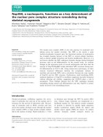
Figure 1. AFAP1-AS was down-regulated and its cytobiology change transfected with AFAP1-AS siRNA. (A) The expression of AFAP1-AS in HSCR
was significantly down-regulated compared with normal tissues. Data were presented as box plot of the median and range of log-transformed relative expression
level. The top and bottom of the box represent the 75th and 25th percentile. The whiskers indicate the 10th and 90th points. (B) AFAP1-AS knockdown affected
abilities of cell migration and proliferation. Representative images of migrated cells were visualized as shown (left panel). Quantifications of cell migration were
presented as percentage migrated cell numbers (middle panel). Absorbance at 450 nm as measured by CCK8 was expressed as Mean ± SE (right panel). (C–D) Cycle
and apoptosis assays were conducted after AFAP1-AS knockdowm by flow cytometer. (E) F-actin cytoskeleton of was visualized with Rhodamine Phalloidin staining
in SH-SY5Y cells.
Int. J. Med. Sci. 2017, Vol. 14
mainly located in the cytoplasm. Cytoplasmic
lncRNAs are well known for modulating gene
expression through interaction with miRNA.
Recently, the competing endogenous RNAs (ceRNA)
hypothesis has been reported to function by
competitively
binding
common
miRNAs.
Bioinformatics prediction according to web server
RegRNA
( />tutorial.html) suggested that four miR-181 family
binding sites were found binding to AFAP1-AS with
high scores [23]. Among the above four miRNAs
(miR-181a/b/c/d), we mainly concentrated on
miR-181a to study the interaction between AFAP1-AS
and miR-181a in HSCR. To validate this hypothesis, a
luciferase reporter including the wild type AFAP1-AS
1026
(pMIR-AFAP1-AS-WT) and mutant AFAP1-AS
(pMIR-AFAP1-AS -MUT) was designed (Figure 2B).
Based on the luciferase assay, we found that miR-181a
significantly reduced luciferase activity for the
wild-type reporter, while mutagenesis of the
predicted miR-181a target sites abolished the previous
suppressive effect (Figure 2C).To verify the physical
interaction between miR-181a and AFAP1-AS at the
endogenous level, RNA immunoprecipitation (RIP)
assay was performed using a specific antibody against
Ago2 protein. Compared with the control group,
AFAP1-AS
was
preferentially
enriched
in
Ago2-coating beads (Figure 2D). Together, our results
revealed that miR-181a directly bind to AFAP1-AS.
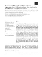
Figure 2. AFAP1-AS acted as a ceRNA by biding miR-181a (A) Expression levels of AFAP1-AS in the nuclear and cytoplasm fractions were determined using
qRT-PCR. GAPDH and U6 were used as cytoplasmic and nuclear markers, respectively. (B) The top and bottom regions are the sequences of AFAP1-AS wild-type
binding to miR-181a and mutations in the 3'-UTR of AFAP1-AS, respectively. (C) The AFAP1-AS wild type or mutant vectors were co-transfected with miR-181a NC
or miR-181a mimics. Firefly luciferase activities were then measured normalized to Renilla. (D) Extracts of 293T cells were applied for RNA binding protein
immunoprecipitation (RIP). Relative RNA levels of AFAP1-AS and miR-181a were measured by qRT-PCR.
Int. J. Med. Sci. 2017, Vol. 14
MiR-181a contributes to the development of
HSCR
Based on the above results, miR-181a was likely
to be involved in the pathogenesis of HSCR. Contrary
to
AFAP1-AS,
miR-181a
was
remarkably
up-regulated in HSCR compared to normal tissues,
showing its potentiating effect on HSCR (Figure 3A).
We then utilized 293T and SH-SY5Y cells to uncover
the functions of miR-181a in HSCR. As was expected,
CCK-8 assays revealed that cell viability was inhibited
1027
after transfection with miR-181a mimics. Meanwhile,
cell migration was markedly reduced after
transfection of miR-181a mimics both in 293T and
SH-SY5Y cells (Figure 3B). No significance was
detected in apoptosis and cell cycle (Figure 3C, D).
Furthermore, the cytoskeleton was disrupted after
transfection with miR-181a mimics (Figure 3E). In
summary, these data presented the promoting effect
of miR-181a on HSCR, in contrast to its ceRNA
AFAP1-AS.
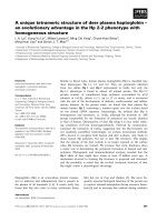
Figure 3. The functional assays of miR-181a were conducted in HSCR cells (A) The expression of miR-181a in HSCR was significantly over-expressed
compared with normal tissues. Data were presented as box plot of the median and range of log-transformed relative expression level. The top and bottom of the box
represent the 75th and 25th percentile. The whiskers indicate the 10th and 90th points. (B) Transwell analysis of 293T and SH-SY5Y cells transfected with negative
control or miR-181a mimics (left panel); Migrated cells stained with crystal violet was shown (middle panel); Cell proliferation were evaluated by CCK8 assay(right
panel).The data are presented as the mean ± SE. (C-D) Cycle and apoptosis assays were conducted after transfection with miR-181a mimics or negative control by
flow cytometer. (E) F-actin cytoskeleton of was stained with Rhodamine Phalloidin staining after transfection with miR-181a mimics or negative control in SH-SY5Y
cells.
Int. J. Med. Sci. 2017, Vol. 14
AFAP1-AS acts as a ceRNA and regulates the
miR-181a mRNA target, RAP1B
To identify the targets of miR-181a, we used
miRsystem
()
which contains several algorithms and predicted
numerous potential direct targets [24]. Gene ontology
analysis revealed relative pathways for candidate
genes. The Gene Ontology results showed that target
genes of mir-181a were involved in multiple
pathways, including MAPK signaling, regulation of
the actin cytoskeleton, neurotrophin signaling and
focal adhesion. Among these putative targets of
1028
miR-181a, we focused on RAP1B, shared by the above
pathways. Previous study has reported that miR-181
targets RAP1B in glioblastoma cells [25]. As a small
GTPaes, Rap1B is involved in cell adhesion which is
mediated by integrin and cadherin, as well as the
cytoskeleton during cell activation [26]. To confirm
whether miR-181a targets RAP1B in HSCR, miRanda
was applied to identify miR-181a recognition sites in
the 3’-UTR of RAP1B (Figure 4A). Luciferase activity
of wild RAP1B reporter was decreased, while mutant
RAP1B reporter was not changed co-transfected with
miR-181a (Figure 4B).
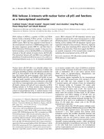
Figure 4. Relationship between AFAP1-AS and the miR-181a target, RAP1B (A) Top: Predicted binding sites between miR-181a and 3'UTR of RAP1B
were constructed as well as mutations in the 3'-UTR of RAP1B. (B) The RAP1B wild type or mutant vectors were co-transfected with negative control or miR-181a
mimics. Firefly luciferase activities were then measured normalized to Renilla. (C) Expression levels of RAP1B in the previous tissues were examined by qRT-PCR
analysis. Data were presented as box plot of the median and range of log-transformed relative expression level. The top and bottom of the box represent the 75th
and 25th percentile. The whiskers indicate the 10th and 90th points. (D) A correlation between AFAP1-AS and RAP1B was observed according to the qRT-PCR
results (r2=0.4726; P<0.0001). (E) Western blots of RAP1B were measured in HSCR and normal tissues. (F) Western blot and qRT-PCR analysis of RAP1B were
conducted after transfection with si-NC, si-AFAP1-AS, and inhibition of AFAP1-AS in combination with miR-181a inhibitor. GAPDH was used as control. All results
were presented as mean ± SE.
Int. J. Med. Sci. 2017, Vol. 14
The above results strongly suggested that
miR-181a directly targeted RAP1B, so we further
explored the association of RAP1B with AFAP1-AS.
Firstly, we measured the expression of RAP1B in the
previous 128 tissues by qRT-PCR analysis and RAP1B
was markedly decreased in HSCR tissue (Figure 4C).
An inverse correlation between AFAP1-AS and
RAP1B expression levels was observed (Figure 4D).
The Western Blot results also revealed that RAP1B
was down-regulated in HSCR tissues in comparison
with normal tissues, which were consistent with the
PCR result (Figure 4E).We further clarified the
regulatory relationship between AFAP1-AS and
RAP1B by AFAP1-AS knockdown against RAP1B in
293T and SH-SY5Y cells. PCR and Western Blot were
carried out and showed that down-regulation of
AFAP1-AS significantly inhibited RAP1B expression,
whereas inhibition of AFAP1-AS in combination with
miR-181a inhibitor did not affect RAP1B expression,
suggesting that AFAP1-AS regulates RAP1B in a
miR-181a-dependent manner (Figure 4F).
Together, these results revealed that through
binding to miR-181a directly, AFAP1-AS acted as a
ceRNA for RAP1B and modulated the expression of
RAP1B in a post-transcriptional regulation.
Discussion
Increasing studies indicated that lncRNAs
engaged in a broad spectrum of cellular processes,
including tumorigenesis and embryogenesis. It has
previously been reported that lncRNA AFAP1-AS
was frequently dysregulated in several diseases, such
as hepatocellular carcinoma, pancreatic ductal
adenocarcinoma and nasopharyngeal carcinoma.
However, the possible role of AFAP1-AS in HSCR
remains to be clarified. In this study, we examined the
expression of AFAP1-AS in HSCR samples and
normal control tissues. The function of AFAP1-AS
was
further
characterized
by
applying
loss-of-function approaches in HSCR cell lines. We
detected that AFAP1-AS was down-regulated in
HSCR tissues in comparison with the normal tissues,
and AFAP1-AS depletion inhibited cell migration and
growth ability while no significant difference was
observed about apoptosis and cell cycle. Furthermore,
down-regulated AFAP1-AS contributed to the actin
degradation and loss of cell polarity. Therefore, it is
concluded that AFAP1-AS participated in the
modulation of neural crest cell colonization.
However, the inhibition of migration and growth
capacity in HSCR by down-regulating AFAP1-AS
needs further verification by in vivo experiments.
We also sought out the molecular mechanism of
AFAP1-AS regulating neural crest cell colonization.
AFAP1-AS exists in the cytoplasm, indicating that it
1029
can regulate gene expression at mRNA levels. As we
known, ceRNAs are recognized as a novel regulatory
mechanism of posttranscriptional gene expression by
competing shared for miRNAs to suppress the
expression of target genes. We integrated online
bioinformatics database, and found that the mir-181a
had the higher scores binding to AFAP1-AS. To
further verify the relationship between miR-181a and
AFAP1-AS, dual-luciferase reporter and RNA
immunoprecipitation (RIP) assays were conducted.
These results revealed that miR-181a can directly bind
to the AFAP1-AS. As a well-known miRNA, miR-181a
functions as a tumor suppressor in several cancers,
such as breast cancer, glioma and gastric cancer [25,
27, 28]. Consistent with the above results, miR-181a
was overexpression in HSCR tissues and negatively
regulatd neural crest cell proliferation and migration
through targeting RAP1B.Here we provide direct
evidence that AFAP1-AS can function as a ceRNA,
which sequesters miR-181a, thereby protecting
RAP1B
transcripts
from
miR-181a-mediated
suppression. AFAP1-AS and RAP1B were validated
as targets for miRNA-181a in our cellular models.
Consistent with AFAP1-AS competing miRNA-181a
with RAP1B, we found that while depression of
AFAP1-AS could reduce expression of RAP1B.
Besides, our studies found miRNA-181a inhibitor
could rescue the down-regulation of RAP1B in
AFAP1-AS knockdown cells, which support the
ceRNA regulatory mechanism of RAP1B by
AFAP1-AS.
Taken
together,
we
reported
that
down-regulated lncRNA AFAP1-AS inhibits HSCR
cell proliferation and migration by competitively
binding the miR-181a, down-regulating RAP1B, and
suppressing neural crest cell colonization. Our studies
revealed a novel ceRNA regulatory network,
AFAP1-AS/miR-181a/RAP1B,
providing
new
insights into understanding the mechanisms of HSCR.
Abbreviations
HSCR: Hirschsprung disease; lncRNA: long
non-coding RNA; miRNA: microRNA; ceRNA:
competing endogenous RNA; CCK-8: Cell counting
kit-8; DMEM: Dubelcco modified Eagle's medium;
FBS:
Fetal
bovine
serum;
GAPDH:
Glyceraldehyde-3-phosphate dehydrogenase; RIP:
RNA immunoprecipitation
Acknowledgements
We thank Dr. Jie Zhang, HuanChen, Xiaofeng
Lv, Weiwei Jiang, Wei Li and Changgui Lu
(Children’s Hospital of Nanjing Medical University)
for sample collection. This study was supported by
Natural Science Foundation of China (NSFC
Int. J. Med. Sci. 2017, Vol. 14
81370473, NSFC 81400574, NSFC 81570467), Priority
Academic Program Development of Jiangsu Higher
Education Institutions (PAPD), Nanjing Medical
Science and Technique Development Foundation
(201405014) and Jiangsu Qing Lan Project.
Competing Interests
The authors have declared that no competing
interest exists.
1030
24. Lu TP, Lee CY, Tsai MH, Chiu YC, Hsiao CK, Lai LC, et al. miRSystem: an
integrated system for characterizing enriched functions and pathways of
microRNA targets. PLoS One. 2012; 7: e42390.
25. She X, Yu Z, Cui Y, Lei Q, Wang Z, Xu G, et al. miR-181 subunits enhance the
chemosensitivity of temozolomide by Rap1B-mediated cytoskeleton
remodeling in glioblastoma cells. Med Oncol. 2014; 31: 892.
26. Schwamborn JC, Puschel AW. The sequential activity of the GTPases Rap1B
and Cdc42 determines neuronal polarity. Nat Neurosci. 2004; 7: 923-9.
27. Neel JC, Lebrun JJ. Activin and TGFbeta regulate expression of the
microRNA-181 family to promote cell migration and invasion in breast cancer
cells. Cell Signal. 2013; 25: 1556-66.
28. Peng W, Si S, Zhang Q, Li C, Zhao F, Wang F, et al. Long non-coding RNA
MEG3 functions as a competing endogenous RNA to regulate gastric cancer
progression. J Exp Clin Cancer Res. 2015; 34: 79.
References
1.
2.
3.
4.
5.
6.
7.
8.
9.
10.
11.
12.
13.
14.
15.
16.
17.
18.
19.
20.
21.
22.
23.
Borrego S, Ruiz-Ferrer M, Fernandez RM, Antinolo G. Hirschsprung's disease
as a model of complex genetic etiology. Histol Histopathol. 2013; 28: 1117-36.
Spouge D, Baird PA. Hirschsprung disease in a large birth cohort. Teratology.
1985; 32: 171-7.
McKeown SJ, Stamp L, Hao MM, Young HM. Hirschsprung disease: a
developmental disorder of the enteric nervous system. Wiley Interdiscip Rev
Dev Biol. 2013; 2: 113-29.
Butler Tjaden NE, Trainor PA. The developmental etiology and pathogenesis
of Hirschsprung disease. Transl Res. 2013; 162: 1-15.
Tang W, Tang J, Qin J, Geng Q, Zhou Z, Li B, et al. Involvement of
down-regulated E2F3 in Hirschsprung's disease. J Pediatr Surg. 2013; 48:
813-7.
Li H, Tang J, Lei H, Cai P, Zhu H, Li B, et al. Decreased MiR-200a/141
suppress cell migration and proliferation by targeting PTEN in Hirschsprung's
disease. Cell Physiol Biochem. 2014; 34: 543-53.
Tang W, Qin J, Tang J, Zhang H, Zhou Z, Li B, et al. Aberrant reduction of
MiR-141 increased CD47/CUL3 in Hirschsprung's disease. Cell Physiol
Biochem. 2013; 32: 1655-67.
Lee S, Kopp F, Chang TC, Sataluri A, Chen B, Sivakumar S, et al. Noncoding
RNA NORAD Regulates Genomic Stability by Sequestering PUMILIO
Proteins. Cell. 2016; 164: 69-80.
Shi X, Sun M, Liu H, Yao Y, Song Y. Long non-coding RNAs: a new frontier in
the study of human diseases. Cancer Lett. 2013; 339: 159-66.
Chen YA, Aravin AA. Non-Coding RNAs in Transcriptional Regulation: The
review for Current Molecular Biology Reports. Curr Mol Biol Rep. 2015; 1:
10-8.
Wan LB, Bartolomei MS. Regulation of imprinting in clusters: noncoding
RNAs versus insulators. Adv Genet. 2008; 61: 207-23.
Wan L, Sun M, Liu GJ, Wei CC, Zhang EB, Kong R, et al. Long Noncoding
RNA PVT1 Promotes Non-Small Cell Lung Cancer Cell Proliferation through
Epigenetically Regulating LATS2 Expression. Mol Cancer Ther. 2016; 15:
1082-94.
Kitagawa M, Kitagawa K, Kotake Y, Niida H, Ohhata T. Cell cycle regulation
by long non-coding RNAs. Cell Mol Life Sci. 2013; 70: 4785-94.
Qi X, Zhang DH, Wu N, Xiao JH, Wang X, Ma W. ceRNA in cancer: possible
functions and clinical implications. J Med Genet. 2015; 52: 710-8.
Wu W, Bhagat TD, Yang X, Song JH, Cheng Y, Agarwal R, et al.
Hypomethylation of noncoding DNA regions and overexpression of the long
noncoding RNA, AFAP1-AS1, in Barrett's esophagus and esophageal
adenocarcinoma. Gastroenterology. 2013; 144: 956-66 e4.
Ye Y, Chen J, Zhou Y, Fu Z, Zhou Q, Wang Y, et al. High expression of
AFAP1-AS1 is associated with poor survival and short-term recurrence in
pancreatic ductal adenocarcinoma. J Transl Med. 2015; 13: 137.
Zeng Z, Bo H, Gong Z, Lian Y, Li X, Li X, et al. AFAP1-AS1, a long noncoding
RNA upregulated in lung cancer and promotes invasion and metastasis.
Tumour Biol. 2016; 37: 729-37.
Bo H, Gong Z, Zhang W, Li X, Zeng Y, Liao Q, et al. Upregulated long
non-coding RNA AFAP1-AS1 expression is associated with progression and
poor prognosis of nasopharyngeal carcinoma. Oncotarget. 2015; 6: 20404-18.
Zhou XL, Wang WW, Zhu WG, Yu CH, Tao GZ, Wu QQ, et al. High
expression
of
long
non-coding
RNA
AFAP1-AS1
predicts
chemoradioresistance and poor prognosis in patients with esophageal
squamous cell carcinoma treated with definitive chemoradiotherapy. Mol
Carcinog. 2016.
Vargiolu M, Fusco D, Kurelac I, Dirnberger D, Baumeister R, Morra I, et al. The
tyrosine kinase receptor RET interacts in vivo with aryl hydrocarbon
receptor-interacting protein to alter survivin availability. J Clin Endocrinol
Metab. 2009; 94: 2571-8.
Kawamoto T, Ohira M, Hamano S, Hori T, Nakagawara A. High expression of
the novel endothelin-converting enzyme genes, Nbla03145/ECEL1alpha and
beta, is associated with favorable prognosis in human neuroblastomas. Int J
Oncol. 2003; 22: 815-22.
Peng H, Luo J, Hao H, Hu J, Xie SK, Ren D, et al. MicroRNA-100 regulates
SW620 colorectal cancer cell proliferation and invasion by targeting RAP1B.
Oncol Rep. 2014; 31: 2055-62.
Huang HY, Chien CH, Jen KH, Huang HD. RegRNA: an integrated web server
for identifying regulatory RNA motifs and elements. Nucleic Acids Res. 2006;
34: W429-34.