Longitudinal micro-endoscopic monitoring of high-success intramucosal xenografts for mouse models of colorectal cancer
Bạn đang xem bản rút gọn của tài liệu. Xem và tải ngay bản đầy đủ của tài liệu tại đây (1.42 MB, 8 trang )
Int. J. Med. Sci. 2019, Vol. 16
Ivyspring
International Publisher
1453
International Journal of Medical Sciences
2019; 16(11): 1453-1460. doi: 10.7150/ijms.35666
Research Paper
Longitudinal micro-endoscopic monitoring of
high-success intramucosal xenografts for mouse models
of colorectal cancer
Bjorn Paulson1*, Ick Hee Kim2*, Jung-Man Namgoong 3*, Young Gyu Kim1, Sanghwa Lee1, Youngjin Moon1,4,
Dong-Myung Shin5,6, Myung-Soo Choo7, Jun Ki Kim1,4
1.
2.
3.
4.
5.
6.
7.
Biomedical Engineering Research Center, Asan Institute for Life Sciences, Asan Medical Center, 88, Olympic-ro 43-gil, Songpa-gu, Seoul 05055, Republic of
Korea
Wake Forest Institute for Regenerative Medicine, Wake Forest School of Medicine, 391 Technology Way, Winston-Salem, NC 27101, USA
Department of Surgery, Asan Medical Center, University of Ulsan College of Medicine, 88, Olympic-ro 43-gil, Songpa-gu, Seoul 05055, Republic of Korea
Department of Convergence Medicine, University of Ulsan College of Medicine, 88, Olympic-ro 43-gil, Songpa-gu, Seoul 05055, Republic of Korea
Department of Biomedical Sciences, Asan Medical Center, University of Ulsan College of Medicine, 88, Olympic-ro 43-gil, Songpa-gu, Seoul 05055, Republic
of Korea
Department of Physiology, Asan Medical Center, University of Ulsan College of Medicine, 88, Olympic-ro 43-gil, Songpa-gu, Seoul 05055, Republic of Korea
Department of Urology, Asan Medical Center, University of Ulsan College of Medicine, 88, Olympic-ro 43-gil, Songpa-gu, Seoul 05055, Republic of Korea
*These authors contributed equally to this work.
Corresponding author: Jun Ki Kim, Ph.D., Biomedical Engineering Center, ASAN Institute for Life Sciences, Asan Medical Center, Pungnap-2 dong,
Songpa-gu, Seoul, 05505, Korea. Email:
© The author(s). This is an open access article distributed under the terms of the Creative Commons Attribution License ( />See for full terms and conditions.
Received: 2019.04.11; Accepted: 2019.08.02; Published: 2019.09.20
Abstract
Colorectal cancer (CRC) is one of the most frequently lethal forms of cancer. Intramucosal injection
allows development of better mouse models of CRC, as orthotopic xenografts allow development
of adenocarcinoma in the submucosa of the mouse colon wall. In this paper, a method of orthotopic
injection is monitored longitudinally using cellular-resolution real-time in vivo fluorescence
microendoscopy, following the injection of three different cell lines: 3T3-GFP to confirm
immunosuppression and HCT116-RFP cells to model CRC. Adenoma formation is first observable
after 7 to 10 days, and by use of 33 G needles a tumor induction rate of greater than 85% is
documented. An additional experiment on the injection of rapamycin reveals drug efficacy and
localization between 24 and 48 hours, and suggests the promise of real-time cellular-resolution
fluorescence micro-endoscopy for developing longitudinal therapy regimes in mural models of CRC.
Key words: Orthotopic injection, Colorectal cancer, mouse models, microendoscopy, side-view endoscopy,
fluorescence imaging
Introduction
Colorectal cancers (CRC) have the second
highest mortality rate among cancers worldwide,
accounting for around 11% of cancer deaths in men
and 7% in women, and exhibit the highest mortality
rate among intestinal diseases [1]. With the goal of
developing novel cancer therapeutics, several
standard methods have been developed for the
precipitation of colorectal cancer (CRC) in mouse
models: genetically engineered mice which
recapitulate known cancerogenic genotypes [2],
chemically induced but non-specific tumorigenesis
[3], xenografts of human tumor tissue into
immune-compromised mice [4], and xenografts of
human cancer tissue into mice with humanized
immune systems [5]. While mural tumors in
genetically engineered mouse models give realistic
tumor expression in a realistic microenvironment,
accurately demonstrating the pathology and
mechanisms of several tumor types, their
performance for the discovery of molecularly-targeted
Int. J. Med. Sci. 2019, Vol. 16
treatment compounds has been limited. In contrast,
the efficacy of compounds on human tumor-derived
cell lines and transplanted cancer tissue in the mural
model has been predictive of phase 2 clinical trial
performance [6]. In addition, transplantation of tumor
tissue allows for fine control of tumor location in
comparison to spontaneously tumorigenic transgenic
mouse models [7]. As a result, xenografts of cancerous
cells and tissues are the primary method for inducing
colorectal cancer in mouse models.
Typical procedure for the xenograft of colorectal
tissue into mouse models is to section fresh surgical
tissue into small pieces, followed by implantation
either subcutaneously by injection, or orthotopically
via surgery. The success or failure of engraftment can
be ascertained after four to six months, and
immunocompromised mouse models have resulted in
tumor induction rates of up to 95%, although most
investigators report take rates of about 75% [6,8].
Surgical implantation has several limitations.
Implantation requires significant surgical time,
feedback on procedure success is slow, failure rates
are high, and transplantation precisely to the colon
epithelium is difficult. In order to overcome these
difficulties, several groups have recently developed
less invasive and more precise methods for orthotopic
xenografts of CRC tissues, including enema-based
acid treatment [9], a specially-designed sponge [10],
and intramucosal (orthotopic) injection [4,11–14].
Where the development of minimally invasive
methods for orthotopic transplantation has enabled
precise and consistent tumor placement, the
development of micro-endoscopy has recently
enabled in situ targeting and evaluation of CRC
models.
Optical
measurement
using
a
micro-endoscope gives real-time data about the
surface optical properties of organs in the living
organism [15].
Using a working channel on a laparoscope, our
group has demonstrated ovarian injection of
chemo-resistant cell cultures [16], while Roper et al.
have successfully used colonoscopy to target
individual submucosal tumors at 0.5 cm intervals
along the mural colon [11]. By avoiding the use of
surgery and reaching the target microenvironment
directly, injection-based orthotopic xenografts
promise a more realistic microenvironment, and the
narrow diameter of the needle minimizes the chance
of pneumoperitoneum even in the case of colon
perforation, while increasing tumor induction and
survival rates. Thereby researcher time, money, and
animals may be saved [14].
In this study, a modified method for the
orthotopic transplantation of colorectal cancer (CRC)
xenographs is presented and studied. In contrast to
1454
previous injection methods [11–14], the method
presented does not require the use of bespoke needles,
yet still achieves CRC adenoma formation in more
than 85% of mouse models. Longitudinal, real-time
monitoring following injection is performed both via
commercially available colonosope, as in previous
work, and via a custom side-view laser-scanning
confocal
micro-endoscope,
which
allows
cellular-resolution imaging of the colon lumen.
Tumor formation is confirmed by confocal
microscopy and histology. Taken together, these
results
demonstrate
the
first
in
vivo
cellular-resolution longitudinal monitoring of
orthotopically injected CRC models, to the best
knowledge of the authors. Finally, feasibility of the
injection procedure for localized treatment and
chemically-enhanced optical biopsy is assessed by
injection of rapamycin into LC3-GFP+ mice, and
found promising for the localized assessment of
future cancer therapies.
Materials and Methods
Cell lines and preparation
Human colorectal cancer cell line HCT116 was
prepared at the Wellman Center for Photomedicine,
Massachusetts General Hospital (MGH), while
3T3-GFP-expressing fibroblast cells were graciously
donated by the Center for Computational and
Integrative Biology (CCIB), Massachusetts General
Hospital (MGH). A red fluorescent protein (RFP)
lentivirus (Lenti-Red, Biogenova, Rockville, USA) was
transfected to induce stable RFP expression in
HCT116. Cells were cultured in RPMI1640 medium
supplemented with 10% fetal bovine serum, 100 U/ml
penicillin, 0.1 mg/ml streptomycin and 2 mM
L-glutamine. The medium was changed every 1-2
days. For each injection, 3.0 × 10⁵ cells were
administered at a density of 104 cell µL-1.
Rapamycin and fluorescent microparticles
Rapamycin and fluorescent micro particles
(Sigma-Aldrich) were prepared for injection.
Fluorescent particles were excited at 636 nm for
fluorescence at 686 nm. Rapamycin was dissolved in
DMSO (dimethyl sulfoxide), then added in 1:24
proportion to a solution of 10% polyethylene glycol
(MW avg. = 400 Da).
Mouse preparation
Female BALB/c nude mice and LC3-GFP mice
(Jackson Laboratories), seven to sixteen weeks old,
were raised in a specific pathogen free environment,
controlling for pathogens, temperature, humidity,
atmospheric contaminants, lighting, and sound. Nude
mice were split into three groups of 7 mice each, and
Int. J. Med. Sci. 2019, Vol. 16
LC3-GFP mice were split into two groups: a
LC3-GFP+ control group, subject to a sham procedure
with injection of fluorescent beads; an LC3-GFP+
treatment group subject to injection of fluorescent
beads and rapamycin. One nude mouse group each
was subject to injection of 3T3 cells, and HCT116–RFP
cells. All animal studies were conducted in
accordance with the policies of the NIH Guide for the
Care and Use of Laboratory Animals and approved
by the Institutional Animal Care and Use Committee
(IACUC) of Massachusetts General Hospital
(MGH2007N0000110).
Intramucosal xenografts and forward-view
colonoscopy
Before xenografts were performed, an extended
needle was prepared for insertion through the
endoscope’s working channel. A 33G needle was
bonded to a 2 Fr diameter hollow stainless steel
sheath using a thermal-curing epoxy (Thorlabs) (Fig
1(a)). The connection to the sheath was then wrapped
with parafilm (Bemis, WI, USA) to protect the epoxy
from mechanical strain during insertion into the
curved 3 Fr diameter working channel of the
colonoscope.
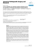
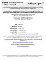
Fig 1. Schematic of the experiments. (a) Minimally invasive transplantation
needle designed from commonly available parts. (b) Needle is inserted through the
working channel of a forward-viewing colonoscope for (c) orthotopic injection of
CRC cells into mouse models. (d) Side-view GRIN lens triplet micro-endoscope
probes are applied for longitudinal monitoring of neoplasm at cellular resolution in
vivo.
For all injections, mice were anesthetized
intraperitoneally with 75 mg kg-1 ketamine and 15 mg
kg-1 xylazine, according to standard protocols, and
placed on a heated pad for the maintenance of
homeostasis. LC3-GFP mice were shaved using
clippers and depilatory cream. The colon was washed
with 0.5 mL phosphate buffered saline (PBS) at 37
degrees centigrade, using a rubber-tipped syringe. A
three-axis micro-stage was used to direct and guide
insertion of a 2.8 mm diameter Coloview® miniature
colonoscope (Karl Storz) 2 cm proximally from the
1455
end of the colon, while a 33G needle was staged near
the end of the working channel (Fig 1(b)). The needle
was then extended into the colon tissue, and the
desired cell solution was injected (Fig 1(c)). All cells
were administered in batches of 3.0 × 10⁵ cells at a
density of 104 cell µL-1. Fluorescent microbeads in
buffered saline solution and rapamycin solutions
were prepared and injected to a total volume of 100
μL.
Side-view confocal micro-endoscopy and
fluorescent cellular imaging
Cellular resolution images were captured in vivo
with
a
custom
built
side-view
confocal
micro-endoscope
(Fig
1(d)).
A
side-view
gradient-index (GRIN) relay micro-endoscopy probe
of length 5.5 cm and diameter 1.2 mm was assembled
using three GRIN lenses and an angled prism,
cementing lenses together as previously described
[17]. The side-view probe has a field of view of 220
microns, and achieves transverse and lateral
resolutions of 1 μm and 11 μm in air, respectively,
which is sufficient for image capture at single-cell
resolution. It was connected to a custom built laser
scanning confocal microscope via a 4f lens relay [18].
A three-axis micro-stage was used to guide the
endoscope along the colon, and allowed measurement
of probe position relative to the distal end of the
colon. Green fluorescent protein (GFP) and red
fluorescent protein (RFP) were excited using
continuous wave 488 nm and 532 nm laser diodes,
respectively, which were scanned over the field of
view at 30 fps via resonant scanning mirrors.
Photomultiplicative detectors (PMT) were used in
tandem with dichroic filters to capture emitted
fluorescence while avoiding noise caused by the
excitation laser and autofluorescence. Green
fluorescent protein has a fluorescence peak at 525 nm,
while RFP emits at 607 ± 15 nm, respectively.
Fluorescent microbeads were excited using a 636 nm
laser and emissions were detected at 680 ± 21 nm.
Large-field-of-view images were generated by
compositing smaller endoscopic images into a mosaic
as previously described [18]. Average fluorescence
intensity was quantified by separating out the color
channel for the relevant fluorescence signal and
taking the average value of that channel for all
nonzero pixels in the image. LC3-GFP cells
undergoing autophagy were counted by thresholding
the GFP channel at 60% of its saturation value and
counting the number of clumps of nine or more
connected pixels above the threshold value. The
autophagy cellular density was then calculated by
dividing by the field of view.
Int. J. Med. Sci. 2019, Vol. 16
Histology
After terminating longitudinal observations, the
distal colons were harvested and tumors were
removed from the colon after sacrifice. Tumor
diameters were measured with digital calipers before
tumor sections were cut, fixed in formalin, and
stained under hematoxylin and eosin dyes for
micrographs. Neoplasms were verified to be of the
injected fluorescent cell lines by ex vivo confocal
fluorescence microscopy. Histology results were
matched with forward colonoscopy images to
calibrate tumor size measurements for longitudinal in
vivo tumor size estimation.
Results and Discussion
The demonstration of consistent orthotopic
tumorigenesis without immune rejection is necessary
prior to the longitudinal study of orthotopic mural
models of CRC. Previously, Zigmond et al. have
reported, using 30G needles, that orthotopic
transplantation of injected cell counts of 105 or higher
result in increased colonic obstruction and
higher-grade tumors over a fixed period of 3 weeks
[12] compared to smaller cell counts. However, Roper
et al demonstrated the use of a smaller needle gauge to
be well suited to infusing CRC-inducing transgenic
compounds in the lamina propria of a mouse model
due to its reduced risk of complications from colon
1456
perforation [11]. In this work, a standard 33G
hypodermic needle with a 30 degree bevel was
applied, necessitating confirmation of tumor
formation efficacy at the desired cell counts.
To verify the efficacy of tumor generation for our
modified injection process, GFP-expressing mouse
embryonic fibroblast 3T3 cells (3T3-GFP), which are
commonly used for xenograft studies of immune
rejection, were injected into BALB/c nude mice. At
distances of 1.0 and 2.0 cm from the distal end of the
colon, the injection locations on the mucosa were
imaged weekly over a period of 3 weeks by
colonoscopy using the Coloview® endoscope and by
side-view
micro-endoscopes.
Representative
longitudinal tumor growth is shown in Fig 2(a).
Distinct neoplasia was evident after a two-week
period, and adenoma formation was clearly observed
after three weeks. For the first two weeks after
orthotopic implantation, mouse colon vasculature
was monitored using the side-view fluorescent
confocal micro-endoscope after rhodamine dextran
intravascular (IV) injection. While bulging was
observed due to formation of tumor in the colon
epithelium immediately after injection, the deformed
microvascular patterns were observed in patches over
the bulged area as shown in Fig 2(b). As the week
progressed, the area affected by this bulging pattern
appeared to become wider.
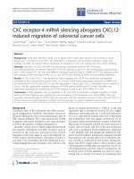
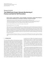
Fig 2. Orthotopic injection and tumorigenesis from 3T3-GFP cells in BALB/c nude mice. (a) Longitudinal forward-view colonoscope observation following injection
shows tumor formation over a period of 3 weeks. (b) In an induced tumor 1.0 cm from the distal end of the colon, patterns of microvascular deformation (white arrows) were
observed by side-view endoscopy after 1 and 2 weeks. Scale bars 100 µm. (c) Size comparison of the harvested tumors. (d) Micrograph of excised tumor tissue following
hematoxylin and eosin stain. Scale bars 250 µm. (e) A plot of the tumor size progression observed longitudinally by front-view colonoscopy and combined with post-sacrifice
direct size measurements. All results are indicative of successful transplantation without immune rejection.
Int. J. Med. Sci. 2019, Vol. 16
After 3 weeks, mice were sacrificed and colons
were harvested for histology as shown in Fig 2(c,d).
Overall, tumor growth was successful and tumor size
ranged between 3 mm (6 mm3) and 8 mm (64 mm3).
Histology showed deformities in the submucosa and
muscularis consistent with neoplasm. All samples
were injected submucosally, and no deaths or colon
perforations were observed. The tumors were
successfully generated at a tumor induction rate of
85% (N=14 in 7 mice), where the tumor induction rate
is defined as the percent of orthotopic injections
which resulted in a tumor within a 14 day period.
While 3T3-GFP cells demonstrate immune
suppression, the study of human cancer cells in
mouse models of CRC is more directly demonstrated
by the HCT116-RFP cell line, a human CRC line
expressing red fluorescent protein (RFP), which is of
interest due to its visibility in side-view fluorescence
confocal endomicroscopy. Identical quantities (3 × 10⁵
cells) of HCT116-RFP were delivered submucosally
into BALB/c nude mice, and examined by fluorescent
side-view endoscopes 10, 17, 24, and 30 days after
implantation, as shown in Fig 3(a).
Using cellular-resolution side-view colonoscopy,
injected HCT116-RFP cells could be observed at 10
days. After 24 days, imaging was complicated by
restriction of the colon around the tumor sites.
While neoplasm was not distinctly evident 10
days after injection in the front-view colonoscope,
cellular-level development of neoplasm was evident
in the side-view images from the same time period. It
should be noted that the expression of fluorescent
protein can be induced in cell and tissue cultures
virally, and thus the monitoring of injection sites by
side-view micro-endoscopy presents a method for the
early detection of CRC model mice for which
tumorigenesis is not successful due to immune
rejection or other factors. This potentially decreases
the time required for orthotopic CRC transplantation
experiments by up to a week. We could confirm that
the growth speed of the tumor is dependent of species
and aggressiveness of the cell lines. This was also
apparent by inspection of colons ex vivo, as large
growths were visible external to the colon, as shown
in Fig 3(c). The HCT116-RFP tumors were also
imaged by histology after one month. As shown in Fig
3(c) and Fig 3(d), the growth of tumors was observed
between the muscularis mucosae and muscularis
propria.
While several authors have demonstrated the
orthotopic transplantation of genetically modified
cells and tissues [12,14], understanding of the
localization properties and cellular behavior of
1457
genetically inert pharmaceuticals in the in vivo
environment is also desirable. To this end, rapamycin
was injected orthotopically into LC3-GFP expressing
transgenic mice and monitored longitudinally at
cellular resolution by side-view micro-endoscopy.
Mice expressing green fluorescent LC3 protein
(LC3-GFP) allow for the highly specific visualization
of autophagy in vivo, as the LC3 membrane protein
binds to autolysosome membranes and is degraded
during autophagy, expressing punctate fluorescence
signals during autophagic processes as a result
[19,20]. Autophagy is chiefly regulated by the
mammalian target of rapamycin (mTOR), although
mTOR-independent autophagy pathways also exist
[21]. Thus, in LC3-GFP expressing transgenic mice,
areas of high rapamycin concentration may be
expected to show cells with significantly increased
fluorescence spots, as negative regulation of
autophagy is inhibited. While the sub-cellular-sized
puncta are not visible in standard colonoscopy,
individual cells with abnormally increased
fluorescence are distinguishable at the resolution of
the side-view GRIN micro-endoscope probe.
By delivering rapamycin to LC3-GFP mice, the in
vivo localization and duration of rapamycin may be
assessed via the fluorescence of cell autophagy, as
shown in Fig 4. In this study, a treatment group of
tumor-free LC3-GFP+ mice was orthotopically
injected with a mix of relatively immobile fluorescent
nanospheres and rapamycin, while a tumor-free
control group was subject to the same procedure
without rapamycin. Although there is a risk that
punctate
GFP
signals
may
result
from
autofluorescence and from LC3-GFP trapped in
protein aggregates [22], these may be limited to the
effects of rapamycin by comparison of fluorescence
relative to the control group. Fluorescent nanospheres
were used to mark the injection location, allowing
repeated monitoring of the induced autophagy, and
thereby the persistence of rapamycin, by side-view
confocal micro-endoscopy. Over a 48-hour period,
strong cellular signals likely indicative of autophagy
were observed in the rapamycin-treatment group, but
not in the control group.
Although the field of view of the GRIN
microendoscope is limited, the measuring and
marking of multiple points as well as the composition
of images from several passes and rotations of the
side-view microendoscope allow the generation of
larger, contiguous images [17,23]. Several such
post-processed images are used in Fig. 4(e-h) to show
the extent of the spread of the rapamycin-derived
autophagy.
Int. J. Med. Sci. 2019, Vol. 16
1458
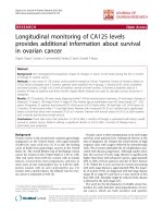
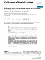
Fig 3. Cellular resolution real-time longitudinal monitoring of HCT116-RFP cells after orthotopic injection. (a) Side-view cellular-resolution micro-endoscopy
post-injection shows visible neoplasm after 10 days. (b) Gross image of excised tumor tissue. (c) Hematoxylin-and-eosin-stained histology of colon section showed tumor
formation in the tissue layers of the colon; M:Musosa, MM:Muscularis mucosae, SM:Submucosa, MP:Muscularis Propria. (d) Higher-magnification images of the circled region (d).
Scale bars: (a) 50 µm, (b) 5 mm, (c) 500 µm.
Fig 4. Cellular resolution side-view microendoscope images following orthotopic injection of rapamycin in the mural colon. (a,c,d,e) Rapamycin injection sites
24 hours post-injection show higher fluorescence than (b,f) after 48 hours. (c,f) Control image without Rapamycin injection. (d) Rapamycin treated mice show higher autophagy
than controls for all time periods. (h) Rapamycin shows slightly increased diffusion compared to concurrently injected fluorescent microparticles after a 24 h period. Green =
GFP, bright green = autophagy, red = rhodamine dextran, blue = fluorescent beads. Scale bars, 40 microns.
Int. J. Med. Sci. 2019, Vol. 16
In the first 24 hours, autophagy spread slightly
beyond the injection region, as marked by fluorescent
beads, to be localized in an area of 105~106 microns2
for the first 24 h. (Fig. 4h). The spread of the
rapamycin may be due to DMSO-assisted perfusion,
or due to diffusion through the extracellular matrix.
Autophagy declined between 24 and 48 h. No mouse
fatalities were observed in either the control group or
the treatment group (7 mice each).
In addition to its autophagic properties,
rapamycin’s inhibition of mTOR interrupts the
signaling cascades of KRAS-negative CRC tumors,
and targeted injection of rapamycin may be promising
for treating these carcinomas in human patients [7].
However, targeted intramucosal injection paired with
longitudinal monitoring is also powerful tool for the
development and testing of other therapeutics
targeted at pre-metastatic CRCs.
Conclusion
Through the process of orthotopic injection and
transplantation of CRC cell cultures and organoids in
mouse models, mural models for colorectal cancers
have recently been developed with high tumor
induction rates and improved accuracy to human
CRC [14]. This work builds on those models,
demonstrating the use of side-view confocal
micro-endoscopy for the minimally invasive,
longitudinal, and cellular-resolution monitoring of
tumor development. The submucosal injection of cell
cultures was performed using standard gauge
hypodermic needles, and monitored longitudinally.
Tumorigenesis is demonstrated with 3T3-GFP and
HCT116-RFP cells in mural models, and real-time
side-view endoscopy is used to demonstrate early
identification
of
neoplasm.
Side-view
endomicroscopy also allows for the observation of
cellular-level features, such as the fluorescence of
LC3-GFP due to autophagy, as demonstrated by the
injection of rapamycin. With rapamycin, the potential
for targeted orthotopic injection of pharmaceuticals is
demonstrated and an alternative method for
evaluation of their efficacy in vivo is shown, which has
potential for application in future mural models of
CRC.
Abbreviations
CRC, Colorectal cancers, GFP, green fluorescent
protein, RFP, red fluorescent protein, DMSO dimethyl
sulfoxide;
MW,
molecular
weight;
PMT,
photomultiplier tube; G, gauge; mTOR, molecular
target of rapamycin.
Acknowledgements
This work was supported by the Basic Science
1459
Research Program (2018R1D1A1B07048562) and MRC
grant (2018R1A5A2020732) through the National
Research Foundation of Korea (NRF) funded by the
Ministry of Science & ICT (MSIT), and by the Ministry
of Trade, Industry & Energy (MOTIE) under
Industrial Technology Innovation Program (10080726,
20000843), and by a grant of the Korea Health
Technology R&D Project through the Korea Health
Industry Development Institute (KHIDI), funded by
the Ministry of Health & Welfare, Republic of Korea
(HI18C2391). This study was also supported by a
grant (2018IE7042) from the Asan Institute for Life
Sciences, Asan Medical Center, Seoul, Korea.
Competing Interests
The authors have declared that no competing
interest exists.
References
1.
2.
3.
4.
5.
6.
7.
8.
9.
10.
11.
12.
13.
14.
15.
16.
17.
18.
Bray F, Ferlay J, Soerjomataram I, Siegel RL, Torre LA, Jemal A. Global cancer
statistics 2018: GLOBOCAN estimates of incidence and mortality worldwide
for 36 cancers in 185 countries. CA Cancer J Clin. 2018 Nov; 68(6): 394–424.
Moser AR, Pitot HC, Dove WF. A dominant mutation that predisposes to
multiple intestinal neoplasia in the mouse. Science. 1990 Jan 19; 247(4940):
322–4.
Tanaka T, Kohno H, Suzuki R, Yamada Y, Sugie S, Mori H. A novel
inflammation-related mouse colon carcinogenesis model induced by
azoxymethane and dextran sodium sulfate. Cancer Sci. 2003 Nov; 94(11):
965–73.
Pocard M, Tsukui H, Salmon RJ, Dutrillaux B, Poupon MF. Efficiency of
orthotopic xenograft models for human colon cancers. Vivo Athens Greece.
1996 Oct; 10(5): 463–9.
Richmond A, Su Y. Mouse xenograft models vs GEM models for human
cancer therapeutics. Dis Model Mech. 2008 Oct; 1(2–3): 78–82.
Morton CL, Houghton PJ. Establishment of human tumor xenografts in
immunodeficient mice. Nat Protoc. 2007 Feb; 2(2): 247–50.
Hung KE, Maricevich MA, Richard LG, Chen WY, Richardson MP, Kunin A,
et al. Development of a mouse model for sporadic and metastatic colon tumors
and its use in assessing drug treatment. Proc Natl Acad Sci. 2010 Jan 26; 107(4):
1565–70.
Tentler JJ, Tan AC, Weekes CD, Jimeno A, Leong S, Pitts TM, et al.
Patient-derived tumour xenografts as models for oncology drug development.
Nat Rev Clin Oncol. 2012 Jun; 9(6): 338–50.
Hite N, Klinger A, Hellmers L, Maresh GA, Miller PE, Zhang X, et al. An
Optimal Orthotopic Mouse Model for Human Colorectal Cancer Primary
Tumor Growth and Spontaneous Metastasis. Dis Colon Rectum. 2018 May; : 1.
Hadac JN, Leystra AA, Paul Olson TJ, Maher ME, Payne SN, Yueh AE, et al.
Colon Tumors with the Simultaneous Induction of Driver Mutations in APC,
KRAS, and PIK3CA Still Progress through the Adenoma-to-carcinoma
Sequence. Cancer Prev Res (Phila Pa). 2015 Oct 1; 8(10): 952–61.
Roper J, Tammela T, Akkad A, Almeqdadi M, Santos SB, Jacks T, et al.
Colonoscopy-based colorectal cancer modeling in mice with CRISPR–Cas9
genome editing and organoid transplantation. Nat Protoc. 2018 Jan 4; 13(2):
217–34.
Zigmond E, Halpern Z, Elinav E, Brazowski E, Jung S, Varol C. Utilization of
Murine Colonoscopy for Orthotopic Implantation of Colorectal Cancer.
Glinskii VV, editor. PLoS ONE. 2011 Dec 12; 6(12): e28858.
Beyaz S, Mana MD, Roper J, Kedrin D, Saadatpour A, Hong S-J, et al. High-fat
diet enhances stemness and tumorigenicity of intestinal progenitors. Nature.
2016 Mar; 531(7592): 53–8.
Roper J, Tammela T, Cetinbas NM, Akkad A, Roghanian A, Rickelt S, et al. In
vivo genome editing and organoid transplantation models of colorectal cancer
and metastasis. Nat Biotechnol. 2017 May 1; 35(6): 569–76.
Angelo JP, van de Giessen M, Gioux S. Real-time endoscopic optical properties
imaging. Biomed Opt Express. 2017 Nov 1; 8(11): 5113.
Choi JW, Lee J-W, Kim JK, Jeon H-K, Choi J-J, Kim DG, et al. Splicing variant
of AIMP2 as an effective target against chemoresistant ovarian cancer. J Mol
Cell Biol. 2012 Jun; 4(3): 164–73.
Kim JK, Lee WM, Kim P, Choi M, Jung K, Kim S, et al. Fabrication and
operation of GRIN probes for in vivo fluorescence cellular imaging of internal
organs in small animals. Nat Protoc. 2012 Jul 5; 7(8): 1456–69.
Köhler M, Paulson B, Kim Y, Lee S, Dicker A, van Krieken P, et al. Integrative
micro-endoscopic system combined with conventional microscope for live
animal tissue imaging. J Biophotonics. 2018 Aug 5; 11(12): e201800206.
Int. J. Med. Sci. 2019, Vol. 16
1460
19. Mizushima N. Chapter 2 Methods for Monitoring Autophagy Using GFP‐LC3
Transgenic Mice. In: Methods in Enzymology [Internet]. Elsevier; 2009 [cited
2018
Oct
5].
p.
13–23.
Available
from:
/>20. Tanida I, Ueno T, Kominami E. LC3 and Autophagy. In: Deretic V, editor.
Autophagosome and Phagosome [Internet]. Humana Press; 2008 [cited 2018
Oct 5]. p. 77–88. (Methods in Molecular Biology; vol. 445). Available from:
/>21. Sarkar S, Ravikumar B, Floto RA, Rubinsztein DC. Rapamycin and
mTOR-independent
autophagy
inducers
ameliorate
toxicity
of
polyglutamine-expanded huntingtin and related proteinopathies. Cell Death
Differ. 2009 Jan; 16(1): 46–56.
22. Mizushima N. Methods for monitoring autophagy using GFP-LC3 transgenic
mice. Methods Enzymol. 2009; 452: 13–23.
23. Kim P, Chung E, Yamashita H, Hung KE, Mizoguchi A, Kucherlapati R, et al.
In vivo wide-area cellular imaging by side-view endomicroscopy. Nat
Methods. 2010 Mar 14; 7(4): 303–5.