Over expression of activated signal transducer and activator of transcription 3 predicts poor prognosis in upper tract urothelial carcinoma
Bạn đang xem bản rút gọn của tài liệu. Xem và tải ngay bản đầy đủ của tài liệu tại đây (937.08 KB, 8 trang )
Int. J. Med. Sci. 2017, Vol. 14
Ivyspring
International Publisher
1360
International Journal of Medical Sciences
2017; 14(13): 1360-1367. doi: 10.7150/ijms.17367
Research Paper
Over-expression of Activated Signal Transducer and
Activator of Transcription 3 Predicts Poor Prognosis in
Upper Tract Urothelial Carcinoma
Wei-Ming Li 1,2,3,4, Chun-Nung Huang 1,2,3,5, Yi-Chen Lee 2,6 , Szu-Han Chen 1,2, Hui-Hui Lin 1,3, Wen-Jeng
Wu 1,2,3,5,7,8, Ching-Chia Li 1,2,3,8, Hsin-Chih Yeh 1,2,3,5,8, Lin-Li Chang 2,9, Wei-Chi Hsu 1,2,3, Hung-Lung Ke
1,2,3
1.
2.
3.
4.
5.
6.
7.
8.
9.
Department of Urology, Kaohsiung Medical University Hospital, Kaohsiung Medical University, Kaohsiung, Taiwan
Graduate Institute of Medicine, College of Medicine, Kaohsiung Medical University, Kaohsiung, Taiwan
Department of Urology, School of Medicine, College of Medicine, Kaohsiung Medical University, Kaohsiung, Taiwan
Department of Urology, Ministry of Health and Welfare Pingtung Hospital, Pingtung, Taiwan
Center for Infectious Disease and Cancer Research, Kaohsiung Medical University, Kaohsiung, Taiwan
Department of Anatomy, School of Medicine, College of Medicine, Kaohsiung Medical University, Kaohsiung, Taiwan
Center for Stem Cell Research, Kaohsiung Medical University, Kaohsiung, Taiwan
Department of Urology, Kaohsiung Municipal Ta-Tung Hospital, Kaohsiung, Taiwan
Department of Microbiology, Kaohsiung Medical University, Kaohsiung, Taiwan
Corresponding author: Hung-Lung Ke, Department of Urology, School of Medicine, College of Medicine, Kaohsiung Medical University, No. 100, TzYou 1st
Road, Kaohsiung City 807, Taiwan; Tel.: +886-7-3208212; Fax: +886-7-3211033; E-mail address:
© Ivyspring International Publisher. This is an open access article distributed under the terms of the Creative Commons Attribution (CC BY-NC) license
( See for full terms and conditions.
Received: 2016.08.27; Accepted: 2017.09.12; Published: 2017.10.15
Abstract
Background: Signal transducer and activator of transcription proteins (STATs) play important roles in
gene regulation, cell proliferation, and cell differentiation. We aimed to establish the relationship
between phosphorylated STAT3 (p-Ser-STAT3) expression and the prognosis of upper tract urothelial
carcinoma (UTUC).
Methods: This study retrospectively reviewed 100 patients with pathologically confirmed UTUC at
Kaohsiung Medical University Hospital. We quantified the expression of p-Ser-STAT3 in cancer cells by
immunohistochemistry, and determined the clinicopathological significance of p-Ser-STAT3 expression
and prognostic outcomes in patients with UTUC.
Results: High p-Ser-STAT3 expression was detected in 52% of UTUC patients. High p-Ser-STAT3
expression was associated with poor recurrence-free survival (p = 0.018) and overall survival (p =
0.026). In advanced cancer samples (stage T3/T4), p-Ser-STAT3 expression is the only independent
prognostic factor for recurrence-free survival (hazard ratio = 5.91, p = 0.01) and cancer-specific survival
(hazard ratio = 8.83, p = 0.039).
Conclusions: The expression of p-Ser-STAT3 can be a potential prognostic marker for cancer
recurrence and survival in UTUC, especially in advanced stage cases.
Key words: Upper tract urothelial carcinoma; signal transducer and activator of transcription 3;
immunohistochemistry; prognosis
Introduction
In Western countries, renal pelvic urothelial
carcinoma accounts for only 5% of all renal tumors,
and upper tract urothelial carcinoma (UTUC)
accounts for 5%-10% of all urinary tract cancers [1].
However, there is an unusually high incidence of
UTUC in Taiwan [2], suggesting that there may be
specific genetic or environmental factors for UTUC
carcinogenesis in the Taiwanese population. There are
many studies about the mechanism of bladder cancer
carcinogenesis, but few studies regarding UTUC have
been conducted. Green et al. called bladder cancer and
UTUC “the disparate twins”, owing to the many
Int. J. Med. Sci. 2017, Vol. 14
different characteristics between the two, including
gender distribution, prognosis, tumor location,
inherent staging, and intra-cavitary therapy [3]. We
have previously proposed several molecules such as
cyclooxygenease-2 (COX2) [4], osteopontin (OPN) [5],
hypoxia-induced factor 1α (HIF-1α) [6], glutathione
S-transferase (GST) [7], and nuclear factor-κB (NFκB)
[8] as prognostic biomarkers associated with UTUC.
However, accurate prognosis prediction of UTUC is
still difficult.
Signal transducer and activator of transcription 3
(STAT3) is an important signaling molecule for many
cytokines and growth factor receptors, and is required
for murine fetal development. The C-terminal
transactivation domain of STAT3 plays an important
role in its activation through a tyrosine residue at
position 705 and a serine residue at position 727 [9].
Published studies have shown that STAT3 is
constitutively activated in many human tumors and
induces oncogenesis and anti-apoptosis [10]. In
normal cells, ligand-dependent activation of STATs is
a transient process, lasting from a few minutes to
several hours. However, in tumor cells, STAT proteins
remain persistently phosphorylated and consequently
remain activated. Phosphorylated STAT3 (pSTAT3)
dimerizes and moves to the nucleus, regulating the
transcription of target genes. STAT3 target genes
include survivin, vascular endothelial growth factor
(VEGF), matrix metalloproteinases (MMPs), and
E-cadherin; these genes regulate cell proliferation,
survival, angiogenesis, metastasis, immune evasion,
inflammation, and drug resistance in a tumor
microenvironment [11, 12].
Recent studies have shown that overexpression
of pSTAT3 significantly correlates with a variety of
human cancers, including breast cancer [13], liver [14],
and head and neck cancer [15]. To our knowledge,
there is only one study about STAT3 expression in
urothelial carcinoma, including bladder cancer and
UTUC [16]. Since bladder cancer and UTUC share the
same histology but different clinical characterisics.
The purpose of this study was to evaluate the
association between pSTAT3 expression and the
clinicopathological characteristics of UTUC.
Patients and Methods
Surgical specimens and clinicopathological
data
One hundred formalin-fixed UTUC samples
were obtained from the Department of Urology,
Kaohsiung Medical University Hospital from
1997-2006. All samples were histologically confirmed
as transitional cell carcinoma. All the patients
received nephroureterectomy and excision of bladder
1361
cuff. The data were retracted from medical records
retrospectively. Follow-Up protocol was decided
according to NCCN guideline. Patients received
cystoscopy by 3-month interval within 2 years after
surgery and then increasing intervals thereafter.
Median follow-up time was 40.39 months and the
range between 1 to 136 months. Bladder recurrence
was defined as UC proved pathologically.
Recurrence-free survival was defined as the time from
the date of surgery to the date of bladder recurrence.
Cancer-specific survival was calculated from the date
of surgery to the date of cancer death. The pathologic
grade was classified according to World Health
Organization (WHO) histologic criteria, and tumor
staging was determined according to the International
Union Against Cancer tumor-node-metastasis
classification. The clinicopathological parameters
were obtained by retrospectively reviewing medical
records. The informed consent was provided to the
patient and signed before surgery. The tumor
specimens were collected from surgical specimen. The
study protocol was reviewed and approved by the
Institutional Review Board of Kaohsiung Medical
University Hospital (KMUH-IRB-20120120).
Immunohistochemical Staining of
phosphorylated STAT3 (p-Ser-STAT3)
Four-micrometer-thick
sections
from
paraffin-embedded blocks were cut onto precoated
slides, followed by deparaffinization, rehydration,
and antigen retrieval. Endogenous peroxidase was
blocked in accordance with the manufacturer’s
protocol. The slides were incubated with
anti-phospho-STAT3 monoclonal antibody (Ser727,
sc-135649, Santa Cruz Biotechnology) at a 1:400
dilution at 4°C for overnight. Primary antibodies were
detected using the DAKO ChemMateEnVision Kit
(K5001; Dako, Carpinteria, CA). Finally, the slides
were counterstained with hematoxylin and examined
by light microscopy. Notably, only the staining in
tumor cells (approximately 1000 cells in 3–4
high-power fields) was calculated.
Evaluation of immunohistochemistry staining
Breast carcinoma samples served as positive
controls owing to their constitutive p-Ser-STAT3
activation, according to the producer’s suggestions.
Sections incubated with no primary antibody were
used as negative controls. For each slide, the nuclear
immunoreaction in tumor cells was scored separately
by two pathologists. The evaluation of p-Ser-STAT3
staining was based on the percentage of positively
stained cells in two categories: low expression, ≤30%
positive cells; high expression >30% positive cells.
Int. J. Med. Sci. 2017, Vol. 14
1362
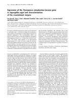
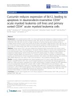
Figure 1. Immunohistochemistry staining for p-Ser-STAT3 in upper tract urothelial carcinoma (UTUC). (A) Low p-Ser-STAT3; (B) High p-Ser-STAT3; (C) negative
control.
Statistical Analysis
Chi-square analysis or Fisher’s exact test were
used to evaluate p-Ser-STAT3 expression in patients
with different age (dichotomized by medium),
gender, tumor stage and grade (low or high),
creatinine level, and hemodialysis. Hazard ratios
(HRs) and 95% confidence intervals (CIs) computed
from univariate and multivariate Cox regression
models were used to investigate the relationship
between clinicopathological characteristics and
survival. Survival analysis was estimated according to
the Kaplan–Meier method from the date of primary
tumor surgery to the time of recurrence of cancer or
death from cancer, and the significance of differences
between curves was evaluated by the log-rank test.
Results were considered statistically significant if the
p-value was less than 0.05. The data were analyzed
using the SPSS package (version 20.0, SPSS, Inc.,
Chicago, IL, USA); all p-values were two-sided.
Results
UTUC specimens were obtained from 100
patients with a male-to-female ratio of 1.04 : 1.00. Of
these, 62 patients had organ-confined T stage (T1/T2)
cancer and 38 had locally advanced T stage (T3/T4)
cancer. The cancer grade was low in 33 patients and
high in 67 patients. Two-thirds of the patients were
over 60 years old. Less than half of the patients had
abnormal creatinine levels, and only a small
proportion (16%) needed hemodialysis for end stage
renal disease. Activated STAT3 was identified by
detection of the phosphorylated form of the protein,
p-Ser-STAT3.
Figure
1
shows
the
immunohistochemistry staining of p-Ser-STAT3 in
UTUC samples. The patients were stratified into two
groups based on low or high p-Ser-STAT3 expression.
The
clinical
parameters
and
pathological
characteristics of the UTUC patients are summarized
in Table 1.
Univariate analysis for recurrence-free survival
demonstrated that men had higher risk for tumor
recurrence (Hazard Ratio [HR] = 2.13, 95% confidence
interval [CI] = 1.06-4.31). Both high grade and late
stage were associated with increased risk of tumor
recurrence (HR = 5.09 and 2.63, respectively). In
addition, high p-Ser-STAT3 expression was associated
with decreased recurrence-free survival (HR = 2.4,
95% CI = 1.13-5.08). However, following multivariate
analysis, only sex and tumor grade were associated
with a significantly higher risk of tumor recurrence
(Table 2). When patients were stratified based on
cancer stage, late stage patients (T3/T4) with high
p-Ser-STAT3 protein expression had decreased
recurrence-free survival in both univariate (HR = 4.31,
p = 0.023) and multivariate (HR = 5.91, p = 0.01)
analyses compared to patients with low p-Ser-STAT3
expression (Table 3).
Table 1. Clinicopathological characteristics of patients with upper
tract urothelial carcinoma and association with p-Ser-STAT3
expression.
Variable
No.
Stage
T1/T2
T3/T4
Grade
Low
High
Gender
Male
Female
Age (years)
<65
≧65
Hemodialysis
No
Yes
Creatinine (mg/dl)
≦1.5
>1.5
Recurrence status
No
Yes
Survival
No
Yes
a
b
p-Ser-STAT3
Low
High
Patient, no. (%) n (%)
n (%)
100 (100.0)
48 (48.0)
52 (52.0)
p-Value
62 (62.0)
38 (38.0)
35 (72.9)
13 (27.1)
27 (51.9)
25 (48.1)
0.031a
33 (33.0)
67(67.0)
19 (39.6)
29 (60.4)
14 (26.9)
38 (73.1)
0.179a
51 (51.0)
49 (49.0)
26 (54.2)
22 (45.8)
25 (48.1)
27 (51.9)
0.543a
34 (34.0)
66 (66.0)
14 (29.2)
34 (70.8)
20 (38.5)
32 (61.5)
0.327a
84 (84.0)
16 (16.0)
37 (77.1)
11 (22.9)
47 (90.4)
5 (9.6)
0.070a
57 (57.0)
43 (43.0)
24 (50.0)
24 (50.0)
33 (63.5)
19 (36.5)
0.174a
68 (68.0)
32 (32.0)
38 (79.2)
10 (20.8)
30 (57.7)
22 (42.3)
0.021a
17 (17.0)
83 (83.0)
4 (8.3)
44 (91.7)
13 (25.0)
39 (75.0)
0.034b
p by the chi-square test.
p by the Fisher’s exact test
Int. J. Med. Sci. 2017, Vol. 14
1363
Table 2. Univariate and multivariate analysis of recurrence-free
survival for patients with upper tract urothelial carcinoma.
Variable
Hazard
ratio
Stage
T3/T4
T1/T2
Grade
High
Low
Gender
Male
Female
Age (years)
≧65
<65
Hemodialysis
Yes
No
Creatinine
(mg/dl)
>1.5
≦1.5
Chemotherapy
Yes
Univariate
95%
Confidence
interval
p-Val
ue
Multivariate*
Hazard 95%
p-Value
ratio
Confidence
interval
2.63
1.00
(1.33-5.23)
0.006 2.81
1.00
(1.21-6.51)
0.016
5.09
1.00
(1.78-14.55)
0.002 2.37
1.00
(0.74-7.57)
0.144
2.13
1.00
(1.06-4.31)
0.035 1.68
1.00
(0.74-3.84)
0.215
0.95
1.00
(0.47-1.92)
0.891 1.14
1.00
(0.49-2.63)
0.767
0.72
1.00
(0.25-2.04)
0.535 1.92
1.00
(0.50-7.39)
0.343
expression was the only risk factor associated with
cancer-specific survival in both univariate (HR = 8.13,
p = 0.045) and multivariate analyses (HR = 8.83, p =
0.039) (Table 5).
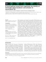
Kaplan-Meier survival curves demonstrate that
low p-Ser-STAT3 expression was associated with a
significantly higher recurrence-free survival (p =
0.018) (Figure 2(A)) and cancer-specific survival (p =
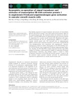
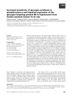
0.026) (Figure 2(B)). For patients with late stage
(T3/T4) disease, high p-Ser-STAT3 expression was
associated with a significantly lower recurrence-free
survival (p = 0.013) (Figure 3(B)) and cancer-specific
survival (p = 0.0016) (Figure 4(B)). However, this
association was not observed in patients with early
stage (T1/T2) disease (Figures 3(A) and 4(B)).
Discussion
As described above, we reported several
proteins served as prognostic factors in UTUC
patients, including COX2, OPN, HIF-1α, GST, and
NFκB. Overexpression of COX2, OPN, HIF-1α and
6.40
(3.12-13.13)
<0.00 5.34
(2.39-11.93) <0.001
1
NFκB were associated with poor cancer-specific
No
1.00
1.00
survival, whereas overexpression of COX2 and
p-Ser-STAT3
High
2.40
(1.13-5.08)
0.022 2.19
(1.00-4.81)
0.051
HIF-1α could predict shorter recurrence free survival.
Low
1.00
1.00
In gastric cancer, patients with high levels of
*Multivariate Cox regression model was adjusted for stage, grade, gender, age,
STAT3
often experience worse outcomes, with a
hemodialysis, creatinine and chemotherapy.
meta-risk for overall survival (risk ratio, RR = 1.845)
[17]. In non-small-cell lung cancer, high STAT3 or
phospho-STAT3
expression is also a strong predictor
Univariate analysis also identified cancer stage
for
poor
prognosis
[18]. High nuclear expression of
and p-Ser-STAT3 expression as being associated with
STAT3 was found to be correlated with poor overall
cancer-specific survival (Table 4). Late stage and high
survival (P = 0.005) in diffuse large B-cell lymphoma
p-Ser-STAT3 expression were associated with a
[19]. Our data indicated that p-Ser-STAT3 expression
significant decrease in the cancer-specific survival
was
also associated with recurrence and survival rates
(HR = 6.28 and HR = 3.32, respectively). Following
in
UTUC
patients. Patients with high p-Ser-STAT3
multivariate analysis, only cancer stage remained a
expression
had a higher risk for cancer recurrence and
significant risk factor for cancer-specific survival (HR
a
lower
cancer-specific
survival. When patients were
= 6.03, p = 0.003). In patients with late stage (T3/T4)
stratified
into
early
and
advanced stage groups, there
UTUC, regardless of cancer grade, high p-Ser-STAT3
were no significant
differences
in
recurrence-free
and
cancer-specific survival
between low and high
p-Ser-STAT3 expression
in early stage patients.
However, in advanced
stage patients, high
p-Ser-STAT3 expression
was associated with a
significantly
poorer
prognosis,
higher
cancer recurrence rate,
and
lower
cancerFigure 2. Kaplan-Meier survival curves for recurrence-free (A) and cancer-specific (B) survival of patients with low and high
p-Ser-STAT3 expression in all cases of upper tract urothelial carcinoma.
specific survival. This
study provides the first
0.73
1.00
(0.36-1.48)
0.385
0.70
1.00
(0.28-1.72)
0.436
Int. J. Med. Sci. 2017, Vol. 14
1364
evidence
suggesting
a
correlation
between
p-Ser-STAT3 expression and UTUC.
STAT3 is a transcription factor with important
roles in cancer formation and progression. STAT3 is
involved in several cellular mechanisms, such as
proliferation, inhibition of apoptosis, immune escape,
epithelial-mesenchymal transition, invasion, and
angiogenesis [20]. In addition, STAT3 is involved in
cellular invasion through regulation of matrix
metalloproteinases (MMPs) [21] and as first reported
by Sano and coworkers. Subsequently, in response to
surrounding tumor cell secretions, stromal cells
upregulate SDF-1/CXCL12 receptors, resulting in
infiltration of endothelial progenitor cells and
enhancing the metastatic spread of tumor cells [22].
Under hypoxic conditions, both STAT3 and HIF1-α
bind simultaneously to the VEGF promoter, leading
to maximum transcriptional activation and
angiogenesis. Increased VEGF expression, due to high
activation of STAT3, induces faster cancer cell
proliferation, and promotes distant metastasis [23].
STAT3, a latent self-signaling transcription factor, has
been implicated as the hallmark of tumor invasion
and metastasis in a wide variety of human
malignancies. Activation of STAT3 is achieved by
phosphorylation of two sites, tyrosine 705 and serine
727 [24]. Tyrosine 705 phosphorylation is mediated by
Jak1, Jak2 and Tyk2, leading to STAT3
homodimerization,
nuclear
translocation
and
downstream transcriptional activities. Serine 727
phosphorylation is mediated by ERK1, ERK2, p38,
JNK and MAP, which is required for the full
activation of STAT3 [25]. Consistent with these
studies, we have shown that high p-Ser-STAT3
expression in UTUC tissues significantly correlated
with advanced cancer stage and predicting poor
prognosis in advanced cancer stage patients. The
present investigation revealed that p-Ser-STAT3
overexpression maybe be a useful biomarker to
predict disease invasion and metastasis.
On the other hand, inhibiting STAT3 signaling in
endothelial cells prohibits cell migration and vessel
formation [26]. As metastatic tumor cells enter the
blood vessels, they are affected by various nonspecific
forces such as mechanical stress, hemodynamic
turbulence, loss of adhesion-induced cell death, and
cell-mediated cytotoxicity. As a result, very few tumor
cells survive and metastasize. STAT3 activation also
plays a major role in
protecting tumor cells
from
immune
surveillance during their
transit
through
the
circulatory system [27]. It
is
not
surprising,
therefore, that advanced
cancer with lymph-node
or
organ
metastasis
requires
the
specific
ability for cell survival
with regard to the
immune system. The
interplay between STAT3
Figure 3. Kaplan-Meier survival curves for recurrence-free survival of patients with low and high p-Ser-STAT3 expression
in stage T1/T2 (A) and stage T3/T4 (B) groups in upper tract urothelial carcinoma.
in cancer and immune
cells in the tumor
microenvironment
is
very
complex
and
remains elusive. Studies
have
shown
that
constitutively
active
STAT3 signaling recruits
immune cells and inhibits
their
function
by
increasing
suppressive
agents
[28].
STAT3
mediates
bidirectional
communication
with
immune cells and is a
Figure 4. Kaplan-Meier survival curves for cancer-specific survival of patients with low and high p-Ser-STAT3 expression
potent negative regulator
in stage T1/T2 (A) and stage T3/T4 (B) groups in upper tract urothelial carcinoma.
Int. J. Med. Sci. 2017, Vol. 14
1365
of T1 helper cells. In addition, inflammatory cytokines
released from tumors, such as IL6 and IL10, are
responsible for the partial differentiation of dendritic
cells, thereby reducing their antigen presenting ability
[29]. By activating the IL-6/STAT3 pathway, the
tumor microenvironment promotes tumorigenesis
and invasion. Finally, the activity of NK cells also is
reduced, thereby protecting circulating tumor cells
[30]. The above activities promote the proliferation of
cancer cells with a more malignant behavior and
contribute to life-threatening disease.
cancers because oncogenesis is known to be a
multigenic process. Future research focused on the
analysis of outcomes and patient prognosis following
different targeted therapies in UTUC is needed.
Table 4. Univariate and multivariate analysis of cancer-specific
survival for patients with upper tract urothelial carcinoma.
Univariate
Hazard 95%
p-Value
ratio
Confidence
interval
Hazard
ratio
Stage
T3/T4
6.28
7.28
Table 3. Univariate and multivariate analysis of recurrence-free
survival for patients with stage T3/T4 in upper tract urothelial
carcinoma.
T1/T2
1.00
High
3.15
Variable
Low
Gender
1.00
Male
1.04
Female
Age (years)
1.00
≧65
0.68
<65
Hemodialysis
1.00
Yes
0.83
No
1.00
Creatinine
(mg/dl)
>1.5
0.87
Univariate
Hazard 95%
p-Value
ratio
Confidence
interval
Multivariate*
Hazard 95%
p-Value
ratio
Confidence
interval
High
5.11
5.43
Low
Gender
1.00
Male
2.48
Female
Age (years)
1.00
≧65
0.55
Grade
<65
Hemodialysis
1.00
Yes
0.41
No
1.00
Creatinine
(mg/dl)
>1.5
0.88
(0.67-38.82) 0.115
(0.64-45.95) 0.120
1.00
(0.96-6.41)
0.062
1.58
(0.49-5.09)
0.446
1.00
(0.22-1.39)
0.208
0.58
(0.19-1.74)
0.327
1.00
(0.06-3.12)
0.392
1.95
(0.19-20.03) 0.574
1.00
(0.33-2.34)
0.795
0.80
(0.25-2.53)
0.697
≦1.5
1.00
Chemotherapy
Yes
4.94
No
1.00
p-Ser-STAT3
1.00
(1.83-13.38)
0.002
3.80
1.00
(1.08-13.43) 0.038
High
4.31
(1.23-15.11) 0.023
6.57
(1.71-25.26) 0.006
Low
1.00
1.00
*Multivariate Cox regression model was adjusted for grade, gender, age,
hemodialysis, creatinine and chemotherapy.
Constitutive activation of STAT3 plays a critical
role in the initiation, progression, and metastases of
cancers. Therefore, inhibition of STAT3 could be a
strategy for cancer treatment. Numerous approaches,
such as anti-sense oligonucleotide targeting of STAT3,
synthetic drugs, small molecules derived from natural
sources, and gene therapy techniques are available to
achieve this goal [31]. In theory, targeting a single
molecular mechanism may be sufficient to be
therapeutically effective. However, in practice
single-target drugs have had little therapeutic impact
and are generally highly ineffective in treating cancer
[32]. An approach targeting multiple genes or
pathways is likely to be particularly beneficial in
Variable
(2.26-17.49)
<0.001
Multivariate*
95%
p-Value
Confidence
interval
(1.82-19.94) 0.002
1.00
Grade
(0.92-10.84)
0.068
1.31
(0.47-6.36)
0.700
(0.34-2.85)
0.601
(0.33-2.47)
0.956
(0.22-7.07)
0.829
(0.34-3.93)
0.726
1.00
(0.42-2.57)
0.927
0.73
1.00
(0.28-1.70)
0.415
1.03
1.00
(0.19-3.59)
0.799
1.21
1.00
(0.34-2.21)
0.770
1.24
≦1.5
1.00
Chemotherapy
Yes
1.71
No
1.00
p-Ser-STAT3
1.00
(0.65-4.48)
0.279
2.18
1.00
(0.64-7.46)
0.215
High
3.32
(1.08-10.18)
0.036
2.65
(0.83-8.46)
0.099
Low
1.00
1.00
*Multivariate Cox regression model was adjusted for stage, grade, gender, age,
hemodialysis, creatinine and chemotherapy.
In this study, while stage, grade, and sex were
useful predictors of prognosis, high p-Ser-STAT3
expression was a particularly useful marker to predict
cancer recurrence and survival. Moreover, high
p-Ser-STAT3 expression was the most useful tool for
predicting outcome in advanced UTUC. However,
there are some limitations to this study. First, this is a
retrospective analysis and it neglected the smoking
behavior of patients in the subgroup analysis. It has
been reported that nicotine can activate STAT3 in
bladder cells, thereby affecting cyclin D1 expression
and promoting cell proliferation [33]. This
confounding factor needs to be addressed in future
research. Second, although there was a significant
correlation between p-Ser-STAT3 expression and poor
outcome, the sample size in this study is small. A
multi-institutional study with a larger sample size is
needed to validate our conclusions. Finally, easily
accessible samples such as blood, urine, or even those
Int. J. Med. Sci. 2017, Vol. 14
1366
obtained
through
minimally
invasive
ureterorenoscopy tumor biopsies could be analyzed
in the future to determine whether similar results are
observed regarding the correlation between
expression of p-Ser-STAT3 and UTUC patient
outcomes. In summary, p-Ser-STAT3 is observed in
UTUC and is positively correlated with advanced
cancer
stage.
Moreover,
over-expression
of
p-Ser-STAT3 is predictive of recurrence and survival
and may be indicative of the need for an aggressive
treatment
plan.
Furthermore,
inhibition
of
p-Ser-STAT3 may provide a new therapeutic
approach for treatment of advanced UTUC.
Competing Interests
The authors have declared that no competing
interest exists.
References
1.
2.
3.
4.
5.
Table 5. Univariate and multivariate analysis of cancer-specific
survival for patients with stage T3/T4 in upper tract urothelial
carcinoma.
Variable
Hazard
ratio
Univariate
95%
p-Value
Confidence
interval
Multivariate*
Hazard 95%
p-Value
ratio
Confidence
interval
(0.39-23.36) 0.289
2.80
Grade
High
3.02
Low
Gender
1.00
Male
1.12
Female
Age (years)
1.00
≧65
0.83
<65
Hemodialysis
1.00
Yes
0.70
No
1.00
Creatinine
(mg/dl)
>1.5
0.842
0.92
0.737
0.98
(0.22-3.87)
0.912
0.735
1.05
10.
11.
12.
(0.23-4.13)
0.978
1.00
(0.09-5.42)
8.
9.
1.00
(0.28-2.48)
7.
(0.32-24.57) 0.354
1.00
(0.38-3.33)
6.
13.
(0.09-12.01) 0.968
1.00
14.
1.06
(0.33-3.46)
0.918
≦1.5
1.00
Chemotherapy
Yes
1.42
No
1.00
p-Ser-STAT3
(0.44-4.61)
High
8.13
(1.05-62.90) 0.045
Low
1.00
1.37
(0.26-7.33)
0.716
1.18
1.00
(0.22-6.42)
0.849
8.93
(1.12-71.30) 0.039
15.
1.00
0.563
16.
1.00
*Multivariate Cox regression model was adjusted for grade, gender, age,
hemodialysis, creatinine and chemotherapy.
Acknowledgment
This study was supported by grants from
Kaohsiung Medical University “Aim for the Top
Universities” (KMU-TP104E31, KMU-TP105G00,
KMU-TP105G01 and KMU-TP105G025), The Health
and Welfare Surcharge of Tobacco Products, Ministry
of Health and Welfare (MOHW106-TDU-B-212
-144007), Ministry of Science and Technology
(MOST106-2314-B-037-092), and Kaohsiung Medical
University Hospital (KMUH102-2R42 and KMUH1033T11).
17.
18.
19.
20.
21.
22.
23.
24.
Roupret M, Babjuk M, Comperat E, Zigeuner R, Sylvester R, Burger M, et al.
European guidelines on upper tract urothelial carcinomas: 2013 update.
European urology. 2013; 63: 1059-71.
Yeh HC, Jan HC, Wu WJ, Li CC, Li WM, Ke HL, et al. Concurrent Preoperative
Presence of Hydronephrosis and Flank Pain Independently Predicts Worse
Outcome of Upper Tract Urothelial Carcinoma. PLoS One. 2015; 10: e0139624.
Green DA, Rink M, Xylinas E, Matin SF, Stenzl A, Roupret M, et al. Urothelial
carcinoma of the bladder and the upper tract: disparate twins. The Journal of
urology. 2013; 189: 1214-21.
Ke HL, Tu HP, Lin HH, Chai CY, Chang LL, Li WM, et al. Cyclooxygenase-2
(COX-2) up-regulation is a prognostic marker for poor clinical outcome of
upper tract urothelial cancer. Anticancer research. 2012; 32: 4111-6.
Ke HL, Chang LL, Yang SF, Lin HH, Li CC, Wu DC, et al. Osteopontin
overexpression predicts poor prognosis of upper urinary tract urothelial
carcinoma. Urologic oncology. 2011; 29: 703-9.
Ke HL, Wei YC, Yang SF, Li CC, Wu DC, Huang CH, et al. Overexpression of
hypoxia-inducible factor-1alpha predicts an unfavorable outcome in urothelial
carcinoma of the upper urinary tract. International journal of urology : official
journal of the Japanese Urological Association. 2008; 15: 200-5.
Chen SH, Wu WJ, Tu HP, Li WM, Huang CN, Li CC, et al. Glutathione
S-transferase expression in upper urinary tract urothelial carcinomas: a
Taiwan study. Asian Pacific journal of cancer prevention : APJCP. 2013; 14:
6475-9.
Yeh HC, Huang CH, Yang SF, Li CC, Chang LL, Lin HH, et al. Nuclear
factor-kappaB activation predicts an unfavourable outcome in human upper
urinary tract urothelial carcinoma. BJU international. 2010; 106: 1223-9.
Wang X, Crowe PJ, Goldstein D, Yang JL. STAT3 inhibition, a novel approach
to enhancing targeted therapy in human cancers (review). Int J Oncol. 2012; 41:
1181-91.
Bromberg JF, Wrzeszczynska MH, Devgan G, Zhao Y, Pestell RG, Albanese C,
et al. Stat3 as an oncogene. Cell. 1999; 98: 295-303.
Darnell JE, Jr. STATs and gene regulation. Science. 1997; 277: 1630-5.
Nkansah E, Shah R, Collie GW, Parkinson GN, Palmer J, Rahman KM, et al.
Observation of unphosphorylated STAT3 core protein binding to target
dsDNA by PEMSA and X-ray crystallography. FEBS Lett. 2013; 587: 833-9.
Dolled-Filhart M, Camp RL, Kowalski DP, Smith BL, Rimm DL. Tissue
microarray analysis of signal transducers and activators of transcription 3
(Stat3) and phospho-Stat3 (Tyr705) in node-negative breast cancer shows
nuclear localization is associated with a better prognosis. Clinical cancer
research : an official journal of the American Association for Cancer Research.
2003; 9: 594-600.
He G, Karin M. NF-kappaB and STAT3 - key players in liver inflammation and
cancer. Cell Res. 2011; 21: 159-68.
Masuda M, Suzui M, Yasumatu R, Nakashima T, Kuratomi Y, Azuma K, et al.
Constitutive activation of signal transducers and activators of transcription 3
correlates with cyclin D1 overexpression and may provide a novel prognostic
marker in head and neck squamous cell carcinoma. Cancer Res. 2002; 62:
3351-5.
Huang WT, Yang SF, Wu CC, Chen WT, Huang YC, Su YC, et al. Expression of
signal transducer and activator of transcription 3 and suppressor of cytokine
signaling 3 in urothelial carcinoma. The Kaohsiung journal of medical
sciences. 2009; 25: 640-6.
Chen J, Liu X, Jiao H, Peng L, Huo Z, Yang W, et al. Prognostic and clinical
significance of STAT3 and MMP9 in patients with gastric cancer: a
meta-analysis of a Chinese cohort. Int J Clin Exp Med. 2015; 8: 546-57.
Xu YH, Lu S. A meta-analysis of STAT3 and phospho-STAT3 expression and
survival of patients with non-small-cell lung cancer. Eur J Surg Oncol. 2014;
40: 311-7.
Wu ZL, Song YQ, Shi YF, Zhu J. High nuclear expression of STAT3 is
associated with unfavorable prognosis in diffuse large B-cell lymphoma. J
Hematol Oncol. 2011; 4: 31.
Yu H, Jove R. The STATs of cancer--new molecular targets come of age. Nat
Rev Cancer. 2004; 4: 97-105.
Xie T-x, Wei D, Liu M, Gao AC, Ali-Osman F, Sawaya R, et al. Stat3 activation
regulates the expression of matrix metalloproteinase-2 and tumor invasion
and metastasis. Oncogene. 2004; 23: 3550-60.
Nguyen-Jackson H, Panopoulos AD, Zhang H, Li HS, Watowich SS. STAT3
controls the neutrophil migratory response to CXCR2 ligands by direct
activation of G-CSF–induced CXCR2 expression and via modulation of
CXCR2 signal transduction. Blood. 2010; 115: 3354-63.
Oh M-K, Park H-J, Kim N-H, Park S-J, Park I-Y, Kim I-S. Hypoxia-inducible
factor-1α enhances haptoglobin gene expression by improving binding of
STAT3 to the promoter. Journal of Biological Chemistry. 2011; 286: 8857-65.
Bishop JL, Thaper D, Zoubeidi A. The Multifaceted Roles of STAT3 Signaling
in the Progression of Prostate Cancer. Cancers (Basel). 2014; 6: 829-59.
Int. J. Med. Sci. 2017, Vol. 14
1367
25. Yang R, Rincon M. Mitochondrial Stat3, the Need for Design Thinking. Int J
Biol Sci. 2016; 12: 532-44.
26. Yahata Y, Shirakata Y, Tokumaru S, Yamasaki K, Sayama K, Hanakawa Y, et
al. Nuclear translocation of phosphorylated STAT3 is essential for vascular
endothelial growth factor-induced human dermal microvascular endothelial
cell migration and tube formation. Journal of Biological Chemistry. 2003; 278:
40026-31.
27. Nguyen DX, Bos PD, Massagué J. Metastasis: from dissemination to
organ-specific colonization. Nature Reviews Cancer. 2009; 9: 274-84.
28. Groner B, Lucks P, Borghouts C. The function of Stat3 in tumor cells and their
microenvironment. Seminars in cell & developmental biology: Elsevier;
2008:341-50.
29. Yu H, Pardoll D, Jove R. STATs in cancer inflammation and immunity: a
leading role for STAT3. Nat Rev Cancer. 2009; 9: 798-809.
30. Wang T, Niu G, Kortylewski M, Burdelya L, Shain K, Zhang S, et al.
Regulation of the innate and adaptive immune responses by Stat-3 signaling in
tumor cells. Nature medicine. 2003; 10: 48-54.
31. Siveen KS, Sikka S, Surana R, Dai X, Zhang J, Kumar AP, et al. Targeting the
STAT3 signaling pathway in cancer: Role of synthetic and natural inhibitors.
Biochimica et Biophysica Acta (BBA)-Reviews on Cancer. 2014; 1845: 136-54.
32. Giordano S, Petrelli A. From single-to multi-target drugs in cancer therapy:
when aspecificity becomes an advantage. Current medicinal chemistry. 2008;
15: 422-32.
33. Chen R-J, Ho Y-S, Guo H-R, Wang Y-J. Rapid activation of Stat3 and ERK1/2
by nicotine modulates cell proliferation in human bladder cancer cells.
Toxicological Sciences. 2008; 104: 283-93.