Hepatocyte growth factor plays a particular role in progression of overall cardiac damage in experimental pulmonary hypertension
Bạn đang xem bản rút gọn của tài liệu. Xem và tải ngay bản đầy đủ của tài liệu tại đây (1.07 MB, 10 trang )
Int. J. Med. Sci. 2019, Vol. 16
Ivyspring
International Publisher
854
International Journal of Medical Sciences
2019; 16(6): 854-863. doi: 10.7150/ijms.31690
Research Paper
Hepatocyte growth factor plays a particular role in
progression of overall cardiac damage in experimental
pulmonary hypertension
Michal Radik, Zuzana Kmecova, Jana Veteskova, Eva Malikova, Gabriel Doka, Peter Krenek, Jan Klimas
Department of Pharmacology and Toxicology, Faculty of Pharmacy, Comenius University in Bratislava, Odbojarov 10, 83232 Bratislava, Slovak Republic
Corresponding author: Peter Krenek, Department of Pharmacology and Toxicology, Faculty of Pharmacy, Comenius University in Bratislava, Odbojarov 10,
83232 Bratislava, Slovak Republic. E-mail:
© Ivyspring International Publisher. This is an open access article distributed under the terms of the Creative Commons Attribution (CC BY-NC) license
( See for full terms and conditions.
Received: 2018.11.21; Accepted: 2019.03.27; Published: 2019.06.02
Abstract
Background: HGF/MET pathway may have a role in pulmonary hypertension (PH). However, the link
between the pathway and development of target organ damage in PH remains elusive. We aimed to
demonstrate the relation between plasma HGF and HGF/MET tissue expressions in affected organs
during PH progression.
Methods: 12 weeks old male Wistar rats were injected with monocrotaline (MCT, 60 mg/kg, s.c.) to
induce PH and sacrificed after 1, 2 and 4 weeks. Controls received saline. mRNA levels of HGF regulatory
complex (Hgf, Met, Hgfa, Hai-1, Hai-2) were determined in right and left ventricles (RV, LV), lungs,
pulmonary artery and liver by RT-qPCR. HGF protein levels in plasma were analysed by ELISA.
Results: PH development was associated with a progressive elevation of HGF plasma levels that
correlated with relative RV mass. Furthermore, Hgf mRNA expressions at week 4 were upregulated
solely in the cardiac ventricles while being downregulated in a. pulmonalis, lungs and liver. Met and
Hai-1/Hai-2 followed a similar pattern and were upregulated in cardiac ventricles, where Hgfa remained
unchanged, but downregulated in lungs.
Conclusion: We suggest that cardiac overexpression of Hgf might contribute to increased plasma HGF
in MCT-induced PH. HGF could be exploited as a cardiospecific biomarker and HGF/MET pathway as a
target in drug discovery for PH.
Key words: HGF; MET receptor; pulmonary hypertension; monocrotaline; biomarker
Introduction
Pulmonary hypertension (PH) is a rare,
progressive disease with poor prognosis and limited
therapeutic options [1]. It is assessed by functional
tests [1], but a reliable and specific prognostic
biomarker is lacking. PH pathogenesis involves
processes, which progressively increase pulmonary
vascular resistance leading to myocardial remodeling
and failure of the right ventricle (RV) [2]. This
outcome is driven by alterations in cytokines and
growth factors [3], including the hepatocyte growth
factor (HGF) that via its receptor MET
(mesenchymal-epithelial
transition)
promotes
proliferation and morphogenesis as well as
anti-apoptotic and anti-fibrotic effects on various
types of cells, including cardiomyocytes [4]. HGF
activity is managed by its set of endogenous
regulators, namely HGF activator [5], and respective
inhibitors of HGF activator, HAI-1 [6] and HAI-2 [7].
HGFA is the main factor responsible for splitting
pro-HGF form to a mature, biologically active HGF
protein [8], while HAI-1/HAI-2 are binding HGFA in
an intrinsic inhibitory mechanism [6, 7]. HGF/MET
are involved in tissue repair [9]. In diseases like
pulmonary arterial hypertension (PAH) [10], lung
fibrosis [11], myocardial infarction [12], and heart
failure [13], exogenous HGF or gene transfer was
protective and attenuated disease progression. HGF
Int. J. Med. Sci. 2019, Vol. 16
activates pathways known to be involved in PH
pathophysiology and target organ damage [14].
Since PH is often diagnosed only at an advanced
stage of disease [15], the lack of specific biomarker for
detection of early stages of PH represents a crucial
problem. HGF has been suggested as a potential
predictor of mortality in heart failure patients [16],
hypertension severity [17] and could also be relevant
in diagnostics of PH [18]. HGF plasma levels
correlated with mean pulmonary arterial pressure
and HGF was detectable already at early stages of the
disease [19], making it a potential biomarker
candidate. However, its relation to target organ
damage during disease progression remains unclear.
We hypothesize, that HGF/MET system
dysregulation could reflect development and
progression of target organ damage in PH. We also
hypothesize, that such a dysregulation would be
accompanied by elevated plasma HGF levels when
right ventricular pressure (RVP) increases. We aim to
evaluate the significance of plasma levels of HGF as a
potential PH biomarker and to investigate a possible
link of HGF plasma level alteration to Hgf and Met
gene expressions in affected tissues in the
monocrotaline (MCT)-induced rat model of PH.
Materials and Methods
Animal experiment design
12 weeks old male Wistar rats (Dobra Voda,
Slovak Republic) were randomized according to
experiment duration: 1, 2, 4 weeks (1W, 2W, 4W) and
type of treatment (saline - CON, monocrotaline MCT). Rats were subcutaneously injected with 60
mg/kg dose of monocrotaline [20], or saline. In
experimental model of MCT-induced PH in rat, males
are preferred over females as they are more
susceptible to MCT toxicity than female rats [21].
Animals were sacrificed 1, 2 and 4 weeks after MCT
injection, to study different PH progression stages.
Procedures involving the use of animals were
approved by the Ethics Committee of the Faculty of
Pharmacy, Comenius University in Bratislava, Slovak
Republic and the State Veterinary and Food
Administration of the Slovak Republic. The
investigations were conducted in accordance with
NIH Guide for the Care and Use of Laboratory
Animals: Eight Edition (2010) published by the US
Committee for the Update of the Guide for the Care
and Use of Laboratory Animals; National Research
Council, the EU adopted Directive 2010/63/EU of the
European Parliament and of the Council on the
protection of animals used for experimental and other
scientific purposes and the Slovak law regulating
animal experiments.
855
Right ventricular pressure measurement
RVP was measured by RV catheterization using
a polyethylene catheter, filled with heparinized saline
and connected to the pressure transducer (Spel
Advanced HaemoSys, Experimetria Ltd., Hungary).
The catheter was advanced into the RV via right
jugular vein under tribromoethanol anaesthesia [22].
Collection of samples
Rats were sacrificed in CO2 atmosphere. Blood
was collected from caudal vena cava using EDTA as an
anticoagulant,
plasma
was
separated
by
centrifugation and stored at -80 °C. Cardiac ventricles,
lungs and livers were blotted dry and weighed.
Relative organ weights were calculated compared to
total body weights and ratios were used as measures
of organ damage. Samples of LV, RV, a. pulmonalis,
lungs and liver were frozen in liquid nitrogen and
stored at -80 °C until further processing.
ELISA
To measure HGF protein plasma concentrations,
the
Quantikine
ELISA
Mouse/Rat
HGF
Immunoassay MHG00 (R&D Systems, USA) was used
according to the manufacturer’s instructions. The
assay uses a quantitative sandwich enzyme
immunoassay technique and detects natural and
recombinant HGF, with less than 0.5% cross-reactivity
and no significant interference with related molecules.
RT-qPCR
Total RNA was isolated from tissues using Tri
Reagent (Sigma-Aldrich, USA). RNA quality was
verified by gel electrophoresis and quantified by
spectrophotometry (NanoDrop ND-1000, Thermo
Fisher Scientific, USA). Reverse transcription was
performed using High Capacity cDNA Reverse
Transcription Kit with RNAse inhibitor (Thermo
Fisher Scientific, USA). Quantitative real-time PCR
was performed using SYBR Select Master Mix
(Thermo Fisher Scientific, USA) on StepOnePlus
Real-Time PCR System (Thermo Fisher Scientific,
USA). Hgf, Met, Hgfa, Hai-1, Hai-2, Nppa, and Nppb
mRNA levels were evaluated using gene-specific
primers (Sigma-Aldrich, USA) verified to yield a
single PCR product with a correct length (Table 1).
Results were normalized to expression of reference
genes beta-2-microglobulin (B2m) and hypoxanthine
phosphoribosyltransferase 1 (Hprt1) and calibrated to
appropriate control groups [23].
Statistical analysis
Data are reported as mean ± standard error of
the mean. Data distribution was determined by
Shapiro-Wilk normality test. Means were compared
Int. J. Med. Sci. 2019, Vol. 16
856
by unpaired Student's t-test for normally distributed
data or non-parametric Mann-Whitney U test for
nonparametric data, with P<0.05 considered
statistically significant. Mean PCR efficiency estimates
(E) per amplicon and quantification cycle (Cq) values
per sample were determined with LinRegPCR
software, version 2015.0 (Heart Failure Research
Center, NL). Relative gene expression ratios were
calculated using Pfaffl method [23]. Statistical
evaluations and correlations were performed using
GraphPad Prism, version 6 (GraphPad Software,
USA).
Table 1. Primer sequences for qRT-PCR.
Gene GenBank Access Primer Sequences (5’→3’)
Name No.
B2m1
Hai-11
Hai-21
Hgf1
Hgfa1
Hprt11
Met1
Nppa1
Nppb1
NM_012512.1
PCR
product
size (bp)
105
forward: ATGGAGCTCTGAATCATCTGG
reverse: AGAAGATGGTGTGCTCATTGC
NM_001004265.2 forward: ACACGCCTGACTGCCCTGAT
128
reverse: TCCAGTGTCTGGCAGCTCTGCA
NM_199087.1
forward: CCTAGACGTCCACGAGAACACCAT 150
reverse: CCAGTGACCGCCTTTGGGACA
NM_017017.2
forward: TCAGCGCTGGGATCAGCAGACA
122
reverse: TGTAGCACCATGGCCTCGGCTT
NM_053320.1
forward: TGTGCTGGGAGCCTCGTCCA
130
reverse: GTGTCACATCCGTCGTGCGGT
NM_012583.2
forward: CAGCTTCCTCCTCAGACCGCTTT
150
reverse: TCACTAATCACGACGCTGGGACTG
NM_031517.1
forward: GTGGCTGGTGCCACAATCGGT
150
reverse: TCCTCCTTCGAGGGGTGCACT
NM_012612.2
forward: GGGGGTAGGATTGACAGGAT
104
reverse: GGATCTTTTGCGATCTGCTC
NM_031545.1
forward: GACCGGATCGGCGCAGTCAGT
78
reverse: GGAGTCTGCAGCCAGGAGGTCT
1 B2m: beta-2-microglobulin, Hai-1: hepatocyte growth factor activator inhibitor 1,
Hai-2: hepatocyte growth factor activator inhibitor 2, Hgf: hepatocyte growth
factor, Hgfa: hepatocyte growth factor activator, Hprt1: hypoxanthine
phosphoribosyltransferase 1, Met: mesenchymal-epithelial transition, Nppa:
natriuretic peptide A, Nppb: natriuretic peptide B.
Results
Progressive increase of RV weight and RV
pressure after MCT
Body weight in MCT groups decreased from
week 2 (-7%) to week 4 (-10%), both P<0.05. This was
accompanied by increased relative weights of RVs
and lungs at weeks 1, 2 and 4, respectively (all P<0.05;
see Table 2). For comparison, relative weights of LVs
were significantly increased in MCT groups solely at
week 4 (P<0.05). Relative liver weight increased at
week 2 and at week 4, both P<0.05 vs CON (Table 2).
This was accompanied by significant increase of RVP
in MCT-treated rats exclusively at week 4 (MCT 4W:
51±5 mmHg; CON 4W: 25±2 mmHg; P<0.05), while
RV function in MCT groups remained unaltered at
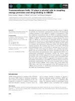
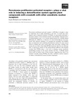
week 1 and 2. Natriuretic peptides genes, Nppa and
Nppb gene (natriuretic peptide A and B), were used
as markers of cardiac damage. We observed
unchanged ventricular expression of these genes at
week 1, but Nppb mRNA in RV was significantly
elevated 4.7-fold already at week 2, i.e. in the stage
with stable RV pressure. At the final week, expression
of both natriuretic peptides was significantly
increased in both heart ventricles (P<0.05, Figure 1),
though it was more pronounced in RV.
Table 2. Relative organ weights when related to their absolute
body weights (BW) of right ventricle (RV), left ventricle (LV), lung
and liver samples after 1, 2 and 4 weeks after MCT injection
(n=6-12 per group; mean±SEM; *P<0.05 vs. CON).
1W
CON
0.44±0.03
2W
MCT
CON
0.53±0.03* 0.52±0.02
MCT
0.62±0.02*
4W
CON
0.53±0.02
RV/BW
[mg/g]
LV/BW 2.16±0.05 2.14±0.06 2.01±0.07 2.00±0.03
2.18±0.04
[mg/g]
lung/BW 3.85±0.08 4.28±0.13* 4.91±0.21 6.97±0.64* 3.90±0.15
[mg/g]
liver/BW 30.08±0.58 31.09±0.57 28.76±0.55 33.07±0.97* 30.51±0.50
[mg/g]
MCT
1.02±0.06*
2.38±0.07*
8.32±0.44*
33.44±1.01*
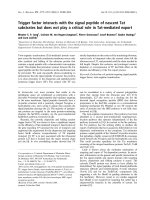
Progressive rise of plasma HGF
concentrations after MCT
One week after MCT we observed no change in
plasma HGF levels. At week 2, significant 3-fold
elevation was detected and this trend further
escalated to a 6-fold increase at week 4 (both P<0.05,
Figure 2).
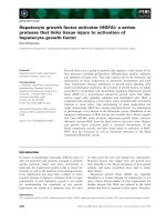
In the pooled group of MCT-treated rats, plasma
HGF levels correlated positively with RV/BW (but
not with LV/BW; see Figure 3). When divided to
subgroups according to disease progression, this
positive significant correlation between plasma HGF
and RV/BW was present in the group of 4W MCT
(Spearman r = 0.7857, P<0.05), but not in subgroups of
2W and 1W MCT (Spearman r = 0.1429, NS and
Spearman r = 0.3143, NS, respectively). In healthy
controls, we observed no relationship between plasma
HGF and RV/BW neither LV/BW, respectively. We
used RV/BW and LV/BW as a measures of organ
remodeling, but all findings were valid also when
using absolute RV and LV weights.
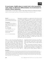
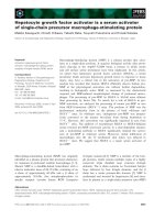
Increased cardiac Hgf mRNA expression at
week 4
At week 1, we observed a decrease of Hgf
mRNA expression in the RV (-11%, P<0.05) and lung
(-33%, P<0.05), no change in the LV and a. pulmonalis
and a significant increase (+46%, P<0.05) in liver. At
week 2, we observed unaltered expression in all
examined tissues except lung, where it remained
decreased (-24%, P<0.05). At week 4, mRNA level of
Hgf gene expression was increased in the left (+64%)
and right (+76%) ventricle and decreased in
a. pulmonalis (-29%), lungs (-19%) and liver (-16%), all
P<0.05 vs CON (Figure 4).
Int. J. Med. Sci. 2019, Vol. 16
857
Figure 1. Relative (when related to B2m and Hprt1) mRNA expression of Nppa gene after 1, 2 and 4 weeks after MCT injection in (A) right ventricle, (B) left ventricle, and Nppb
gene in (C) right ventricle (D) left ventricle samples (n=6-12 per group; mean±SEM; *P<0.05 vs. CON).
Figure 2. Relative plasma concentration of HGF protein after 1, 2 and 4 weeks after MCT injection (n=4-8 per group; mean±SEM; *P<0.05 vs. CON).
Figure 3. Relationship between plasma HGF concentrations and right and left ventricular weight to body weight ratio (RV/BW and LV/BW) in pooled group of all MCT-treated
rats.
Int. J. Med. Sci. 2019, Vol. 16
858
Figure 4. Relative (when related to B2m and Hprt1) mRNA expression of Hgf gene after 1, 2 and 4 weeks after MCT injection in (A) right ventricle, (B) left ventricle, (C) lung
and (D) liver samples (n=6-12 per group; mean±SEM; *P<0.05 vs. CON).
Increased cardiac Met mRNA expression at
week 4
Met receptor gene expressions followed an
almost identical pattern as Hgf. When compared to
controls, at week 1 we observed decreases of Met
expression in left (-28%) and right (-15%) ventricle,
lung (-48%), all P<0.05, but no change in a. pulmonalis
and liver. At week 2, expressions of Met were similar
in all tissues except for lung, where it remained
significantly decreased (-35%, P<0.05). Finally, at
week 4 we observed an increased expression of Met in
both left (+119%) and right (+81%) ventricle,
decreased in lung (-40%), all P<0.05, and unchanged
in a. pulmonalis and liver (Figure 5).
Reflecting mRNA expression of HGF
regulators
Expression of Hgfa was significantly (P<0.05)
supressed in lung tissue from week 1 to week 4 as Hgf
and Met, no change was observed in RV/LV and liver
tissues. In case of HGFA inhibitors, Hai-1 gene
showed the same significant changes (P<0.05) as Hgf
gene in RV and in liver only at week 1, no changes
were observed in LV and lung tissue. Hai-2 gene
expression was similar to Hai-1, however significant
(P<0.05) increase was seen at week 4 in both heart
ventricles together with a decrease in lung tissue
(Figures 6, 7, 8).
Discussion
The main finding of our study are the elevated
plasma levels of HGF and increased mRNA levels
solely in cardiac ventricles in the monocrotaline
model of PH in rats. HGF could be an indicator of
cardiac damage and, along with simultaneous
upregulation of Met, also suggest the role of
HGF/MET pathway in the progression cardiac
damage in advanced PH.
Progression of the MCT-induced PH was
accompanied by gradual rise of plasma HGF
concentrations with robust elevations mainly at the
late stage of PH. Plasma HGF positively correlated
with RV/BW, but not with LV/BW ratio, further
indicating a causal relationship with sustained
pressure overload-related RV damage. Interestingly,
we detected a significant increase of plasma HGF
already two weeks after MCT administration, i.e. at a
stage with as yet stable RV performance and only a
moderately increased RV/BW ratio along with
significantly upregulated mRNA expression for
Nppb. These alterations collectively suggest very
early cardiac damage reflected in plasma HGF levels,
which could be a result of starting pressure overload
from damaged pulmonary arteries, although we
cannot
exclude
also
direct
monocrotaline
cardiotoxicity [24]. Increased plasma HGF were
reported in patients with various cardiovascular and
pulmonary disorders [16, 17, 25, 26], as well as in
Int. J. Med. Sci. 2019, Vol. 16
patients with PH [19]. In patients with PAH, plasma
levels of HGF correlated with PAH severity, although
a relation between HGF and duration of PAH was not
studied [19]. Our results confirm these findings and
reveal that a rise of plasma HGF precedes the
859
deterioration of RV function during the disease
development and is in line with significantly rising
Nppb expression in RV even in settings of stable RV
pressure.
Figure 5. Relative (when related to B2m and Hprt1) mRNA expression of Met gene after 1, 2 and 4 weeks after MCT injection in (A) right ventricle, (B) left ventricle, (C) lung
and (D) liver samples (n=6-12 per group; mean±SEM; *P<0.05 vs. CON).
Figure 6. Relative (when related to B2m and Hprt1) mRNA expression of Hgfa gene after 1, 2 and 4 weeks after MCT injection in (A) right ventricle, (B) left ventricle, (C) lung
and (D) liver samples (n=6-12 per group; mean±SEM; *P<0.05 vs. CON).
Int. J. Med. Sci. 2019, Vol. 16
860
Figure 7. Relative (when related to B2m and Hprt1) mRNA expression of Hai-1 gene after 1, 2 and 4 weeks after MCT injection in (A) right ventricle, (B) left ventricle, (C) lung
and (D) liver samples (n=6-12 per group; mean±SEM; *P<0.05 vs. CON).
Figure 8. Relative (when related to B2m and Hprt1) mRNA expression of Hai-2 gene after 1, 2 and 4 weeks after MCT injection in (A) right ventricle, (B) left ventricle, (C) lung
and (D) liver samples (n=6-12 per group; mean±SEM; *P<0.05 vs. CON).
Although a link between increased plasma HGF
and tissue damage was suggested [16, 17, 19, 25, 26],
the source of detected HGF is mostly unknown. The
cytokine is produced by cells of mesenchymal origin
[9], while MET receptor is expressed mainly by
epithelial cells, but is also found in other cell types
including cardiomyocytes [27]. We showed that
elevated plasma HGF mirrored, at least partially, the
increased cardiac Hgf mRNA expression exclusively
at week 4, the advanced stage of PH. Hgf gene in
cardiomyocytes is normally silent, but can be
enhanced [28] and this is known to have
Int. J. Med. Sci. 2019, Vol. 16
cardioprotective
effects:
anti-apoptosis
[29],
angiogenesis [30] and cell regeneration [31]. In cardiac
ischemia/reperfusion injury, increased cardiac
mRNA expression of Hgf is in line with elevated
plasma levels of HGF [32]. The found correlation of
cardiac Hgf expression and plasma HGF in our study
is a novel finding. It suggests a role of HGF in cardiac
compensatory processes and also, the possibility that
cardiac tissue might contribute to increased HGF
plasma levels in PH. One of possible alternative
mechanisms of plasma HGF elevation may involve
influence of erythropoietin, another prognostic
indicator of cardiovascular mortality [33], which plays
a role in PH development [34] and it can increase Hgf
expression in stem cells [35]. Importantly, plasma
levels of erythropoietin are known to be increased in
PH patients [36] and MCT-injected rats as we reported
previously [37]. Whether this is relevant also for the
increased cardiac expression of Hgf remains to be
elucidated, but it might provide an explanation for the
surprising Hgf mRNA increase in both cardiac
ventricles, i.e. in damaged RV as well as in “less
affected” LV. This further supports the use of plasma
HGF as a biomarker of cardiac damage in PH.
Contrariwise, we detected a sustained decrease
of Hgf gene expression in lungs and stable or
depressed (at week 4) Hgf mRNA in a. pulmonalis in
MCT rats. Decreased HGF protein and mRNA levels
in lungs in PH were reported [38, 39]. Interestingly,
the finding of suppressed Hgf mRNA expression in
lungs already one week after MCT injection is novel
and it indicates that Hgf expression is impaired at
early stages of PH. We speculate that reduced
expression of Hgf, which is important in lung repair
[39], mirrors its repressed protective activity [40] and
therefore the organ is more susceptible to damage.
MCT causes predominantly interstitial pulmonary
fibrosis [20] and pulmonary arterial medial
hypertrophy [11]. Importance of HGF is suggested by
reports that MCT-induced vascular and lung injuries
are, at least partially, preventable by HGF treatment,
since Hgf overexpression attenuates medial
hyperplasia and matrix accumulation in the
pulmonary arteries [39] and prevents lung
inflammation in rat models of PH [38]. As decrease of
Hgf mRNA is present early, i.e. after one week
following MCT injection, we can only speculate about
a possible predominant MCT-related damage of cell
types with more pronounced HGF expression.
Nevertheless, progressive lung damage that is caused
by MCT to induce PH leads to suppressed expression
of HGF since the early stages and remains that way as
the condition further develops [39], therefore
promotion of its potential regenerative effects is most
likely suppressed as well.
861
HGF is secreted from liver mesenchymal cells
and is an important mediator of liver regeneration
[41]. Consequently, plasma levels of HGF in our study
might be influenced by hepatic expression. However,
we observed increased Hgf mRNA levels exclusively
at an early stage while in advanced PH the hepatic
Hgf expression was reduced. Initial upregulation
could be a result of MCT hepatotoxicity [42] as MCT
undergoes hepatic metabolism leading to generation
of a reactive dehydromonocrotaline. Proposed
mechanism of hepatotoxicity involves inhibition of
NADH oxidase activity of respiratory chain complex
I, which causes liver damage [43]. The probable
mechanism of MCT lung toxicity lies in the metabolic
activation in the liver, which plays a crucial role in the
development of this model. After the injection, MCT
is quickly metabolized in the liver with CYP450
enzyme
family
involved
to
produce
dehydromonocrotaline, the main active metabolite of
MCT responsible for its damaging effects in
pulmonary endothelium [44] probably by targeting
the extracellular calcium–sensing receptor [45]. After
metabolization, the metabolite is bound to
erythrocytes and delivered to the pulmonary vascular
endothelium [46] to initiate endothelial cell injury
probably by various mechanisms [47, 48]. Thus, the
early upregulation of Hgf mRNA might be associated
with hepatic self-repair processes [49] after MCT
administration as HGF stimulates hepatocytes
proliferation in settings of liver injury [47, 48], while
the late downregulation could be similar to chronic
liver damage [50]. Although the mechanism remains
unclear, we can likely exclude the hepatic
overproduction as a source of elevated plasma HGF in
rats with advanced PH.
We also explored the expression of Met receptor,
the only HGF receptor [4]. In addition to HGF, MET
receptor tissue and plasma levels are known to be
increased in cardiac [51], liver [52], or lung disorders
[39]. The initial decrease of Met in both heart
ventricles found in our study could be associated with
decreased Hgf mRNA expressions, at least in the RV.
In accordance with elevated cardiac Hgf expression,
we also detected an increased Met expression in both
heart ventricles in advanced PH what could result
from increased cardiac Hgf expression. Similarly, also
the significant decrease of Met expression in lungs is
related to depressed Hgf expression. These findings
also suggest that lungs are predominantly affected by
damage in the model of MCT-induced PH because of
lacking regenerative capacity of HGF/MET pathway.
In overall, little is known about the mechanisms
of HGF regulatory system (HGFA, HAI-1, HAI-2). In
our experiment we detected changes reflecting the
HGF/MET pathway behaviour. Expression of Hgfa
Int. J. Med. Sci. 2019, Vol. 16
showed no significant differences in cardiac and liver
tissues, however it was significantly supressed in lung
tissue since week 1 to week 4 in the same way as
Hgf/Met in this compartment. This result may
indicate that Hgfa gene could be one of the reasons for
supressed expression of HGF/MET axis in lung
tissue. It is considered a major regulator of HGF active
form availability to the MET receptor as a substrate
[8], but probably does not play a role in heart and
liver in this condition. In case of HGFA inhibitors,
Hai-1 and Hai-2, we observed a similar pattern to
Hgf/Met in RV at week 4, where they were both
significantly upregulated. This could be interpreted
also as a compensatory reaction to an excessive
mRNA expression of Hgf in cardiac ventricles and
respective amounts of HGF in plasma at this stage of
the disease progression. Hai-1 has two isoforms with
dual roles, where one does not possess the inhibitory
properties and therefore could lead to an enhanced
HGF activation [53]. We also observed that Hai-1 was
significantly upregulated also in liver at week 1 and
Hai-2 was significantly upregulated also in LV and
downregulated in lungs at week 4 in the same way as
Hgf in these tissues. These results support, at least
partially, our compensatory mechanism hypothesis,
which could be potentially specific for this condition.
Remarkably, we observed no upregulation of the
Hgf/Met pathway or the intrinsic regulators in any of
the analysed tissues in the week 2 after MCT
administration. Therefore, the question of potential
plasma HGF source at this time point remains elusive.
We can only speculate about alterations in HGF
systemic clearance.
This study has certain limitations. We measured
only mRNA levels of Hgf/Met in tissues while
protein levels would be desirable. However, studies
reported a tight relation between mRNA and protein
expression of HGF and also MET [38, 39, 54].
We conclude that increased HGF plasma levels
temporally
coincided
and
correlated
with
development of RV hypertrophy in the monocrotaline
model of PH. This was associated with increased
cardiac expression of Hgf and Met as PH advanced
indicating that increased plasma HGF may be of
cardiac origin underlining its role in disease
development and also supporting its use as a
cardiac-specific biomarker in PH.
Acknowledgements
The authors gratefully acknowledge the
excellent technical assistance of Ms. Alena Hnizdova,
Ms. Lenka Svobodova and Ms. Elena Vilemova. This
research was funded by the Slovak Research and
Development Agency (APVV-15-0685) and Science
Grant Agency (VEGA 1/0294/15).
862
Competing Interests
The authors have declared that no competing
interest exists.
References
1.
2.
3.
4.
5.
6.
7.
8.
9.
10.
11.
12.
13.
14.
15.
16.
17.
18.
19.
20.
21.
22.
23.
24.
Galiè N, Humbert M, Vachiery JL, et al. 2015 ESC/ERS Guidelines for the
diagnosis and treatment of pulmonary hypertension: The Joint Task Force for
the Diagnosis and Treatment of Pulmonary Hypertension of the European
Society of Cardiology (ESC) and the European Respiratory Society (ERS)
Endorsed by: Association for European Paediatric and Congenital Cardiology
(AEPC), International Society for Heart and Lung Transplantation (ISHLT).
Eur Heart J. 2016; 37: 67–119.
Humbert M, Morrell NW, Archer SL, et al. Cellular and molecular
pathobiology of pulmonary arterial hypertension. J Am Coll Cardiol. 2004; 43:
13S–24S.
Archer SL, Weir EK, Wilkins MR. Basic science of pulmonary arterial
hypertension for clinicians: new concepts and experimental therapies.
Circulation 2010; 121: 2045–2066.
Rubin JS, Bottaro DP, Aaronson SA. Hepatocyte growth factor/scatter factor
and its receptor, the c-met proto-oncogene product. Biochim Biophys Acta
1993; 1155: 357–371.
Miyazawa K, Shimomura T, Kitamura A et al. Molecular-cloning and
sequence-analysis of the cDNA for a human serine protease responsible for
activation of hepatocyte growth-factor - structural similarity of the protease
precursor to blood-coagulation factor-xii. J Biol Chem. 1993; 268: 10024–10028.
Shimomura T, Denda K, Kitamura A et al. Hepatocyte growth factor activator
inhibitor, a novel Kunitz-type serine protease inhibitor. J Biol Chem. 1997; 272:
6370–6376.
Kawaguchi T, Qin L, Shimomura T et al. Purification and cloning of
hepatocyte growth factor activator inhibitor type 2, a Kunitz-type serine
protease inhibitor. J Biol Chem. 1997; 272: 27558–27564.
Shimomura T, Kondo J, Ochiai M et al. Activation of the Zymogen of
Hepatocyte Growth Factor Activator by Thrombin. J Biol Chem. 1993; 268:
22927–22932.
Saito S, Sakakura S, Enomoto M, et al. Hepatocyte growth factor promotes the
growth of cytotrophoblasts by the paracrine mechanism. J Biochem. 1995; 117:
671–676.
Pang Y, Liang MT, Gong Y, et al. HGF Reduces Disease Severity and
Inflammation by Attenuating the NF-κB Signaling in a Rat Model of
Pulmonary Artery Hypertension. Inflammation 2018; 41: 924-931.
Panganiban RAM, Day RM. Hepatocyte growth factor in lung repair and
pulmonary fibrosis. Acta Pharmacol Sin. 2011; 32: 12–20.
Ueda H, Nakamura T, Matsumoto K, et al. A potential cardioprotective role of
hepatocyte growth factor in myocardial infarction in rats. Cardiovasc Res.
2001; 51: 41–50.
Jayasankar V, Woo YJ, Bish LT, et al. Gene Transfer of Hepatocyte Growth
Factor Attenuates Postinfarction Heart Failure. Circulation 2003; 108: 230-236.
Li M, Jiang D, Wang Y, et al. Signal Mechanisms of Vascular Remodeling in
the Development of Pulmonary Arterial Hypertension. J Cardiovasc
Pharmacol. 2016; 67: 182–190.
Hoeper MM, Bogaard HJ, Condliffe R, et al. Definitions and diagnosis of
pulmonary hypertension. J Am Coll Cardiol. 2013; 62: D42–D50.
Rychli K, Richter B, Hohensinner PJ, et al. Hepatocyte growth factor is a strong
predictor of mortality in patients with advanced heart failure. Heart 2011; 97:
1158–1163.
Morishita R, Moriguchi A, Higaki J, Ogihara T. Hepatocyte growth factor
(HGF) as a potential index of severity of hypertension. Hypertens Res. 1999;
22: 161–167.
Sung YK, Zamanian RT, Wagner CA, et al. Differential expression of
hepatocyte growth factor in patients with systemic sclerosis-associated
pulmonary arterial hypertension. J Scleroderma Relat Disord. 2017; 2: 225–230.
Liang M, Pang Y, Zhang S, Zhang M. Utility of Hepatocyte Growth Factor as a
Biomarker for Early Diagnosis of Pulmonary Artery Hypertension. Mol Diagn
Ther. 2016; 20: 463–468.
Hayashi S, Mitsumori K, Imaida K, et al. Establishment of an animal model for
pulmonary fibrosis in mice using monocrotaline. Toxicol Pathol. 1995; 23:
63–71.
Ratnoff OD, Mirick GS. Influence of sex upon the lethal effects of an
hepatotoxic alkaloid, monocrotaline. Bull Johns Hopkins Hosp. 1949; 84:
507-525.
Sun X, Ku DD. Rosuvastatin provides pleiotropic protection against
pulmonary hypertension, right ventricular hypertrophy, and coronary
endothelial dysfunction in rats. Am J Physiol Heart Circ Physiol. 2008; 294:
H801–H809.
Pfaffl MW. A new mathematical model for relative quantification in real-time
RT-PCR. Nucleic Acids Res. 2001; 29: e45.
Akhavein F, St-Michel EJ, Seifert E et al. Decreased Left Ventricular Function,
Myocarditis, and Coronary Arteriolar Medial Thickening Following
Monocrotaline Administration in Adult Rats. J Appl Physiol. 2007; 103:
287-295.
Int. J. Med. Sci. 2019, Vol. 16
25. Jin H, Yang R, Li W, et al. Early Treatment with Hepatocyte Growth Factor
Improves Cardiac Function in Experimental Heart Failure Induced by
Myocardial Infarction. J Pharmacol Exp Ther. 2003; 304: 654–660.
26. Ziora D, Adamek M, Czuba Z, et al. Increased Serum Hepatocyte Growth
Factor (HGF) Levels in Patients with Idiopathic Pulmonary Fibrosis (IPF) or
Progressive Sarcoidosis. J Mol Biomark Diagn. 2014; 5: 1-4.
27. Arechederra M, Carmona R, González-Nuñez M, et al. Met signaling in
cardiomyocytes is required for normal cardiac function in adult mice. Biochim
Biophys Acta 2013; 1832: 2204-2215.
28. Riess I, Sala V, Leo C, et al. A mouse model for spatial and temporal
expression of HGF in the heart. Transgenic Res. 2011; 20: 1203–1216.
29. Wang Y, Liu J, Tao Z, et al. Exogenous HGF Prevents Cardiomyocytes from
Apoptosis after Hypoxia via Up-Regulating Cell Autophagy. Cell Physiol
Biochem. 2016; 38: 2401–2413.
30. Ruvinov E, Leor J, Cohen S. The effects of controlled HGF delivery from an
affinity-binding alginate biomaterial on angiogenesis and blood perfusion in a
hindlimb ischemia model. Biomaterials 2010; 31: 4573–4582.
31. Deuse T, Peter C, Fedak PWM, et al. Hepatocyte Growth Factor or Vascular
Endothelial Growth Factor Gene Transfer Maximizes Mesenchymal Stem
Cell-Based Myocardial Salvage After Acute Myocardial Infarction. Circulation
2009; 120: S247–S254.
32. Nakamura T, Mizuno S, Matsumoto K, et al. Myocardial protection from
ischemia/reperfusion injury by endogenous and exogenous HGF. J Clin
Invest. 2000; 106: 1511–1519.
33. van der Meer P, Voors AA, Lipsic E, et al. Prognostic value of plasma
erythropoietin on mortality in patients with chronic heart failure. J Am Coll
Cardiol. 2004; 44: 63–67.
34. Onal EM, Sag AA, Sal O, et al. Erythropoietin mediates brain-vascular-kidney
crosstalk and may be a treatment target for pulmonary and resistant essential
hypertension. Clin Exp Hypertens. 2017; 39: 197–209.
35. Tari K, Atashi A, Kaviani S, et al. Erythropoietin induces production of
hepatocyte growth factor from bone marrow mesenchymal stem cells in vitro.
Biologicals 2017; 45: 15–19.
36. Karamanian VA, Harhay M, Grant GR, et al. Erythropoietin Upregulation in
Pulmonary Arterial Hypertension. Pulm Circ. 2014; 4: 269–279.
37. Malikova E, Galkova K, Vavrinec P, Vavrincova-Yaghi D, Kmecova Z, Krenek
P, Klimas J. Local and systemic renin–angiotensin system participates in
cardiopulmonary–renal interactions in monocrotaline-induced pulmonary
hypertension in the rat. Mol Cell Biochem. 2016; 418: 147–157.
38. Chen J, Zhang H, Zhang R, et al. Transfer of human hepatocyte growth factor
reduces inflammation and prevents pulmonary arterial remodeling in
monocrotaline-induced pulmonary arterial hypertensive rats. Int J Clin Exp
Pathol. 2014; 7: 8763-8769.
39. Ono M, Sawa Y, Mizuno S, et al. Hepatocyte Growth Factor Suppresses
Vascular Medial Hyperplasia and Matrix Accumulation in Advanced
Pulmonary Hypertension of Rats. Circulation 2004; 110: 2896–2902.
40. Hiramine K, Sata N, Ido A, et al. Hepatocyte growth factor improves the
survival of rats with pulmonary arterial hypertension via the amelioration of
pulmonary hemodynamics. Int J Mol Med. 2011; 27: 497-502.
41. Bottaro D, Rubin J, Faletto D, et al. Identification of the hepatocyte growth
factor receptor as the c-met proto-oncogene product. Science 1991; 251:
802–804.
42. Schoental R, Head M. Pathological changes in rats as a result of treatment with
monocrotaline. Br J Cancer 1955; 9: 229-237.
43. Mingatto FE, Dorta DJ, dos Santos AB, et al. Dehydromonocrotaline inhibits
mitochondrial complex I. A potential mechanism accounting for
hepatotoxicity of monocrotaline. Toxicon 2007; 50: 724–730.
44. Mattocks AR. Toxicity of pyrrolizidine alkaloids. Nature 1968; 217: 723-728.
45. Xiao R, Su Y, Feng T et al. Monocrotaline Induces Endothelial Injury and
Pulmonary Hypertension by Targeting the Extracellular Calcium–sensing
Receptor. J Am Heart Assoc. 2017; 6: e004865.
46. Pan LC, Lame MW, Morin D et al. Red blood cells augment transport of
reactive metabolites of monocrotaline from liver to lung in isolated and
tandem liver and lung preparations. Toxicol Appl Pharmacol. 1991; 110:
336-346.
47. Wilson DW, Segall HJ, Pan LC, et al. Mechanisms and pathology of
monocrotaline pulmonary toxicity. Crit Rev Toxicol. 1992; 22: 307–325.
48. Lee J, Reich R, Xu F, Sehgal PB. Golgi, trafficking, and mitosis dysfunctions in
pulmonary arterial endothelial cells exposed to monocrotaline pyrrole and NO
scavenging. Am J Physiol Lung Cell Mol Physiol. 2009; 297: L715–L728.
49. Zarnegar R, De Frances MC, Kost DP, et al. Expression of hepatocyte growth
factor mRNA in regenerating rat liver after partial hepatectomy. Biochem
Biophys Res Commun. 1991; 177: 559-565.
50. Masuhara M, Yasunaga M, Tanigawa K, et al. Expression of hepatocyte
growth factor, transforming growth factor α, and transforming growth factor
β 1 messenger RNA in various human liver diseases and correlation with
hepatocyte proliferation. Hepatology 1996; 24: 323–329.
51. Ono K, Matsumori A, Shioi T, et al. Enhanced expression of hepatocyte growth
factor/c-Met by myocardial ischemia and reperfusion in a rat model.
Circulation 1997; 95: 2552–2558.
52. Noguchi O, Enomoto N, Ikeda T, et al. Gene expressions of c-met and
hepatocyte growth factor in chronic liver disease and hepatocellular
carcinoma. J Hepatol. 1996; 24: 286–292.
53. Kataoka H, Shimomura T, Kawaguchi T et al. Hepatocyte Growth Factor
Activator Inhibitor Type 1 Is a Specific Cell Surface Binding Protein of
863
Hepatocyte Growth Factor Activator (HGFA) and Regulates HGFA Activity in
the Pericellular Microenvironment. J Biol Chem. 2000; 275: 40453–40462.
54. Mesarosova L, Ochodnicky P, Leemans JC, Florquin S, Krenek P, Klimas J.
High glucose induces HGF-independent activation of Met receptor in human
renal tubular epithelium. J Recept Signal Transduct Res. 2017; 37: 535-542.