Ebook ABC of dermatology (4/E): Part 2
Bạn đang xem bản rút gọn của tài liệu. Xem và tải ngay bản đầy đủ của tài liệu tại đây (2.58 MB, 76 trang )
14 The sun and the skin
R StC Barnetson
People with darkly pigmented skin very rarely get skin cancer.
Those of a Celtic constitution, when exposed to strong sunlight
in countries such as Australia, get skin cancer very readily.
Australia has the highest incidence of skin cancer in the world,
with 140 000 new cases per year, and 1200 deaths per year,
mainly from melanoma.
It is therefore important to understand that there is a
variation in skin sensitivity to sunlight. This is rated from one to
six (Fitzpatrick classification). Skin type one subjects have red
hair and do not tan, burn very easily in the sun and develop
skin cancer readily, whereas skin type six subjects have black
skin (with an inbuilt sun protection factor of 10) and very
rarely develop skin cancer. This is a useful guide in assessing
the risk of sun damage and in determining the dose of
ultraviolet B in treatment.
Skin types and sun
•
•
•
•
•
•
Type 1—Never tans, freckles, red hair, blue eyes
Type 2—Tans with difficulty, less freckled
Type 3—Tans easily, dark hair, brown eyes
Type 4—Always tans, Mediterranean skin
Type 5—Brown skin (for example, Indian)
Type 6—Black skin (for example, African)
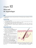
Ultraviolet radiation
There are three types of ultraviolet radiation—the short
wavelength ultraviolet C (100–280 nm), ultraviolet B (290–
320 nm), and long wavelength ultraviolet A (320–400 nm).
Beyond this is visible light then infrared, and radiowaves.
ultraviolet C does not penetrate beyond the stratosphere as it is
absorbed by the ozone layer. Ultraviolet B is very important in
both sunburn and the development of skin cancer. Ultraviolet A
is thought to be of increasing importance in the development
of skin cancer, and causes tanning but not sunburn. It is also
important in people with photosensitivity. The effects of
ultraviolet radiation may be classified as short term
(sunburn, photosensitivity) or long term (skin cancer,
wrinkling, solar elastosis, solar keratoses, seborrhoeic warts).
There is general awareness that the sun causes cancer in
the skin, with some people becoming obsessively fearful of any
exposure to sun. A sensible approach with emphasis on
reasonable precautions is called for. Useful points are:
Aborigines do not get skin
cancer
UVC
UVB*
UVA†
Visible
Infrared
Wavelength:
(280 nm)
Short wave
sunburn
spectrum
(280-320 nm)
Long wave:
(320-400 nm)
(380-770 nm)
(700 nm)
* The UVB band (280-320 nm) is responsible for erythema, sunburn, tanning and skin malignancy
† UVA light (320-400 nm) has the greatest penetration into the dermis and augments UVB
erythema and perhaps skin malignancy
Light spectrum (UVCϭultraviolet C, UVBϭultraviolet B,
UVAϭultraviolet A)
• Most moles are entirely harmless.
• Detecting the changes in moles or early melanoma enables
the diagnosis to be made at an early stage with a good chance
of curative treatment.
• The non-melanotic, epidermal cancers—basal cell and
squamous cell carcinomas—grow slowly and are generally not
life threatening. But squamous cell carinoma arising at sites
of trauma, on the extremities, or in ulcers may metastasise.
Exposure to sun has usually occurred many years previously.
Noon
Sun
6 am
6 pm
Subject
Sun
Sun
Earth
Prevention of sun damage and
skin cancer
Prevention of sun damage and skin cancer will depend on
reducing exposure to ultraviolet radiation. This can be
achieved in a number of ways:
Intensity of
UV light
• Covering the skin with clothes. It must be remembered
however that light clothes such as shirts or blouses may only
have a sun protection factor of four. A wide-brimmed hat is
essential to protect the face and neck.
• Sunscreens will greatly reduce sun exposure for exposed
parts such as the face and hands. Sunscreens are much more
6 am
Noon
6 pm
Diurnal variation in UV intensity of light from sun
65
ABC of Dermatology
efficient than previously, particularly those with a sun
protection factor greater than 30; they are now water
resistant, and most have a broad spectrum, protecting against
ultraviolet B and ultraviolet A. This is important because
there is now increasing evidence that ultraviolet A is
important in the development of skin cancer.
• Exposure to midday sun, particularly in tropical or
subtropical latitudes, should be avoided. At this time of the
day the sunlight passes vertically through the atmosphere and
there is less filtering of dangerous ultraviolet light. So
remember the adage: “Between eleven and three, stay under
a tree” in the summer months.
Effects of sun
Short term
• Sunburn
• Photosensitivity
Long term
• Skin wrinkling
• Telangiectasia
• Hyper and hypopigmentation
• Solar elastosis
• Actinic keratosis
• Seborrhoeic warts
• Skin cancers
Development of skin cancers
Sun-damaged skin
A number of different features characterise sun-damaged skin,
which is often seen in the elderly particularly if they have lived
in a sunny climate such as Australia. The skin has many fine
wrinkles and often has a sallow yellowish discoloration
particularly on the face and other exposed parts of the body.
Hyperpigmentation occurs as result of recent sun exposure,
which may be diffuse or localised in the form of solar lentigo.
In some areas there may be hypopigmentation, particularly
where solar keratoses have been treated with liquid nitrogen
(cryotherapy). There may be marked telangiectasia and
numerous blood vessels are seen. In some, there may be
thickening and a yellow hue of the skin, particularly of the
neck, due to elastin deposition in the upper dermis;
this is known as solar elastosis.
Sun damaged skin
Forms of skin cancer
There are three common forms of skin cancer caused by
ultraviolet light: basal cell carcinoma, squamous cell carcinoma,
and malignant melanoma. Whereas there seems to be a direct
relationship with the amount of ultraviolet exposure and basal
cell carcinoma and squamous cell carcinoma, the relationship
with ultraviolet exposure and melanoma is more complex and
it seems likely that intermittent exposure to ultraviolet light is
the main factor (for example, exposure to sunlight on
holidays). These different types of neoplastic change that
occur in the skin are discussed in chapters 13 and 15.
Photosensitivity
Exposure to sun in non-pigmented races causes inflammation
in the skin, depending on the skin type and amount of
exposure. In some individuals there is an abnormal sensitivity
to sunlight. This may arise because of an idiopathic reaction to
sunlight or allergic reaction that is activated by sunlight. Some
chemicals seen to induce photosensitivity without causing an
allergy. Other causes are metabolic diseases and inflammatory
conditions that are made worse by sun exposure.
Solar elastosis
Causes of photosensitivity
• Idiopathic—for example, polymorphic light eruption, actinic
prurigo, solar urticaria
• Photoaggravated dermatoses—for example, lupus
erythematosus, eczema
• Metabolic—porphyria—for example, erythropoietic, hepatic
• Drug induced—for example, sulphonamides, phenothiazines
• Chemical induced (topical)—for example, tar, anthracene
Polymorphic light eruption
This is the most common of the idiopathic photosensitive
rashes and occurs predominantly in women. It is due to
both the shorter (ultraviolet B) and longer (ultraviolet A)
wavelength types of sunlight. The eruption occurs from hours
to days after exposure and varies in severity from a few
inflamed papules to extensive inflamed oedematous lesions.
There may be only a few trivial lesions initially, but increasingly
severe reactions can develop restricting the patients ability to
venture outside. A useful measure of seventy is to ask the
66
Photosensitivity caused by drugs
The sun and the skin
patient if they cross to the shady side of the street to avoid the
sun. Treatment includes topical or systemic steroids for the
acute rash and prevention by using sunscreens. Desensitisation
by narrow waveband phototherapy before exposure is effective.
Solar urticaria
This is a much less common condition and may be induced by
longer wavelength (ultraviolet A) and visible radiation as well as
ultraviolet B. It is characterised by rapidly developing irritation
and in the exposed skin is followed by urticarial wheals. It can
occur as part of a photoallergic reaction, in which case
avoidance of the relevant allergen will prevent the condition.
Treatment is with antihistamines and sunscreens. In some
cases phototherapy with ultraviolet B, narrow waveband or
psoralen with ultraviolet A (PUVA), is helpful.
67
15 Black spots in the skin
There has been a great increase in public awareness of
melanoma, and any dark lesions of the skin are sometimes
regarded with the same dread as Long John Silver’s “black
spot” in Treasure Island—a sign of imminent demise. However,
the vast majority of pigmented lesions are simply moles or
harmless pigmented naevi. The most important thing is to
know which moles can be safely ignored and which should be
removed. Benign moles are described first, then malignant
melanoma, followed by a discussion of the differences between
these two.
Benign mole
Benign moles
Benign moles are naevi with a proliferation of melanocytes and
a variable number of dermal naevus cells. Some moles are
congenital and are present from birth, but most develop in
early childhood and adolescence. The number of moles
remains constant during adult life with a gradual decrease
from the sixth decade onwards.
There is often an increase in both the number of moles and
the degree of pigmentation during pregnancy.
Benign moles
Benign pigmented naevus
Acquired melanocytic naevi
Acquired melanocytic naevi are the familiar moles and present in
a number of different ways depending on the type of cells and
the depth in the skin.
Junctional naevi are flat macules with melanocytes
proliferating along the dermo-epidermal border.
Compound naevi have pigmented naevus cells at the dermoepidermal border and in the dermis, producing a raised brown
lesion. The dermal melanocytes may accumulate around the
skin appendages and blood vessels and form a band of cells
without melanin or more deeply penetrating strands of spindle
cells. Proliferating naevus cells may throw the overlying
epidermis into folds, giving a papillary appearance.
In a purely intradermal naevus the junctional element is lost,
with the deeper cells showing characteristics of neural tissue.
Other types of acquired pigmented naevi include the following.
Blue naevus is a collection of deeply pigmented melanocytes
situated deep in the dermis, which accounts for the deep
slate-blue colour.
Spitz naevus presents as a fleshy pink papule in children. It is
composed of large spindle cells and epitheloid cells with
occasional giant cells, arranged in “nests”. It is benign and the
old name of juvenile melanoma should be abandoned.
Halo naevus consists of a melanocytic naevus with a
surrounding halo of depigmentation associated with the
presence of antibodies against melanocytes in some cases.
The whole naevus gradually fades in time.
Becker’s naevus is an area of increased pigmentation, often
associated with increased hair growth, which is usually seen on
the upper trunk or shoulders. It is benign.
Freckles or ephelides are small pigmented macules, less than
0·5 cm in diameter, that occur in areas exposed to the sun in fair
skinned people. These macules fade during the winter months.
Blue naevus
Spitz naevus
Halo naevus
Congenital pigmented naevi
Congenital pigmented naevi are present at birth, generally over
1 cm in diameter, and vary from pale brown to black in colour.
They often become hairy and more protuberant, possibly with
68
Becker’s naevus
Black spots in the skin
an increased risk of malignant change. Larger lesions can cover
a considerable area of the trunk and buttocks, such as the
bathing trunk naevi, and their removal may present a
considerable problem.
Dysplastic naevi
These show very early malignant change and may progress to
malignant melanoma. They are deeply pigmented often with
an irregular margin.
In dysplastic naevus syndrome multiple pigmented naevi that
occur predominantly on the trunk, becoming numerous during
adolescence. They vary in size—many being over 0·5 cm—and
tend to develop into malignant melanoma, particularly if there
is a family history of this condition.
Congenital hairy naevus
Dysplastic naevus syndrome
Melanoma
Melanoma is an invasive malignant tumour of melanocytes.
Most cases occur in white adults over the age of 30, with
a predominance in women.
Incidence
The incidence of melanoma has doubled over the past 10 years
in Australia (currently 40/100 000 population) and shown a
similar increase in other countries. In Europe twice as many
women as men develop melanoma—about 12/100 000 women
and 6/100 000 men.
Prognosis
The prognosis is related to the thickness of the lesion,
measured histologically in millimetres from the granular layer
to the deepest level of invasion. Lesions less than 0·76 mm thick
have a 100% survival at five years, 0·76–1·5 mm thick an 80%
survival at five years, and lesions over 3·5 mm less than 40%
survival. These figures are based on patients in whom the
original lesion had been completely excised. A recent study in
Scotland has shown an overall five year survival of 71·6–77·6%
for women and 58·7% for men.
Melanoma
Melanoma
Sun exposure
The highest incidence of melanoma occurs in countries with
the most sunshine throughout the year. However, skin type
and the regularity of exposure to sun are also important. The
incidence is much greater in fair skinned people from higher
latitudes who have concentrated exposure to sun during
holidays than in those with darker complexions who have more
regular exposure throughout the year. Severe sunburn may also
predispose to melanoma.
Genetic factors
Since melanin protects the skin from ultraviolet light it is not
surprising that melanoma occurs most commonly in fair
skinned people who show little tanning on exposure to sun,
particularly those of Celtic origin. Members of families with the
dysplastic naevus syndrome are more likely to develop
melanoma in their moles. These patients have multiple naevi
from a young age.
Pre-existing moles
It is rare for ordinary moles to become malignant but
congenital naevi and multiple dysplastic naevi are more likely
to develop into malignant melanoma.
Nodular melanoma
Superficial melanoma with
nodules
Lentigo maligna
Nodule developing in superficial
spreading melanoma
69
ABC of Dermatology
Types of melanoma
There are four main types of melanoma.
Superficial spreading melanoma is the more common variety.
It is common on the back in men and on the legs in women.
As the name implies the melanoma cells spread superficially in
the epidermis, becoming invasive after months or years.
The margin and the surface are irregular, with pigmentation
varying from brown to black. There may be surrounding
inflammation and there is often clearing of the central portion.
The invasive phase is associated with the appearance of
nodules and increased pigmentation. The prognosis is
correspondingly poor.
Lentigo maligna melanoma occurs characteristically in areas
exposed to sun in elderly people. Initially there is a slowly
growing, irregular pigmented macule that is present for many
years before a melanoma develops.
Nodular melanoma presents as a dark nodule from the start
without a preceding in situ epidermal phase. It is more
common in men than women and is usually seen in people in
their fifties and sixties. Because it is a vertical invasive growth
phase from the beginning there is a poor prognosis.
Acral melanoma occurs on the palm and soles and near or
under the nails. Benign pigmented naevi may also occur in
these sites and it is important to recognise early dysplastic
change by using the criteria set out below. A very important
indication that discoloration of the nail is due to melanoma is
Hutchinson’s sign—pigmentation of the nail fold adjacent to
the nail. It is important to distinguish talon noir, in which a
black area appears on the sole or heel. It is the result of
trauma—for example sustained while playing squash—causing
haemorrhage into the dermal papillae. Paring the skin gently
with a scalpel will reveal distinct blood filled papillae, to the
relief of doctor and patient alike.
Other types of melanoma
As the melanoma cells become more dysplastic and less well
differentiated they lose the capacity to produce melanin and
form an amelanonitic melanoma. Such non-pigmented nodules
may be regarded as harmless but are in fact extremely
dangerous.
Superficial spreading melanoma
Nodular melanoma in a lentigo
Benign lentigo
Acral melanoma
Talon noir of left heel
Dysplastic melanoma
Amelanotic melanoma
Malignant melanoma in a black person; note the surrounding “halo”
Progressive
growth in depth
of malignant
melanoma
70
Black spots in the skin
Prognosis
This depends on the depth to which the melanoma has
penetrated below the base of the epidermis—lesions confined
to the epidermis having better prognosis than those
penetrating into the dermis. The Clark classification describes
the depth of penetration as follows:
Level I—within the epidermis
Level II—few melanoma cells within the dermal papillae
Level III—many melanoma cells in the papillary dermis
Level IV—invasion of the reticular dermis
Level V—invasion of the subcutaneous tissues
The Breslow classification is based on measurements of tumour
thickness from the granular layer overlying epidermis. A depth
of less than 1·5 mm is associated with a 90% five year survival,
1·5–3·5 mm with a 75% five year survival, and greater than
3·5 mm with only a 50% five year survival.
In deeper tumours “sentinel lymph node” biopsy may be
carried out to assess whether lymphatic spread has occurred.
How to tell the difference
Benign moles show little change and remain static for years.
Any change may indicate that a mole is in fact a melanoma or
that a mole is becoming active. Size, shape, and colour are the
main features and it is change in them that is most important.
Patients with moles should have these changes explained to
them, in particular that they indicate activity of the cells, not
necessarily malignant change.
Criteria for suspecting malignant changes in pigmented
lesions
• Growth—Benign pigmented naevi continue to appear in
adolescents and young adults. Any mole increasing in size in
an adult over the age of 30 may be a melanoma
• Shape—Moles usually have a symmetrical, even outline, any
indentations being quite regular; melanomas usually have an
irregular edge with one part advancing more than the others
• Colour—Variation in colour of benign moles is even but a
melanoma may be intensely black or show irregular
coloration varying from white to slate blue, with all shades of
black and brown. Inflammation may give a red colour as well.
The amelanotic melanoma shows little or no pigmentation
• Size—Apart from congenital pigmentation naevi most benign
moles are less than 1 cm in diameter. Any lesion growing to
over 0·5 cm should be carefully checked
• Itching—Normally a mole does not itch but a melanoma may.
Irritated seborrhoeic warts also itch
• Bleeding and crusting occur in an actively growing melanoma
If more than two of these features are present refer the patient
for specialist opinion
A simple summary:
A—Asymmetry of the lesion
C—Variations in colour
B—Irregularity of the border
D—Diameter larger
than 0·5 cm
Further reading
Ackerman AB. Malignant melanoma and other melanocytic neoplasms.
Baltimore: Williams and Wilkins,1984
Mackie R (ed). Primary and secondary prevention of malignant
melanoma. Basle: Karger
Roses DF. Diagnosis and management of cutaneous malignant melanoma.
Philadelphia: Saunders, 1983
Seigler HE. Clinical management of melanoma. The Hague: Nijhoff,
1982
71
16 The skin and systemic disease—Genetics and
skin disease (JA Savin)
When a man has on the skin of his body a swelling or an eruption or a
spot … and the disease appears to be deeper than the skin it is a leprous
disease.
Leviticus 13: 2–3
In ancient times changes in the skin were taken to indicate
that the whole body was diseased and although arguments
continue about what the Old Testament writers understood
by “leprous”, there was clearly an appreciation of the
connection between the skin and systemic illness. Clinical
signs in the skin may give valuable diagnostic clues to
underlying disease. The cutaneous signs of systemic disease
is a very large subject; and what follows is only an outline
of the more common skin changes that may be associated
with systemic illness.
A disease affecting internal organs may produce the same
changes in both the skin and other organs—as in the connective
tissue diseases. However, underlying conditions may be
associated with skin changes brought about by quite different
processes, as in acanthosis nigricans or dermatomyositis in which
there is an underlying neoplasm with characteristic skin signs.
Sometimes severe skin disease itself may be the cause of
generalised illness.
The skin is also a common site for allergic reactions to
drugs, with a rash being the first clinical sign. The florid skin
lesions of AIDS illustrate the results of infections when the
immune response is impaired.
Conditions affecting
both the skin and the
internal organs
When to suspect an underlying systemic disease
• An unusual rash which does not have the features of one of
the common primary inflammatory skin conditions
• Evidence of systemic illness—weight loss, and other symptoms
such as breathlessness, altered bowel function or painful
joints
• Erythema of the skin due to inflammation around the blood
vessels, usually without epidermal changes—reactive
erythema. Vasculitis, in which there are palpable
erythematous lesions which may be painful or nodular
• Unusual changes in pigmentation or texture of the skin
• Palpable dermal lesions that may be due to granuloma,
metastases, lymphoma, or deposits of fat or minerals
Rash from penicillin
Immune reactions
Allergic reactions to drugs such as penicillin can occur.
In this case the penicillin molecule attaches to serum protein.
This compound acts as an antigen and may form a complex
with IgG antibody. It is this complex which attaches to blood
vessel walls to produce an inflammatory reaction. This presents
as a rash developing a few days to two weeks after treatment on
the skin, but if it occurs in the kidneys the resulting tissue
damage can have serious consequences. This is an example of
Type III allergy with antigen–antibody complexes being
deposited in the small blood vessels. Sometimes a much more
acute anaphylactic reaction develops A fixed drug eruption is
characterised by a localised patch of erythema that flares up
whenever the drug is taken. Erythema multiforme can occur in
drug reactions.
Connective tissue diseases involve complex immunological
processes that affect both internal organs and the skin. This
means that it is particularly important to realise the
significance of any associated skin changes.
Lupus erythematosus
This condition has been described as “a disease with a
thousand faces” because of the wide range of organs involved
72
Erythema multiforme
The skin and systemic disease—Genetics and skin disease
and the numerous ways in which it can present. In three
quarters of the patients the skin is involved. There are four
main types, with numerous variations.
In systemic lupus erythematosus (SLE) the commonest skin
change is an acute erythematous eruption occurring bilaterally
on the malar area of the face in a “butterfly” distribution.
There may also be photosensitivity, hair loss, and areas of
vasculitis in the skin. There is often intolerance of sunlight. It is
more common in females with a female:male ratio of 8:1.
The systemic changes include fever, arthritis and renal
involvement, but there may be involvement of a wide range of
organs. The criteria for diagnosing the condition include
at least four of the features in the box on the right.
Subacute lupus erythematosus is a variant in about 10% of
patients with lupus erythematosus that presents with
non-scarring erythematosus plaques mainly on the face, hands
and arms. Papulo squamous lesions also occur. They may be
annular. Systemic involvement is less common and severe than
in SLE. It is associated with a high incidence of neonatal lupus
erythematosus in children born to mothers with the condition.
The antinuclear factor test is positive in 60% and
anticytoplasmic antibodies are present in 80% of patients.
Discoid lupus erythematosus (DLE) is a condition in which
circulating antinuclear antibodies are very rare. There are quite
well defined photosensitive inflammatory lesions, with some
degree of atrophy and hyperkeratosis of the follicles, giving a
“nutmeg grater” feel. It occurs predominantly on the face or
areas exposed to the sun, becoming worse in the summer
months. Scarring is common causing hair loss in lesions on the
scalp.
Treatment of SLE with the threatened or actual involvement
of other organs is important. Prednisolone is usually required
and sometimes immunosuppressant drugs such as azathioprine
as well. Treatment of DLE is generally with topical steroids.
Hydroxychloroquine by mouth is also used, generally in a dose
of 200 mg daily. This drug can diminish visual acuity in higher
doses and this should be checked every few months. A simple
chart, the Amsler Chart, is available for patients to use, consisting
of a central dot with a grid which becomes blurred when held at
arm’s length when there is any impairment of acutity.
Clinical variants of lupus erythematosus
•
•
•
•
Systemic
Subacute cutaneous
Discoid (neonatal)
Systemic sclerosis
Criteria for diagnosing systemic lupus erythematosus
•
•
•
•
•
•
Malar rash
Discoid plaques
Photosensitivity
Arthritis
Mouth ulcers
Renal changes
•
•
•
•
•
Serositis
Neurological involvement
Haematological changes
Immunological changes
Antinuclear antibodies
Systemic lupus
erythematosus
Dermatomyositis
This condition is associated in adults with underlying
carcinoma—commonly of the breasts, lung, ovary, or
gastrointestinal tract. It is characterised by localised erythema
with a purple hue (heliotrope), predominantly on the eyelids,
cheeks, and forehead. There may be similar changes on the
dorsal surface of the fingers, often with dilated nail fold
capillaries. These changes may precede the discovery of an
underlying tumour and may also fade away once it is removed.
There is a variable association with muscle discomfort and
weakness, mainly in the upper limb girdle. The finding of
muscle weakness together with specific electromyographic
changes and an inflammatory infiltrate in the muscle means
there is almost certainly an underlying malignancy, so suitable
investigation is indicated.
Dermatomyositis
Systemic sclerosis
As the name implies, there is extensive sclerosis of the
connective tissue of the lungs, gastrointestinal tract, kidneys,
and heart. Endothelial cell damage in the capillaries results in
fibrosis and sclerosis of the organs concerned. The skin
becomes tethered to the subcutaneous tissues and immobile,
leading to fixed claw like hands, constricted mouth with
furrowed lips, and beak-like nose. There are vascular changes
producing Raynaud’s phenomenon and telangiectasia around
Persistent dermatomyositis
73
ABC of Dermatology
the mouth and on the fingers. There are also flat “mat-like”
telangiectasia on the face.
Workers manufacturing polyvinyl chloride can develop skin
changes similar to systemic sclerosis with erosions of the bones,
hepatic and pulmonary lesions. Pesticides and epoxy resin can
also produce scleroderma-like changes.
It is associated with antinuclear antibodies (speckled or
nucleolar), and in about 50% of cases, circulating immune
complexes may be present.
A variant is the CREST syndrome. In this type of scleroderma
there is Calcinosis with calcium deposits below the skin on the
fingers and toes, Raynaud’s phenomenon with poor peripheral
circulation, immobility of the oEsophagus, dermal Sclerosis of
the fingers and toes, and Telangiectasia of the face and lips and
adjacent to the toe and finger nails. It has a better prognosis
than systemic sclerosis. Antinuclear antibodies at the
centromere are frequently present.
Morphoea is a benign form of localised systemic sclerosis in
which there is localised sclerosis with very slight inflammation.
There is atrophy of the overlying epidermis. The early changes
often consist of a dusky appearance to the skin.
CREST syndrome
•
•
•
•
•
C—Calcinosis cutis
R—Raynaud’s phenomenon
E—Esophagus
S—Scleroderma
T—Telangiectasia
CREST syndrome
Lichen sclerosus
The full name is lichen sclerosis et atrophicus—or LSA. This is
a relatively uncommon condition seen mainly in women in
whom well defined patches of superficial atrophy of the
epidermis occur with a white colour. There is fibrosis of the
underlying tissues. It frequently occurs in the vulva and
perineum and may also appear on the penis as balanitis
xerotica obliterans. Extragenital lesions may occur anywhere on
the skin. It may occur in a more acute form in children where
it tends to resolve, but in adults it is a very chronic condition.
There is an increased incidence of squamous cell carcinoma.
Treatment is with topical steroids and excision of any areas that
appear to be developing tumours.
The cause of the hyalinized collagen and epidermal atrophy
is unknown, but in early lesions there is an infiltrate of
lymphocytes with CD3, CD4, CD8, and CD68 markers. There is
also an increase in Langerhans cells, so there may well be an
immunological basis for these changes.
Calcinosis cutis
Vascular changes
Vascular lesions are associated with a wide range of conditions
including infections, neoplasia, and allergic reactions.
Hormones, particularly oestrogen, may affect the small blood
vessels of the skin to produce telangiectasia and small
angiomas, such as spider naevi.
Vasculitis and purpura, described in chapter 7, may be
associated with disease of the kidneys and other organs.
“Splinter haemorrhages” under the nails are usually the result
of minor trauma but may be associated with a wide range of
conditions, including subacute bacterial endocarditis and
rheumatoid arthritis.
Livedo reticularis is a cyanotic, net-like discoloration of the
skin over the legs. It may be idiopathic or associated with
arteritis or changes in blood viscosity.
Erythrocyanosis is a dusky, red, cyanotic change in the skin
over the legs and thighs, where there is a deep layer of
underlying fat. The condition becomes worse in the winter
months. It is most common in young women and usually
resolves over the years. Lupus erythematosus, sarcoidosis, and
tuberculous infection may localise in affected areas.
Telangiectasia and clubbing may be features of scleroderma
in the CREST syndrome described above.
In carcinoid and phaeochromocytoma vasoactive substances
cause episodes of flushing and telangiectasia.
74
Vasculitis
Livedo reticulosis
The skin and systemic disease—Genetics and skin disease
In hereditary haemorrhagic telangiectasia thin walled ectatic
blood vessels develop in the mucous membranes and the skin—
generally on the upper half of the body and the nail beds.
Erythemas
Erythema is macular redness of the skin due to congestion in
the capillaries. It occurs as part of immunological reactions in
the skin as in drug allergies and specific patterns of viral
infections, such as measles. There are other types that show
a specific pattern but are associated with a wide range of
underlying conditions, such as erythema multiforme.
Erythema multiforme is associated with herpes and other viral
infections or streptococcal and various bacterial infections, but
also with many other conditions, particularly connective tissue
disease, sarcoidosis, and reactions to drugs such as
sulphonamides.
The lesions consist of erythematous macules becoming
raised and typically developing into “target lesions” in which
there is a dusky red or purpuric centre with a pale indurated
zone surrounded by an outer ring of erythema. The lesions
may be few or multiple and diffuse, often involving the hands,
feet, elbows, and knees. Blisters may develop.
In the more severe forms there may be dermal changes and
blister formation with involvement of the mucous membranes
(Stevens–Johnson syndrome). There is often pyrexia with
gastrointestinal and renal lesions. It can progress to toxic
epidermal necrolysis, which some consider a form of erythema
multiforme.
Erythema annulare is a specific pattern in the skin with a
large number of reported associations, ranging from fungal
and viral infections to sarcoidosis and carcinoma. It consists of
a small erythematous macule that enlarges to form an
expanding ring, usually on the trunk.
Erythema chronicum migrans is associated with Borrelia
infection and Lyme disease—it is described on page 106.
There are many other types of “figurate erythemas”. Erythema
gyratum repens is associated with underlying carcinoma and
erythema marginatum, which is now rare, with rheumatic fever.
Erythemas associated with systemic
conditions
•
•
•
•
•
Erythema multiforme
Erythema annulare
Erythema chronicum migrans
Erythema gyratum repens
Erythema marginatum
Figurate erythema
Angiomas
Spider naevi, which show a central blood vessel with radiating
branches, are frequently seen in women (especially during
pregnancy) and children. If they occur in large numbers,
particularly in men, they may indicate liver failure. Palmar
erythema and yellow nails may also be present.
Congenital angiomas
Eruptive angiomas may be associated with systemic angiomas
of the liver, lung, and brain. Port wine stain due to abnormality
of the dermal capillaries commonly develops on the head and
neck. It may be associated with congenital vascular abnormalities
of the meningies and epilepsy. Vascular abnormalities of the eye,
and also glaucoma, occur with lesions on the face.
Erythema of the nailbeds
This may be associated with connective tissue disease, such as
lupus erythematosus, scleroderma, and dermatomyositis.
Erythema of nailbeds and
clubbing
Changes in pigmentation
Hypopigmentation
Hormonal
A widespread partial loss of melanocyte functions with loss
of skin colour is seen in hypopituitarism and is caused by an
absence of melanocyte stimulating hormone.
75
ABC of Dermatology
Genetic
In albinism, an autorecessive condition, there is little or no
production of melanin with loss of pigment from the skin, hair,
and eyes. Other genetic conditions with loss of skin pigment
include piebaldism, phenylketonuria, and tuberous sclerosis.
Localised depigmentation is most commonly seen in
vitiligo, in which a family history of the condition is found in
one third of the patients. In the sharply demarcated,
symmetrical macular lesions there is loss of melanocytes and
melanin. There is an increased incidence of organ specific
antibodies and their associated diseases.
Other causes of hypopigmented macules include:
postinflammatory conditions after psoriasis, eczema, lichen
planus, and lupus erythematosus; infections, for example, tinea
versicolor and leprosy; chemicals, such as hydroquinones,
hydroxychloroquine, and arsenicals, reactions to pigmented
naevi, seen in halo naevus; and genetic diseases, such as
tuberous sclerosis (“ash leaf” macules).
Autoimmune associations with vitiligo
•
•
•
•
•
Thyroid disease
Pernicious anaemia
Hypoparathyroidism
Addison’s disease
Diabetes
•
•
•
•
Myasthenia gravis
Alopecia areata
Halo naevus
Morphoea and
lichen sclerosus
Hyperpigmentation
There is wide variation in the pattern of normal pigmentation
as a result of heredity and exposure to the sun. Darkening of
the skin may be due to an increase in the normal pigment
melanin or to the deposition of bile salts in liver disease, iron
salts (haemochromatosis), drugs, or metallic salts from
ingestion. In agyria ingested silver salts are deposited in the skin.
Causes of hyperpigmentation include the following factors.
Vitiligo
Hormonal
An increase in circulating hormones that have melanocyte
stimulating activity occurs in hyperthyroidism, Addison’s
disease, and acromegaly. In women who are pregnant or taking
oral contraceptives there may be an increase in melanocytic
pigmentation of the face. This is known as melasma
(or chloasma) and occurs mainly on the forehead and cheeks.
It may fade slowly. Sometimes a premenstrual darkening of the
face occurs.
Increased deposition of haemosiderin is generalised in
haemochromatosis. Localised red-brown discoloration of the
legs is seen with longstanding varicose veins. It also occurs
in a specific localised pattern in Schamberg’s disease,
when there is a “cayenne pepper” appearance of the legs
and thighs.
Neoplasia
Lymphomas may be associated with increased pigmentation.
Acanthosis nigricans, characterised by darkening and
thickening of the skin of the axillae, neck, nipples, and
umbilicus, occurs with internal cancers, usually
adenocarcinoma of the stomach. It is also seen in acromegaly.
There is a benign juvenile type. Pseudoacanthosis nigricans is
much more common, consisting of simple darkening of the
skin in the flexures of obese individuals; it is not associated with
malignancy.
Agyria (silver salts in skin)
Melasma
Acanthosis nigricans
Carcinoma left upper zone
Pseudoacanthosis nigricans
76
Acanthosis nigricans
The skin and systemic disease—Genetics and skin disease
Drugs
Chlorpromazine, other phenothiazines, and minocycline may
cause an increased pigmentation in areas exposed to the sun.
Phenytoin can cause local hyperpigmentation of the face
and neck.
Inflammatory reactions
Postinflammatory pigmentation is common, often after acute
eczema, fixed drug eruptions, or lichen planus. Areas of
lichenification from rubbing the skin are usually darkened.
Malabsorption and deficiency states
In malabsorption syndromes, pellagra, and scurvy there is
commonly increased skin pigmentation.
Congenital conditions
There is clearly a marked variation in pigmentation and in the
number of freckles in normal individuals. There may be
localised well defined pigmented areas in neurofibromatosis
with “cafe au lait” patches. Increased pigmentation with a blue
tinge occurs over the lumbosacral region in the condition
known as Mongolian blue spot.
Peutz–Jeghers syndrome is described under the section
“The gut and the skin”, below. There are pigmented macules
associated with intestinal polyposis in the oral mucosa, lips,
and face.
Neurofibromatosis
Neurofibromatosis
Increased pigmentation in
malabsorption syndrome
Peutz–Jeghers syndrome
Skin markers of internal malignancy
Malignant lesions
Malignant lesions may cause skin changes such as acanthosis
nigricans and dermatomyositis or produce secondary deposits.
Lymphomas can arise in or invade the skin. Pruritus may be
associated with Hodgkin’s disease.
Mycosis fungoides is a T cell lymphoma of cutaneous origin.
Initially well demarcated erythematous plaques develop on
covered areas with intense itching. In many cases there is a
gradual progression to infiltrated lesions, nodules, and
ulceration. In others the tumour may occur de novo or be
preceded by generalised erythema.
Poikiloderma, in which there is telangiectasia, reticulate
pigmentation, atrophy, and loss of pigment, may precede
mycosis fungoides, but it is also seen after radiotherapy and in
connective tissue diseases.
Parapsoriasisis a term used for well defined maculopapular
erythematous lesions that occur in middle and old age. Some
cases undoubtedly develop into mycosis fungoides and a biopsy
specimen should be taken of any such fixed plaques that do not
clear with topical steroids.
•
•
•
•
•
Acanthosis nigricans—usually intra-abdominal lesions
Erythematous rashes, “figurate erythema”
Pruritus—usually lymphoma
Dermatomyositis in the middle aged and elderly
Acquired ichthyosis
Non-specific skin changes associated with malignant
disease
• Secondary deposits
• Secondary hormonal effects
•
•
•
•
Acne (adrenal tumours)
Flushing (carcinoid)
Pigmentation
Generalised pruritus (particularly lymphoma)
Figurate erythema
Superficial thrombophlebitis
Various eruptive skin lesions seen in Gardener’s and Bazex
syndromes
Parapsoriasis
Lymphoma
Poikiloderma
B cell lymphoma
77
ABC of Dermatology
The gut and the skin
Vasculitis of various kinds, periarteritis nodosa, connective
tissue diseases such as scleroderma, and many metabolic
diseases produce both cutaneous and gastrointestinal lesions.
There are, however, some specific associations.
Dry skin, asteatosis, and itching, with superficial eczematous
changes and a “crazy paving” pattern, occur in malabsorption
and cachectic states. Increased pigmentation, brittle hair and
nails may also be associated.
Pyoderma gangrenosum gives rise to an area of non-specific
inflammation and pustules break down to form a necrotic ulcer
with hypertrophic margins. There is an underlying vasculitis.
There is a strong association with ulcerative colitis and also with
Crohn’s disease, rheumatoid arthritis, abnormal gamma
globulins, and leukaemia.
Dermatitis herpetiformis, which has already been discussed, is
an intensely itching, chronic disorder with erythematous and
blistering lesions on the trunk and limbs. It is more common in
men than women. Most patients have a gluten sensitive
enteropathy with some degree of villus atrophy. There is an
associated risk of small bowel lymphoma.
Peutz–Jeghers syndrome is inherited as an autosomal dominant
characterised by the appearance in infancy of pigmented
macules of the oral mucosal membranes, lips, and face.
Benign intestinal polyps, mainly in the ileum and jejunum,
which rarely become malignant, are associated with the
condition.
Other conditions include congenital disorders with
connective tissue and vascular abnormalities that affect
the gut, such as Ehlers–Danlos syndrome and
pseudoxanthoma elasticum (arterial gastrointestinal bleeding),
purpuric vasculitis (bleeding from gastrointestinal lesions), and
neurofibromatosis (intestinal neurofibromas).
In Crohn’s disease (regional ileitis) perianal lesions and sinus
formation in the abdominal wall often occur. Glossitis and
thickening of the lips and oral mucosa and vasculitis may be
associated.
Liver disease may affect the skin, hair, and nails to a variable
degree. Obstructive jaundice is often associated with
itching which is thought to be due to the deposition of bile
salts in the skin. Evidence of this is the fact that drugs which
combine with bile salts such as cholestyramine improve pruritus
in some patients. Jaundice is the physical manifestation of bile
salts in the skin.
Liver failure is characterised by a number of skin signs,
particularly vascular changes causing multiple spider naevi
and palmar erythema due to diffuse telangiectasia. It is not
unusual to see spider naevi on the trunk in women but large
numbers in men should raise suspicion of underlying hepatic
disease.
Porphyria cutanea tarda as a result of chronic liver
disease produces bullae, scarring, and hyperpigmentation
in sun exposed areas of the skin. Xanthomas may be
associated with primary biliary cirrhosis and in chronic liver
disease asteotosis, with dry skin producing a “crazy paving”
pattern.
Diabetes and the skin
In diabetes the disturbances of carbohydrate–lipid metabolism,
small blood vessel lesions, and neural involvement may be
associated with skin lesions. The more common of these
include the following.
78
Diseases that pyoderma gangrenosum
may occur with
•
•
•
•
•
Ulcerative colitis
Crohn’s disease
Rheumatoid arthritis
Monoclonal gammopathy
Leukaemia
Early pyoderma gangrenosum
Pyoderma gangrenosum
Dermatitis herpetiformis
Liver disease and the skin
Obstructive
• Jaundice
• Pruritus
Liver failure
• Multiple spider naevi (in men)
• Palmar erythema
• White nails—hypoalbuminaemia
• Porphyria cutanea tarda
Cirrhosis
• Xanthomas (primary biliary cirrhosis)
• Asteatosis
The skin and systemic disease—Genetics and skin disease
Infection
Diabetic patients have an increased susceptibility to
staphylococcal, coliform, and pseudomonal infection. Candida
albicans infection is also more common in diabetics.
Vascular lesions
“Diabetic dermopathy”, due to a microangiopathy, consists of
erythematous papules which slowly resolve to leave a scaling
macule on the limbs. Atherosclerosis with impaired peripheral
circulation is often associated with diabetes. Ulceration due to
neuropathy (trophic ulcers) or impaired blood supply may
occur, particularly on the feet.
Specific skin lesions
Diabetic ulcer
Necrobiosis lipoidica
Necrobiosis lipoidica
Between 40% and 60% of patients with this condition may
develop diabetes, but it is not very common in diabetic patients
(0·3%). As the name indicates, there is necrosis of the
connective tissue with lymphocytic and granulomatous
infiltrate. There is replacement of degenerating collagen fibres
with lipid material. It usually occurs over the shin but may
appear at any site.
Granuloma annulare
This usually presents with localised papular lesions on the
hands and feet but may occur elsewhere. The lesions may be
partly or wholly annular and may be single or multiple.
There is some degree of necrobiosis, with histiocytes forming
“palisades” as well as giant cells and lymphocytes. It is seen more
commonly in women, usually those aged under 30. There is
an association with insulin dependent diabetes. In itself it is
a harmless and self limiting condition that slowly clears
but may recur.
Granuloma annulare
Porphyria
Other diseases
Porphyrias are due to the accumulation of intermediate
metabolites in the metabolic pathway of haem synthesis.
There are several types. In hepatic porphyrias there is skin
fragility leading to blisters from exposure to the sun or minor
trauma. In erythropoietic and erythrohepatic photoporphyrias
there is intense photosensitivity. They are sometimes associated
with sensitivity to long wavelength ultraviolet light that
penetrates window glass.
Porphyria cutanea tarda usually occurs in men, with a genetic
predisposition, who have liver damage as a result of an
excessive intake of alcohol. There is impaired porphyria
metabolism leading to skin fragility and photosensitivity, with
blisters and erosions, photosensitivity on the face and the
dorsal surface of the hands.
Xanthomas are due to the deposition of fat in connective
tissue cells. They are commonly associated with
hyperlipidaemia—either primary or secondary to diabetes, the
nephrotic syndrome, hypothyroidism, or primary biliary
cirrhosis. Four of the primary types are associated with an
increased risk of atherosclerosis; type I is not. Diabetes may be
associated with the eruptive type.
Necrotising fasciitis is an area of cellulitis that develops
vesicles. Necrosis of the skin may indicate much more
extensive, life threatening necrosis of the deeper tissues.
Urgent surgical debridement is indicated.
Amyloid deposits in the skin occur in primary systemic
amyloidosis and myeloma.
Xanthomas
Common types of xanthoma
Clinical type
Xanthelasma of the
eyelids—yellow plaques
Tuberous nodules on
elbows and knees
Eruptive—small yellow papules
on buttocks and shoulders
Plane—yellow macules,
palmar creases involved
Generalised—widespread
macules
Tendons—swelling
on fingers or ankles
Association with hyperlipidaemia
Primary
Secondary
II (may be
normal)
II, III
ϩ
I, III, IV, V
ϩ
I, III
ϩ
myeloma
II, III
ϩ
79
ABC of Dermatology
Pregnancy
Pregnancy may be associated with pruritus, in which the skin
appears normal in 15–20% of women (prunigo gestationis). It
is generally more severe in the first trimester.
Polymorphic eruptions also present with pruritis with urticaria
papules and plaques (the PUPP syndrome). It usually occurs on
the abdomen in the third trimester and then becomes
widespread. There may be a postpartum flare up. It can be a
distressing condition for the mother but the baby is not
affected, and it rarely recurs in subsequent pregnancies. Topical
steroids can be used, but systemic steroids should be avoided.
Pemphigoid gestationis is a rare disorder that may resemble
PUPP initially but develops pemphigoid-like vesicles, spreading
over the abdomen and thighs: autoantibodies to the basement
membrane are present.
Polymorphic eruption
Pemphigoid gestationis
Sarcoidosis
Pulmonary and other systemic manifestation of sarcoidosis may
occur without involvement of the skin. The most common
changes are:
• Erythema nodosum, which is often a feature of early
pulmonary disease.
• Papules, nodules, and plaques are associated with acute and
subacute forms of the disease.
• Scar sarcoidosis, with papules occurring in scars.
• Lupus pernio is characterised by dusky red infiltrated lesions
on the nose and fingers.
Nodule in sarcoidosis
Thyroid disease
Thyroid disease is associated with changes in the skin, which
may sometimes be the first clinical signs. There may be
evidence of the effect of altered concentrations of thyroxine on
the skin, with changes in texture and hair growth. Associated
increases in thyroid stimulating hormone concentration may
lead to pretibial myxoedema. In autoimmune thyroid disease
vitiligo and other autoimmune conditions may be present.
Sarcoid granuloma
Genetics and skin disease
by JA Savin
Though many genetic disorders of the skin are inherited in
a classically Mendelian way (single gene disorders), others are
genetically more complex. As a general rule, the common skin
disorders that run in families, such as psoriasis, atopic eczema,
and acne, tend to belong to the latter group.
Single gene disorders
Recent advances in genetic technology have been relatively easy
to apply to these, usually rather uncommon, disorders, most of
which had already been classified accurately on clinical
grounds. Several things followed from this:
(1) The next step has often been an improvement in current
clinical classifications, which can now be based logically on
the underlying molecular abnormalities of the disorders in
question. A good example of this is the modern
classification of the inherited mechano-bullous disorders
(also known as epidermolysis bullosa).
(2) New skin constituents were quickly recognized, their function
being understood after studying the disorders in which they
80
Clinical signs of thyroid disease
Hypothyroidism
Dry skin
Oedema of eyelids and hands
Absence of sweating
Coarse, thin hair—loss of pubic,
axillary, and eyebrow hair
Pale “ivory” skin
Brittle poorly growing nails
Purpura, bruising, and
telangiectasia
Hyperthyroidism
Soft, thickened skin
Pretibial myxoedema
Increased sweating (palms and
soles)
Thinning of scalp hair
Diffuse pigmentation
Rapidly growing nails
Palmar erythema
Facial flushing
The skin and systemic disease—Genetics and skin disease
are abnormal. Soon it was realized that the same molecules
could be the targets both for genetic abnormalities and for
acquired skin diseases. One example of this is the way in
which autoantibodies directed against one of the constituents
of hemidesmosomes (BP180) cause pemphigoid, whereas
mutations in the gene responsible for BP180 are the basis of
the junctional type of epidermolysis bullosa.
(3) Advances have been made too in our understanding of
the structure and function of normal skin and its
appendages—for example, the finding that melanocortin-1
receptor gene variants are associated with fair skin, red
hair, and skin tumours.
(4) Mosaics were soon recognized. Clinically these are linear
abnormalities in the skin, usually present at birth, which
often contain cells with the same genetic abnormalities as
those of known generalised genodermatoses. A good
example of this is the way the same abnormalities in the
genes controlling the production of keratins 1 and 10 can
be responsible both for a generalised skin condition
(epidermolytic hyperkeratosis) and for warty linear naevi.
The mosaic areas follow Blashko’s lines, a bizarre pattern
of lines and whorls, which are not the same as
dermatomes.
(5) The prenatal diagnosis of severe genodermatoses has
become more accurate, though gene therapy has not yet
fulfilled its early promise.
Genetically complex disorders
Psoriasis is a good example. It clusters in some families but does
not follow a classical Mendelian pattern of inheritance.
Environmental triggers are important, as well as genetic factors.
Over the last few years, several wide scans of the genome have
been undertaken with the aim of identifying the location of the
genes that determine susceptibility to psoriasis. Five have been
confirmed, all on different chromosomes, and now designated
as Psors1 to Psors5. A further six loci may have similar effects,
but the evidence for them is less strong. Psors1, on
chromosome 6p21.3, is an especially important gene for
psoriasis susceptibility in many populations and lies within the
area of the major histocompatibility complex (MHC). However
it is not itself an HLA class 1 gene, and may belong to the
newly described MHC class 1 chain-related (MIC) gene family.
The possession of one allele (A5.1) of this gene seems to lead
to a type of psoriasis that starts especially early, and is more
common in familial than in sporadic cases.
In atopic eczema, matters are equally complicated.
Environmental factors may well be responsible for the recent
rise in its prevalence as the gene pool within the population is
not likely to have changed greatly, but a genetic component is
obvious too, even though affected children can be born to
clinically normal parents. Within each family, atopic disorders
tend to run true to type, so that, in some, most affected
members will have eczema, in others, respiratory allergy
predominates. The inheritance of atopic eczema probably
involves genes that predispose to the state of atopy, and others
that determine whether it is asthma, eczema, or hay fever that
develops. One plausible gene for the inheritance of atopy
encodes for the  subunit of the high affinity IgE receptor, and
lies on chromosome 11q13. However several groups have failed
to confirm earlier reports of this linkage, and a gene linked to
atopic eczema has recently been found on chromosome 3q21.
Blashko’s lines
The abnormality underlying some inherited skin disorders
Skin disorder
Ehlers–Danlos syndrome
Dystrophic epidermolysis bullosa
Pseudoxanthoma elasticum
Xeroderma pigmentosum
Simple epidermolysis bullosa
Epidermolytic hyperkeratosis
Palmoplantar keratoderma
Junctional epidermolysis bullosa
X-linked recessive ichthyosis
Darier’s disease
Albinism (tyrosinase negative type)
Abnormality in
Collagen and the
extracellular matrix
Type VII collagen
Elastic tissue
DNA repair
Keratins 5 and 14
Keratins 1 and 10
Keratins 9 and 16
Laminins
Steroid sulphatase
Epidermal cell adhesion
Tyrosinase
Further reading
Fine JD, Eady RA, Bauer EA, et al. Revised classification system for
inherited epidermolysis bullosa. J Am Acad Dermatol 2000;42:
1051–66
Harper J. Inherited skin disorders. Oxford: Butterworth–Heinemann
1999
Moss C, Savin JA. Dermatology and the new genetics. Oxford: Blackwell
Science, 1995
81
17 Cutaneous immunology—Autoimmune disease
and the skin (DJ Gawkrodger)
Types of allergic reaction
Allergic and other immune reactions may occur in the
skin—the “immunological battleground of the body”—rather
than involving internal organs. An acute vasculitis occurring in
the skin is unpleasant and requires treatment but the same
reaction occurring in the kidneys can be life threatening.
The pattern of skin changes can indicate the type of
immune process involved and also whether there is likely
to be systemic involvement. The immune response of the
skin is also used clinically in the tuberculin skin test to
detect the level of immunity to tuberculosis. It is also
the means of immunisation when an injection of inactivated
organisms induces an immune response that protects the
entire body.
The different types of immune reaction are all manifested
in the skin as part of a normal response to pathogens or as an
allergic reaction. The difference is expressed by the word
“allergy”, first used by Von Pirquet in 1906, derived from the
Greek (␣␥ ⑀␥), meaning literally “other work”. In other
words it is a response that is appropriate for pathogenic
organisms such as a tubercle bacillus but is misdirected against
a harmless substance such as a rubber glove or the metal of a
watch strap buckle.
Immunological reactions are of four types—five if
autoimmunity is counted—of responses mediated by antibodies
known as the humoral response and one by the lymphocytes
known as the cell mediated response.
Reaction to fish protein
Ig E
Fc receptor
Immediate hypersensitivity
This type of reaction is caused by “reagin” antibodies, which
consist mainly of lgE, that react with allergens such as
housedust mite, animal dander, or grass pollens. These
reactions may occur in both the skin or the lung to produce
asthma. Allergic reactions to insect stings can cause severe
systemic effects—“anaphylaxis”, which literally means “without
protection”. Food proteins can also cause an immediate type of
hypersensitivity reaction. The IgE molecule is attached to
specific receptors on the surface of mast cells and when
activated by linkage to specific allergen inflammatory
mediators are released. This is an acute process, hence the
name “immediate hypersensitivity”.
The initial response occurring within five minutes is due to
by the release of histamine, heparin, tryptophan. This is followed
by inflammatory mediators—released in five to 30 minutes—
leukotrien, prostaglandin. The later response, occurring after
some hours, is caused by cytokines—predominantly tumour
necrosis factor ␣ (TNF-␣) and interleukin 4 (IL4).
Severe reactions cause shock that is made worse by stress
and exercise, as in the case of a young woman, allergic to wasp
stings, who had a wasp sting when picnicking by a lake. She
then plunged into the cold water, swimming vigorously, leading
to a fatal anaphylactic reaction. Acute anaphylactic reactions to
peanuts may be life threatening.
Mast cell
Type I—immediate hypersensitivity
Antibody
Target cell
+
Complement
Lysis
Cytotoxic reactions
In this case cells become the target of attack by circulating
antibodies. There are a number of causes, such as drugs or
proteins attached to the cell surface that act as haptens so they
82
Histamine
+ inflammatory
mediators
Antigen
Fc receptor
K cell
Type II—cytotoxic
Cutaneous immunology—Autoimmune disease and the skin
become antigenic. This occurs in drug induced haemolysis
from drugs. Alternatively, immune complexes are attached to
the surface of the cell with the incorporation of complement
leading to lysis. In haemolytic anaemia and incompatible blood
transfusions antibodies are formed against erythrocytes. They
may also be destroyed by killer cells. A typical example is
haemolytic anaemia.
This immune response is the means of destroying cells that
become antigenic as a result of being infected with virus.
In autoimmune diseases antibodies are directed against
specific structures.
Antigen
+ Complement
+ Polymorphs
Damaged basement membrane
Antigen–antibody complex reactions
As a result of antibody production to antigens in the
circulation, complexes form in the blood and these may be
deposited in capillaries resulting in inflammatory changes.
Similar changes may occur in the lung. This involves the
activation of complement and the release of mediators of
inflammation, producing vasodilatation and the accumulation
of polymorphs.
Delayed hypersensitivity
This type of reaction results from lymphocytes known as
T cells, because of their derivation from the thymus, which
react with antigen in the skin. The reaction is initiated by
antigen attached to Langerhan’s cells in the epidermis being
transported to the paracortical area of the regional lymph node
with the production of lymphocytes sensitised specifically for
that antigen. There is also the production of interleukin which
has a feedback effect in stimulating the production of more
sensitised lymphocytes.
The reaction of the T lymphocytes in the epidermis results in
the accumulation of macrophages and the release of
inflammatory mediators.
Antibody
Type III—circulating immune complexes
Antigen on skin
Epidermal
Langerhans
cell
Antigen presenting
cell (Veiled cell)
Sensitised T cell
reacts with antigen
Inflammation
associated with:
Interferon
Cytokines
Macrophages
Lymph node
Paracortical area
T cell sensitised to antigen
Interleukins stimulate proliferation
Autoimmune disease and the skin
by DJ Grawkrodger
Type IV—delayed hypersensitivity
There is always the risk that the well developed human immune
system may react against the body’s own tissues, with a failure to
distinguish between “self” and “non-self”. An immune response
develops which may be specific for a particular organ, such as
the thyroid gland, or react against a number of different
organs, as in connective tissue diseases. The skin can manifest
both types of autoimumme response. The results of such
reactions can be destruction of the cells concerned and the
production of inflammation. There is an inherited tendency to
autoimmune disease, marked by specific HLA (human
lymphocyte antigen) in some cases.
The most common types of skin disease in which this
autoimmune mechanism occurs are the blistering disorders,
pemphigoid and pemphigus, as well as dermatitis
herpetiformis.
Pemphigoid
In this condition large, tense blisters develop in which there are
antibodies attached to the upper layer of the basement
membrane at the dermo-epidermal junction, with an
underlying inflammatory reaction producing a split above the
basement membrane. Lysosomal enzymes are released
damaging the basement membrane, resulting in separation of
the epidermis and blister formation. The presence of
Reaction to metal
Blistering disorder as a result of an
autoimmune response
Split at dermo-epidermal junction
83
ABC of Dermatology
antibodies, usually IgG, can be shown by an antihuman IgG
antibody labelled with fluorescein. When viewed under the
microscope with ultraviolet light illumination, the presence of
the IgG antibody is shown by fluorescence. The presence of
circulating antibasement membrane antibodies in the serum
can be shown either by direct immunofluorescence using a
specimen of the patient’s skin or by incubation by attachment
to skin which has been incubated in serum from the patient.
The clinical features are described in chapter 8. The blisters
develop, frequently with an erythematous background, on the
limbs, trunk, and flexures. It is mainly seen in the elderly and is
slightly more common in women.
Fluorescein labelled antibody
to human immunoglobulin
Antibody (immunoglobulin) in situ
Normal antigen
Skin section from patient
Direct immunofluorescence
Fluorescein labelled antibody
to human immunoglobulin
Antibody in patient's serum,
placed on section
Antigen similar to human
Indirect immunofluorescence
Pemphigus
Intraepidermal split
Direct immunofluorescence
Substrate (usually animal tissue)
Indirect immunofluorescence
Pemphigus
In this condition, antibodies are found to have developed
against the epidermis above the basement membrane.
The main antibody is IgG, but IgM and IgA may also be
found. As a result of this reaction, there is separation of the
epidermal cells with the formation of a superficial blister. A row
of basal cells remains attached to the basement membrane.
Direct immunofluorescence of the skin from affected patients
shows that antibodies are deposited on the intercellular
substance of the epidermis. Circulating antibodies are often
present. Oral lesions are much more common than in
pemphigoid.
Other organ-specific autoimmune diseases of the skin
Range of autoimmune disease
Vitiligo
In this condition there is a loss of pigment as a result of
antibodies developing against melanocytes in the skin in a
limited area. However, the areas affected tend to gradually
increase. There may be other autoimmune diseases in the
same patient, causing, for example, pernicious anaemia, and
thyroid disease.
Organ specific
Hashimoto's thyroiditis
Pernicious anaemia
Addison's disease
Myasthenia gravis
Pemphigus
Pemphigoid
Vitiligo
Primary biliary cirrhosis
Alopecia areata
There is evidence that this condition may be associated with
an immune reaction against the hair follicle. The increased
incidence of antibodies to the thyroid gland and gastric parietal
cells in patients with alopecia areata provides circumstantial
support for an autoimmune aetiology.
Rheumatoid arthritis
Dermatomyositis
Systemic sclerosis
Non-organ specific
Lupus erythematosus
Range of autoimmune disease
Non-organ-specific skin autoimmune disease
Systemic lupus erythematosus (SLE)
The hallmark of this condition is the presence of antibodies
against various components of the cell nucleus. Although a
wide range of organs may be affected, in three quarters of the
patients the skin is involved, generally with an erythematous
eruption occurring bilaterally on the face in a “butterfly”
distribution. There may also be photosensitivity, hair loss, and
areas of vasculitis in the skin. There is often intolerance of
84
Clinical variants of lupus erythematosus
•
•
•
•
Systemic
Subacute cutaneous
Discoid
(Neonatal)
Cutaneous immunology—Autoimmune disease and the skin
sunlight. Subacute lupus erythmatosus is a variant that presents
with an erythematous eruption in the skin and anticytoplasmic
RNA molecules.
Discoid lupus erythematosus (DLE)
This is a condition in which circulating antinuclear antibodies
are very rare. There are quite well defined inflammatory
lesions, with some degree of atrophy occurring on the face and
occasionally on the arms as well.
Treatment of SLE with the threatened or actual
involvement of organs is important. Prednisolone is usually
required and sometimes immunosuppressant drugs such as
azathioprine as well. Treatment of DLE is generally with topical
steroids. Hydroxychloroquine by mouth is also used, generally
in a dose of 200 mg daily. This drug can diminish visual
acuity and this should be checked every few months. A simple
chart, the Amsler Chart, is available for patients to use,
consisting of a central dot with a grid which becomes blurred
when held at arm’s length when there is any impairment of
acuity.
Systemic sclerosis
This is a condition in which there is extensive sclerosis of the
subcutaneous tissues in the fingers and toes as well as around
the mouth (scleroderma), with similar changes affecting the
internal organs, particularly the lung and kidneys. There are
vascular changes producing Raynaud’s phenomenon and
telangiectasia around the mouth and fingers. It is associated
with antinuclear antibodies (speckled or nucleolar), and in
about 50% of cases circulating immune complexes may be
present. Endothelial cell damage in the capillaries results in
fibrosis and sclerosis of the organs concerned. There is
considerable tethering of the skin on the fingers and toes,
which becomes very tight with a waxy appearance and
considerable limitation of movement. A variant is the CREST
syndrome.
Morphoea is a benign form of localised systemic sclerosis
in which there is localised sclerosis with very slight
inflammation. There is atrophy of the overlying epidermis.
The early changes often consisit of a dusky appearance to
the skin.
The clinical features are described in chapter 16.
Subacute lupus erythematosus
Subacute lupus erythematosus
Discoid lupus erythematosus
Systemic sclerosis
Morphoea
Dermatomyositis
This condition is described in chapter 16, but the main
immunological features are deposition of IgG, IgM, and C3 at
the dermo-epidermal junction in about half the cases in the
early stages, as well as a lymphocytic infiltrate with CD4ϩ cells
and macrophages. There are reports of autoantibodies in some
patients. Dermatomyositis may represent an immune reaction
to an underlying mechanism or derangement of the normal
immune response.
Dermatomyositis
Lichen sclerosus
This condition is also described in chapter 16 and is
characterised by atrophic patches of skin. It occurs mainly in
females and predominantly involves the genitals and perineum.
The cause is unknown but in early lesions there is a band of
lymphocytes, mainly CD3, CD4, and CD8. Immunoglobulins
and complement accumulate in the affected areas. There is an
association with vitiligo, morphoea, alopecia, and pernicious
anaemia, suggesting an autoimmune association.
Lichen sclerosus
85
ABC of Dermatology
Graft versus host disease
This reaction occurs following bone marrow transplantation in
immunosuppressed patients. T lymphocytes produced by the
graft react against the body’s own tissues, producing a skin
eruption which may resemble measles. There is lysis of the
basement membrane with shedding of the skin and sometimes
lichen planus-like eruption. In the more chronic form,
localised lesions develop, with immunoglobulins deposited in
the walls of blood vessels with the activation of complement.
Graft versus host disease
Further reading
Roitt IM, Brostoff J, Male D. Immunology, 6th ed. St Louis: Mosby,
2001
86
18 Bacterial infection
RJ Hay
The process of infection involves the interaction between two
organisms—the host and the invader. The clinical changes
result from mechanisms involved in this process, notably the
micro-organism, its virulence, and the patient’s immune
defenses. The lesions produced often have a well defined
appearance, such as impetigo or tinea cruris, but the changes
may be less specific.
Several features enable us to recognise that infection is a
possible cause of the patient’s condition. Acute bacterial
infections generally produce some or all of the classical
characteristics of acute inflammation.
Cardinal signs of infection
•
•
•
•
Erythema
Swelling and oedema
Heat or warmth
Pain and discomfort
Erythema of the face
Usually
unilateral
Usually
bilateral
Photosensitive
ϩ
ϩ
or ϩ
ϩ
ϩ or Ϫ
ϩ or Ϫ
ϩ or Ϫ
Acute
(1) Allergic reactions
Cosmetics
Plants
Drugs
(2) Urticaria
Reactions to light
(3) Photodermatitis
Solar urticaria
(4) Infection
Erysipelas
Fifth disease
(“slapped cheek”)
(5) Rosacea
Chronic—recurrent
(6) Lupus erythematosus
Systemic
Discoid
(7) Seborrhoeic dermatitis
(8) Acne
(9) Perioral dermatitis
(10) Vascular naevus
ϩ
ϩ
ϩ
Ϫ
ϩ
ϩ
ϩ
ϩ
ϩ
ϩ
Ϫ
Ϫ
ϩ
ϩ or Ϫ
ϩ
or ϩ
ϩ
ϩ
ϩ
Ϫ
ϩ
ϩ
Ϫ
ϩ
Ϫ
Ϫ
Allergic reactions to cosmetics
Erysipelas
Fifth disease (“slapped cheek”)
Photodermatitis
Rosacea
Systemic lupus erythematosus
Perioral dermatitis
Discoid lupus erythematosus
Seborrhoeic dermatitis
Vascular naevus
87
ABC of Dermatology
Clinical presentation
The woman shown in the photographs had acute erysipelas
due to streptococcal infection, and all four features of
inflammation were present. She was referred to the clinic with
a diagnosis of an acute allergic response, which, from the
appearance alone, was understandable. However, malaise and
fever were also present and the lesions were warm and tender.
The condition responded well to antibiotic treatment.
The point of entry in such cases is thought to be a small
erosion on the face. Erysipelas of the leg or foot may
follow the development of a small fissure between the
toes, but often there is no discernible portal
of entry.
Erysipelas is the local manifestation of a Group A
streptococcal infection, in the case illustrated the infection is
confined to deep dermis as a form of cellulitis. However the
same organism at distal sites, through the production of
toxins or superantigens, can cause other skin lesions
such as: (a) the rash of scarlet fever; (b) erythema
nodosum; (c) guttate psoriasis; and (d) an acute generalised
vasculitis.
Other forms of local bacterial infection include impetigo,
folliculitis, and furuncles (boils). These conditions are caused by
Staphylococcus aureus and in the case of folliculitis or boils the
infection is associated with a local abscess. Staph. aureus
colonises the anterior nares or perineum of normal people; it
also commonly colonises eczema and may cause an acute
exacerbation of atopic dermatitis.
Impetigo is a superficial infection of the skin of which there
are two forms. In the non-bullous form the affected skin is
covered with crusts. Both staphylococci and streptococci are
responsible. However the bullous form which presents with
blisters is due to staphylococci. Folliculitis, an inflammation
of the hair follicle, is commonly caused by Staph. aureus.
Infection of the scalp or beard hair (sycosis barbae) is
uncommon but may become chronic. Abscess formation
around the hair follicles may result in furuncles or boils;
where several furuncles coalesce the lesion is known as a
carbuncle.
Ecthyma, which is most common on the leg, is due
to bacterial infection penetrating through the epidermis
to the dermis causing a necrotic lesion with a superficial
crust and surrounding inflammation. Both streptococci and
staphylococci are responsible.
Acute erysipelas: presentation
Acute erysipelas: patient shown in
same patient two weeks later
Clinical presentation—points to note
• In any patient with a localised area of acute erythema,
swelling, and fever—consider infection
• Remember that a generalised erythematous rash may be the
manifestation of a localised infection. Scarlet fever arises from
streptococcal sore throat, and herpes simplex of the lip may
be associated with erythema multiforme
• The common pathogens are also commensals—recent studies
showed that 69% of individuals are nasal carriers of
Staphylococcus aureus and some may carry Group A
streptococci in the throat
Mycobacterial disease
The clinical presentation of infections due to mycobacteria,
a specific group of organisms that includes the causes of
tuberculosis and leprosy, reflects the success of the host’s
response in eradicating organisms. There are clear differences,
for instance, between disseminated miliary tuberculosis and
lupus vulgaris or, for example, tuberculoid and lepromatous
leprosy. These are discussed in chapter 23. As these infections
are not common only lupus vulgaris and non-tuberculous or
“atypical” mycobacterial infection are described.
Lupus vulgaris
Tuberculous mycobacterial infections
Lupus vulgaris presents as a very slowly growing indolent
plaque. It usually represents a localised skin infection
disseminated from a deep focus of infection. Squamous
carcinomas may develop in long standing cases.
Mycobacterial disease—histology
88
Bacterial infection
Non-tuberculous mycobacterial infections
The most common is “fishtank” or “swimming pool”
granuloma, acquired from tropical fish or rarely swimming
pools, respectively, and caused by Mycobacterium marinum.
Nodular lesions develop slowly with ulceration and may spread
along local lymphatics to give a chain of nodules (sporotrichoid
spread). Injection abscesses may be caused by mycobacteria
such as M. chelonei. Buruli ulcer, an extensive ulcerating
condition due to M. ulcerans, is confined to the tropics.
Swimming pool granuloma
Other infections
Rochalimea infections include bacillary angiomatosis, which
presents in AIDS patients with small haemangioma-like papules,
and cat scratch disease where crusted nodules appear at the site
of the scratch associated with the development of regional
lymphadenopathy one or two months later. A maculopapular
eruption on the face and limbs or erythema multiforme may
occur.
Psittacosis and ornithosis may be associated with a rash.
Rickettsial infections, including typhus, Rocky Mountain
spotted fever, and rickettsial pox, are all associated with rashes,
often purpuric.
Secondary syphilis
Syphilis
There is a disseminated erythematous rash in secondary
syphilis that is followed by a papulosquamous eruption, which
affects the trunk, limbs, and mucous membranes. The palms
and soles may be involved. There are also small clustered
mouth ulcers. In patients with AIDS the rash of secondary
syphilis is florid and often crusted or scaly.
Further reading
Carnwres O, Harman RM. Clinical tropical dermatology. 2nd ed.
Oxford: Blackwell Scientific, 1992
Noble WC. The skin: microflora and microbial skin disease. Cambridge:
Cambridge University Press
89