Ebook Surgical pathology of the head and neck (Vol 1 - 3/E): Part 1
Bạn đang xem bản rút gọn của tài liệu. Xem và tải ngay bản đầy đủ của tài liệu tại đây (20.79 MB, 294 trang )
Volu m e
1
Surgical
Pathology
of the
Head and Neck
Third Edition
EDITED BY
LEON BAR NES
Surgical
Pathology
of the
Head and Neck
Volu m e
1
Surgical
Pathology
of the
Head and Neck
Third Edition
EDITED BY
LEON BARNES
University of Pittsburgh Medical Center
Presbyterian-University Hospital
Pittsburgh, Pennsylvania, USA
Printed in India by Replika Press Pvt. Ltd.
Preface to Third Edition
Seven years have elapsed since the second edition of Surgical Pathology of the Head
and Neck was published. During this interval there has been an enormous amount
of new information that impacts on the daily practice of surgical pathology.
Nowhere is this more evident than in the area of molecular biology and genetics.
Data derived from this new discipline, once considered to be of research interest
only, have revolutionized the evaluation of hematolymphoid neoplasms and are
now being applied, to a lesser extent, to the assessment of mesenchymal and
epithelial tumors. While immunohistochemistry has been available for almost
30 years, it has not remained static. New antibodies are constantly being
developed that expand our diagnostic and prognostic capabilities.
Although these new technologies are exciting, they only supplement and do
not replace the ‘‘H&E slide,’’ which is, and will continue to be, the foundation of
surgical pathology and this book particularly. This edition has been revised to
incorporate some of these new technologies that further our understanding of the
pathobiology of disease and improve our diagnostic acumen, while at the same
time retaining clinical and pathological features that are not new but remain
useful and important.
Due to constraints of time and the expanse of new knowledge, it is almost
impossible for a single individual to produce a book that adequately covers the
pathology of the head and neck. I have been fortunate, however, to secure the aid
of several new outstanding collaborators to assist in this endeavor and wish to
extend to them my sincere thanks and appreciation for lending their time and
expertise. In addition to new contributors, the illustrations have also been
changed from black and white to color to enhance clarity and emphasize
important features.
This edition has also witnessed changes in the publishing industry. The two
previous editions were published by Marcel Dekker, Inc., which was subsequently acquired by Informa Healthcare, the current publisher. At Informa
Healthcare, I have had the pleasure of working with many talented individuals,
including Geoffrey Greenwood, Sandra Beberman, Alyssa Fried, Vanessa Sanchez, Mary Araneo, Daniel Falatko, and Joseph Stubenrauch. I am especially
indebted to them for their guidance and patience.
I also wish to acknowledge the contributions of my secretary, Mrs. Donna
Bowen, and my summer student, Ms. Shayna Cornell, for secretarial support and
Ms. Linda Shab and Mr. Thomas Bauer for my illustrations. Lastly, this book
would not have been possible without the continued unwavering support of my
family, Carol, Christy, and Lori, who have endured yet another edition!
Leon Barnes
Contents
Preface to Third Edition . . . . iii
Contributors . . . . vii
Volume 1
1. Fine Needle Aspiration of the Head and Neck . . . . . . . . . . . . . . . . . . . . . . . . . . . . . 1
Tarik M. Elsheikh, Harsharan K. Singh, Reda S. Saad, and Jan F. Silverman
2. Uses, Abuses, and Pitfalls of Frozen-Section Diagnoses of Diseases
of the Head and Neck . . . . . . . . . . . . . . . . . . . . . . . . . . . . . . . . . . . . . . . . . . . . . . . . . . . . . . . . 95
Mario A. Luna
3. Diseases of the Larynx, Hypopharynx, and Trachea . . . . . . . . . . . . . . . . . . . . . . 109
Leon Barnes
4. Benign and Nonneoplastic Diseases of the Oral Cavity
and Oropharynx . . . . . . . . . . . . . . . . . . . . . . . . . . . . . . . . . . . . . . . . . . . . . . . . . . . . . . . . . . . . . 201
Robert A. Robinson and Steven D. Vincent
5. Noninfectious Vesiculoerosive and Ulcerative
Lesions of the Oral Mucosa . . . . . . . . . . . . . . . . . . . . . . . . . . . . . . . . . . . . . . . . . . . . . . . . . 243
Susan M€
uller
6. Premalignant Lesions of the Oral Cavity . . . . . . . . . . . . . . . . . . . . . . . . . . . . . . . . . . 267
Pieter J. Slootweg and Thijs A.W. Merkx
7. Cancer of the Oral Cavity and Oropharynx
Samir K. El-Mofty and James S. Lewis, Jr.
. . . . . . . . . . . . . . . . . . . . . . . . . . . . . . . 285
8. Diseases of the Nasal Cavity, Paranasal Sinuses,
and Nasopharynx . . . . . . . . . . . . . . . . . . . . . . . . . . . . . . . . . . . . . . . . . . . . . . . . . . . . . . . . . . . . 343
Leon Barnes
9. Diseases of the External Ear, Middle Ear, and Temporal Bone . . . . . . . . . . 423
Bruce M. Wenig
10. Diseases of the Salivary Glands . . . . . . . . . . . . . . . . . . . . . . . . . . . . . . . . . . . . . . . . . . . . 475
John Wallace Eveson and Toshitaka Nagao
Volume 2
11. Midfacial Destructive Diseases . . . . . . . . . . . . . . . . . . . . . . . . . . . . . . . . . . . . . . . . . . . . . 649
Leon Barnes
12. Tumors of the Nervous System . . . . . . . . . . . . . . . . . . . . . . . . . . . . . . . . . . . . . . . . . . . . . 669
Beverly Y. Wang, David Zagzag, and Daisuke Nonaka
13. Tumors and Tumor-Like Lesions of the Soft Tissues . . . . . . . . . . . . . . . . . . . . 773
Julie C. Fanburg-Smith, Jerzy Lasota, Aaron Auerbach, Robert D. Foss,
William B. Laskin, and Mark D. Murphey
14. Diseases of the Bones and Joints . . . . . . . . . . . . . . . . . . . . . . . . . . . . . . . . . . . . . . . . . . . 951
Kristen A. Atkins and Stacey E. Mills
vi
Contents
15. Hematolymphoid Lesions of the Head and Neck . . . . . . . . . . . . . . . . . . . . . . . . . 997
Alexander C. L. Chan and John K. C. Chan
16. Pathology of Neck Dissections
Mario A. Luna
...........................................
1135
17. The Occult Primary and Metastases to and
from the Head and Neck . . . . . . . . . . . . . . . . . . . . . . . . . . . . . . . . . . . . . . . . . . . . . . . . . . 1147
Mario A. Luna
18. Cysts and Cyst-like Lesions of the Oral Cavity,
Jaws, and Neck . . . . . . . . . . . . . . . . . . . . . . . . . . . . . . . . . . . . . . . . . . . . . . . . . . . . . . . . . . . . 1163
Steven D. Budnick and Leon Barnes
Volume 3
19. Odontogenic Tumors . . . . . . . . . . . . . . . . . . . . . . . . . . . . . . . . . . . . . . . . . . . . . . . . . . . . . . 1201
Finn Prætorius
20. Maldevelopmental, Inflammatory, and Neoplastic
Pathology in Children . . . . . . . . . . . . . . . . . . . . . . . . . . . . . . . . . . . . . . . . . . . . . . . . . . . . . 1339
Louis P. Dehner and Samir K. El-Mofty
21. Pathology of the Thyroid Gland . . . . . . . . . . . . . . . . . . . . . . . . . . . . . . . . . . . . . . . . . 1385
Lori A. Erickson and Ricardo V. Lloyd
22. Pathology of the Parathyroid Glands . . . . . . . . . . . . . . . . . . . . . . . . . . . . . . . . . . . . 1429
Raja R. Seethala, Mohamed A. Virji, and Jennifer B. Ogilvie
23. Pathology of Selected Skin Lesions of the Head and Neck . . . . . . . . . . . . 1475
Kim M. Hiatt, Shayestah Pashaei, and Bruce R. Smoller
24. Diseases of the Eye and Ocular Adnexa . . . . . . . . . . . . . . . . . . . . . . . . . . . . . . . . . 1551
Harry H. Brown
25. Infectious Diseases of the Head and Neck . . . . . . . . . . . . . . . . . . . . . . . . . . . . . . 1609
Panna Mahadevia and Margaret Brandwein-Gensler
26. Miscellaneous Disorders of the Head and Neck . . . . . . . . . . . . . . . . . . . . . . . . 1717
Leon Barnes
Index . . . . I-1
Contributors
Kristen A. Atkins Department of Pathology, University of Virginia Health
System, Charlottesville, Virginia, U.S.A.
Aaron Auerbach Department of Hematopathology, Armed Forces Institute of
Pathology, Washington D.C., U.S.A.
Leon Barnes Department of Pathology, University of Pittsburgh Medical
Center, Presbyterian-University Hospital, Pittsburgh, Pennsylvania, U.S.A.
Margaret Brandwein-Gensler Department of Pathology, Albert Einstein
College of Medicine, Montefiore Medical Center—Moses Division, Bronx,
New York, U.S.A.
Harry H. Brown Departments of Pathology and Ophthalmology, Harvey and
Bernice Jones Eye Institute, University of Arkansas for Medical Sciences, Little
Rock, Arkansas, U.S.A.
Steven D. Budnick Emory University School of Medicine Atlanta, Georgia, U.S.A.
Alexander C. L. Chan
Hong Kong
Department of Pathology, Queen Elizabeth Hospital,
Department of Pathology, Queen Elizabeth Hospital,
John K. C. Chan
Hong Kong
Louis P. Dehner Lauren V. Ackerman Laboratory of Surgical Pathology,
Barnes-Jewish and St. Louis Children’s Hospitals, Washington University
Medical Center, Department of Pathology and Immunology, St. Louis, Missouri,
U.S.A.
Samir K. El-Mofty Department of Pathology and Immunology, Washington
University, St. Louis, Missouri, U.S.A.
Samir K. El-Mofty Lauren V. Ackerman Laboratory of Surgical Pathology,
Barnes-Jewish and St. Louis Children’s Hospitals, Washington University
Medical Center, Department of Pathology and Immunology, St. Louis, Missouri,
U.S.A.
Tarik M. Elsheikh
Lori A. Erickson
PA Labs, Ball Memorial Hospital, Muncie, Indiana, U.S.A.
Mayo Clinic College of Medicine, Rochester, Minnesota, U.S.A.
John Wallace Eveson Department of Oral and Dental Science, Bristol Dental
Hospital and School, Bristol, U.K.
Julie C. Fanburg-Smith Department of Orthopaedic and Soft Tissue Pathology,
Armed Forces Institute of Pathology, Washington D.C., U.S.A.
Robert D. Foss Department of Oral and Maxillofacial Pathology, Armed Forces
Institute of Pathology, Washington D.C., U.S.A.
Kim M. Hiatt Department of Pathology, University of Arkansas for Medical
Sciences, Little Rock, Arkansas, U.S.A.
William B. Laskin Surgical Pathology, Northwestern Memorial Hospital,
Feinberg School of Medicine, Northwestern University, Chicago, Illinois, U.S.A.
viii
Contributors
Jerzy Lasota Department of Orthopaedic and Soft Tissue Pathology, Armed
Forces Institute of Pathology, Washington D.C., U.S.A.
James S. Lewis, Jr. Department of Pathology and Immunology, Washington
University, St. Louis, Missouri, U.S.A.
Ricardo V. Lloyd
U.S.A.
Mayo Clinic College of Medicine, Rochester, Minnesota,
Mario A. Luna Department of Pathology, The University of Texas,
M.D. Anderson Cancer Center, Houston, Texas, U.S.A.
Susan Muăller Department of Pathology and Laboratory Medicine and
Department of Otolaryngology-Head & Neck Surgery, Emory University School
of Medicine, Atlanta, Georgia, U.S.A.
Panna Mahadevia Department of Pathology, Albert Einstein College of
Medicine, Montefiore Medical Center—Moses Division, Bronx, New York, U.S.A.
Thijs A.W. Merkx Department of Oral and Maxillofacial Surgery, Radboud
University Nijmegen Medical Center, Nijmegen, The Netherlands
Stacey E. Mills Department of Pathology, University of Virginia Health System,
Charlottesville, Virginia, U.S.A.
Mark D. Murphey Department of Radiologic Pathology, Armed Forces
Institute of Pathology, Washington D.C., U.S.A.
Toshitaka Nagao Department of Diagnostic Pathology, Tokyo Medical
University, Tokyo, Japan
Daisuke Nonaka Department of Pathology, New York University School of
Medicine, New York University Langone Medical Center, New York, New York,
U.S.A.
Jennifer B. Ogilvie University of Pittsburgh Medical Center, Pittsburgh,
Pennsylvania, U.S.A.
Shayesteh Pashaei Department of Pathology, University of Arkansas for
Medical Sciences, Little Rock, Arkansas, U.S.A.
Finn Prætorius Department of Oral Pathology, University of Copenhagen,
Copenhagen, Denmark
Robert A. Robinson Department of Pathology, The University of Iowa, Roy
J. and Lucille A. Carver College of Medicine, Iowa City, Iowa, U.S.A.
Reda S. Saad
Canada
Sunnybrook Hospital, University of Toronto, Toronto, Ontario,
Raja R. Seethala University of Pittsburgh Medical Center, Pittsburgh,
Pennsylvania, U.S.A.
Jan F. Silverman Department of Pathology and Laboratory Medicine,
Allegheny General Hospital, and Drexel University College of Medicine,
Pittsburgh, Pennsylvania, U.S.A.
Harsharan K. Singh University of North Carolina-Chapel Hill School of
Medicine, Chapel Hill, North Carolina, U.S.A.
Pieter J. Slootweg Department of Pathology, Radboud University Nijmegen
Medical Center, Nijmegen, The Netherlands
Bruce R. Smoller Department of Pathology, University of Arkansas for Medical
Sciences, Little Rock, Arkansas, U.S.A.
Contributors
Steven D. Vincent Department of Oral Pathology, Oral Radiology and Oral
Medicine, The University of Iowa College of Dentistry, Iowa City, Iowa, U.S.A.
Mohamed A. Virji University of Pittsburgh Medical Center, Pittsburgh,
Pennsylvania, U.S.A.
Beverly Y. Wang Departments of Pathology and Otolaryngology, New York
University School of Medicine, New York University Langone Medical Center,
New York, New York, U.S.A.
Bruce M. Wenig Department of Pathology and Laboratory Medicine,
Beth Israel Medical Center, St. Luke’s and Roosevelt Hospitals, New York,
New York, U.S.A.
David Zagzag Department of Neuropathology, New York University School of
Medicine, Bellevue Hospital, New York, New York, U.S.A.
ix
1
Fine Needle Aspiration of the Head and Neck
Tarik M. Elsheikh
PA Labs, Ball Memorial Hospital, Muncie, Indiana, U.S.A.
Harsharan K. Singh
University of North Carolina-Chapel Hill School of Medicine, Chapel Hill, North Carolina, U.S.A.
Reda S. Saad
Sunnybrook Hospital, University of Toronto, Toronto, Ontario, Canada
Jan F. Silverman
Department of Pathology and Laboratory Medicine, Allegheny General Hospital, and
Drexel University College of Medicine, Pittsburgh, Pennsylvania, U.S.A.
I. THYROID GLAND
A.
Introduction
Although clinically palpable thyroid nodules are only
present in 4% to 7% of the adult population in the
United States, the increasing use of diagnostic imaging has greatly increased the frequency of incidentally
discovered thyroid nodules with a prevalence as high
as 50% in many studies (1,2). These incidental thyroid
nodules are usually detected with head and neck
ultrasound (US) studies for carotid or parathyroid
disease, and on computed tomography (CT), magnet
resonance imaging (MRI), or positron emission
tomography (PET) scans for the work-up of metastatic
disease unrelated to the thyroid. However, the prevalence of cancer appears to be similar in palpable and
nonpalpable thyroid nodules, ranging from 5% to 8 %,
with microinvasive papillary carcinomas being
increasingly diagnosed at earlier stages because of
widespread use of high-resolution US (1).
Fine needle aspiration (FNA) cytology has
become the standard of care for the initial diagnostic
workup of thyroid nodules. FNA of the thyroid gland
is primarily a ‘‘screening test,’’ but can be diagnostic
in many conditions. Most clinicians use FNA results in
conjunction with clinical findings to guide patient
management. Clinical factors reported to be associated with increased risk of malignancy include head
and neck irradiation, family history of medullary
carcinoma or MEN2, extremes of age, male sex, a
growing or fixed nodule, firm/hard consistency, cervical lymphadenopathy, and persistent hoarseness,
dysphonia, dysphagia, or dyspnea (1). However,
most patients with thyroid nodules have few or no
symptoms, and usually no clear relationship exists
between nodule histologic features or size and the
reported symptoms. Clinicians generally use FNA to
provide a relative risk of malignancy from which they
can base upon their management decisions, i.e., surgery versus clinical follow-up (3). FNA has achieved a
35 to 75% reduction in the number of patients requiring
surgery, while doubling or tripling the incidence of
malignancy detected at thyroidectomy (4). Therefore,
the main priority of FNA is not to miss a cancer.
Accordingly, high sensitivity coupled with a low
false-negative rate is what most pathologists and
clinicians stride towards achieving.
All patients with a palpable thyroid nodule
should undergo US examination. Solitary palpable
nodules may be sampled by freehand or US-guided
FNA (US-FNA). In multinodular glands, sampling
should be focused on lesions characterized by suspicious US features, rather than the larger or clinically
dominant nodule (1). US-FNA is recommended for
nonpalpable nodules larger than 1 cm (1). FNA is not
recommended for nodules smaller than 1 cm, unless
they demonstrate suspicious US features or the patient
has a high-risk history.
Diagnostic Categories
In general, FNA results fall into one of four major
diagnostic categories, with the relative frequency of
diagnoses noted in parenthesis: benign (70%), indeterminate or suspicious (10–15%), malignant (5%), and
nondiagnostic/unsatisfactory (10–15%) (5). The cytologic findings can be grouped into four to six categories, depending on whether or not the indeterminate
category is further subdivided. We prefer to further
subclassify the indeterminate category into three subgroups, as was recommended by the Papanicolaou
Society of Cytopathology (PSC) in 2006 and National
Cancer Institute (NCI) thyroid FNA state of the science conference in 2007 (Table 1) (6,7). The use of this
terminology is presented later in the discussion of
2
Elsheikh et al.
Table 1 Thyroid Diagnostic Categories, as proposed by PSC
and NCI State of the Science Conference
Category
Thyroid diagnosis
1
2
Benign, nonneoplastic
Follicular lesion of undetermined
significance
Follicular neoplasm/Hurthle cell
neoplasm
Suspicious for malignancy
Malignant
Unsatisfactory
3
4
5
6
Abbreviations: NCI, National Cancer Institute; PSC, Papanicolaou
Society of Cytopathology
Source: Modified from Refs. 6, 7.
‘‘follicular lesions.’’ The rendered cytologic diagnosis
should be specific enough to help the clinician appropriately manage the thyroid condition. Increased communication between the clinician and the pathologist,
and standardization of terminology within the pathology department will greatly improve patient care.
Although the utilization of diagnostic categories is
encouraged, they should not be used alone. These
diagnostic categories need to be qualified with a
specific diagnosis when possible, or with a differential
diagnosis, in order to give the clinician the clearest
idea possible of the likely risk of malignancy, i.e., a
diagnosis of follicular lesion alone is not encouraged,
but rather should be further qualified as hyperplastic
nodule versus follicular neoplasm (FN) (8).
Diagnostic Accuracy
Sensitivity of thyroid FNA ranges from 65% to 98%,
and specificity ranges from 72% to 100%. The falsepositive rate varies from 0% to 7%, but is usually
reported as below 1% if unsatisfactory, suspicious,
and indeterminate diagnoses are excluded (3). The
majority of false-positive diagnoses are due to overinterpretation of repair, hyperplastic papillae, and reactive nuclear changes as features of papillary carcinoma
(3). False-negative rates range from 1% to 11%, and are
predominately due to unsatisfactory/nondiagnostic
specimens, sampling error, misinterpretation, and
cystic neoplasms (especially cystic papillary thyroid
carcinoma) (5,8).
Adequacy Criteria
The goal of FNA is to provide a specimen from which
the pathologist can render an accurate and meaningful
interpretation, which the clinicians can then use in
their management of the patient. This implies a specimen with sufficient cellularity to yield a specific
diagnosis that falls within one of the major diagnostic
categories listed above (Table 1), i.e., benign/nonneoplastic, FN, malignancy. A diagnosis of ‘‘rare or few
benign follicular cells’’ without qualification is, in our
opinion, not considered an appropriate interpretation.
An adequate sample should be representative of the
lesion (appropriate cellularity) and technically well
prepared, i.e., good fixation, thin smear, adequate
staining. Unfortunately, judging specimen adequacy
tends to be subjective and differs among pathologists.
As many as one-third of the cases initially diagnosed
as ‘‘adequate and benign’’ were thought to be ‘‘nondiagnostic’’ on second review by other pathologists
(9). Currently, there are no standardized criteria for
judging aspirate sufficiency or cellularity. The most
commonly cited adequacy criteria in the published
literature are the presence of 5 to 6 groups of well
preserved follicular cells with 10 or more cells per
group (10); 10 or more clusters of follicular cells with
more than 20 cells per cluster (11); 6 groups of
follicular cells on at least 2 of 6 passes (12); and 8 to
10 clusters of follicular cells on each of two slides (13).
With the application of these criteria, approximately
10% to 20% of thyroid aspirates fall in the nondiagnostic category. Repeat FNA, especially under US
guidance, will result in an adequate specimen in
about half of these cases (14). Despite repeat FNAs,
however, 5% to 10% of patients will continue to have
persistently nondiagnostic FNAs. Studies evaluating
this subset of patients, with persistent nondiagnostic
FNAs, have found an incidence of 2% to 37% of
malignancy (14–16). Therefore, repeatedly nondiagnostic FNAs should be surgically excised (1).
The main purpose of establishing the adequacy
criteria is to minimize the number of false-negative
diagnoses. In our view, at least two passes are needed,
as a single pass may not be representative. In our
practice, we obtain at least three to four passes from
each nodule to insure sampling of different areas.
Exceptions include cystic lesions that collapse
completely following aspiration (1). We employ a
combined Kini-Hamburger criterion in our practice—
at least six to eight groups of well-preserved follicular
cells (10 or more cells per group) on at least each of two
slides, preferably from two separate passes. We do not,
however, restrict ourselves to these numbers when
aspirates are predominately cystic or contain abundant
colloid, as the presence of few benign follicular cells
(3–4 groups) is sufficient to issue a diagnosis of benign/
colloid nodule in these situations. However, in cystic
change alone, i.e., macrophages without follicular cells,
we generate a nondiagnostic interpretation, as up to
20% of cystic papillary carcinomas show only cystic
change. Although abundant colloid alone represents a
cytologically unsatisfactory specimen, we usually add a
qualifier that colloid nodule is suggested if clinically
benign, and recommend repeat US-FNA in six months.
Using our criteria, follow-up thyroidectomy showed a
12% cancer rate in nondiagnostic specimens (17). This
rate, however, may be slightly overestimated because
of a selection bias, reflected by the fact that patients
with clinically suspicious lesions are more likely to be
referred for surgical excision. Individual centers should
monitor their own diagnostic accuracy for guidance to
their clinicians, and make it unambiguous when the
specimen is unsatisfactory. Nondiagnostic FNAs
should neither be considered benign nor be interpreted
as ‘‘negative for malignancy.’’ Repeat FNA is encouraged, or excision may be indicated, especially in persistently nondiagnostic cases and high-risk patients
(14). A specimen should not be interpreted as ‘‘unsatisfactory’’ in the presence of any degree of significant
cytologic atypia, and should warrant an interpretation
of ‘‘atypical’’ or ‘‘suspicious’’.
Chapter 1: Fine Needle Aspiration of the Head and Neck
3
Role of Repeat FNA in Thyroid
Indications for repeat FNA of thyroid nodules include
follow-up of benign nodules, enlarging nodules, nodules larger than 4 cm, recurrent cyst, nonshrinkage of
the nodule after levothyroxine therapy, and an initial
unsatisfactory/nondiagnostic FNA (1). Repeat conventional (freehand) FNA in patients with initial
unsatisfactory specimens results in an adequate sample in approximately 50% of cases (14). Conversely,
repeat US-FNA of previously nondiagnostic biopsies
can lead to a definitive diagnosis in up to 90% of cases
and have resulted in a reduction in the unsatisfactory
rate from 15% to 3% in some studies (18,19). This is
especially helpful in small nodules less than 1.5 cm
and complex cystic lesions, where needles can target
the more solid component (4). Baloch et al. evaluated
the role of repeat FNA in 50 unsatisfactory and
‘‘indeterminate for follicular neoplasm’’ cases, with
surgical follow-up (20). Repeat US-FNA has led to a
more definitive diagnosis in 80% of cases. Unsatisfactory diagnoses were associated with a 51% cancer rate,
while ‘‘indeterminate’’ diagnoses were associated with
a 48% cancer rate. Repeat FNA, however, is not recommended for ‘‘follicular neoplasm’’ diagnoses, as it
creates confusion and does not provide additional
useful management information (1). Repeat FNA
should be performed at least three months following
the initial FNA, to prevent post-FNA reparative
changes (21). Flanagan et al. evaluated the role of
repeat FNA in 267 patients with initial benign cytologic
diagnoses, excluding unsatisfactory specimens, who
had follow-up surgery (22). The use of one repeat
FNA significantly increased the sensitivity for detecting
malignancy from 82% to 90%, and decreased the falsenegative rate from 17% to 11%. There was, however, no
significant improvement in sensitivity or specificity
when more than one repeat FNA were performed.
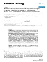
Figure 1 Normal thyroid follicular cells arranged in a honeycomb configuration. The nuclei have finely granular chromatin
and small or inconspicuous nucleoli and show slight anisonucleosis (Papanicolaou stain; magnification, 600Â).
Normal Cytology and General Cytologic Features
to Assess in Thyroid FNA
Generally, the presence of abundant colloid is associated with benign conditions, whereas scant to absent
colloid is seen in neoplasms. Architecturally, a predominant honeycomb configuration of the thyroid
cells and minimal microfollicle formation is associated
with benign nonneoplastic conditions. Conversely, a
predominant syncytial and/or microfollicular pattern
is correlated with neoplasia. The normal thyroid cell
nucleus is about the size of a red blood cell (RBC) (7–9 m),
and has finely granular chromatin, and small or inconspicuous nucleoli (Fig. 1). In nonneoplastic conditions,
slight uniform enlargement and anisonucleosis can be
observed. On the other hand, neoplasms may show
considerable nuclear enlargement (3–4 times size of
RBC), and variable pleomorphism.
B.
Nonneoplastic Disease
Thyroiditis
Lymphocytic (Hashimoto’s) thyroiditis. Lymphocytic
thyroiditis is the most common type of thyroiditis
encountered, and is characterized by variable numbers
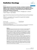
Figure 2 Hashimoto’s thyroiditis. There is intimate admixture of
oncocytes and lymphocytes (DQ stain; magnification, 400Â).
of admixed lymphocytes and follicular cells (Fig. 2).
Oncocytes may predominate in an aspirate, raising the
differential diagnosis of oncocytic neoplasm. There is
usually limited colloid, and the lymphoid population is
polymorphic, showing admixture of small mature lymphocytes, larger reactive lymphoid cells, and occasional
plasma cells. The inflammatory cells are often intimately
admixed with the follicular cells. The differential diagnosis includes lymphoma, which is characterized by a
monomorphic population of lymphoid cells. The follicular cells occasionally demonstrate reactive changes and
4
Elsheikh et al.
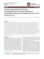
Figure 3 Hashimoto’s thyroiditis associated with reactive
changes, including nuclear enlargement and occasional nuclear
grooves (Papanicolaou stain; magnification, 600Â).
atypia, including nuclear grooves and nuclear enlargement (Fig. 3). Therefore, the diagnostic threshold for
papillary thyroid carcinoma (PTC) should be raised in
the presence of lymphocytic thyroiditis, and only considered when nuclear features of papillary carcinoma are
diffusely present in a population of cells devoid of
infiltrating lymphocytes (23). Nonspecific chronic
inflammation may also be associated with nodular
goiter.
The differential diagnosis of lymphocytic thyroiditis also includes thyroid lesions that may demonstrate lymphoepithelial features (Table 2). Thyroid
tumors with thymus-like features are extremely rare
and include a spectrum of tumors ranging from
benign to malignant, such as ectopic thyroid thymoma
and carcinoma showing thymus-like differentiation
(CASTLE) (24). These tumors are identical in morphology to their mediastinal counterparts (25). CASTLE may demonstrate variable atypia ranging from
bland morphology resembling thymoma, to highly
atypical features resembling nasopharyngeal carcinoma, anaplastic carcinoma, or squamous carcinoma.
The epithelial cells usually possess abundant eosinophilic/pale cytoplasm and large vesicular nuclei with
prominent nucleoli and may be arranged in single
cells and loosely cohesive groups (24,26). FNA of
benign lymphoepithelial lesion is characterized by
scant cellularity of the epithelial component and predominance of the lymphoid component. The squamous and mucinous epithelial component may show
degenerative changes, but is devoid of significant
atypia. PTC variants, such as Warthin-like, tall cell,
and oncocytic types, arising in a background of lymphocytic thyroiditis should also be considered in the
differential diagnosis (27). These tumors show oncocytic features including prominent nucleoli, granular
cytoplasm, and admixed lymphocytes (20). The
absence of nuclear features of PTC, however, excludes
these entities from the differential diagnosis.
Subacute granulomatous (Dequervain’s) thyroiditis.
Subacute thyroiditis rarely presents for FNA, but with
the presence of epithelioid granulomas, the cytologic
features can be diagnostic. Nonspecific findings such
as mixed inflammatory cells, proteinaceous debris,
multinucleated giant cells, and scant degenerated
follicular cells might be seen. The biopsy procedure,
however, may be quite painful for the patient, preventing adequate sampling. In the late fibrotic stages
of the disease, FNA is often nondiagnostic.
Acute thyroiditis. Acute thyroiditis is another
cause of painful thyroid aspiration, and it is rarely
sampled by FNA. The aspirates are rich in neutrophils,
and are associated with scant reactive follicular cells,
fibrin, macrophages, and blood. Bacteria or fungal
organisms are occasionally seen in the background.
Nodular Goiter/Colloid Nodule
Nodular goiter or colloid nodule is the lesion most
commonly sampled by FNA. Characteristically, there
is abundant colloid and variable number of follicular
cells (Fig. 4). Romanowsky/Diff-Quik1 (DQ) stain best
demonstrates the presence of colloid (especially watery
Table 2 Thyroid Lesions with Lympho-Epithelial Features
Lymphocytic thyroiditis
Lymphoma
Papillary thyroid carcinoma, i.e., Warthin-like and tall cell variants
Ectopic thymoma
Benign lymphoepithelial lesion
Carcinoma with thymus-like features
Metastatic carcinoma to intra or perithyroid lymph node
Neoplasm arising in a background of lymphocytic thyroiditis
Figure 4 Colloid nodule showing abundant colloid and admixed
benign thyroid follicular cells (Papanicolaou stain; magnification,
200Â).
Chapter 1: Fine Needle Aspiration of the Head and Neck
Figure 5 Dense colloid has a hyaline quality and stains blueviolet on air-dried smears (A) and dark orange on fixed preparations (B). Note prominent cracks [(A)DQ stain; magnification,
200Â (B) Papanicolaou stain; magnification, 200Â].
colloid). Colloid, when dense, is easy to recognize, has a
hyaline quality, and often shows cracks. It has a dark
blue-violet-magenta appearance on DQ stain, while
stains dark green-orange with Papanicolaou (Fig. 5)
(8). Thin watery colloid has a blue-violet appearance
on DQ, and pale green-orange look on Papanicolaou
stain (Figs. 4 and 6). It often forms a ‘‘thin membrane/
cellophane’’ coating or film, frequently with folds
imparting a ‘‘crazy pavement’’ appearance and/or
cracks (Fig. 6) (8). Thin colloid, however, maybe difficult to recognize in Papanicolaou-stained specimens
and may also disappear completely in thin-layer preparations (28). Thin colloid can also be confused with
serum in bloody specimens. Helpful clues are the
recognition of cracking and folding in colloid, as well
as its tendency to surround follicular cells, whereas
serum accumulates at the edges of the slide and around
platelets, fibrin, and blood clots (3).
The individual thyroid follicular cells are small in
size, arranged in monolayered sheets (Fig. 7) and/or
large balls (Fig. 8), and show no significant nuclear
overlapping or crowding. Occasional microfollicles can
5
Figure 6 Thin watery colloid showing crazy pavement appearance (A) and cellophane-like coating with folds (B) (DQ stain,
magnification, 200Â).
be seen. The cytoplasm is delicate, scant, and may have
small blue-black granules that are of no diagnostic
significance, as they can be observed in benign and
malignant conditions (29). Flat sheets of oncocytic cells
with mild anisonucleosis may be observed in some
aspirates, but no significant atypia is demonstrated.
The number of macrophages present in the background
usually coincides with the extent of cystic degeneration. Many of the macrophages may contain hemosiderin pigment granules. Focal reparative changes might
also be observed in cystic lesions, including the presence of spindle cells and cells with tissue culture
medium appearance (30). Hyperplastic/adenomatoid
nodules show increased cellularity compared to colloid
nodules and may be confused with FNs. Detailed
cytologic features of hyperplastic nodules are later
discussed with follicular lesions.
Diffuse Goiter/Hyperthyroidism (Graves’ Disease)
Most patients with Graves’ disease have diffuse
enlargement of the thyroid gland, and do not require
biopsy. Occasionally, however, prominent nodules
develop, that prompt FNA. The cytologic features of
Graves’ disease are nonspecific, and clinical correlation
6
Elsheikh et al.
Figure 7 Colloid nodule. Flat sheets of benign thyroid cells with
delicate cytoplasm and no significant nuclear overlapping (DQ
stain; magnification, 400Â).
Figure 9 Hyperthyroidism (Graves’ disease). The follicular cells
are commonly arranged in flat sheets and have foamy delicate
cytoplasm with distinctive flame cells. These flame cells, however,
represent a nonspecific finding (DQ stain; magnification, 400Â).
overdiagnose these changes as malignancy or neoplasia, and inquiry should be sought regarding prior
radioactive iodine therapy (32). Sometimes the follicular cells display focal nuclear chromatin clearing, but
other diagnostic nuclear features of papillary carcinoma such as grooves and inclusions are commonly
absent (33).
C.
Figure 8 Colloid nodule. The follicular cells are arranged in
large balls (microtissue fragments), but show no significant
overlapping or atypia (Papanicolaou stain; magnification, 200Â).
is needed for a definitive diagnosis. Specimens are
often cellular, containing numerous follicular cells
and thin colloid. Oncocytes and lymphocytes may be
found in the background (3). The follicular cells are
commonly arranged in flat sheets and have foamy
delicate cytoplasm with distinctive flame cells. Flame
cells are best appreciated on DQ stain and represent
marginal cytoplasmic vacuoles with red to pink frayed
edges (Fig. 9) (31). Flame cells, however, are not specific
to Graves’ disease as they may be encountered in other
nonneoplastic thyroid conditions, FN, and papillary
carcinoma. Occasionally, treated Graves’ disease
shows prominent microfollicular architecture, significant nuclear overlapping and crowding, and considerable atypia. Therefore, care must be taken to not
Follicular Lesions
Follicular lesions of the thyroid represent the most
problematic area in thyroid FNA cytology. The major
entities included in the differential diagnosis comprise
hyperplastic/adenomatoid nodule, FN (adenoma and
carcinoma), and follicular variant of PTC (Table 3).
Below is a discussion of the differential diagnosis of
follicular lesions, cytologic criteria, terminology commonly used as well as terminology recently suggested
by the PSC and NCI thyroid FNA state of the science
conference, and the clinical implications of various
diagnoses rendered (6,7). In general, smears containing abundant colloid are more likely to be benign,
whereas markedly cellular aspirates are more likely to
be neoplastic.
Table 3 Differential Diagnosis of Thyroid Follicular Lesions
Hyperplastic/adenomatoid nodule
Follicular Neoplasm
Follicular adenoma
Follicular carcinoma
Follicular variant of papillary carcinoma
Chapter 1: Fine Needle Aspiration of the Head and Neck
7
Hyperplastic/Adenomatoid Nodule
Hyperplastic/adenomatoid nodule is characterized by
the presence of abundant colloid and variable number
of follicular cells. Often there is evidence of oncocytic
metaplasia and degenerative changes including macrophages and old blood. Hyperplastic nodule is considered in the differential diagnosis of FN when
aspirates are cellular and contain scant colloid. Similar
to colloid nodules, the follicular cells are arranged
mainly in flat sheets with a honeycomb configuration
(Fig. 7). A few microfollicular structures can be seen.
Occasionally, balls and microtissue fragments are
present (Fig. 8), especially when larger gauge needles
are used. The nuclei are uniform in appearance and
approximate the size of RBCs. They show finely
granular chromatin with rare small nucleoli (Fig. 1).
There is minimal nuclear overlapping and crowding.
FN (Adenoma and Carcinoma)
Using specific cytologic criteria, Kini et al. reported a
75% accuracy rate in the diagnosis of follicular carcinoma (34). Most other studies, however, could not
reproduce such accuracy (35). In our opinion, and
those of most expert cytopathologists, FNA cannot
distinguish follicular adenoma from follicular carcinoma, since histologic confirmation is needed to demonstrate the presence of capsular and/or vascular space
invasion. There are, however, several cytologic features reported to be associated with an increased
cancer risk (40–60% cancer risk) (36). These features
include an enlarged nuclei (at least twice the size of
RBC), marked nuclear atypia including significant
nuclear pleomorphism and irregularity, significant
nuclear overlapping, and predominance of microfollicular structures (involving >75% of thyroid clusters)
(36–40). Most follicular carcinomas (>90%) have
prominent microfollicular architecture, while 10% to
15% of cases show significant cytologic atypia (41). It
is important to emphasize, however, that the mere
presence of microfollicles is not equated with neoplasia. In fact, studies have shown that microfollicles
associated with no atypia had a low cancer risk
comparable to that of benign FNA diagnoses (6%
cancer risk) (36), and that microfollicles lacking nuclear overlap and mixed with abundant colloid had a 0%
chance of harboring cancer (37). However, microfollicles with atypia were associated with a 44% cancer
risk, while atypia with or without microfollicles was
associated with malignancy in 60% of the cases, most
of which represented follicular variant of papillary
carcinoma (FVPC) (36).
FNAs of FN are typically highly cellular with
scant colloid. There is prominent microfollicular
and/or syncytial arrangement, involving greater
than 50% to 75% of the cellular groups. Microfollicles
are defined as groups of cells (6–12 cells) arranged in
a ring or rosette-like configuration, and often display
a repetitive pattern (Fig. 10) (41). The syncytial
groups exhibit a three-dimensional appearance with
loss of cell borders (Fig. 11). The nuclei are uniform
and slightly enlarged, but usually demonstrate prominent overlapping and crowding. The chromatin is
Figure 10 Follicular neoplasm showing prominent microfollicular architecture (DQ stain; magnification, 200Â).
Figure 11 Follicular neoplasm showing syncytial groups of
follicular cells. The cells have a uniform appearance and display
prominent nuclear overlapping and crowding (Papanicolaou
stain; magnification, 400Â).
finely to coarsely granular, and nucleoli are infrequent (Fig. 11) (41). Significant nuclear atypia (which
may or may not be present) is characterized by
nuclear enlargement that is greater than twice the
size of RBCs, coarse and clumped chromatin, and
prominent enlarged nucleoli (Fig. 12). FVPC and
medullary carcinoma should also be considered in
the differential diagnosis.
Challenges in the Diagnosis of Hyperplastic/
Adenomatoid Nodule and FN
Clearly, one of the most difficult problems in thyroid
cytology is distinguishing hyperplastic nodule with
little colloid from FN with some colloid (41). As
8
Elsheikh et al.
FN diagnosis has ranged in the literature from none to
predominant. There was no clear definition of how
cellular an aspirate needed to be in order to be classified as ‘‘hypercellular.’’ There were also major disagreements in recognizing colloid, especially when it
had a watery-thin appearance or was associated with
considerable blood in the background (46). A wide
range of interobserver variability (fair to substantial)
has been reported, even among pathologists from the
same institution, when cases diagnosed as follicular
lesion and FN were examined (45). Clary et al. reported
a higher accuracy in predicting neoplastic (84% sensitivity) over nonneoplastic (66% specificity), which supported the utility of FNA as more of a screening test in
these conditions (45). Other studies analyzed clinical
factors that may complement FNA in predicting malignancy, including size more than 4 cm, fixed lesions,
younger patients, male sex, solitary nodule, etc. Clinical
history, however, is provided to the pathologist in only
up to one-third of the cases (45).
Follicular Lesions, Grey Zone, and Terminology
Figure 12 Follicular neoplasm with significant atypia, including
nuclear enlargement, coarse and clumped chromatin, and prominent nucleoli (Papanicolaou stain; magnification, 600Â).
previously mentioned, the mere presence of microfollicles is not diagnostic of FN, as microfollicles may
be focally seen in 5% to 10% of hyperplastic nodules.
Up to 30% of hyperplastic nodules are highly cellular,
and 15% to 20% show scant colloid (42,43). Although
degenerative changes are often associated with hyperplastic nodule, they may be found in up to 30% of FN.
A definitive diagnosis of hyperplastic nodule should
not be made in the absence of colloid (3,41–43). Low
cellularity may be encountered in aspirates of FN
because of poor biopsy techniques or because of a
macrofollicular architecture yielding abundant colloid
and scant follicular cells. Some FNs are highly vascular, yielding abundant blood, and rare follicular
groups with prominent nuclear overlapping and/or
microfollicular architecture (44). Oncocytic change
and flame cells may also be found in FN (benign and
malignant), in addition to hyperplastic nodule and
colloid nodule.
Several studies have evaluated the variability in
reporting and diagnosing FN and cellular hyperplastic
nodules (40,45,46). The areas of greatest debate and
confusion included terminology and criteria employed
in diagnosing FN. Differences in terminology involved
mainly the use of two diagnostic categories (i.e., follicular lesion and FN) versus one category. Some pathologists apply the terms follicular lesion and FN
interchangeably, while others require more stringent
criteria for the diagnosis of FN. Other than increased
cellularity, the criteria used by cytopathologists in the
diagnosis of FN vary from strict to none. For example,
the proportion of microfollicles needed to establish a
Although the terms ‘‘follicular lesion’’ and ‘‘follicular
neoplasm’’ are used interchangeably by some authors,
we do not consider them synonymous. According to
literature review, lesions categorized as indeterminate
account for 5% to 42% of FNA diagnoses. We do not
recommend the use of ‘‘indeterminate’’ as a stand-alone
diagnosis, as its meaning has not been standardized
and may be interpreted in different ways. Indeterminate has been used by different authors and institutions
to refer to a variety of diagnoses, including FN, follicular lesion, suspicious for malignancy, and atypia not
otherwise specified. Redman et al. surveyed 133 clinicians (endocrinologists, surgeons, and thyroid specialists) in order to determine the implications of FNA
diagnoses on management options (47). In this study,
clinicians appropriately opted for repeat FNA in 98% of
the nondiagnostic cytologic terminology, and elicited a
96% surgical excision response to ‘‘suspicious’’ diagnoses. However, clinicians chose repeat FNA (58%) or
surgery (32%) for indeterminate diagnoses, and selected
repeat FNA (37%) or surgery (52%) for ‘‘atypical’’
designations (47). The study clearly demonstrated that
confusion arose with the atypical and indeterminate
diagnoses. Indeterminate was confused with nondiagnostic in some cases, while atypical was too ambiguous
and treated as suspicious in many other cases. The
majority of clinicians, on the other hand, correctly
interpreted the nondiagnostic and suspicious diagnoses. The indeterminate category, in reality, includes two
types of lesions with different clinical implications:
(i) truly indeterminate, showing features intermediate
between hyperplastic/colloid nodule and FN which
may be best managed by repeat FNA, clinical followup, and correlation with US and ancillary studies; and
(ii) follicular patterned lesion consistent with FN, in
which repeat biopsy is unlikely to clarify the situation,
and surgery would be indicated (8).
Two European studies found no malignancy on
follow-up of FNAs diagnosed as follicular lesion and
FN (48,49). The authors advocated a less aggressive
Chapter 1: Fine Needle Aspiration of the Head and Neck
9
approach to management, i.e., clinical follow-up. The
authors, however, did not apply strict criteria, and FN
was loosely defined as hypercellular smears associated
with scant colloid and microfollicles, with no mention
of the percentage of microfollicle formation, nuclear
atypia, or other architectural patterns. Architectural
features and nuclear atypia, in addition to colloid and
cellularity, should be evaluated in these hypercellular
specimens, so pathologists can better define and classify those gray-zone lesions and lessen the number of
cases classified as indeterminate. We recommend subdividing the indeterminate category, as was recommended by the PSC and NCI, into (i) Follicular lesion
of undetermined significance (FLUS) and (ii) FN (6,7).
Follicular Lesion of Undetermined Significance
In 2006, the PSC introduced the terminology ‘‘cellular
lesion cannot rule out FN,’’ addressing lesions falling in
the gray zone (6). In 2007, The NCI thyroid state-of-thescience conference suggested similar terminologies,
including ‘‘FLUS’’, ‘‘a typical follicular lesion’’, and
‘‘atypia of undetermined significance’’ (7). These terminologies were chosen in order to avoid the confusing
terms of ‘‘follicular lesion,’’ ‘‘indeterminate,’’ ‘‘atypical,’’ etc. as stand alone diagnoses. This designation of
‘‘FLUS’’ is employed when the major differential diagnosis is between hyperplastic nodule and FN. These
aspirates are often highly cellular and have scant
colloid. There is admixture of flat sheets and microfollicles/syncytial fragments, with minimal nuclear
overlapping and crowding (Fig. 13A). This diagnosis
is also rendered when smears from different passes
show mixed cytologic findings ranging from ‘‘benign’’
to ‘‘possible FN.’’ Bloody specimens of low cellularity,
but containing microfollicles and prominent nuclear
overlap (highly vascular lesions) would also be included in this category (Fig. 13 B). This latter cytologic clue,
although important in recognizing some FN, has rarely
been emphasized in the literature (40).
D.
Oncocytic (Hurthle Cell) Neoplasms
Similar to FN, the separation of oncocytic adenoma
from carcinoma requires histologic evaluation for evidence of capsular and/or vascular space invasion.
Oncocytic neoplasms usually render highly cellular
aspirates with scant colloid and rare to absent lymphocytes. The oncocytes are arranged mainly in large and
small clusters and isolated single cells (Fig. 14). Occasional microfollicles may be seen. The cells show
uniform appearance, and have abundant granular cytoplasm with well-defined borders, round to oval nuclei,
granular chromatin, and prominent nucleoli. Cytologic
atypia may be observed in oncocytic lesions, including
scattered nuclear enlargement and pleomorphism (50).
A variety of other neoplastic and nonneoplastic lesions
may show oncocytic/granular features (Table 4). The
main differential diagnosis includes oncocytic nodule
in association with a nodular goiter or lymphocytic
thyroiditis. Admixture of benign thyroid follicular cells
and colloid favors nodular goiter, while a prominent
Figure 13 Follicular lesion of undetermined significance
(A) This specimen showed admixture of flat sheets and microfollicles, with no significant atypia, and scant to absent colloid.
(B) This bloody aspirate had rare clusters of follicular cells with
prominent nuclear overlapping, and occasional microfollicles [(A)
DQ stain; magnification, 400Â (B) DQ stain; magnification,
200Â].
lymphoid cell component favors lymphocytic thyroiditis. Endocrine neoplasms such as medullary carcinoma, paraganglioma, and parathyroid adenoma may
show considerable variability in cell size and shape,
including polygonal to spindle cells with ill-defined
granular cytoplasm and frayed borders. In medullary
carcinoma, the nuclei typically have a ‘‘salt and pepper’’ chromatin, but may show pleomorphism with
prominent nucleoli, and scattered stripped atypical
nuclei. The cytologic features of metastatic renal cell
carcinoma (RCC) are discussed later in the chapter.
E. Malignant Neoplasms
Papillary Thyroid Carcinoma
PTC is the most commonly encountered thyroid
malignancy. It can be partially cystic, or entirely cystic
10
Elsheikh et al.
Figure 14 Oncocytic (Hurthle cell) neoplasm showing sheets of
oncocytic cells and minimal to absent colloid (DQ stain; magnification, 200Â).
Figure 15 Papillary carcinoma, classic type. Tightly cohesive
clusters with papillary architecture are seen (Papanicolaou stain;
magnification, 400Â).
Table 4 Thyroid Lesions with Oncocytic (Hurthle)/Granular Cell
Features
Oncocytic neoplasm
Lymphocytic thyroiditis
Nodular goiter
Medullary carcinoma
Papillary carcinoma: oncocytic, tall cell, and Warthin-like variants
Parathyroid adenoma
Paraganglioma
Metastatic renal cell carcinoma
in up to 10% of cases. Classic PTC shows distinctive
architectural and cytologic features. Architectural features include the presence of papillary and tightly
cohesive three-dimensional clusters of neoplastic cells
(Fig. 15). Many of the neoplastic groups demonstrate
prominent nuclear crowding and overlapping. Thick
colloid with a ‘‘bubble-gum’’ appearance may be
present in the background in up to one quarter of
the cases (Fig. 16) (42). In most cases, the cytoplasm
has a thick ‘‘metaplastic’’ consistency, with welldefined borders (51). Occasionally, especially in cystic
PTC, the neoplastic cells show prominent cytoplasmic
microvacuolization resembling macrophages (52). The
hallmark for diagnosing PTC, however, is based on its
distinctive nuclear features. The diffuse presence of
nuclear grooves, nuclear enlargement, and finely
granular (powdery) chromatin must be present, before
a definitive diagnosis is rendered (Fig. 17). The
ground glass appearance of the nuclei seen in histologic sections is an artifact of formalin fixation and is
not recognized in cytologic preparations. Nuclear
grooves may traverse the entire longitudinal axis of
the nucleus, or may appear as invaginations of the
nuclear membrane. Intranuclear pseudoinclusions,
which are also distinctive of PTC, are not present in
Figure 16 Papillary carcinoma showing thick colloid with a
‘‘bubble-gum’’ appearance in the background (DQ stain; magnification, 200Â).
all cases. These inclusions usually have sharp margins, and should reflect the color of the cytoplasm of
that cell (not just a clear hole in the nucleus) (53).
Nucleoli are often small and peripherally situated
against a thickened nuclear membrane. The presence
of psammoma bodies, although not diagnostic by
itself, should raise a red flag for PTC, and may be
seen in up to 40% of cases (13). A proportion of cases
show multinucleated giant cells, the presence of which
should increase awareness about the possibility of
PTC, and invoke a more thorough search for diagnostic nuclear features (Fig. 18). It is important to note
that there is no single feature that is diagnostic of PTC,
Chapter 1: Fine Needle Aspiration of the Head and Neck
11
of entirely cystic PTC (56). This is mainly due to scant
cellularity and prominent degenerative changes. The
aspirate may consist predominately of foamy macrophages, blood, and reparative changes. Other neoplastic and nonneoplastic lesions may also be associated
with prominent cystic change (discussed later in the
section on cystic lesions). When nuclear features of
PTC are diffusely present in the neoplastic cells, a
specific diagnosis can be rendered, but, occasionally,
PTC nuclear features are only found focally. However,
the presence of nuclear grooves and nuclear inclusions in association with fine powdery chromatin,
even focally, should always raise a warning flag,
and elicit a ‘‘suspicious for PTC’’ diagnosis.
Variants of Papillary Thyroid Carcinoma
Figure 17 Papillary carcinoma demonstrating characteristic
nuclear features, including powdery chromatin, grooves, and
intranuclear pseudoinclusions (Papanicolaou stain; magnification, 600Â).
There are several described variants of PTC, including
follicular, tall cell, oncocytic, and columnar cell.
Follicular variant of PTC (FVPC) is by far the most
common of these subtypes. By definition, no papillary
structures are identified, and the neoplasm consists
predominately of follicular structures. The aspirates
mainly display branching monolayered sheets, which
are considered to be a significant low-power discriminating feature from other follicular-patterned neoplasms (Fig. 19) (57). The combination of flat
syncytial sheets, nuclear enlargement, and fine powdery chromatin, were found to be the most sensitive
criteria, whereas the combination of nuclear enlargement, fine chromatin, and nuclear grooves were the
most specific, in establishing the diagnosis of FVPC
(Fig. 20) (58). Intranuclear pseudoinclusions are seen
in less than half the cases. Fulciniti et al. also emphasized the presence of nuclear grooves and nucleoli
over intranuclear inclusions in the diagnosis of FVPC
(57). The importance of both architectural and nuclear
features in establishing the diagnosis of FVPC cannot
Figure 18 Multinucleated giant cells associated with papillary
carcinoma (Papanicolaou stain; magnification, 400Â).
and that a definitive diagnosis should be based on a
constellation of cytologic features. Several studies
have attempted to determine the most sensitive cytologic criteria for diagnosing classic PTC, and found
nuclear inclusions, nuclear grooves, papillary structures, and metaplastic cytoplasm, when present in
combination, to be the most reliable cytologic features
(13,42,51,54). PTC may show prominent cystic change,
and this accounts for PTC being the most common
cystic neoplasm of the thyroid (55). FNA is unable to
establish a diagnosis of malignancy, however, in 50%
Figure 19 Follicular variant of papillary carcinoma showing a
distinctive low power appearance of branching monolayered
sheets (DQ stain; magnification, 200Â).
12
Elsheikh et al.
Figure 20 Follicular variant of papillary carcinoma. The combination of nuclear enlargement, fine chromatin, and nuclear
grooves were found to be most specific in establishing the
diagnosis. Softer criteria include nuclear membrane thickening
and eccentric placement of nucleoli against the nuclear membrane (Papanicolaou stain; magnification, 600Â).
be overstressed. The predominance of microfollicles in
some cases can lead to misclassification as FN, and a
predominant monolayered sheet pattern may be misinterpreted as honeycomb sheets associated with nodular goiter. However, careful high-power examination
of the nuclei and recognition of the nuclear features of
PTC in these cases will usually establish the diagnosis
of FVPC, and prevent those potential pitfalls. Not
infrequently, FVPC may show abundant colloid or
paucity of nuclear features of PTC, leading to a misdiagnosis of benign thyroid disease or FN. In fact, FVPC
is only second to sampling error as the most common
cause of false-negative diagnoses in thyroid FNA. Wu
et al. reported 11 false-negative cases of FVPC, where 6
cases were attributed to sampling error (micropapillary
carcinoma), and 5 cases showed focal atypia in a
background of abundant colloid and cystic change (17).
The oncocytic (Hurthle cell) variant of PTC is characterized by large tumor cells with abundant dense
cytoplasm, well-defined cytoplasmic borders, eccentric nuclei, and nuclear features of PTC (Fig. 21). The
tall cell variant of PTC shows elongated tumor cells
with enlarged nuclei, overlapping and stratification of
nuclei, and dense eosinophilic cytoplasm (Fig. 22). The
nuclear features of PTC, in tall cell variant, are often
diffuse and extensive, compared with classic PTC. The
columnar cell variant of PTC demonstrates elongated to
columnar cells with ill-defined cell borders, delicate
cytoplasm with focal vacuolization/clearing, nuclear
pseudostratification, and oval to elongated hyperchromatic nuclei (59). Architecturally, it may resemble
Figure 21 Oncocytic (Hurthle cell) variant of papillary carcinoma. The neoplastic cells show cytoplasmic oncocytic features,
but demonstrate nuclear features of papillary carcinoma, including a rare intranuclear inclusion (at 4:00) (DQ stain; magnification, 400Â).
Figure 22 Tall cell variant of papillary carcinoma. Elongated
tumor cells with abundant cytoplasm, enlarged overlapping
nuclei, and characteristic nuclear features of papillary carcinoma
(Papanicolaou stain; magnification, 600Â).
colonic adenocarcinoma. Cytologic classification of
PTC is accurate in over 90% of classic PTC, and in
most FVPC, but is much less reproducible in other
PTC variants (59). Tall cell and oncocytic variants of
PTC, in particular, show significant overlap in their
Chapter 1: Fine Needle Aspiration of the Head and Neck
13
cytologic appearance. It is more important, in our
opinion, to be familiar with the spectrum of cytologic
appearances of PTC than it is to render a specific PTCvariant diagnosis.
Suspicious for PTC
We issue a diagnosis of suspicious for PTC when
nuclear features of PTC are only focally present.
These nuclear features, however, must be present in
combination (Fig. 23), and include rare nuclear
grooves, nuclear enlargement, and powdery chromatin. Focal nuclear grooves and/or intranuclear inclusions (by themselves) can be found in a variety of
other neoplastic and nonneoplastic conditions,
including nodular hyperplasia, lymphocytic thyroiditis, FN, and medullary carcinoma (3). It is important
to recognize ‘‘suspicious for PTC’’ as a distinct category, and not to lump it with other indeterminate or
FN diagnoses, because of its substantially greater
association with malignancy on surgical follow-up.
Logani et al. and Wu et al. reported cancer follow-up
rates of 77% and 75%, respectively, when rendering
such diagnoses (60). This is in contrast to the cancer
follow-up rate of 10% to 30% typically associated
with indeterminate or FN diagnoses. With such an
increased risk of malignancy, clinicians and patients
may consider total thyroidectomy as an alternative
option to lobectomy. Another management choice
includes lobectomy with intraoperative consultation,
which has been shown to be helpful in an additional
30% of cases (23).
Figure 23 Suspicious for papillary carcinoma. This case
showed nuclear enlargement, powdery chromatin, rare nuclear
grooves, and no nuclear pseudoinclusions (Papanicolaou stain;
magnification, 600Â).
Figure 24 Medullary carcinoma. There are many single cells,
as well as clusters with a microacinar configuration (carcinoidlike appearance). Amyloid is present in the background (DQ
stain; magnification, 200Â).
Medullary Carcinoma
Medullary carcinoma is notorious for its variable cytologic appearances, ranging from a monomorphic to a
pleomorphic cell population. Typically, medullary
carcinoma presents mostly as isolated cells with occasional clusters (Fig. 24). The neoplastic cells may have a
plasmacytoid, spindle, epithelioid, or carcinoid-like
appearance, but often a mixture of different cell types
is encountered in the same specimen (Figs. 24–26). The
cytoplasm has a delicate lacy quality, and may show
Figure 25 Medullary carcinoma showing variable appearance,
including loosely cohesive groups of uniform cells with delicate
cytoplasm, round to oval nuclei, and occasional spindling. The
nuclei have a salt and pepper chromatin pattern (Papanicolaou
stain; magnification, 400Â).
14
Elsheikh et al.
Figure 26 Medullary carcinoma consisting predominately of
single cells with abundant cytoplasm and eccentric nuclei
(plasmacytoid appearance). There is also significant nuclear
pleomorphism and prominent nucleoli (Papanicolaou stain;
magnification, 600Â).
background (Fig. 24). Amyloid has an appearance
similar to thick colloid, but is confirmed with Congo
red stain. Immunocytochemical stains for calcitonin
and neuroendocrine markers such as chromogranin
and synaptophysin are extremely helpful in establishing the diagnosis. The tumor cells are usually positive
for carcinoembroyonic antigen (CEA), but negative for
thyroglobulin. Serum calcitonin levels are elevated in
most patients, and help establish the diagnosis. The
differential diagnosis includes oncocytic neoplasm,
papillary carcinoma, plasmacytoma, and mesenchymal
tumors. Oncocytic tumors show prominent nucleoli
and lack the neuroendocrine chromatin pattern.
Although medullary carcinoma may display intranuclear holes, it will not show other diagnostic nuclear
features of PTC such as nuclear grooves and powdery
chromatin (62). In addition, PTC is negative for calcitonin and chromogranin, and is positive for thyroglobulin. Mesenchymal tumors and spindle cell melanoma
are included in the differential diagnosis of predominately spindle medullary carcinoma, while plasmacytoma and melanoma may be confused with
plasmacytoid medullary carcinoma. Immunohistochemistry (IHC) can play a pivotal role in establishing a definitive diagnosis in those cases.
Insular Carcinoma/Poorly Differentiated Carcinoma
pink cytoplasmic granules. Sometimes, the neoplastic
cells show a predominately oncocytic appearance,
mimicking oncocytic neoplasms (Fig. 27). However,
oncocytic neoplasm usually has cells with well-defined
borders, in contrast to ill-defined wispy borders of
medullary carcinoma. Clear cell change may be
observed, but is often only focally present. The nuclei
are round to oval, with finely to coarsely granular
chromatin (salt and pepper), and have small or inconspicuous nucleoli (Fig. 25). Binucleation and multinucleation are common. Occasional intranuclear
inclusions are appreciated in some cases (Fig. 27) (61).
Variable amount of amyloid may be present in the
Insular carcinoma is a rare aggressive malignancy
characterized histologically by the presence of focal
or diffuse insular pattern. FNA smears are usually
highly cellular and composed of monomorphic
appearing small follicular cells, occurring singly and
in clusters (63). Occasionally, intact insulae of follicular cells surrounded by hyaline stroma are seen. The
neoplastic cells show scant delicate cytoplasm with
round hyperchromatic nuclei, coarsely granular chromatin, and mild-to-moderate nuclear irregularities.
Many naked nuclei are often present in the background. Microfollicles as well as infrequent grooves
and inclusions can be seen, mimicking FN and PTC. In
our experience, and those of others, a definitive diagnosis of insular carcinoma cannot be established by
FNA (63). Most cases are diagnosed as FN or suspicious for malignancy. A helpful clue as to the aggressive nature of this tumor is the presence of increased
mitotic activity and individual cell necrosis.
Anaplastic Carcinoma
Figure 27 Medullary carcinoma with a predominant oncocytic
appearance. Notice scattered intranuclear inclusions (DQ stain;
magnification, 600Â).
Anaplastic carcinoma is the most aggressive of all
primary thyroid cancers and is usually unresectable
or present with metastatic disease. Therefore, an accurate FNA diagnosis may spare the patient unnecessary
surgery, in favor of radiation therapy (3). Cytologically, anaplastic carcinoma presents as isolated cells and
loose groups. The malignant cells show extreme
nuclear pleomorphism and atypia, and may have a
spindle, giant cell, or small cell appearance (Fig. 28).
Occasional multinucleated forms are seen. Typically,
the nuclei have coarsely irregular chromatin and
prominent macronucleoli. Differential diagnosis
includes poorly differentiated components of papillary, follicular, and medullary carcinoma. Anaplastic
Chapter 1: Fine Needle Aspiration of the Head and Neck
15
Table 5 Differential Diagnosis of Lymphoid Lesions of Thyroid
Lymphocytic thyroiditis
Lymphoid hyperplasia in peri- or intrathyroid lymph node
Lymphoma
Medullary carcinoma
Small cell carcinoma
Suspicious for Malignancy
Figure 28 Anaplastic carcinoma. There are loosely cohesive
groups of extremely pleomorphic cells with spindle cell appearance (DQ stain; magnification, 600Â).
carcinoma, therefore, can only be considered in the
absence of well-differentiated components. Cytokeratin stain may be employed to exclude sarcoma, lymphoma, and melanoma. In contrast to FNs, PTC, and
medullary carcinoma, anaplastic carcinoma is infrequently positive for thyroid transcription factor-1
(TTF-1). The differential diagnosis also includes granulation tissue, repair, I131 therapy effect, and metastatic carcinoma. Abundant necrosis may be present in
the background, rendering an unsatisfactory diagnosis. Therefore, the presence of rare pleomorphic cells
associated with necrosis in an elderly patient should
raise the possibility of anaplastic carcinoma and trigger additional studies.
Lymphoma
FNA cytology of thyroid lymphoma is similar to its
lymph node counterpart. Most patients have an
already established history of lymphoma. NonHodgkin’s lymphoma (NHL) is by far the most common type, with Hodgkin’s disease representing a
rarity in the thyroid. Diffuse large B-cell lymphoma
(DLBCL) and extranodal marginal zone B-cell lymphoma are the most common types. The smears have
a monomorphic lymphoid appearance and show
many lymphoglandular bodies in the background.
Flow cytometry and/or immunocytochemistry are
necessary for establishing a definitive diagnosis, particularly in patients with no previous history. Differential diagnosis includes lymphoid hyperplasia and
lymphocytic thyroiditis (Table 5), both of which are
characterized by a polymorphic population of
lymphoid cells. Flow cytometry is often required to
separate low-grade lymphoma from lymphoid hyperplasia. CD45, CD20, CD3, calcitonin, chromogranin,
CEA, and cytokeratin may be needed in difficult cases,
to distinguish lymphoma from medullary carcinoma
and small cell carcinoma.
We use this diagnostic category when the cytologic
features are suggestive of a specific malignancy, but a
definitive diagnosis cannot be rendered. A definitive
diagnosis of malignancy may not be rendered due to
quantitative reasons (i.e., malignant appearing cells,
but limited cellularity) or qualitative reasons (i.e.,
focal or less than well developed features of malignancy). The most commonly encountered example of
this diagnostic category is ‘‘suspicious for PTC’’
(Fig. 23). It is imperative to establish ‘‘suspicious for
malignancy’’ as a distinct and separate diagnostic
category and not to combine it with indeterminate or
FN diagnoses because of its significantly increased
association with malignancy on follow-up surgery.
‘‘Suspicious for PTC’’ has a reported cancer followup rate of approximately 75% (58,60). With such a
high risk of malignancy, clinicians and patients may
consider total thyroidectomy as an alternative management option to lobectomy. Intraoperative consultation may also be suggested. Careful attention to
cytologic details is especially needed when examining
cystic lesions, as atypical reparative changes in benign
cysts may be confused with malignancy (41).
F.
Cystic Lesions
Thyroid cysts account for approximately 15% to 25 %
of all thyroid nodules, and are most commonly due to
cystic degeneration in nodular goiter. Thyroid cyst
fluid may be clear yellow, hemorrhagic, or dark
brown. It usually contains macrophages (Fig. 29),
inflammatory cells, colloid, and degenerated follicular
cells. Features associated with benign cysts include
complete drainage of the cyst with no residual mass,
no recurrence, and absence of cytologic atypia. Lining
cells from benign cysts often show reparative features,
including flat sheets of evenly spaced cells with elongated shape, dense cytoplasm, distinct cell borders,
and prominent nucleoli. The major differential diagnostic consideration in these cases is cystic PTC vs.
benign cyst. Studies have shown that 7% to 29% of
thyroid cysts are malignant (64). Cystic PTC often
lacks the repair-like spindle morphology and shows
atypia characterized by papillary architecture, nuclear
enlargement and crowding, nuclear grooves, and rare
intranuclear inclusions (64). Atypical features may
also be focally encountered in benign cysts and
include rare cells with nuclear grooves and fine pale
chromatin, which could raise the suspicion for PTC
(Fig. 30). Other atypical features associated with cystic
change such as fibroblastic proliferation and atypical
repair, including atypical elongated round or bizarre
cells, can be confused with malignancy (41). Some