Ebook ECG interpretation made incredibly easy: Part 2
Bạn đang xem bản rút gọn của tài liệu. Xem và tải ngay bản đầy đủ của tài liệu tại đây (10.46 MB, 204 trang )
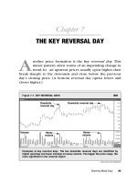
Part III Treating arrhythmias
ECG_Chap09.indd 173
9 Nonpharmacologic treatments
175
10 Pharmacologic treatments
205
7/8/2010 4:30:58 PM
ECG_Chap09.indd 174
7/8/2010 4:30:59 PM
9
Nonpharmacologic treatments
Just the facts
In this chapter, you’ll learn:
nonpharmacologic treatments of arrhythmias and how
they work
ways to identify and treat complications of nonpharmacologic treatments
nursing care for patients receiving nonpharmacologic
treatments
patient teaching points for nonpharmacologic treatments.
A look at pacemakers
A pacemaker is an artificial device that electrically stimulates the
myocardium to depolarize, which begins a contraction.
Pacemakers may be used when a patient has an arrhythmia,
such as certain bradyarrhythmias and tachyarrhythmias, sick
sinus syndrome, or atrioventricular (AV) blocks. The device may
be temporary or permanent, depending on the patient’s condition.
Pacemakers are commonly necessary following myocardial infarction or cardiac surgery.
Keep up to
pace with pacemaker
information!
And the beat goes on…
Pacemakers work by generating an impulse from a power source
and transmitting that impulse to the heart muscle. The impulse
flows throughout the heart and causes the heart muscle to depolarize. Pacemakers consist of three components: the pulse generator, the pacing leads, and the electrode tip.
ECG_Chap09.indd 175
7/8/2010 4:30:59 PM
NONPHARMACOLOGIC TREATMENTS
176
Making the pacer work
The pulse generator contains the pacemaker’s power source and
circuitry. The lithium batteries in a permanent or implanted pacemaker are its power source and last about 10 years. The circuitry
of the pacemaker is a microchip that guides heart pacing.
A temporary pacemaker, which isn’t implanted, is about the
size of a small radio or a telemetry box and is powered by alkaline batteries. These units also contain a microchip and are programmed by a touch pad or dials.
A stimulus on the move
An electrical stimulus from the pulse generator moves through
wires or pacing leads to the electrode tips. The leads for a pacemaker designed to stimulate a single heart chamber are placed in
A look at pacing leads
Pacing leads have either one electrode (unipolar) or two (bipolar). These illustrations show the difference between the
two leads.
Unipolar lead
In a unipolar system, electric current moves from the
pulse generator through the leadwire to the negative pole.
From there, it stimulates the heart and returns to the pulse
generator’s metal surface (the positive pole) to complete
the circuit.
Bipolar lead
In a bipolar system, current flows from the pulse generator through the leadwire to the negative pole at the tip. At
that point, it stimulates the heart and then the positive pole
within the lead to complete the circuit.
From pulse generator
Pulse generator (+)
Direction of current flow
Pacing lead
Pacing lead
Electrode (–)
Electrode (+)
Electrode (–)
ECG_Chap09.indd 176
7/8/2010 4:31:00 PM
WORKING WITH PACEMAKERS
177
either the atrium or the ventricle. For dual-chamber, or AV, pacing, the leads are placed in both chambers, usually on the right
side of the heart.
One lead or two
The electrodes—one on a unipolar lead or two on a bipolar lead—
send information about electrical impulses in the myocardium
back to the pulse generator. The pulse generator senses the
heart’s electrical activity and responds according to how it has
been programmed.
A unipolar lead system is more sensitive to the heart’s intrinsic
electrical activity than is a bipolar system. A bipolar system isn’t
as easily affected by electrical activity outside the heart and the
generator (for example, from skeletal muscle contraction or magnetic fields). (See A look at pacing leads.)
Working with pacemakers
On an ECG, you’ll notice a pacemaker spike right away. (See
Pacemaker spikes.) It occurs when the pacemaker sends an electrical impulse to the heart muscle. That impulse appears as a vertical line or spike.
Depending on the position of the electrode, the spike appears
in different locations on the waveform.
• When the atria are stimulated by the pacemaker, the spike is
followed by a P wave and the patient’s baseline QRS complex and
T wave. This series of waveforms represents successful pacing, or
capture, of the myocardium. The P wave may look different from
the patient’s normal P wave.
• When the ventricles are stimulated by a pacemaker, the spike is
followed by a QRS complex and a T wave. The QRS complex appears wider than the patient’s own QRS complex because of the
way the ventricles are depolarized.
• When the pacemaker stimulates both the atria and the ventricles, the first spike is followed by a P wave, then a spike, and then
a QRS complex. Be aware that the type of pacemaker used and
the patient’s condition may affect whether every beat is paced.
Permanent and temporary pacemakers
Depending on the patient’s signs and symptoms, a permanent or a
temporary pacemaker can be used to maintain heart rhythm. Lead
placement varies according to the patient’s specific needs.
ECG_Chap09.indd 177
Pacemaker
spikes
Pacemaker impulses—
the stimuli that travel
from the pacemaker to
the heart—are visible on
the patient’s ECG tracing as spikes. Large or
small, pacemaker spikes
appear above or below
the isoelectric line. This
example shows an atrial
and a ventricular pacemaker spike.
P wave
QRS complex
Ventricular
pacemaker
spike
Atrial
pacemaker
spike
7/8/2010 4:31:01 PM
178
NONPHARMACOLOGIC TREATMENTS
Permanent pacemakers
A permanent pacemaker is used to treat chronic heart conditions
such as AV block. It’s surgically implanted, usually under local
anesthesia. The leads are placed transvenously, positioned in the
appropriate chambers, and then anchored to the endocardium.
(See Placing a permanent pacemaker.)
Pocket generator
The generator is then implanted in a pocket made from subcutaneous tissue. The pocket is usually constructed under the clavicle.
Permanent pacemakers are programmed during implantation.
The programming sets the conditions under which the pacemaker
functions and can be adjusted externally if necessary.
Temporary pacemakers
A temporary pacemaker is commonly inserted in an emergency.
The patient may show signs of decreased cardiac output, such as
hypotension or syncope. The temporary pacemaker supports the
patient until the condition resolves.
A temporary pacemaker can also serve as a bridge until a permanent pacemaker is inserted. Temporary pacemakers are used
for patients with heart block, bradycardia, or low cardiac output.
Several types of temporary pacemakers are available, including
transvenous, epicardial, and transcutaneous.
Going the transvenous way
Doctors may use the transvenous approach—inserting the pacemaker through a vein, such as the subclavian or internal jugular
vein—when inserting a temporary pacemaker at the bedside or
in other nonsurgical environments. The transvenous pacemaker
is probably the most common and reliable type of temporary
pacemaker. It’s usually inserted at the bedside or in a fluoroscopy
suite. The leadwires are advanced through a catheter into the right
ventricle or atrium and then connected to the pulse generator.
A temporary
pacemaker serves
as a bridge until a
permanent one can
be placed. Types
include transvenous,
epicardial, and
transcutaneous.
Taking the epicardial route
Epicardial pacemakers are commonly used for patients undergoing cardiac surgery. The doctor attaches the tips of the leadwires
to the surface of the heart and then brings the wires through the
chest wall, below the incision. They’re then attached to the pulse
generator. The leadwires are usually removed several days after
surgery or when the patient no longer requires them.
ECG_Chap09.indd 178
7/8/2010 4:31:01 PM
WORKING WITH PACEMAKERS
179
Placing a permanent pacemaker
The doctor who implants the endocardial pacemaker usually selects a transvenous route and begins lead placement
by inserting a catheter percutaneously or by venous cutdown. Then, with a stylet and fluoroscopic guidance, the doctor
threads the catheter through the vein until the tip reaches the endocardium.
Lead placement
For lead placement in the atrium, the tip must lodge in
the right atrium or coronary sinus, as shown here. For
placement in the ventricle, it must lodge within the right
ventricular apex in one of the interior muscular ridges, or
trabeculae.
Implanting the generator
When the lead is in the proper position, the doctor secures
the pulse generator in a subcutaneous pocket of tissue
just below the clavicle. Changing the generator’s battery
or microchip circuitry requires only a shallow incision over
the site and a quick component exchange.
Subclavian vein
Generator in
subcutaneous pocket
Right atrial lead
Right ventricular lead
Following the transcutaneous path
Use of an external or transcutaneous pacemaker has become commonplace in the past several years. In this noninvasive method,
one electrode is placed on the patient’s anterior chest wall, and
a second is applied to his back. An external pulse generator then
emits pacing impulses that travel through the skin to the heart
muscle. Transcutaneous pacing is also built into many defibrillators for use in an emergency. In this case, the electrodes are built
into the same electrode patches used for defibrillation.
Transcutaneous pacing is a quick and effective method of pacing heart rhythm and is commonly used in an emergency until
a transvenous pacemaker can be inserted. However, some alert
ECG_Chap09.indd 179
7/8/2010 4:31:02 PM
NONPHARMACOLOGIC TREATMENTS
180
A look at a pulse generator
This is an illustration of a single-chamber temporary pulse generator with brief descriptions of its various parts.
The pace meter registers every
pacing stimulus delivered to the
heart.
The sensing meter registers
every time an intrinsic
depolarization is recognized
The rate control sets the number
of pulses to be given each minute.
The pacemaker sensitivity
control adjusts pacemaker
sensitivity to the patient’s
heart rate.
The output controls determine the
amount of electricity sent to the
heart (in milliamperes).
The on-off buttons activate and
deactivate the pulse generator.
patients can’t tolerate the irritating sensations produced from prolonged pacing at the levels needed to pace the heart externally.
Setting the controls
When your patient has a temporary pacemaker, you’ll notice
several types of settings on the pulse generator. The rate control
regulates how many impulses are generated in 1 minute and is
measured in pulses per minute (ppm). The rate is usually set at
60 to 80 ppm. (See A look at a pulse generator.) The pacemaker
ECG_Chap09.indd 180
7/8/2010 4:31:02 PM
WORKING WITH PACEMAKERS
fires if the patient’s heart rate falls below the preset rate. The rate
may be set higher if the patient has a tachyarrhythmia that’s being
treated with overdrive pacing.
Measuring the output
The electrical output of a pacemaker is measured in milliamperes.
First, an assessment is made of the stimulation threshold, or how
much energy is required to stimulate the cardiac muscle to depolarize. The stimulation threshold is sometimes referred to as the
energy required for capture. The pacemaker’s output is then set
higher than the stimulating threshold to ensure capture.
Sensing the norm
You can also program the pacemaker’s sensing threshold, measured in millivolts. Most pacemakers let the heart function naturally and assist only when necessary. The sensing threshold allows
the pacemaker to do this by sensing the heart’s normal activity.
181
Ages
and stages
Pacemakers in
elderly patients
Older adults with active
lifestyles who require a
pacemaker may respond
best to atrioventricular
synchronous pacemakers. That’s because
older adults have a
greater reliance on atrial
contraction, or atrial
kick, to complete ventricular filling.
Demand pacemakers
A demand pacemaker responds to the heart’s activity by monitoring the intrinsic rhythm and pacing only when the heart can’t do
so itself. (See Pacemakers in elderly patients.)
Pacemaker codes
The capabilities of permanent pacemakers may be described by a
generic five-letter coding system, although three letters are more
commonly used. (See Pacemaker coding system, page 182.)
Don't
be puzzled by
pacemaker codes.
Use a five- or threeletter system.
Introducing letter 1
The first letter of the code identifies the heart chambers being
paced. These are the options and the letters used to signify those
options:
• V = Ventricle
• A = Atrium
• D = Dual (ventricle and atrium)
• O = None.
ECG_Chap09.indd 181
7/8/2010 4:31:03 PM
182
NONPHARMACOLOGIC TREATMENTS
Pacemaker coding system
A coding system for pacemaker functions can
provide a simple description of pacemaker capabilities. One commonly used coding system
employs three letters to describe functions.
The first letter refers to the chamber
paced by the pacemaker. The second refers
to the chamber sensed by the pacemaker.
The third refers to the pacemaker’s response
to the sensed event.
In the example shown here, both chambers (represented in the code by D, for dual)
are paced and sensed. If no intrinsic activity
is sensed, the pacemaker responds by firing
impulses to both chambers.
Chamber sensed
Chamber
paced
Response
to sensing
Learning about letter 2
The second letter of the code signifies the heart chamber in which
the pacemaker senses the intrinsic activity:
• V = Ventricle
• A = Atrium
• D = Dual (ventricle and atrium)
• O = None.
A threeletter code, rather
than a five-letter code,
is typically used to
describe pacemaker
function.
Looking at letter 3
The third letter shows the pacemaker’s response to the intrinsic
electrical activity it senses in the atrium or ventricle:
• T = Triggers pacing (For instance, if atrial activity is sensed,
ventricular pacing may be triggered.)
• I = Inhibits pacing (If the pacemaker senses intrinsic activity in
a chamber, it won’t fire in that chamber.)
• D = Dual (The pacemaker can be triggered or inhibited depending on the mode and where intrinsic activity occurs.)
• O = None. (The pacemaker doesn’t change its mode in response
to sensed activity.)
ECG_Chap09.indd 182
7/8/2010 4:31:04 PM
WORKING WITH PACEMAKERS
Figuring out letter 4
The fourth letter of the code describes rate modulation, also
known as rate responsiveness or rate adaptive pacing:
• R = Rate modulation (A sensor adjusts the programmed paced
heart rate in response to patient activity.)
• O = None. (Rate modulation is unavailable or disabled.)
Finally, letter 5
The final letter of the code is rarely used but specifies the location
or absence of multisite pacing:
• O = None (No multisite pacing is present.)
• A = Atrium or atria (Multisite pacing in the atrium or atria is
present.)
• V = Ventricle or ventricles (Multisite pacing in the ventricle or
ventricles is present.)
• D = Dual site. (Dual site pacing in both the atrium and ventricles
is present.)
183
Ages
and stages
Pediatric
pacemakers
In children, the demand
rate of programmable
pacemakers can be set
to a heart rate appropriate for the child’s age.
As the child grows,
the heart rate can be
adjusted to a lower rate.
Pacemaker modes
The mode of a pacemaker indicates its functions. Several different
modes may be used during pacing, and they may not mimic the
normal cardiac cycle. Here are three of the more commonly used
modes and their three-letter abbreviations. (A three-letter code,
rather than a five-letter code, is typically used to describe pacemaker function.) Pacemaker rates may vary by age. (See Pediatric
pacemakers.)
AAI mode
The AAI, or atrial demand, pacemaker is a single-chambered
pacemaker that paces and senses the right atrium. When the pacemaker senses intrinsic atrial activity, it inhibits pacing and resets
itself. Only the atria are paced.
Not in block or brady
Because AAI pacemakers require a functioning AV node and ventricular conduction, they aren’t used in AV block or ventricular
bradycardia. An AAI pacemaker may be used in patients with
sinus bradycardia, which may occur after cardiac surgery, or
with sick sinus syndrome as long as the His-Purkinje system isn’t
diseased.
ECG_Chap09.indd 183
7/8/2010 4:31:06 PM
NONPHARMACOLOGIC TREATMENTS
184
VVI mode
The VVI, or ventricular demand, pacemaker paces and senses the
ventricles. (See AAI and VVI pacemakers.) When it senses intrinsic ventricular activity, it inhibits pacing. This single-chambered
AAI and VVI pacemakers
AAI and VVI pacemakers are single-chamber pacemakers. Typically, the electrode for
an AAI is placed in the right atrium; the right electrode for a VVI is placed in the right
ventricle. These rhythm strips show how each pacemaker works.
AAI pacemaker
Note how the AAI pacemaker senses and paces the atria only. The QRS complex that
follows occurs as a result of the heart’s own conduction.
Each atrial spike…
…is followed by a P wave
(atrial depolarization).
The QRS complex results
from normal conduction.
VVI pacemaker
The VVI pacemaker senses and paces the ventricles. When each spike is followed by a
depolarization, as shown here, the rhythm is said to reflect 100% pacing.
These
rhythm strips
show how
AAI and VVI
pacemakers
work.
Each ventricular spike…
…is followed by a QRS
complex (ventricular
depolarization).
ECG_Chap09.indd 184
7/8/2010 4:31:06 PM
WORKING WITH PACEMAKERS
185
pacemaker benefits patients with complete heart block and those
needing intermittent pacing. Because it doesn’t affect atrial activity, it’s used for patients who don’t need an atrial kick—the extra
15% to 30% of cardiac output that comes from atrial contraction.
Unsynchronized activity
If the patient has spontaneous atrial activity, the VVI pacemaker
won’t synchronize the ventricular activity with it, so tricuspid and
mitral regurgitation may develop. Sedentary patients may receive this
pacemaker, but it won’t adjust its rate for more active patients.
DDD mode
A DDD, or universal, pacemaker is used with severe AV block.
(See DDD pacemaker rhythm strip.) However, because the pacemaker possesses so many capabilities, it may be hard to troubleshoot problems. Its advantages include its:
• versatility
• programmability
DDD pacemaker rhythm strip
On this DDD pacemaker rhythm strip, complexes 1, 2, 4, and 7 reveal the atrial-synchronous mode, set at a rate of 70. The
patient has an intrinsic P wave; the pacemaker serves only to make sure the ventricles respond.
Complexes 3, 5, 8, 10, and 12 are intrinsic ventricular depolarizations. The pacemaker senses these depolarizations
and inhibits firing. In complexes 6, 9, and 11, the pacemaker is pacing both the atria and the ventricles in sequence. In
complex 13, only the atria are paced; the ventricles respond on their own.
1
2
3
The pacemaker is pacing
the ventricles only.
ECG_Chap09.indd 185
4
5
6
7
This is the patient’s own
ventricular depolarization.
8
9
10
11
12
13
The pacemaker is pacing
both the atria and the ventricles.
7/8/2010 4:31:07 PM
186
NONPHARMACOLOGIC TREATMENTS
• ability to change modes automatically
• ability to mimic the normal physiologic cardiac cycle, maintaining AV synchrony
• ability to sense and pace the atria and ventricles at the same
time according to the intrinsic atrial rate and the maximal rate
limit.
Home, home on the rate range
Unlike other pacemakers, the DDD pacemaker is set with a rate
range, rather than a single critical rate. It senses atrial activity and
ensures that the ventricles track or respond to each atrial stimulation, thereby maintaining normal AV synchrony.
Firing and pacing
The DDD pacemaker fires when the ventricle doesn’t respond on
its own, and it paces the atria when the atrial rate falls below the
lower set rate. (See Evaluating a DDD pacemaker rhythm strip.)
In a patient with a high atrial rate, a safety mechanism allows the
pacemaker to follow the intrinsic atrial rate only as far as a preset
upper limit. That limit is usually set at about 130 beats/minute and
helps to prevent the ventricles from tracking atrial fibrillation,
atrial tachycardia, or atrial flutter.
Evaluating pacemakers
Now you’re ready to find out if your patient’s pacemaker is working correctly. To do this, follow the procedure described below.
1. Read the records
First, determine the pacemaker’s mode and settings. If your
patient had a permanent pacemaker implanted before admission,
ask him whether he has a wallet card from the manufacturer that
notes the mode and settings.
If the pacemaker was recently implanted, check the patient’s
records for information. Don’t check only the ECG tracing—you
might misinterpret it if you don’t know the pacemaker type.
2. Look at the leads
Next, review the patient’s 12-lead ECG. If it isn’t available, examine lead V1 or MCL1 instead. If there is only one ventricular lead,
it is usually in the right ventricle. Therefore, expect a negatively
deflected paced QRS complex here, just as with a left bundlebranch block. An upright QRS complex may mean that the
ECG_Chap09.indd 186
Evaluating a
DDD pacemaker
rhythm strip
Look for these possible
events when examining
a rhythm strip showing
the activities of a DDD
pacemaker:
• Intrinsic rhythm—No
pacemaker activity
occurs because none is
needed.
• Intrinsic P wave followed by a ventricular
pacemaker spike—The
pacemaker is tracking the atrial rate and
ensuring a ventricular
response.
• Pacemaker spike
before a P wave, then an
intrinsic ventricular QRS
complex—The atrial
rate is falling below the
lower rate limit, causing the atrial channel to
fire. Normal conduction
to the ventricles then
ensues.
• Pacemaker spike
before a P wave and a
pacemaker spike before
the QRS complex—No
intrinsic activity occurs
in either the atria or the
ventricles.
7/8/2010 4:31:07 PM
EVALUATING PACEMAKERS
187
leadwire is out of position, perhaps even perforating the septum
and lodging in the left ventricle.
3. Scrutinize the spikes
Then select a monitoring lead that clearly shows the pacemaker
spikes. Make sure the lead you select doesn’t cause the cardiac
monitor to mistake a spike for a QRS complex and then doublecount the heart rate monitor. This may cause the alarm to go off,
falsely signaling a high heart rate. If the monitor has a “paced
mode,” select this mode to reduce errors.
4. Mull over the mode
When looking at the ECG tracing of a patient with a pacemaker,
consider the pacemaker mode. Then interpret the paced rhythm.
Does it match what you know about the pacemaker?
5. Unravel the rhythm
Check out these
5 procedure points
to find out if your
patient’s pacemaker
is working correctly.
Look for information that tells you which chamber is paced. Is
there capture? Is there a P wave or QRS complex after each atrial
or ventricular spike? Or do the P waves and QRS complexes stem
from intrinsic activity?
Look for information about the pacemaker’s sensing ability. If
intrinsic atrial or ventricular activity is present, what’s the pacemaker’s response? Look at the rate. What’s the pacing rate per
minute? Is it appropriate given the pacemaker settings? Although
you can determine the rate quickly by counting the number of
complexes in a 6-second ECG strip, a more accurate method is to
count the number of small boxes between complexes and divide
this into 1,500.
Troubleshooting problems
Malfunction of a pacemaker can lead to arrhythmias, hypotension,
and syncope. (See When a pacemaker malfunctions, page 188.)
Common problems with pacemakers that can lead to low cardiac
output and loss of AV synchrony include:
• failure to capture
• failure to pace
• undersensing
• oversensing.
Failure to capture
Failure to capture is indicated on an ECG by a pacemaker spike
without the appropriate atrial or ventricular response—a spike
without a complex. Think of failure to capture as the pacemaker’s
inability to stimulate the chamber.
ECG_Chap09.indd 187
7/8/2010 4:31:07 PM
188
NONPHARMACOLOGIC TREATMENTS
Mixed signals
When a pacemaker malfunctions
Occasionally, pacemakers fail to function properly. When that happens, you need to take immediate action to correct the
problem. The strips shown below are examples of problems that can occur with a temporary pacemaker and corrective
actions to take in response.
Failure to capture
• If the patient’s condition has
changed, notify the practitioner and
ask for new settings. Be prepared to
initiate cardiopulmonary resuscitation (CPR) if needed.
• If pacemaker settings have been
altered by the patient or someone
else, return them to their correct
positions. Make sure the face of the
pacemaker is covered with its plastic shield. Remind the patient not to
touch the dials.
• If the heart still doesn’t respond,
carefully check all connections. You
can also increase the milliampere
setting slowly (according to your
facility’s policy or the practitioner’s
orders), turn the patient from side to
side, or change the battery. Keep in
mind that the practitioner may order a
chest X-ray to determine the position
of the electrode.
Failure to pace
• If the pacing or indicator light
flashes, check the connections to
the cable and the position of the pacing electrode in the patient (done by
X-ray).
• If the pulse generator is turned on
but the indicators aren’t flashing,
change the battery. If that doesn’t
help, use a different pulse generator.
• Decrease the sensitivity by increasing the millivolts. The pacemaker may
be inhibiting pacing due to oversensing electrical activity from another
heart chamber or muscle.
• Make sure atropine is available in
case the patient’s heart rate drops,
and be prepared to initiate CPR if
needed.
There is a pacemaker spike but no
response from the heart.
ECG_Chap09.indd 188
Failure to sense intrinsic beats
• If the pacemaker is undersensing (it
fires but at the wrong times or for the
wrong reasons), turn the sensitivity
control to a smaller number.
• Change the battery or pulse generator.
• Remove items in the room that
might cause electromechanical interference. Check if the bed is grounded.
Unplug each piece of equipment, and
then check to see if the interference
stops.
• If the pacemaker still fires on the
T wave, turn off the pacemaker (per
facility policy or practitioner’s order).
Make sure atropine is available in
case the patient’s heart rate drops,
and be prepared to initiate CPR if
needed.
A pacemaker
spike should
appear here but
doesn’t.
The pacemaker fires anywhere
in the cycle.
7/8/2010 4:31:08 PM
EVALUATING PACEMAKERS
Causes include hypoxia, acidosis, an electrolyte imbalance, fibrosis, an incorrect lead position, a low milliampere setting, depletion of the battery, a broken or cracked leadwire, or perforation of
the leadwire through the myocardium.
Failure to pace
Failure to pace is indicated by no pacemaker activity on an ECG.
The problem is caused by battery or circuit failure, cracked or
broken leads, loose connections, oversensing, or the millivolts set
too low. It can lead to asystole.
Failure to sense
Undersensing is indicated by a pacemaker spike when intrinsic
cardiac activity is already present. Think of it as help being given
when none is needed. When undersensing occurs in synchronous
pacemakers, spikes occur on the ECG where they shouldn’t.
Although they may appear in any part of the cardiac cycle, the
spikes are especially dangerous if they fall on the T wave, where
they can cause ventricular tachycardia or fibrillation.
In synchronous pacemakers, the problem is caused by
millivoltage set too high, electrolyte imbalances, disconnection or
dislodgment of a lead, improper lead placement, increased sensing
threshold from edema or fibrosis at the electrode tip, drug interactions, or a depleted or dead pacemaker battery.
189
Memory
jogger
Malfunction of a
pacemaker can lead
to arrhythmias,
hypotension, and
syncope. To help you
remember common
pacemaker problems
think “failure times
two, under, over”:
failure to capture—
spike without a complex
failure to pace—no
ECG activity
undersensing—spike
when intrinsic activity already present
oversensing—no
pacing when patient
needs it.
Oversensing
If the pacemaker is too sensitive, it can misinterpret muscle movement or events in a chamber other than the one that it should be
sensing as depolarization. Then it won’t pace when the patient
actually needs it, and heart rate and AV synchrony won’t be maintained.
How you intervene
Familiarize
yourself with the
various types of
pacemakers and how
they work.
Make sure you’re familiar with different types of pacemakers and
how they function. This will save you time and worry during an
emergency. When caring for a patient with a pacemaker, follow
these guidelines.
Checks and balances
• Assist with pacemaker insertion as appropriate.
• Regularly check the patient’s pacemaker settings, connections,
and functions.
ECG_Chap09.indd 189
7/8/2010 4:31:09 PM
NONPHARMACOLOGIC TREATMENTS
190
• Monitor the patient to see how well he tolerates the pacemaker.
• Reposition the patient with a temporary pacemaker carefully.
Turning may dislodge the leadwire.
• Avoid potential microshocks to the patient by ensuring that
electrical equipment is grounded properly, including the patient’s
bed.
• Remember that pacemaker spikes on the monitor don’t mean
your patient is stable. Be sure to check his vital signs and assess
for signs and symptoms of decreased cardiac output, such as hypotension, chest pain, dyspnea, and syncope.
On the alert
• Be alert for signs of infection.
• Watch for subcutaneous air around the pacemaker insertion
site. Subcutaneous tissue that contains air feels crunchy under
your fingers.
• Look for pectoral muscle twitching or hiccups that occur in
synchrony with the pacemaker. Both are signs of stimulation of a
structure other than the heart, which may be serious. Notify the
practitioner if you note either condition.
• Watch for a perforated ventricle and cardiac tamponade. Signs
and symptoms include persistent hiccups, distant heart sounds,
pulsus paradoxus (a drop in the strength of a pulse or a drop in
systolic blood pressure greater than 10 mm Hg during inspiration),
hypotension with narrowed pulse pressure, cyanosis, distended
jugular veins, decreased urine output, restlessness, and complaints of fullness in the chest. Notify the practitioner immediately
if you note any of these signs and symptoms.
• Watch for pneumothorax signs and symptoms, including shortness of breath, restlessness, and hypoxia. Mental status changes
and arrhythmias may also occur. Auscultate for diminished breath
sounds over the pneumothorax, usually at the apex of the lung on
the side where the pacemaker was placed. Notify the practitioner
if you suspect pneumothorax.
What to teach the patient
When a patient receives a pacemaker, be sure to cover these
points:
• Explain to the patient and his family why a pacemaker is needed, how it works, and what they can expect.
• Warn the patient with a temporary pacemaker not to get out of
bed without assistance.
• Warn the patient with a transcutaneous pacemaker to expect
twitching of the pectoral muscles. Reassure him that he’ll receive
medication if he can’t tolerate the discomfort.
ECG_Chap09.indd 190
7/8/2010 4:31:10 PM
A LOOK AT BIVENTRICULAR PACEMAKERS
• Instruct the patient not to manipulate the pacemaker wires or
pulse generator.
• Give the patient with a permanent pacemaker the manufacturer’s identification card, and tell him to carry it at all times.
• Emphasize the importance of identifying pacemaker problems
or battery depletion if your patient receives pacemaker checks
over the telephone.
• Teach the patient and his family how to care for the incision,
how to take a pulse, and what to do if the pulse drops below the
pacemaker rate.
• Advise the patient to avoid tight clothing or other direct pressure over the pulse generator, to avoid magnetic resonance imaging scans and certain other diagnostic studies, and to notify the
practitioner if he feels confused, light-headed, or short of breath.
The patient should also notify the practitioner if he has palpitations, hiccups, or a rapid or unusually slow heart rate.
191
Teach your patient
the ABCs of life with
a pacemaker.
A look at biventricular pacemakers
Biventricular pacing is used in the treatment of some patients with
class III and IV heart failure, with both systolic heart failure and
intraventricular conduction delay. Also called cardiac resynchronization therapy, biventricular pacing reduces symptoms and
improves the quality of life in patients with advanced heart failure.
Two ventricles, three leads
Unlike other pacemakers, a biventricular pacemaker has three
leads rather than two: one to pace the right atrium, one to pace
the right ventricle, and one to pace the left ventricle. Both ventricles are paced at the same time, causing them to contract simultaneously, increasing cardiac output.
An important tip
Unlike traditional lead placement, the electrode tip for the left
ventricle is placed in the coronary sinus to a branch of the cardiac
vein. Because this electrode tip isn’t anchored in place, lead displacement may occur. (See Biventricular lead placement, page
192.)
Improves symptoms and quality of life
Biventricular pacing produces an improvement in the patient’s
symptoms and activity tolerance. Moreover, biventricular pacing
improves left ventricular remodeling and diastolic function and
reduces sympathetic stimulation. As a result, in many patients,
ECG_Chap09.indd 191
7/8/2010 4:31:10 PM
NONPHARMACOLOGIC TREATMENTS
192
Biventricular lead placement
The biventricular pacemaker uses three leads: one to pace the right atrium, one to pace
the right ventricle, and one to pace the left ventricle. The left ventricular lead is placed
in the coronary sinus. Both ventricles are paced at the same time, causing them to contract simultaneously, improving cardiac output.
Subclavian vein
Generator
Right atrial lead
Right atrium
Left ventricular lead
(in coronary sinus vein)
Right ventricle
Left ventricle
Right ventricular lead
the progression of heart failure is slowed and quality of life is
improved.
Biventricular
pacing produces
an improvement
in quality of life.
Different ventricles, different timing
Under normal conditions, the right and left ventricles contract
simultaneously to pump blood to the lungs and body,
respectively. However, in heart failure, the damaged
ventricles can’t pump as forcefully and the amount
of blood ejected with each contraction is reduced.
If the ventricular conduction pathways are also
damaged, electrical impulses reach the ventricles
at different times, producing asynchronous contractions. This condition, called intraventricular
conduction defect, further reduces the amount of
blood that the heart pumps, worsening the patient’s
symptoms.
ECG_Chap09.indd 192
7/8/2010 4:31:11 PM
A LOOK AT BIVENTRICULAR PACEMAKERS
193
Sympathetic response
To compensate for reduced cardiac output, the sympathetic
nervous system releases neurohormones, such as aldosterone,
norepinephrine, and vasopressin, to boost the amount of blood
ejected with each contraction. The resultant tachycardia and
vasoconstriction increase the heart’s demand for oxygen, reduce
diastolic filling time, promote sodium and water retention, and
increase the pressure that the heart must pump against. The effect
on the patient is a worsening of symptoms.
Who’s a candidate?
Not all patients with heart failure benefit from biventricular pacing. Candidates should have both systolic heart failure and intraventricular conduction delay along with these characteristics:
• symptomatic heart failure despite maximal medical therapy
• moderate to severe heart failure (New York Heart Association
class III or IV)
• QRS complex greater than 0.13 second
• left ventricular ejection fraction of 35% or less.
Ask the patient
if he has a shellfish
allergy before
pacemaker insertion.
Caring for the patient
Provide the same basic care for the patient with a biventricular
pacemaker that you would for a patient with a standard permanent pacemaker. Specific care includes these guidelines:
• Before the procedure, ask the patient if he has an allergy to
iodine or shellfish because contrast medium is used to visualize the
coronary sinus and veins. Notify the practitioner if an allergy exists.
• Because of the position of the left ventricular lead, watch for
stimulation of the diaphragm and left chest wall. Notify the practitioner if this occurs because the left ventricular lead may need
repositioning or the pacing output may need to be reprogrammed.
• Observe the ECG for pacemaker spikes. Although both ventricles are paced, usually only one pacemaker spike is seen.
• Note the presence of positive R waves in leads V1, I, and aVL.
Notify the practitioner if this isn't the case or if the R wave direction changes at any time.
ECG_Chap09.indd 193
7/8/2010 4:31:12 PM
194
NONPHARMACOLOGIC TREATMENTS
What to teach the patient
Provide the same basic teaching that you would for the patient
receiving a permanent pacemaker. Additionally, when a patient
gets a biventricular pacemaker, be sure to cover these points:
• Explain to the patient and his family why a biventricular pacemaker is needed, how it works, and what they can expect.
• Tell the patient and his family that it’s sometimes difficult to
place the left ventricular lead and that the procedure can take 3
hours or more.
• Stress the importance of calling the practitioner immediately if
the patient develops chest pain, shortness of breath, swelling of
the hands or feet, or a weight gain of 3 lb (1.4 kg) in 24 hours or
5 lb (2.3 kg) in 72 hours.
A look at radiofrequency ablation
Radiofrequency ablation is an invasive procedure that may be
used to treat arrhythmias in patients who haven’t responded to
antiarrhythmic drugs or cardioversion or can’t tolerate antiarrhythmic drugs. In this procedure, bursts of radiofrequency energy
are delivered through a catheter to the heart tissue to destroy the
focus of the arrhythmia or block the conduction pathway.
With
radiofrequency
ablation, a burst of
energy is sent right
to the part of me
that’s causing the
arrhythmia.
Who’s a candidate?
Radiofrequency ablation is effective in treating patients with atrial
tachycardia, atrial fibrillation and flutter, ventricular tachycardia,
AV nodal reentry tachycardia, and Wolff-Parkinson-White (WPW)
syndrome.
Understanding the procedure
The patient first undergoes an electrophysiology study to identify
and map the specific areas of the heart that’s causing the arrhythmia. The ablation catheters are inserted into a vein, usually the
femoral vein, and advanced to the heart where short bursts of
radiofrequency waves destroy small targeted areas of heart tissue.
The destroyed tissue can no longer conduct electrical impulses.
Other types of energy may also be used, such as microwave,
sonar, or cryo (freezing).
ECG_Chap09.indd 194
7/8/2010 4:31:13 PM
A LOOK AT RADIOFREQUENCY ABLATION
195
Hitting the target
In most patients with atrial fibrillation, the tissue inside the pulmonary vein is responsible for the arrhythmia. Targeted radiofrequency ablation is used to block these abnormal impulses. (See
Destroying the source.)
Destroying the source
In radiofrequency ablation, special catheters are inserted in a vein and advanced to the heart. After the source of the
arrhythmia is identified, radiofrequency energy is used to destroy the source of the abnormal electrical impulses or abnormal conduction pathway.
AV node ablation
If a rapid arrhythmia originates above the atrioventricular
(AV) node, the AV node may be destroyed to block impulses from reaching the ventricles.
Pulmonary vein isolation and ablation
If ectopic foci in the pulmonary vein are the source of the
atrial fibrillation, radiofrequency energy is used to destroy
the tissue at the base of the pulmonary vein.
Pulmonary vein
SA node
SA node
Radiofrequency
catheter
Radiofrequency
catheter
Right atrium
AV node
Radiofrequency
energy is used to
destroy the AV node.
ECG_Chap09.indd 195
Radiofrequency energy
is used to destroy the tissue
where the atrium connects to
the pulmonary vein.
7/8/2010 4:31:13 PM
196
NONPHARMACOLOGIC TREATMENTS
If a rapid arrhythmia that originates above the AV node (such
as atrial fibrillation) isn’t terminated by targeted ablation, AV
nodal ablation may be used to block electrical impulses from being conducted to the ventricles. After ablation of the AV node, the
patient may need a pacemaker because impulses can no longer
be conducted from the atria to the ventricles. If the atria continue
to beat irregularly, anticoagulation therapy will also be needed to
reduce the risk of stroke.
If the patient has WPW syndrome, electrophysiology studies
can locate the accessory pathway and ablation can destroy it.
When reentry is the cause of the arrhythmia, such as AV nodal
reentry tachycardia, ablation can destroy the pathway without affecting the AV node.
Caring for
the patient after
radiofrequency
ablation requires
specific guidelines as
discussed here.
How you intervene
When caring for a patient after radiofrequency ablation, follow
these guidelines:
• Provide continuous cardiac monitoring, assessing for arrhythmias and ischemic changes.
• Place the patient on bed rest for 8 hours, or as ordered, and
keep the affected extremity straight. Maintain the head of the bed
between 15 and 30 degrees.
• Assess the patient’s vital signs every 15 minutes for the first
hour, then every 30 minutes for 4 hours, unless the patient’s condition warrants more frequent checking.
• Assess peripheral pulses distal to the catheter insertion site as
well as the color, sensation, temperature, and capillary refill of the
affected extremity.
• Check the catheter insertion site for bleeding and hematoma
formation.
• Monitor the patient for complications, such as hemorrhage,
stroke, perforation of the heart, cardiac tamponade, arrhythmias,
phrenic nerve damage, pericarditis, pulmonic vein stenosis or
thrombosis, and sudden death.
What to teach the patient
When a patient undergoes radiofrequency ablation, be sure to
cover these points:
• Discuss with the patient and his family why radiofrequency ablation is needed, how it works, and what they can expect.
• Warn the patient and his family that the procedure can be
lengthy, up to 6 hours if electrophysiology studies are being done
first.
ECG_Chap09.indd 196
7/8/2010 4:31:24 PM
A LOOK AT ICDS
197
• Explain that the patient may be hospitalized for 24 to 48 hours
to monitor his heart rhythm.
• Provide pacemaker teaching if the patient had a pacemaker
inserted. (For more information about pacemaker teaching, see
“What to teach the patient,” page 190.)
A look at ICDs
An implantable cardioverter-defibrillator (ICD) is an electronic
device implanted in the body to provide continuous monitoring of
the heart for bradycardia, ventricular tachycardia, and ventricular
fibrillation. The device then administers either paced beats or
shocks to treat the dangerous arrhythmia. In general, ICDs are
indicated for patients for whom drug therapy, surgery, or catheter
ablation has failed to prevent the arrhythmia.
The procedure for ICD insertion is similar to that of a permanent pacemaker and may be inserted in a cardiac catheterization
or electrophysiology laboratory. Occasionally, a patient who requires other cardiac surgery, such as coronary artery bypass, may
have the device implanted in the operating room.
What it is
An ICD consists of a programmable pulse generator and one or more
leadwires. The pulse generator is a small battery-powered computer
that monitors the heart’s electrical signals and delivers electrical
therapy when it identifies an abnormal rhythm. The leads are insulated wires that carry the heart’s signal to the pulse generator and
deliver the electrical energy from the pulse generator to the heart.
Storing and retrieving information
An ICD also stores information about the heart’s activity before,
during, and after an arrhythmia, along with tracking which treatment was delivered and the outcome of that treatment. Devices
also store electrograms (electrical tracings similar to ECGs). With
an interrogation device, a practitioner or technician can retrieve
this information to evaluate ICD function and battery status and to
adjust ICD system settings.
Automatic response
Today’s advanced devices can detect a wide range of arrhythmias
and automatically respond with the appropriate therapy, such as
bradycardia pacing (both single- and dual-chamber), antitachycardia pacing, cardioversion, and defibrillation. ICDs that provide
ECG_Chap09.indd 197
7/8/2010 4:31:25 PM