Ebook Practical differential diagnosis in surgical neuropathology: Part 2
Bạn đang xem bản rút gọn của tài liệu. Xem và tải ngay bản đầy đủ của tài liệu tại đây (11.69 MB, 92 trang )
19
Meningioma
I
N 1922, HARVEY CUSHING ADOPTED the term “meningioma” to include a variety of meningeal based neoplasms which had been previously described under a variety of names including meningothelioma, endothelioma,
arachnothelioma, meningocytoma, leptomeningioma,
dural exothelioma, arachnoidal fibroblastoma, and fungus
of the dura mater (1,2). The morphologic heterogeneity
of this group of neoplasms has been recognized for a long
time. Despite the wide variety of phenotypic appearances
of meningioma, it is thought that this group of neoplasms
is similar in that they are derived from arachnoidal cap
cells which are most frequently situated within the leptomeninges and that they share certain immunohistochemical and ultrastructural features which allow their identification. However, they continue to provide a challenge
from a differential diagnostic standpoint because of the
wide variation in appearance. They also continue to challenge the efforts of most to reliably predict, based on
histopathology, which tumors are more likely to behave
in an aggressive manner.
The etiology of meningioma still remains unknown in
most cases. Clearly, a subset of tumors appear to arise
as a result of prior radiation therapy (3,4). In cytogenetic
studies, an association with neurofibromatosis type II has
pointed to an abnormality of chromosome 22 as an underlying etiology in a number of these neoplasms (5,6). Alterations in other chromosomes have been described in a
subset of these tumors (7,8).
Meningiomas comprise anywhere from 10-20% of all
adult intracranial tumors (6). The vast majority of meningiomas arise in adults; however, pediatric-aged patients
may also be affected. Intracranial meningiomas clearly
show a female predominance. Some studies have suggested that growth of meningiomas may be accelerated
during the luteal phase of the menstrual cycle and during
pregnancy (9,10). An association between meningiomas
and other hormonally dependent tumors, in particular
89
breast carcinoma and certain gynecologic malignancies,
has also been documented (11,12). These findings have
prompted some to examine the potential role of estrogen
and progesterone and their receptors, as well as androgen
receptors in meningiomas (13–15). Despite the presence
of estrogen and progesterone receptors in a subset of
meningiomas, attempts at hormonal manipulation of the
tumor, utilizing a variety of agents, have proven to be
generally unsatisfactory and are not utilized in the routine
management of these neoplasms (15).
Meningiomas have been described in a variety of locations and generally are seen arising in association with
the dura and leptomeninges. The most common sites of
origin include the parasagittal region, cavernous sinus
region, tubercullum sella, lamina cribrosa, foramen magnum, and torcular zones. Less commonly, they can occur
in other locations including the optic nerve sheath, spinal
cord region, intraventricular region and a variety of
ectopic sites throughout the body. Clinical presentation
is often dependent on the location, size, and rate of growth
of the neoplasm. Focal neurologic deficits, signs and
symptoms associated with increased intracranial pressure
and seizures are the most common presentations.
The gross appearance of most meningiomas is that of
a well-circumscribed, dural based mass which typically
compresses rather than infiltrates the underlying brain
parenchyma (Fig. 19-1). The gross appearance is dependent upon the histologic subtype of meningioma. A variety
of gross features including cystic degeneration, prominent
calcification, metaplastic bone or cartilage formation, and
pigmentation may all be present. Rarely meningiomas
grow in an en plaque fashion. Hyperostosis of the skull
overlying the tumor is sometimes encountered. Radiographically, the appearance of the tumor mirrors the gross
appearance of the lesion. Meningiomas are generally contrast enhancing, fairly discrete lesions. Often there is
extension of the contrast enhancement along the inner
90
PRACTICAL DIFFERENTIAL DIAGNOSIS IN SURGICAL NEUROPATHOLOGY
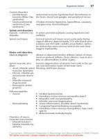
Fig. 19-1. Well circumscribed meningioma attached to the dura.
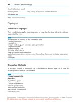
Fig. 19-2. Syncytial meningioma composed of lobules of of plump
meningothelial cells.
surface of the dura at the lateral borders of the meningioma
which has been referred to as a “dural tail”. Edema of
the underlying parenchyma may be quite prominent, particularly in more aggressive behaving neoplasms (16,17).
In the rare tumors that invade the underlying parenchyma
(malignant meningiomas), the circumscription that is
characteristic of most ordinary types of meningioma may
be absent. Areas of necrosis and peritumoral edema are
often more prominent in these cases as well.
Table 19-1 summarizes the histologic subtypes of meningioma that are currently recognized by the World Health
Organization Histological Classification of Tumours of
the Central Nervous System (18). Most meningiomas fall
into one of the first four categories which include meningothelial or syncytial, fibrous or fibroblastic, transitional
or mixed, and psammomatous types. Briefly, meningothelial meningiomas are comprised of lobules of plump
meningioma cells with ill-defined cell borders (Figs. 19-
2 and 19-3). Cells are often arranged in a whorled configuration. Intranuclear pseudoinclusions, which represent
cytoplasmic invaginations into the nucleus, are most commonly seen in association with this type. Fibrous meningioma is characterized by a fascicular architecture and is
composed of elongated cells with increased collagen and
reticulin deposition (Fig. 19-4). The so-called transitional
meningioma represents a combination of both the meningothelial and fibrous patterns. Exact criteria as to how
much of a minor component needs to be present in order
to use this designation do not exist. Psammomatous
meningiomas often have a background meningotheliomatous meningioma pattern with an abundance of psammoma bodies (Fig. 19-5). In general, distinction of one
of the aforementioned types of meningioma from another
is not of clinical significance.
Other less commonly encountered subtypes of meningioma which similarly act in a low-grade fashion include
Table 19-1
Meningioma Classification-Variants
Meningothelial (syncytial)
Fibrous (fibroblastic)
Transitional (mixed)
Psammomatous
Angiomatous (angioblastic)
Microcystic (humid)
Secretory (pseudopsammomatous)
Chordoid
Lymphoplasmacyte-rich
Metaplastic
*Rhabdoid
*Papillary
*Clear cell
*Atypical meningioma
*Malignant/anaplastic meningioma
*Histologic variants associated with more aggressive behavior.
Fig. 19-3. Cytologic preparation of syncytial meningioma.
CHAPTER 19 / MENINGIOMA
Fig. 19-4. Spindled arrangement of cells in a fibrous meningioma.
91
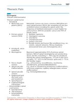
Fig. 19-6. Scattered large hyaline-like cytoplasmic inclusions in a
secretory meningioma.
the so-called microcystic meningioma, secretory meningioma, lymphoplasmocyte rich meningioma, metaplastic
variants of meningioma and chordoid meningioma. As
its name suggests, the microcystic (humid) meningioma
is characterized by cystic spaces with scattered meningothelial cells often demonstrating elongated cell processes
(19–22). Differential diagnostic considerations particular
to this meningioma variant include pilocytic astrocytoma
and rarely hemangioblastoma (in cases when one also has
lipidized meningothelial cells). The secretory (pseudopsammomatous) meningioma is characterized by eosinophilic, hyalinelike cytoplasmic inclusions which ultrastructurally represent microvillous-lined spaces filled with
membranous debris (23–25) (Fig. 19-6). The lymphoplasmocyte-rich or lymphofollicular variant is marked
by a prominent lymphoplasmocytic infiltrate, frequently
accompanied by lymphoid follicles (26,27). Metaplastic
variants contain a variety of mesenchymal elements which
have included bone, cartilage, fat and myxoid tissue
(2,28,29). The rare chordoid variant is histologically char-
acterized by cords and small clusters of epithelioid cells
arranged against a mucinous background (30) (Fig. 197). Although previously melanotic meningioma was recognized as a distinct entity, the current thinking is that
many of these tumors represent examples of so-called
melanocytoma.
Angiomatous or angioblastic meningiomas deserve
special mention from a historical viewpoint. In many of
the earlier classification schemas for meningioma, hemangiopericytomas and hemangioblastomas were grouped
together with a subset of meningiomas rich in blood vessels under the designation of angiomatous or angioblastic
meningioma. In more recent years, both hemangioblastomas and hemangiopericytomas have been separated out
as distinct entities because of differences in terms of
cell of origin, prognosis, and associations. Whether the
remaining small number of so-called angiomatous meningiomas are more aggressive behaving tumors or not is still
debatable. De le Monte’s study on meningioma recurrence
Fig. 19-5. Numerous psammoma bodies with interspersed nests of
meningothelial cells in a psammomatous meningioma.
Fig. 19-7. Chordoid meningioma with cords and clusters of cells
arranged against a mucinous background.
92
PRACTICAL DIFFERENTIAL DIAGNOSIS IN SURGICAL NEUROPATHOLOGY
Fig. 19-8. Meningothelial cells arranged around fibrovascular cores
in a papillary meningioma.
Fig. 19-9. Vague lobules of meningothelial cells with cleared cytoplasm in a clear cell meningioma.
following subtotal resection noted that hypervascularity
and hemosiderin deposition are two histologic features
which were more likely to be present in meningiomas
that recurred as opposed to those tumors which did not
recur (31).
Three particular histologic variants of meningioma
which are thought by many to be associated with more
aggressive behavior and include the papillary meningioma, the clear cell meningioma, and rhabdoid meningioma. In 1975, Ludwin et al reported 17 cases of socalled papillary meningioma (32). These tumors were
characterized by distinctive pseudorosette arrangement of
meningothelial cells around blood vessels (Fig. 19-8).
Eight of the 17 cases arose in childhood and 10 of the
patients (59%) had local recurrence of the tumor anywhere
from 4 to 16 months after surgery. Distant metastasis
occurred in 5 of the 17 patients. Others have reported
similarly aggressive behavior for this subset of meningioma (33). Fortunately, most of these cases demonstrate
a clearly recognizable meningioma component in association with the papillary areas, which allow for their recognition.
More recently, the clear cell meningioma has been
reported to be a potentially more aggressive variant. In
1995, Zorludemir et al reported 14 examples of so-called
clear cell meningioma consisting of sheet-like or lobulated
proliferations of polygonal cells with clear cytoplasm (34)
(Fig. 19-9). Nuclei are generally uniform and round with
delicate chromatin and inconspicuous nucleoli. Tumor
cells contain abundant cytoplasmic glycogen as evidenced
by strong PAS positivity. Mitotic figures were only rarely
identified; foci of necrosis were seen in three of the
tumors. Eight patients developed tumor recurrence. Local
discontinuous spread was noted in two of those eight
cases. Three patients died of disease.
Most recently, Kepes et al. (35) reported four cases of
meningioma which contained areas in which the cells
assumed a rhabdoid morphology. These cells are round to
oval with prominent eosinophilic cytoplasm and eccentric
nuclei (Fig. 19-10). Three of the four patients developed
a tumor recurrence within 20 months of the initial surgery;
the fourth patient died in the immediate postoperative
period. Others have confirmed the aggressive nature of
this subgroup of meningiomas (36).
In recent years, considerable literature has been
afforded meningiomas, attempting to predict tumor
behavior based on the presence of certain histopathologic
features. A number of studies have shown that tumors
which are characterized by prominent nuclear pleomorphism, necrosis, increased mitotic activity, disorganized
architectural pattern, macronucleoli, small cell formation,
brain invasion and distant metastasis are more frequently
aggressive behaving neoplasms (16,37–46). Unfortunately, not all aggressive behaving meningiomas display
worrisome histologic features. In 1986, de la Monte et
al. (31) outlined a useful approach to these atypical menin-
Fig. 19-10. Meningioma with cells demonstrating rhabdoid features.
CHAPTER 19 / MENINGIOMA
Fig. 19-11. Loss of architectural pattern and mitosis figure in a
meningioma with aggressive features or atypical meningioma.
giomas. In this study, a number of histopathologic features
were examined, specifically looking for those which correlated with tumor recurrence. The histologic features
which were found to be statistically significant in terms
of association with tumor recurrence included hypervascularity, hemosiderin deposition, loss of architectural
pattern or sheeting, prominent nucleoli, mitotic figures,
single cell and small group necrosis, nuclear pleomorphism, and overall atypical or malignant tumor grade
(Figs. 19-11 and 19-12). Many of these same histologic
features were also observed in the nonrecurrent tumor
group. In general, meningiomas with two or more of the
above-mentioned histologic features can be designated as
atypical meningiomas or meningiomas with aggressive
features. Others have established slightly different thresholds. Maier et al. (43) defined atypical meningiomas as
tumors exhibiting hypercellularity and 5 or more mitotic
figures per 10 high-power fields. Perry et al. (47,48) lowered the mitotic threshold to four or more per ten high
power fields; in the absence of sufficient mitotic activity,
Fig. 19-12. Necrosis in an aggressive/atypical meningioma.
93
Fig. 19-13. Parenchymal invasion in a malignant meningioma.
brain invasion or the presence of three of four histologic
parameters including sheeting architecture, hypercellularity, small cell formation, and prominent nucleoli were
sufficient for the designation. As always, clinical history
is important in the evaluation of any lesion, particularly
with regard to the presence of necrosis. A tumor that has
been recently operated on, embolized, or irradiated may
demonstrate necrosis that should not necessarily be interpreted as intrinsic to the neoplasm (49).
So-called malignant or anaplastic meningiomas are relatively uncommon lesions and represent the high grade
end of the meningioma spectrum. There is still some
debate as to what exactly constitutes a malignant meningioma. Most agree that brain invasion or metastasis are
features of malignancy (Fig. 19-13). The precise histologic definition of what constitutes brain invasion however
is still debated, e.g., whether or not extension of tumor
into Virchow-Robin spaces constitutes invasion. Most
malignant meningiomas in one series (50) demonstrated
most of the histologic features which had been previously
associated with aggressive behavior: nuclear pleomorphism in 20 of 23 tumors, disorganized architecture in
22 of 22 tumors, necrosis in 20 of 23 tumors, prominent
nucleoli in 17 of 23 tumors, and mitotic figures in 22 of
23 tumors ranging from 1 to 18 mitotic figures per 10 highpower fields (mean 6.1). Six of the patients developed
metastasis which were most commonly to bone, lung, and
skin. Of the 20 patients in whom follow-up information
was available in that series, six died of tumor (mean followup: 27 months), nine were alive with residual tumor (mean:
35 months) and five were alive with no evidence of tumor
(median: 12 months). Recently, Perry et al. (48) have stated
that brain invasion alone is not enough to define malignant
meningioma. They defined anaplastic meningioma as a
tumor marked by either excessive mitotic activity (≥20
mitosis figures/10 high-power fields) or at least focal loss
of meningothelial differentiation, resulting in a sarcoma,
94
PRACTICAL DIFFERENTIAL DIAGNOSIS IN SURGICAL NEUROPATHOLOGY
carcinoma or melanoma-like appearance (48). Use of the
term meningosarcoma in reference to malignant meningioma should be abandoned, because of the incorrect inference that these tumors are somehow sarcomatous in nature.
Because of the problem associated with trying to predict tumor behavior based on histopathology, a number
of individuals have attempted to utilize a variety of cell
proliferation markers in order to predict tumor behavior.
A number of studies employing a variety of modalities
have generally indicated a tendency for higher grade
tumors to demonstrate higher levels of cell proliferation
(51–59). Most of these studies demonstrate an overlap in
terms of the degree of cell proliferation between benign,
aggressive, and malignant tumors. Differences in methodology between laboratories, differences in interpretation
of staining, and variability within a given tumor related
to tumor heterogeneity are all factors which make interpretation of a labeling index or value in a particular case
potentially misleading. As a prospective independent predictor of aggressive behavior, these studies generally fall
short. However, in conjunction with other histologic features, such data may serve as additional evidence for
potentially aggressive or malignant behavior.
In general, electron microscopic examination of meningiomas adds little to the routine evaluation. In selected
cases, it may be useful in distinguishing meningiomas
from other dural based lesions of fibroblastic or smooth
muscle origin. Characteristic ultrastructural features
include the presence of interdigitating processes, cytoplasmic intermediate filaments, and well-formed cell junctions.
Most cases of meningioma do not require immunohistochemical staining for confirmation of diagnosis. Similar
to electron microscopy, immunohistochemical staining
may be useful in rare cases in distinguishing certain tumor
types from meningioma. Meningiomas characteristically
demonstrate diffuse positive immunoreactivity for
vimentin. Most meningiomas show focal positive staining
with epithelial membrane antigen (EMA). A minority of
meningiomas stain positively for S-100 protein in a focal
pattern and may demonstrate focal positive staining with
a variety of cytokeratin markers.
The differential diagnosis of meningioma is widespread, given the marked variability with regard to histology one can encounter in this group of neoplasms. Distinction of meningioma from hemangiopericytoma and
meningeal sarcomas will be discussed in chapter 20. Many
of the remaining differential diagnostic considerations can
be fairly easily resolved utilizing immunohistochemistry.
Distinction of meningioma from glioma is generally not
difficult from a light microscopic standpoint. Most astrocytomas will stain positively for glial fibrillary acidic
protein (GFAP), as compared to meningiomas which are
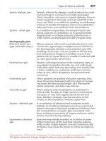
Fig. 19-14. Proliferation of meningothelial cells around parenchymal
vessels in meningioangiomatosis.
GFAP negative. Occasionally, an infiltrating squamous
cell carcinoma may involve the leptomeningeal region
and may ostensibly mimic a meningioma. In general, the
histologic appearance of the carcinoma, in particular, the
anaplastic appearance, as compared with the ordinary
meningioma, and often diffuse positive cytokeratin immunostaining should allow for easy distinction. Meningiomas may occasionally stain very focally for cytokeratin
markers. Distinction of the fibroblastic variant of meningioma from schwannoma may be a diagnostic issue, particularly in small biopsies from the cerebellopontine angle
and spinal cord regions. Lesions that may be obviously
schwannoma or meningioma, based on radiographic or
intraoperative appearances, may be more challenging,
particularly at the time of intraoperative consultation. In
general, schwannomas are characterized by a mixture of
loose, Antoni B and more compact, Antoni A patterns, a
feature that is generally not observed in meningiomas.
Verocay bodies, although not always noted in schwannomas, are a particularly useful histologic feature, when
present, in distinguishing the two lesions. In general, the
nuclei in the fibrous meningioma tend to be more elongated with rounder ends, as opposed to the longer, club
shaped nuclei of schwannoma. From an immunohistochemical standpoint, schwannomas stain diffusely and
strongly for S-100 protein; whereas in meningiomas, S100 immunoreactivity, if present, is focal and somewhat
limited. The membraneous pattern of staining with epithelial membrane antigen which marks meningiomas is generally absent in schwannomas.
A somewhat unusual lesion that can closely mimic a
meningioma is an entity referred to as meningioangiomatosis. Meningioangiomatosis is a rare condition characterized histologically by a proliferation of blood vessels
and perivascular cuffs of meningothelial cells (60–61)
(Fig. 19-14). The adjacent brain parenchyma often shows
95
CHAPTER 19 / MENINGIOMA
REFERENCES
Fig. 19-15. Generally spindled cells set against a collagen background in a solitary fibrous tumor of the meninges.
some degree of gliosis and the lesion is often accompanied
by psammoma bodies or calcifications. The association
of meningioangiomatosis with von Recklinghausen’s disease has been well documented. Distinction of meningioangiomatosis from an ordinary type meningioma is predicated on recognition of the predominantly parenchymal
based blood vessel and meningothelial cell proliferation
and the lack of discrete mass.
Rare cases of other spindled cell proliferations which
may mimic meningiomas have been described. Many of
these lesions have probably been designated as meningioma in the past and have been only recently recognized
as distinct entities. Although the numbers of these cases
reported in the literature are somewhat limited, most of
these lesions have behaved in a generally benign fashion.
These tumors can arise from a whole variety of mesenchymal cell types including fibroblasts, myofibroblasts, and
smooth muscle cells. This list of lesions includes entities
which have been referred to as fibromas and fibro-osseous
lesions (62,63), solitary fibrous tumor (64,65) (Fig. 1915), leiomyoma (66,67), and myofibroblastoma (68).
Table 19-2 summarizes the differential immunohistochemical features of these lesions.
1. Cushing, H. (1922) The meningiomas (dural endotheliomas).
Their source and favored seats of origin. Brain 45:282–316.
2. Chou, S.M., Miles, J.M. (1991) The pathology of meningioma.
In: Meningiomas. Al-Mefty O editor. Raven Press, New York,
NY. pp 37–57.
3. Harrison, M.J., Wolfe, D.E., Lau, T.S., Mitnick, R.J., Sachdev,
V.P. (1991) Radiation-induced meningiomas: experience at
the Mount Sinai Hospital and review of the literature. J. Neurosurg. 75:564–574.
4. Mack, E., Wilson, C. (1993) Meningioma induced by highdose cranial irradiation. J. Neurosurg. 79:28–31.
5. Dumanski, J.P., Rouleau, G.A., Nordenskjo¨ld, M., Collins, P.
(1990) Molecular genetic analysis of chromosome 22 in 81
cases of meningioma. Cancer Res. 50:5863–5867.
6. Ruttledge, M.H., Xie, Y-G., Han, F-Y., Peyrard, M., Collins,
P., Nordenskjo¨ld, M., Dumanski, J.P. (1994) Deletion on chromosome 22 in sporadic meningioma. Genes Chromosome
Cancer 10:122–130.
7. Deprez, R.H.L., Riegman, P.H., von Drunen, E., Warringa,
U.L., Groen, N.A., Stefanko, S.Z., Koper, J.W., Avezaat,
C.J.J., Mulder, P.G.H., Zwarthoff, E.C., Hagemeijer, A.
(1995) Cytogenetic, molecular genetic and pathological analyses in 126 meningiomas. J. Neuropathol. Exp. Neurol.
54:224–235.
8. Griffin, C.A., Hruban, R.H., Long, P.P., Miller, N., Volz, P.,
Carson, B., Brem, H. (1994) Chromosome abnormalities in
meningeal neoplasms: do they correlate with histology? Cancer Genet. Cytogenet. 78:46–52.
9. Bickerstaff, E.R., Small, J.M., Guest, I.A. (1958) The relapsing
course of certain meningiomas in relation to pregnancy and
menstruation. J. Neurol. Neurosurg. Psych. 21:89–91.
10. Roelvink, N.C.A., Kamphorst, W., van Alphen, H.A.M., Rao,
B.R. (1987) Pregnancy-related primary brain and spinal
tumors. Arch. Neurol. 44:209–215.
11. Jacobs, D.H., McFarlane, M.J., Holmes, F.F. (1987) Female
patients with meningioma of the sphenoid ridge and additional
primary neoplasms of the breast and genital tract. Cancer
60:3080–3082.
12. Mehta, D., Khatib, R., Patel, S. (1983) Carcinoma of the breast
and meningioma: association and management. Cancer
51:1937–1940.
13. Carroll, R.S., Zhang, J., Dashner, K., Sar, M., Wilson, E.M.
Black, P.McL. (1995) Androgen receptor expression in meningiomas. J. Neurosurg. 82:453–460.
14. Khalid, H. (1994) Immunohistochemical study of estrogen
Table 19-2
Differential Diagnosis by Immunohistochemistry of Spindled, Meningeal-Based Tumors
Vimentin
EMA(membranous)
S-100 protein
CD34
GFAP
Cytokeratins
Desmin
Smooth muscle actin
Meningioma
Schwannoma
Myofibroblastoma
Solitary Fibrous Tumor
Leiomyoma
+
+
±
±(weak)
−
±
−
−
+
−
+
−
−
−
−
−
+
−
−
−
−
−
±
±
+
−
−
+(strong)
−
−
−
−
+
−
−
−
−
−
+
+
96
15.
16.
17.
18.
19.
20.
21.
22.
23.
24.
25.
26.
27.
28.
29.
30.
31.
32.
PRACTICAL DIFFERENTIAL DIAGNOSIS IN SURGICAL NEUROPATHOLOGY
receptor-related antigen, progesterone and estrogen receptors
in human intracranial meningiomas. Cancer 74:679–685.
Smith, D.A., Cahill, D.W. (1994) The biology of meningiomas. Neurosurg. Clin. North Am. 5:201–215.
Crone, K.R., Challa, V.R., Kute, T.E., Moody, D.M., Kelly
Jr., D.L. (1988) Relationship between flow cytometric features
and clinical behavior of meningiomas. Neurosurgery 23:720–
724.
Inamura, T., Nishio, S., Takeshita, I., Fujiwara, S., Fukui, M.
(1992) Peritumoral brain edema in meningiomas-influence of
vascular supply on its development. Neurosurgery 31:179–
185.
Kleihues, P., Burger, P.C., Scheithauer, B.W. (1993) Histological Typing of Tumours of the Central Nervous System. 2nd
Ed. New York: Springer-Verlag.
Kleinman, G.M., Liszczak, T., Tarlow, E., Richardson Jr.,
E.P. (1980) Microcystic variant of meningioma: a light-microscopic and ultrastructural study. Am. J. Surg. Pathol. 4:383–
389.
Michaud, J., Gagne, F. (1983) Microcystic meningioma: clinicopathologic report of eight cases. Arch. Pathol. Lab. Med.
197:75–80.
Ng, H.K., Tse, C.C., Lo, S.T. (1989) Microcystic meningiomas - an unusual morphological variant of meningiomas. Histopathology 14:1–9.
Nishio, S., Takeshita, I., Marioka, T., Fukui, M. (1994) Microcystic meningioma: Clinicopathological features of 6 cases.
Neurol. Res. 16:251–256.
Budka, H. (1982) Hyaline inclusions (pseudopsammoma bodies) in meningiomas: immunocytochemical demonstration of
epithelial-like secretion of secretory component and immunoglobulins A and M. Acta Neuropathol. (Berl) 56:294–298.
Kepes, J.J. (1961) Observations of the formation of psammoma
bodies and pseudopsammoma bodies in meningiomas. J. Neuropathol. Exp. Neurol. 20:255–262.
Probst-Cousin, S., Villagren-Lillo, R., Lahl, R., Bergmann,
M., Schmid, K.W., Gullotta, F. (1997) Secretory meningioma.
Clinical, histologic, and immunohistochemical findings in 3
cases. Cancer 79:2003–2015.
Horten, B.C., Urich, H., Stefoski, D. (1979) Meningiomas with
conspicuous plasma cell-lymphocytic components: a report of
five cases. Cancer 43:258–264.
Stam, F.C., van Alphen, H.A.M., Boorsma, D.M. (1987) Meningioma with conspicuous plasma cell components. A histopathological and immunohistochemical study. Acta Neuropathol. (Berl) 1980 49:241–243.
Begin, L.R. (1990) Myxoid meningioma. Ultrastruct. Pathol.
14:367–374.
Lattes, R., Bigott, G. (1991) Lipoblastic meningioma: “vacuolated meningioma.” Hum. Pathol. 111:164–171.
Kepes, J.J., Chen, W.Y., Connors, M.H., Vogel, F.S. (1988)
“Chordoid” meningeal tumours in young individuals with peritumoral lymphoplasmacellular infiltrates causing systemic
manifestations of the Castleman syndrome: a report of seven
cases. Cancer 62:391–406.
de la Monte, S.M., Flickinger, J., Linggood, R.M. (1986)
Histopathologic features predicting recurrence of meningiomas following subtotal resection. Am. J. Surg. Pathol. 10:836–
843.
Ludwin, S.K., Rubinstein, L.J., Russell, D.S. (1975) Papillary
meningiomas: a malignant variant of meningioma. Cancer
36:1363–1373.
33. Pasquier, B., Gasnier, F., Pasquier, D., Keddari, E., Morens,
A., Couderc, P. (1986) Papillary meningioma. Clinicopathologic study of seven cases and review of the literature. Cancer 58:299–305.
34. Zorludemir, S., Scheithauer, B.W., Hirose, T., VanHouten,
C., Miller, G., Meyer, F.B. (1995) Clear cell meningioma: a
clinicopathologic study of a potentially aggressive variant of
meningioma. Am. J. Surg. Pathol. 19:493–505.
35. Kepes, J.J., Moral, L.A., Wilkinson, S.B., Abdullah, A., Llena,
J.F. (1998) Rhabdoid transformation of tumor cells in meningiomas: a histologic indication of increased proliferative activity.
Report of four cases. Am. J. Surg. Pathol. 22:231–238.
36. Perry, A., Scheithauer, B.W., Stafford, S.L., Abell-Aleff, P.C.,
Meyer, F.B. (1998) “Rhabdoid” meningioma: an aggressive
variant. Am. J. Surg. Pathol. 22:1482–1490.
37. Boker, D.K., Meurer, H., Gullota, F. (1985) Recurring intracranial meningiomas: evaluation of some factors predisposing
for tumor recurrence. J. Neurosurg. Sci. 29:11–17.
38. Crompton, M.R., Gautier-Smith, P.C. (1970) The prediction
of recurrence in meningiomas. J. Neurol. Neurosurg. Psych.
33:80–87.
39. Deen Jr, H.G., Scheithauer, B.W., Ebersold, M.J. (1982) Clinical and pathological study of meningiomas of the first two
decades of life. J. Neurosurg. 56:317–322.
40. Ja¨a¨kskela¨inen, J. (1986) Seemingly complete removal of histologically benign intracranial meningioma: late recurrence rate
and factors predicting recurrence in 657 patients: a multivariate
analysis. Surg. Neurol. 26:461–469.
41. Ja¨a¨kskela¨inen, J, Haltia, M., Laasonen, E., Wahlstro¨m, T.,
Valtonen, S. (1985) The growth rate of intracranial meningiomas and its relationship to histology: an analysis of 43 patients.
Surg. Neurol. 24:165–172.
42. Jellinger, K., Slowik, F. (1975) Histological subtypes and
prognostic problems in meningiomas. J. Neurol. 208:279–298.
43. Mahmood, A., Caccamo, D.V., Tomecek, F.J., Malik, G.M.
(1993) Atypical and malignant meningiomas: a clinicopathological review. Neurosurgery 33:955–963.
44. Maier, H., Ofner, D., Hittmair, A., Kitz, K., Budka, H. (1992)
Classic, atypical, and anaplastic meningioma: three histopathological subtypes of clinical relevance. J. Neurosurg. 77:616–
623.
45. Miller, D.C. (1994) Predicting recurrence of intracranial
meningiomas. A multivariate clinicopathologic model: Interim
report of the New York University Medical Center Meningioma Project. Neurosurg. Clin. North Am. 5:193–200.
46. Skullerud, K., Lo¨ken, A.C. (1974) The prognosis in meningiomas. Acta. Neuropathol. (Berl.) 29:337–344.
47. Perry, A., Stafford, S.L., Scheithauer, B.W., Suman, V.J.,
Lohse, C.M. (1997) Meningioma grading: an analysis of histologic parameters. Am. J. Surg. Pathol. 21:1455–1465.
48. Perry, A., Scheithauer, B.W., Stafford, S.L., Lohse, C.M.,
Wollan, P.C. (1999) “Malignancy” in meningiomas: a clinicopathologic study of 116 patients with grading implications.
Cancer 85:2046–2056.
49. Ng, H., Poon, W., Goh, K., Chan, M.S.Y. (1996) Histopathology of post-embolized meningiomas. Am. J. Surg. Pathol.
20:1224–1230.
50. Prayson, R.A. (1996) Malignant meningioma: a clinicopathologic study of 23 patients including MIB1 and p53 immunohistochemistry. Am. J. Clin. Pathol. 105:719–726.
51. Hsu, D.W., Pardo, F.S., Efird, J.T., Linggood, R.M., HedleyWhyte, E.T. (1994) Prognostic significance of proliferative
CHAPTER 19 / MENINGIOMA
52.
53.
54.
55.
56.
57.
58.
59.
indices in meningiomas. J. Neuropathol. Exp. Neurol. 53:247–
255.
Iwaki, T., Takeshita, I., Fukui, M., Kitamura, K. (1987) Cell
kinetics of the malignant evolution of meningothelial meningiomas. Acta Neuropathol. 74:243–247.
Lee, K.S., Hoshino, T., Rodriguez, L.A., Bederson, J., Davis,
R.L., Wilson, C.B. (1990) Bromodeoxyuridine labeling study
of intracranial meningiomas: proliferative potential and recurrence. Acta Neuropathol. 80:311–317.
Ohta, M., Iwaki, T., Kitamoto, T., Takeshita, I., Tateishi, J.,
Fukui, M. (1994) MIB1 staining index and scoring of histologic features in meningioma: Indicators for the prediction of
biologic potential and postoperative management. Cancer
74:3176–3189.
Roggendorf, W., Schuster, T., Peiffer, F. (1987) Proliferative
potential of meningiomas determined with the monoclonal
antibody Ki–67. Acta. Neuropathol. 73:361–364.
Salmon, I., Kiss, R., Levivier, M., Remmelink, M., Pasteels,
J-L., Brotchi, J., Flament-Durand, J. (1993) Characterization
of nuclear DNA content, proliferation index, and nuclear size
in a series of 181 meningiomas, including benign primary,
recurrent, and malignant tumors. Am. J. Surg. Pathol. 17:239–
247.
Zimmer, C., Gottschalk, J., Cervos-Navarro, J. (1992) Proliferating cell nuclear antigen (PCNA) in atypical and malignant
meningiomas. Pathol. Res. Pract. 188:951–958.
Perry, A., Stafford, S.L., Scheithauer, B.W., Suman, V.J.,
Lohse, C.M. (1998) The prognostic significance of MIB–1,
p53 and DNA flow cytometry in completely resected primary
meningiomas. Cancer 82:2262–2269.
Abramovich, C.M., Prayson, R.A. (1998) MIB–1 labeling
indices in benign, aggressive, and malignant meninomas: a
study of 90 tumors. Hum Pathol 29:1420–1427.
97
60. Prayson, R.A. (1995) Meningioangiomatosis: a clinicopathologic study including MIB1 immunoreactivity. Arch. Pathol.
Lab. Med. 119:1061–1064.
61. Halper, J., Scheithauer, B.W., Chazaki, H., Laws Jr., E.R.
(1986) Meningio-angiomatosis: a report of six cases with special reference to the occurrence of neurofibrillary tangles. J.
Neuropathol. Exp. Neurol. 45:426–446.
62. Russell, D.S., Rubinstein, L.J. (1989) Pathology of Tumours
of the Nervous System. Williams and Wilkins. Baltimore, MD.
pp 506–507.
63. Rhodes, R.H., Davis, R.L. (1978) An unusual fibro-osseous
component in intracranial lesions. Hum. Pathol. 9:309–319.
64. Carneiro, S.S., Scheithauer, B.W., Nascimento, A.G., Hirose,
T., Davis, D.H. (1996) Solitary fibrous tumor of the meninges:
a lesion distinct from fibrous meningioma: a clinicopathologic
and immunohistochemical study. Am. J. Clin. Pathol. 16:217–
224.
65. Prayson, R.A., McMahon, J.T., Barnett, G.H. (1997) Solitary
fibrous tumor of the meninges: case report and review of the
literature. J. Neurosurg. 86:1049–1052.
66. Lin, S.L., Wang, J.S., Huang, C.S., Tseng, H.H. (1996) Primary intracerebral leiomyoma: a case with eosinophilic inclusions of actin filaments. Histopathology 28:365–369.
67. Lach, B., Duncan, E., Rippstein, P., Benoit, B.G. (1994).
Primary intracranial pleomorphic angioleiomyoma—a new
morphologic variant: an immunohistochemical and electron
microscopic study. Cancer 74:1915–1920.
68. Prayson, R.A., Estes, M.L., McMahon, J.T., Kalfas, I., Sebek,
B.A. (1993) Meningeal myofibroblastoma. Am. J. Surg.
Pathol. 17:931–936.
20
H
Meningeal Sarcoma
has been
grouped together with a subset of meningiomas and
hemangioblastoma under the designation of angioblastic
meningioma. It is the general consensus now that the
hemangiopericytoma is a distinct lesion from meningioma
and generally more aggressive in behavior. Most frequently, the tumor is seen proximal to the leptomeninges,
but may on rare occasion arise in the brain parenchyma
and commonly in the spinal cord region. The tumor is
most frequently encountered in adults; however, rare cases
have presented in the second and third decades of life.
In contrast to meningiomas, there is no definite gender
predilection for hemangiopericytomas; the single largest
series to date, showed only a slight male predominance
(1). The hemangiopericytoma most frequently presents
as a fairly discrete, nonencapsulated mass. The tumor
generally does not elicit the same degree of osteoblastic
reaction and hyperostosis that is frequently encountered
in meningiomas. The gross and radiographic appearance
of the lesion may be altered by areas of hemorrhage,
cystic degeneration, or necrosis.
Histologically, hemangiopericytomas are often cellular
lesions accompanied by a rich vascular background. Vessels are classically described as having a staghorn configuration (Fig. 20-1). Cells with variable degrees of nuclear
pleomorphism are haphazardly arranged (Fig. 20-2).
Nucleoli are generally inconspicuous and cytoplasm
scant. Cell borders are often not clearly defined. Psammomatous calcifications or tight whorls, which may be
encountered in meningiomas, are not seen in hemangiopericytomas. Mitotic activity may be quite variable and
range from few to greater than ten mitotic figures per 10
high-power fields (1). Foci of necrosis and hemorrhage
are observed in a significant percentage of hemangiopericytomas. Similar to meningiomas, hemangiopericytomas
are reticulin rich lesions.
Although hemangiopericytomas are thought to be peri-
cytic in origin, ultrastructural examination of these tumors
demonstrates a range of tumor cell differentiation including pericytic, myoid, and fibroblastic (2–4). There are,
however, ultrastructural features which allow distinction
of this tumor from meningiomas, including the lack of
well-formed desmosomes or interdigitating cell membranes. There have been a number of studies that have
examined the immunohistochemical profile of hemangiopericytomas (3,5–10). Similar to meningioma, hemangiopericytoma will stain positively for vimentin. Meningiomas generally stain negatively for epithelial membrane
antigen (EMA), S-100 protein, and glial fibrillary acidic
protein (GFAP). Positive staining with CD34 and factor
XIIIa has been reported. MIB-1 labeling indices in one
study ranged between 0.2% and 9.9% and appeared to
be unrelated to tumor grade (11).
Based on a series of 94 cases, Mena et al stratified
central nervous system hemangiopericytomas into differentiated and anaplastic categories (1). Anaplastic hemangiopericytomas were characterized by necrosis and/or
ISTORICALLY, THE HEMANGIOPERICYTOMA
Fig. 20-1. Hemanigopericytoma with prominent staghorn vascular
pattern.
99
100
PRACTICAL DIFFERENTIAL DIAGNOSIS IN SURGICAL NEUROPATHOLOGY
Fig. 20-2. Moderate nuclear pleomorphism and disorganized
arrangement of cells in a hemangiopericytoma.
greater than 5 mitotic figures per 10 high-power fields
and at least two additional histologic features including
hemorrhage, moderate to high nuclear atypia and moderate to high cellularity. Median survival in the differentiated tumor group was 144 months versus 62 months for the
anaplastic group. In contrast to meningiomas, a significant
percentage of hemangiopericytomas, 60.6% in Mena’s
series, experienced one or more tumor recurrences and
metastasis developed in 23.4% (1). The most common
sites of metastasis included bone, liver, lung, central nervous system, and abdominal cavity in descending order
of frequency. It has been suggested that postoperative
radiation therapy may increase the time to recurrence and
extend survival (12).
The major differential diagnostic consideration of
hemangiopericytoma is meningioma, particularly atypical
meningioma. Table 20-1 outlines a number of clinicopathologic features that may be useful in distinguishing the
two lesions, many of which have already been discussed.
The other major differential diagnostic consideration is
distinguishing hemangiopericytoma from other sarcomas
with hemangiopericytomatous pattern. The key to distinguishing hemangiopericytomas from these other forms of
sarcoma often rests in recognizing defining features which
allows one to more definitively characterize the lesion as
another form of sarcoma e.g. finding areas of cartilage
differentiation in a chondrosarcoma.
Involvement of the central nervous system by primary
sarcoma is relatively uncommon. Well-known is the association of cranial sarcomas with prior radiation therapy
(13). Criteria for diagnosis and classification should be
the same as for sarcomas arising elsewhere in the body.
Although many of these sarcomas appear to be skullbased, occasional examples of primarily parenchymal
lesions have also been described. Care should be taken
not to misdiagnose a sarcoma as primary in the central
nervous system, when it is metastatic. The sarcoma types
that have been described are quite myriad and
have included examples of chondrosarcoma (14,15),
mesenchymal chondrosarcoma (16,17) (Fig. 20-3),
rhabdomyosarcoma (18,19), fibrosarcoma (20,21), malignant fibrous histiocytoma (22,23) (Fig. 20-4), leiomyosarcoma (24,25) (Fig. 20-5), osteosarcoma (26,27) and
angiosarcoma (28,29) (Fig. 20-6). Use of the term meningosarcoma in reference to meningeal based sarcomas
should be abandoned in favor of specific sarcoma classification. Unfortunately, the term meningosarcoma has also
been used in reference to malignant meningiomas. Occasionally, sarcomas may not demonstrate specific histologic features which allow their classification, in which
case designation of the lesion as a sarcoma without differentiation or not otherwise specified may be appropriate.
Occasionally, one may also encounter benign
Table 20-1
Hemangiopericytoma Versus Meningioma
Age
Gender
Hyperostosis
Calcification/psammoma bodies
Cell of origin
Staghorn vascular pattern
Nuclear atypia
Mitoses
Intranuclear pseudoinclusions
Necrosis
Reticulin rich
Vimentin
EMA
CD34
Prognosis
Hemangiopericytoma
Meningioma
Adult >> children
Females = males
−
−
Pericyte
+
±
Generally +
−
±
+
+
−
+
Generally more aggressive
Adult >> children
Females > males
±
±
Arachnoidal cap cell
−
±
+
+
±
+
+
+
± (weak)
Generally less aggressive
CHAPTER 20 / MENINGEAL SARCOMA
Fig. 20-3. Mesenchymal chondrosarcoma composed of undifferentiated cells and foci of cartilagenous differentiation.
101
Fig. 20-6. Vascular channels lined by tumor cells in an angiosarcoma.
mesenchymal lesions in the central nervous system. Mention of fibromas, myofibroblastomas and solitary fibrous
tumors was made in the previous chapter in the discussion
of differential diagnosis with meningiomas. Occasionally,
low grade vascular lesions (30) and hemangiomas have
been described. Lipomas, chondromas, and osteochondromas have also been reported to rarely involve the central
nervous system (31–32).
REFERENCES
Fig. 20-4. Storiform, pleomorphic malignant fibrous histiocytoma
of the meninges.
Fig. 20-5. Leiomyosarcoma characterized by smooth muscle actin
and desmin positive spindled cells.
1. Mena, H., Ribas, J.L., Pazeshkpour, G.H., Cowan, D.N., Parisi
J.E. (1991) Hemangiopericytoma of the central nervous system: a review of 94 cases. Hum. Pathol. 22:84–91.
2. Dardick, I., Hammar, S.P., Scheithauer, B.W. (1989) Ultrastructural spectrum of hemangiopericytoma: a comparative
study of fetal, adult, and neoplastic pericytes. Ultrastruct.
Pathol. 13:111–154.
3. d’Amore, E.S., Manivel, J.C., Sung, J.H. (1990) Soft-tissue
and meningeal hemangiopericytomas: an immunohistochemical and ultrastructural study. Hum. Pathol. 21:414–423.
4. Pen˜a, C.E. (1977) Meningioma and intracranial hemangiopericytoma: a comparative electron microscopic study. Acta Neuropathol. (Berl.) 39:69–74.
5. Winek, R.R., Scheithauer, B.W., Wick, M.R. (1989) Meningioma, meningeal hemangiopericytoma (angioblastic meningioma), peripheral hemangiopericytoma, and acoustic schwannoma: a comparative immunohistochemical study. Am. J. Surg.
Pathol. 13:251–261.
6. Porter, P.L., Bigler, S.A., McNutt, M., Gown, A.M. (1991)
The immunophenotype of hemangiopericytomas and glomus
tumors, with special reference to muscle protein expression:
an immunohistochemical study and review of the literature.
Mod. Pathol. 4:46–52.
7. Nemes, Z. (1992) Differentiation markers in hemangiopericytoma. Cancer 669:133–140.
8. Nakamura, M., Inoue, H.K., Ono, N., Kunimine, H., Tamada,
J. (1987) Analysis of hemangiopericytic meningiomas by
immunohistochmistry, electron microscopy and cell culture.
J. Neuropathol. Exp. Neurol. 46:57–71.
9. Kawano, H., Hayashi, M., Kabuto, M., Kobayashi, H., Handa,
102
10.
11.
12.
13.
14.
15.
16.
17.
18.
19.
20.
PRACTICAL DIFFERENTIAL DIAGNOSIS IN SURGICAL NEUROPATHOLOGY
Y., Kubota, T., Satsh, K. (1988) An immunohistochemical
and ultrastructural study of cultured intracranial hemangiopericytoma. Clin. Neuropathol. 7:105–110.
Iwaki, T., Fukui, M., Takeshita I., Tsuneyoshi, M., Tateishi,
J. (1988) Hemangiopericytoma of the meninges: a clinicopathologic and immunohistochemical study. Clin. Neuropathol.
7:93–99.
Perry, A., Scheihauer, B.W., Nascimento, A.G. (1997) The
immunophenotypic spectrum of meningeal hemangiopericytoma: comparison with fibrous meningioma and solitary
fibrous tumors of meninges. Am. J. Surg. Pathol 21:1354–
1360.
Guthrie, B.L., Ebersold, M.J., Scheithauer, B.W., Shaw, E.G.
(1989) Meningeal hemangiopericytoma: histopathological
features, treatment, and long-term follow-up of 44 cases. Neurosurgery 25:514–522.
Chang, S.M., Barker II, F.G., Larson, D.A., Bollen, A.W.,
Prados, M.D. (1995) Sarcomas subsequent to cranial irradiation. Neurosurgery 36:685–690.
Adegbite, A.B., McQueen, J.D., Paine, K.W. Rozdilsky, B.
(1985) Primary intracranial chondrosarcoma: a report of two
two cases. Neurosurgery 17:490–494.
Hassounah, M., Al-Mefty, O.L., Akhtar, M., Jinkins, J.R., Fox,
J.L. (1985) Primary cranial and intracranial chondrosarcoma: a
survey. Acta Neurochir. (Wien) 78:123–132.
Scheithauer, B.W., Rubinstein, L.J. (1978) Meningeal
mesenchymal chondrosarcoma: report of 8 cases with review
of the literature. Cancer 42:2744–2752.
Rushing, E.J., Armonda, R.A., Ansari, Q., Mena, H. (1996)
Mesenchymal chondrosarcoma: a clinicopathologic and flow
cytometric study of 13 cases presenting in the central nervous
system. Cancer 77:1884–1891.
Korinthenberg, R., Edel, G., Palm D., Mu¨ller K.M., Brandt,
M., Mu¨eller, R.P. (1984) Primary rhabdomyosarcoma of the
leptomeninges: clinical, neuroradiological and pathological
aspects. Clin. Neurol. Neurosurg. 86:301–305.
Taratuto, A.L., Molina, H.A., Diez, B., Zu´ccaro, G., Monges,
J. (1985) Primary rhabdomyosarcoma of brain and cerebellum:
report of four cases in infants: an immunohistochemical study.
Acta Neuropathol. (Berl.) 66:98–104.
Okeda, R., Mochizuki, T., Terao, E., Matsutani, M. (1980)
The origin of intracranial fibrosarcoma. Acta Neuropathol.
(Berl.) 52:223–230.
21. Gaspar, L.E., Mackenzie, I.R.A., Gilbert, J.J., Kaufmann,
J.C.E., Fisher, B.F., Macdonald, D.R., Cairncross J.G. (1993)
Primary cerebral fibrosarcomas: clinicopathologic study and
review of the literature. Cancer 72:3277–3281.
22. Sima, A.A., Ross, R.T., Hoag, G., Rozdilsky, B., Diocee, M.
(1986) Malignant intracranial fibrous histiocytomas: histologic, ultrastructural and immunohistochemical studies of two
cases. Can. J. Neurol. Sci. 13:138–145.
23. Roosen, N., Cras, P., Paquier, P., Martin, J.J. (1989) Primary
thalamic malignant fibrous histiocytoma of the dominant hemisphere causing severe neuropsychological symptoms. Clin.
Neuropathol. 8:16–21.
24. Asai, A., Yamada, H., Murata, S., Matsuno, A., Tsutsumi, K.,
Takemura, T., Matsutani, M., Takakura, K. (1988) Primary
leiomyosarcoma of the dura mater: Case report. J. Neurosurg. 68:308–311.
25. Louis, D.N., Richardson Jr., E.P., Dickersin, R.F., Petrucci,
D.A., Rosenberg A.E., Ojemann, R.G. (1989) Primary intracranial leiomyosarcoma: case report. J. Neurosurg. 171:279–
282.
26. Lam, R.M., Malik, G.M., Chason, J.L. (1981) Osteosarcoma
of meninges: clinical, light, and ultrastructural observations
of a case. Am. J. Surg. Pathol. 5:203–208.
27. Reznik, M., Lenelle, J. (1991) Primary intracerebral osteosarcoma. Cancer 68:793–797.
28. Charman, H.P., Lowenstein, D.H., Cho, K.G., DeArmond,
S.J., Wilson, C.B. (1988) Primary cerebral angiosarcoma: case
report. J. Neurosurg. 68:806–810.
29. Mena, H., Ribas, J.L. Enzinger, F.M., Parisi, J.E. (1991) Primary angiosarcoma of the central nervous system: study of
eight cases and review of the literature. J. Neurosurg.
75:73–76.
30. Chow, L.T., Chow, W., Fong, D.T. (1992) Epithelioid
hemangioendothelioma of the brain. Am. J. Surg. Pathol.
16:619–625.
31. Harrison, M.J., Mitnick, R.J., Rosenblum, B.R., Rothman,
A.S. (1990) Leptomyelolipoma: analysis of 20 cases. J. Neurosurg. 73:360–367.
32. Dutton, J. (1978) Intracranial solitary chondroma: case report.
J. Neurosurg. 49:460–463.
21
T
Hemangioblastoma
HE (CAPILLARY) HEMANGIOBLASTOMA has the dubious
distinction of comprising the sole entity listed under
“Tumors of Uncertain Histogenesis” in the 1993 W.H.O.
classification of central nervous system tumors (1). In
addition, it does not possess an obvious counterpart outside the nervous system, and therefore might prove particularly perplexing when first encountered. Although an
uncommon tumor, it represents a major differential diagnostic consideration in young to middle-aged adults with
either intracerebellar or intraspinal masses. While
hemangioblastomas are among the histopathologic hallmarks of von Hippel-Lindau disease (VHL, discussed
below), most are encountered as sporadic tumors, which
often then prompt evaluation for VHL.
Hemangioblastomas generally occur in patients 30–50
years of age. There is a tendency for earlier presentation
in tumors associated with VHL. Hemangioblastomas are
most frequently encountered within the cerebellum, where
they typically present as a cystic mass with a contrastenhancing mural nodule (similar to cerebellar juvenile
pilocytic astrocytomas). Less common locations include
the cerebrum, brainstem, and spinal cord, with the latter
predominating (2). Spinal cord hemangioblastomas are
classically associated with a syrinx extending rostrally
from the tumor (a characteristic shared with ependymal,
but usually not astrocytic, tumors of the spinal cord). In
addition, prominent leptomeningeal feeding vessels may
simulate a vascular malformation. While hemangioblastomas have been considered by some to be vanishingly rare
in the supratentorial compartment, this is largely a result
of their previous classification as a subtype of angioblastic
meningioma. Such tumors are now considered to be
meningeal hemangioblastomas, and may be encountered
anywhere along the neuraxis, including the optic
nerve (3).
Intraoperatively, these tumors appear as discrete,
highly vascular nodules. Although not always apparent,
the tumors usually abut the leptomeninges. On section,
which surgeons generally try to avoid, hemangioblastomas are spongy and tend to exude blood. Depending on
their content of lipid-laden stromal cells, they may appear
somewhat to strikingly yellow. Microscopically,
hemangioblastomas are composed of varying proportions
of primitive, thin-walled blood vessels and lipid-laden
stromal cells. It is the resistance of these latter cells to
ultrastructural and immunohistochemical characterization
that is responsible for this tumor’s condemnation to nosologic purgatory. They seem to be strongly to weakly
negative with antibodies to endothelial, glial, and neuroectodermal antibodies and show no defining features
by electron microscopy. Interestingly, studies of vasculogenesis in the chick embryo (during the early 1930s)
demonstrated a stage characterized by lipid-laden cells
appearing remarkably similar to those seen in hemangioblastomas. However, deforestation continues as the
attempt to classify these elusive cells presses on.
Two histopathologic variants are recognized based on
the relative proportion of stromal cells and capillaries
(1). In the reticular variant (Fig. 21-1), stromal cells are
uniformly distributed within an intricate network of capillaries, while in the cellular variant (Fig. 21-2), the stromal
cells are clustered and delimited by the capillaries. Like
many meningioma subtypes, these variants have no prognostic or syndromic importance. However, awareness of
this histopathologic variation is important in preventing
misdiagnoses. Specifically, the cellular variant may be
confused with renal cell carcinoma (another cardinal feature of von Hippel-Lindau disease) (Table 21-1). Useful
differentiating histopathologic characteristics of hemangioblastomas are the similarity in nuclear morphology
between the capillary endothelial cells and stromal cells,
and a more xanthomatous than clear cytoplasm (the latter
being more characteristic of glycogen-rich metastatic
renal cell carcinomas). Histochemical staining for reti-
103
104
PRACTICAL DIFFERENTIAL DIAGNOSIS IN SURGICAL NEUROPATHOLOGY
Table 21-1
Hemangioblastoma Versus Renal Cell Carcinoma
Intracellular lipid
Intracellular glycogen
Reticulin
Cytokeratin
EMA
Von Hippel-Lindau
Fig. 21-1. Lipid laden stromal cells distributed within an intricate
network of capillaries in the reticular variant of hemangioblastoma.
culin will highlight the abundant thin-walled blood vessels
in hemangioblastomas (Fig. 21-3), while showing a
weaker nested pattern in renal cell carcinoma (4). Immunohistochemical staining with antibodies to cytokeratins
and epithelial membrane antigen will be reactive with
renal cell carcinomas and non-reactive with hemangioblastomas (5).
During intraoperative consultation, the other main differential diagnostic consideration is astrocytoma. Confusion may arise as a result of sampling error (surrounding
gliotic tissue or syrinx wall) or as a result of compression
of the delicate capillary component during the preparation
of frozen sections. Cytologic (touch) preparations may
be a valuable diagnostic aid, allowing appreciation of the
lipid-laden stromal cells (6). This differential diagnostic
dilemma is virtually never a problem on permanent sections, as these two tumors appear so distinct as to be
sometimes painfully embarrassing.
Two other interesting histopathologic features occa-
Fig. 21-2. Nests of xanthomatous stromal cells dominate the cellular
variant of hemangioblastoma.
Hemangioblastoma
Renal Cell
Carcinoma
+
−
Individual cells
−
−
+/−
−
+
Cell nests
+
+
+/−
sionally encountered in hemangioblastomas are significant numbers of mast cells (7) and extramedullary erythropoiesis (8). The latter presumably results from
erythropoietin production by the tumor, which may also
cause polycythemia, seen in approximately 10% of
patients at presentation.
Approximately 25% of patients diagnosed with CNS
hemangioblastoma will have von Hippel-Lindau disease
(9). An earlier age of onset and/or multifocally favors
VHL, which may be defined syndromically by a minimum
of CNS hemangioblastoma or retinal angioma with at
least one other typical VHL lesion or an affected firstdegree relative. Interestingly, the mean age of onset varies
for the various syndromic manifestations (10). While
VHL associated hemangioblastomas tend to be seen in
patients in their 30’s, retinal angiomatosis usually develops several years earlier. Therefore, careful funduscopic
examination of hemangioblastoma patients (and their
first-degree relatives) may cinch the diagnosis. Renal cell
carcinomas, which are often bilateral, tend to occur somewhat later in the syndrome, although still at a much
younger age than sporadic renal cell carcinomas. Renal
cysts, adrenal pheochromocytomas, and pancreatic and
Fig. 21-3. Histochemical staining for reticulin demonstrates envelopment of individual stromal cells by reticulin and highlights thin walled
vascular spaces.
CHAPTER 21 / HEMANGIOBLASTOMA
epididymal cysts also occur in many patients with VHL,
although the prevalence of these manifestations varies
quite widely from family to family.
The VHL gene is a tumor suppressor gene found on
the short arm of chromosome 3. The gene product is a
protein which has been observed to inhibit the binding
of transcriptional elongation factors. When the gene is
mutated, transcriptional regulation is impaired. Germ line
mutations have been identified in 85 of 114 VHL families
(75%). It also appears that the types of mutations responsible for VHL with pheochromocytoma differ from those
responsible for VHL without pheochromocytoma.
Amongst patients with VHL, cerebellar hemangioblastoma is the most common presenting manifestation.
The overall prevalence of tumors in patients with VHL
varies from pedigree to pedigree, but approximates:
Tumor
Prevalence
Cerebellar hemangioblastoma
Retinal angioma
Renal cell carcinoma
Spinal hemangioblastoma
Pheochromocytoma
60%
40%
25%
15%
15%
In past decades, patients with VHL tended to die at around
40 years of age, most commonly as a result of cerebellar
hemangioblastomas. Although this outcome has been dramatically ameliorated by modern microsurgical techniques, the development of multiple CNS tumors is still
a major problem, as is the development of metastatic renal
105
cell carcinoma, which is currently the proximate cause
of death in up to 50% of VHL patients.
REFERENCES
1. Kleihues, P., Burger, P.C., Scheithauer, B.W. (1993) Histological typing of tumours of the central nervous system. 2nd ed.
New York: Springer-Verlag
2. Filling-Katz, M.R., Choyke, P.L., Oldfield, E., Charnas, L.,
Patronas, N.J., Glenn, G..M, Gorin, M.B., Morgan, J.K., Linehan, W.M., Seizinger, B.R., Zbar, B. (1991) Central nervous
system involvement in von Hippel-Lindau disease. Neurology 41:41–46.
3. Kerr, D.J., Scheithauer, B.W., Miller, G.M., Ebersold, M.J.,
McPhee, T.J. (1995) Hemangioblastoma of the optic nerve:
case report. Neurosurgery 36:573–581.
4. Clelland, C.A., Streip, C. (1989) Histological differentiation
of metastatic renal carcinoma in the cerebellum from cerebellar
haemangioblastoma in von Hippel-Lindau’s disease. J. Neurol.
Neurosurg. Psychiatry 52:162–166.
5. Hufnagel, T.J., Kim, J.H., True, L.D., Manuelidis, E.E. (1989)
Immunohistochemistry of capillary hemangioblastoma. Am.
J. Surg. Pathol. 13:207–216.
6. Commins, D.L., Hinton, D.R. (1998) Cytologic features of
hemangioblastoma. Comparison with meningioma, anaplastic
astrocytoma and renal cell carcinoma. Acta Cytol. 42:1104–
1110.
7. Ho, K.L. (1984) Ultrastructure of cerebellar capillary hemangioblastoma. Acta Neuropathol. (Berl.) 64:308–318.
8. Zec, N., Cera, P., Towfighi, J. (1991) Extramedullary hematopoiesis in cerebellar hemangioblastoma. Neurosurgery 29:
34–37.
9. Neumann, H.P., Eggert, H.R., Scheremet, R., Schumacher,
M., Mohadjer, M., Wakhloo, A.K., Volk, B., Hettmannsperger,
U., Riegler, P., Schollmeyer, P., Wiestler, O. (1992) Central
nervous system lesions in von Hippel-Lindau syndrome. J.
Neurol., Neurosurg. Psychiatry 55:898–901.
10. Neumann, H.P.H., Lips, C.J.M., Hsia, Y.E., Zbar, B. (1995)
Von Hippel-Lindau syndrome. Brain Pathol. 5:181–193.
22
I
Central Nervous System Primitive
Neuroectodermal Tumors
1910, JAMES HOMER WRIGHT (of Homer Wright
rosette fame) first separated the medulloblastoma from
other CNS tumors. His concept was further refined by
Percival Bailey, who defined a group of 29 tumors arising
in the cerebellar vermis, primarily in children. Following
the lead of the nineteenth century German pathology
school, where tumors were named based on the concept
of a cell of origin, Bailey named these tumors medulloblastomas. The cell of origin model then continued to be
used in CNS tumor nomenclature, leading to the definition
of a variety of “embryonal” tumors of the nervous system.
This nomenclatural system is predicated on the assertion
by the late Dr. Lucien Rubenstein that the central nervous
system contains several unique types of neuroepithelial
precursor cells in different locations which may undergo
transformation giving rise to a variety of morphologically
similar, but biologically distinct, CNS tumors (1). In 1973,
Hart and Earle described a group of small round blue cell
tumors of the central nervous system in children and
introduced the diagnostic appellation “primitive neuroectodermal tumor” (PNET) (2). Dr. Lucy Rorke subsequently suggested that the term be broadened to include
all primary CNS tumors composed of primitive neuroepithelial cells regardless of their location within the CNS.
The codification of these previously disparate entities into
a unique class of tumors is eloquently supported in her
recent review (3)
While this conceptual/nomenclatural debate still rages
(4), current therapy and prognosis appears to be determined
primarily by phenotypic rather than histogenetic parameters. However, this may in part be due to the rarity of nonmedulloblastoma embryonal tumors of the central nervous
system, precluding adequate biologic distinctions.
Medulloblastomas (PNET-MBs) comprise 15% to 25%
of brain tumors in children and account for one-third
N
of pediatric posterior fossa neoplasms. Three-quarters of
medulloblastomas occur before age 15, 50% occur during
the first decade, and the peak incidence is around age
5. Along with supratentorial primitive neuroectodermal
tumors, PNET-MBs represent one of the most common
CNS tumors encountered in the first years of life.
The typical medulloblastoma presents an appearance
all too familiar to the pediatric surgical pathologist—a
monotonous sea of cells with small, relatively round,
hyperchromatic nuclei and virtually unidentifiable cytoplasm (the “small, round, blue cell tumor of childhood”)
(Fig. 22-1). Similar to other small round blue cell tumors,
mitoses, apoptotic cells and geographic tumor necrosis
are typical. The classic Homer Wright rosette, consisting
of a ring of nuclei surrounding a fibrillary core composed
of eosinophilic cell processes (neurites) is a fairly unusual
finding in medulloblastomas, and represents primitive
neuronal (neuroblastic) differentiation (Fig. 22-2). Neu-
Fig. 22-1. A typical undifferentiated medulloblastoma composed of
a monotonous sea of primitive tumor cells.
107
108
PRACTICAL DIFFERENTIAL DIAGNOSIS IN SURGICAL NEUROPATHOLOGY
Fig. 22-4. Cords of tumor cells in a desmoplastic medulloblastoma.
Fig. 22-2. Homer Wright rosette in a medulloblastoma.
ronal differentiation in medulloblastomas may also manifest as nodules of pale, synaptophysin-positive islands
floating within an otherwise undifferentiated sea of tumor
cells (Fig. 22-3). Tumors expressing this histopathologic
pattern have occasionally been referred to as “cerebellar
neuroblastomas,” though we prefer to sign such cerebellar
tumors out as PNET-MB with neuroblastic differentiation.
It is a small step from this appearance to that of the
desmoplastic medulloblastoma, where cords of primitive
tumor cells between the pale synaptophysin-positive
islands are embedded in a dense reticulin meshwork (Fig.
22-4).
While the biologic behavior of tumors with neuroblastic differentiation does not appear to differ significantly from typical medulloblastomas, desmoplastic
medulloblastomas represent the predominant type of
medulloblastomas in children with the nevoid basal cell
carcinoma syndrome, and show a loss of heterozygosity
(LOH) on chromosome 9q corresponding to deletion of
the PTCH gene locus. This LOH of chromosome 9q has
Fig. 22-3. Pale neuroblastic islands in a medulloblastoma.
also been demonstrated in desmoplastic medulloblastomas not associated with the nevoid basal cell carcinoma
syndrome, and contrasts with chromosome 17p abnormalities seen in 30–40% of nondesmoplastic medulloblastomas (5,6). With very rare exceptions, the t(11;22) translocation typical of peripheral PNETs is not present within
central nervous system PNETs (7).
Unlike peripheral neuroblastomas, where n-myc amplification and trk-B expression carry important prognostic
significance, efforts to identify biologic factors of prognostic significance for CNS PNETs, including oncogene
amplification, DNA ploidy, and mitotic index have been
unsuccessful or inconsistent. Similarly, the prognostic relevance of astrocytic differentiation within PNET-MBs
has been controversial. This has been true in part due to
difficulties in distinguishing trapped, dysmorphic astrocytes from astrocytic differentiation within neoplastic
cells. While there will always be cases in which distinguishing reactive from neoplastic astrocytes will prove
either extremely difficult or impossible, glial fibrillary
acidic protein immunostaining generally discloses two
patterns of immunopositivity (Figs. 22-5 and 22-6): 1)
scattered perivascular forms with extensive branching
processes—this pattern is seen most frequently and represents astroglial reaction to the medulloblastoma, and 2)
clumps or compact sheets of small poorly differentiated
cells with scant GFAP immunopositivity. This uncommon
pattern is felt to represent true glial differentiation within
the tumor. Primitive neuroectodermal tumors containing
such clumps or sheets of GFAP positive cells are associated with a three-fold increased risk of relapse compared
with tumors demonstrating either no GFAP immunoreactivity or scattered GFAP immunopositive cells (8).
Distinctly less common, but no less confusing, is “oligodendroglial” and “ependymal” differentiation. The former is manifest as foci of cells with round dark nuclei
and perinuclear halos. While the absence of a reliable
CHAPTER 22 / CNS PRIMITIVE NEUROECTODERMAL TUMORS
Fig. 22-5. Scattered, reactive GFAP-positive cells in a medulloblastoma.
marker for neoplastic oligodendroglia precludes definitive
assessment, such cells are generally felt to represent neuroblastic rather than true oligodendroglial differentiation.
Perivascular pseudorosettes identical to those seen in
ependymal tumors may also rarely be encountered in
PNETs. When the perivascular processes react with antibodies to GFAP, we consider such structures to represent
true ependymal differentiation, and sign such tumors out
as ependymoblastoma or PNET with ependymal differentiation. A word of caution is in order, however, in that we
have also seen cases in which the perivascular processes
reacted with synaptophysin, and not with GFAP, in which
case we make a note of it, but do not further subclassify
the tumor.
Two exceedingly rare PNET-MB variants recognized
by the WHO are the medullomyoblastoma (or PNET with
muscle elements) and the melanotic medulloblastoma
(PNET with melanin pigment). Fewer than 40 medullo-
Fig. 22-6. Clusters of GFAP-positive tumor cells in a medulloblastoma with glial differentiation.
109
myoblastomas have been reported (9,10). In nearly all
cases cross-striations have been evident on light microscopic examination. Melanotic medulloblastomas are similarly defined based on light microscopic examination.
Both of these subtypes have been reported to exhibit
aggressive behavior compared with typical medulloblastomas.
The treatment of CNS-PNETs centers around local
and craniospinal radiation therapy, often combined with
various chemotherapy regimens. The latter is particularly
important in very young patients, where chemotherapy is
often used in an attempt to keep the neoplasm at bay while
the nervous system develops to a stage where radiation
therapy will be somewhat less devastating. While the
addition of chemotherapy, particularly in high-risk
patients (incomplete resections, CSF seeding at diagnosis)
appears to have markedly improved short-term survival
of children with PNET-MBs, long-term follow-up data
is just beginning to become available, and will likely
determine the optimal treatment of patients with medulloblastomas/CNS-PNETs (11). Long-term follow-up of
PNET-MB patients treated with craniospinal irradiation
therapy during the computed tomography era (approximately 1980 to present) reveals a median survival of 58
months, with 25% survival at 10 years. Patients with nonlocalized disease (positive CSF cytology) do significantly
worse, with a 30% 5-year survival (12). While there are
exceptions, the risk of tumor recurrence for PNET-MBs
in children aged 8 and younger closely adheres to Collins’
Law, which defines the period of risk for tumor recurrence
as equal to the patient’s age at diagnosis in months plus
nine months (originally derived from observations of children with congenital Wilms tumors) (13). Failure usually
occurs at the primary tumor site, but supratentorial metastases, diffuse leptomeningeal seeding, and systemic
metastases may each be seen in approximately 20% of
patients. While systemic metastases have often been
blamed on seeding through ventricular shunts, this complication also occurs in the absence of CSF shunting. In
fact, a recent review of the literature indicates that of 160
cases of systemic PNET-MB metastases, only 11(7%)
could have occurred through or been facilitated by ventriculosystemic shunts (14). The most common locations for
extraneural PNET-MB metastases are bone/bone marrow,
lymph nodes, lungs, and liver.
Dr. Rorke has recently defined a clinicopathologic
entity closely related to CNS-PNETs, which she has
named the atypical teratoid/rhabdoid tumor (ATT/RhT)
(15). These tumors generally present in the first two years
of life, and their confusion with PNETs may account in
large part for the worse prognosis generally ascribed to
PNETs in children under the age of two. The diagnostic
confusion arises not in the 10–15% of tumors consisting
110
PRACTICAL DIFFERENTIAL DIAGNOSIS IN SURGICAL NEUROPATHOLOGY
Fig. 22-7. Rhabdoid cells in an atypical teratoid/rhabdoid tumor.
Fig. 22-8. Epithelial differentiation in an atypical teratoid/
rhabdoid tumor.
entirely of rhabdoid cells, similar to those seen in extraneural malignant rhabdoid tumors (Fig. 22-7), but in the
two-thirds of cases where rhabdoid cells are admixed with
classic primitive neuroectodermal tumor cells, or where
the teratoid (teratoma-like) components consist of
mesenchymal or epithelial (usually adenomatous) differentiation (Table 22-1 and Fig. 22-8). Mesenchymal differentiation, seen in about a third of ATT/RhTs, consists of
loosely arrayed spindle-shaped cells separated by pale
“ground substance,” (Fig. 22-9) and should not be confused with the reticulin-rich spindle cell elements of the
desmoplastic medulloblastoma (Figs. 22-9 and 22-4).
As a supplement to careful light microscopic examination of PNETs obtained from very young children, the
following patterns of immunohistochemical staining are
characteristic of atypical teratoid/rhabdoid tumors:
1. Epithelial membrane antigen is always positive and
is primarily expressed in the rhabdoid cells, and
less consistently in the epithelioid cells.
2. Strong vimentin immunopositivity is seen within
the cytoplasm of the rhabdoid cells.
3. Smooth muscle actin is seen in nearly all cases;
while this immunopositivity is also usually localized to the rhabdoid cells (and the blood vessels),
the mesenchymal component is occasionally
stained.
4. GFAP and neurofilament immunostains may be
positive in both the PNET fields and the
rhabdoid cells.
5. Cytokeratins are confined to the epithelial elements
and the rhabdoid cells.
6. Desmin expression is absent or weak, and is typically confined to the mesenchymal and PNET elements.
Further evidence for separating this aggressive neoplasm from other PNETs of childhood is cytogenetic:
most ATT/RhTs examined so far have shown abnormalities of chromosome 22, which contrasts with the
chromosome 17 abnormalities usually associated with
CNS-PNETs (15,16). Recent studies have demonstrated
Table 22-1
PNET Versus Atypical Teratoid/Rhabdoid Tumor
Age
Small, blue cells
Large cells/prominent nucleoli
Spindle cells
Epithelial differentiation
Synaptophysin
Vimentin
EMA
Smooth muscle actin
Chromosomal Abnormalities
PNET
AT/RT
Peak = 5
75% < 20
+
Rare
Desmoplasia
−
±
+
−
−
17,9
Peak = 1.5
75% < 3
±
+
Sarcomatoid
±
±
Inclusions
+
+
22
Fig. 22-9. Mesenchymal differentiation in an atypical teratoid/
rhabdoid tumor.
CHAPTER 22 / CNS PRIMITIVE NEUROECTODERMAL TUMORS
Fig. 22-10. Primitive neural tube formations in a medulloepithelioma.
abnormalities of the INI1 gene on chromosome 22 in both
CNS and non-CNS ATT/RhTs (17,18).
ATT/RhTs usually present in very early childhood,
with three quarters diagnosed in patients less than 3 years
old. While most are infratentorial, supratentorial tumors
are not uncommon, and predominate in older children.
Radiologic findings are not distinctive. Approximately a
third of patients with ATT/RhTs demonstrate leptomeningeal seeding at diagnosis. Unfortunately, the majority of
patients with ATT/RhT rapidly progress both at the primary site and via leptomeningeal dissemination, with a
median survival of less than a year. In contrast to children
with CNS-PNETs, among whom there is at least a transient response to chemotherapy, patients with ATT/RhT
often don’t respond even to aggressive chemo- and/or
radiation therapy.
In 1992, Giangaspero et al. reported four highly aggressive infantile cerebellar tumors composed of cells with
relatively abundant cytoplasm and large vesicular nuclei
with prominent nucleoli, which they termed “large cell
medulloblastoma” (19). Review of the Pediatric Oncology
Group’s experience with PNET-MBs supports the existence of this aggressive subtype (independent of ATT/
RhT) which comprised approximately 4% of their PNETMBs (20).
A final rare, but aggressive and poorly responsive primitive CNS tumor of early childhood is the medulloepithelioma. The name derives from its epithelioid appearance
as it pathologically recapitulates the primitive neural tube.
Children with this tumor generally present with nonenhancing periventricular hemispheric masses during the
first five years of life. The characteristic neural tube like
structures are composed of a pseudostratified arrangement
of primitive neural cells with an external and sometimes
internal PAS positive limiting membrane (Fig. 22-10).
Divergent differentiation along neuronal, glial and
mesenchymal lines may also be seen. As with the ATT/
111
RhTs and large cell medulloblastomas, early tumor progression with leptomeningeal dissemination and poor
response to therapy sets these tumors apart from conventional CNS-PNETs (21).
While this chapter has concentrated on PNET-MB as
a cerebellar tumor, PNETs may be encountered, albeit
much less frequently, in many other locations within the
neuraxis, including the spinal cord (22). However, due
to their relative rarity in these other locations, considerably less is known regarding their biologic behavior. A
recent, small institutional study found a decreased overall
survival and recurrence free survival in supratentorial
PNETs compared with PNET-MBs (23), and comparative
genomic hybridization studies indicate differing genetic
aberrations between these two histologically similar
groups of tumors (24). In general, supratentorial PNETs
are treated similarly to PNET-MBs.
PNET-MBs are rare in adults, comprising only about
1% of primary central nervous system tumors in patients
over 18 years old (25). While 80% of these occur between
the ages of 21 and 40 years, cases have been reported in
the over 50 crowd, with the eldest so far being a 73-year
old woman. 5 and 10-year survival rates are similar to
those observed in pediatric populations. Absence of fourth
ventricular floor involvement and a high radiation dose
to the spinal cord are correlated with a good prognosis.
One recent study found that adults with desmoplastic
medulloblastomas demonstrated significantly better 5 and
10 year survival rates (75%) than did similar patients with
classical medulloblastomas (60% and 40%), but no central
histopathologic review was performed (26).
The main problem in adult PNET-MB is distinguishing
it from metastatic small cell carcinoma. In practice this
is difficult, if not impossible, to accomplish due to tremendous overlap in their ultrastructural and immunocytochemical features. We find the following guidelines useful
in signing out these cases:
1. If the tumor is not in the cerebellum, consider
metastatic small cell carcinoma.
2. If there are multiple lesions, consider metastatic
small cell carcinoma.
3. If there is no neoplastic glial differentiation, consider metastatic small cell carcinoma.
4. If there is a lung lesion, consider metastatic small
cell carcinoma.
5. Consider metastatic small cell carcinoma.
On the other side of the biologic coin is a recently
described intracerebellar tumor of adults referred to variably as lipidized medulloblastoma, lipomatous medulloblastoma, or medullocytoma (27). As the latter designation implies, this is a primary neuroectodermal tumor with
a favorable prognosis. Approximately a dozen cases have
112
PRACTICAL DIFFERENTIAL DIAGNOSIS IN SURGICAL NEUROPATHOLOGY
been reported, with a mean age of 50 years. These tumors
are characterized by
1. a neuroectodermal component with low proliferative activity resembling the cerebral neurocytoma,
2. areas of lipomatous differentiation, and
3. an apparently favorable prognosis without the need
for adjuvant therapy.
REFERENCES
1. Rubinstein, L.J. (1972) Presidential address: cytogenesis and
differentiation of primitive central neuroepithelial tumors. J.
Neuropathol. Exp. Neurol. 31:7–26.
2. Hart, M.N., Earl, K.M. (1973) Primitive neuroectodermal
tumors of the brain in children. Cancer 32:890–987.
3. Rorke, L.B., Trojanowski, J.Q., Lee, V.M.-Y., Zimmerman,
R.A., Sutton, L.N., Biegel, J.A., Goldwein, J.W., Packer, R.J.
(1997) Primitive neuroectodermal tumors of the central nervous system. Brain Pathol. 7:765–784.
4. Katsetos, C.D., Burger, P.C. (1994) Medulloblastoma. Semin.
Diagn. Pathol. 11:85–97.
5. Schofield, D., West, D.C., Anthony, D.C., Marshal, R., Sklar,
J. (1995) Correlation of loss of heterozygosity at chromosome
9q with histological subtype in medulloblastomas. Am. J.
Pathol. 146:472–480.
6. Vortmeyer, A.O., Stavrou, T., Selby, D., Li, G., Weil, R.J.,
Park, W.S., Moon, Y.W., Chandra, R., Goldstein, A.M., Zhuang, Z. (1999) Deletion analysis of the adenomatous polyposis
coli and PTCH gene loci in patients with sporadic and nevoid
basal cell carcinoma syndrome associated medulloblastoma.
Cancer 85:2662–7.
7. Jay, V., Pienkowska, M., Becker, L. Zielenska, M. (1995)
Primitive neuroectodermal tumors of the cerebrum and cerebellum: absence of t(11;22) translocation by RT-PCR analysis.
Mod. Pathol. 8:488–491.
8. Janss, A.J., Yachnis, A.T., Silber, J.H., Trojanawski, J.Q.,
Lee, V.M., Sutton, L.N., Perilongo, G., Rorke, L.B., Phillips,
P.C. (1996) Glial differentiation predicts poor clinical outcome
in primitive neuroectodermal brain tumors. Ann. Neurol.
39:481–489.
9. Bergmann, M., Pietsch, T., Herms, J., Janus, J., Spaar, H.-J.,
Terwey, B. (1998) Medulloblastoma: a histological, immunohistochemical, ultrastructural and molecular genetic study.
Acta Neuropathol. 95:205–212.
10. Mahapatra, A.K., Sinha, A.K., Sharma, M.C. (1998) Medullomyoblastoma: a rare cerebellar tumour in children. Child.
Nerv. Syst. 14:312–316.
11. David, K.M., Casey, A.T.H., Hayward, R.D., Harkness,
W.F.J., Phipps, K., Wade, A.M. (1997) Medulloblastoma: is
the 5-year survival rate improving? J. Neurosurg. 86:13–21.
12. Merchant, T.E., Wang, M.H., Haida, T., Lindsley, K.L.,
Finlay, J., Dunkel, I.J., Rosenblum, M.K., Leibel, S.A. (1996)
Medulloblastoma: long-term results for patients treated with
definitive radiation therapy during the computed tomography
era. Int. J. Radiat. Oncol. Biol. Phys. 36:29–35.
13. Brown, W.D., Tavare, J., Sobel, E.L., Gilles, F.H. (1995)
Medulloblastoma and Collins’ law: a critical review of the
concept of a period of risk for tumor recurrence and patient
survival. Neurosurgery 36:691–697.
14. Jamjoom, Z.A.B., Jamjoom, A.B., Sulaiman, A.-H., Rahman,
N.-U., Al-Rabiaa, A. (1993) Systemic metastasis of medulloblastoma through ventriculoperitoneal shunt: report of a case
and critical analysis of the literature. Surg. Neurol. 40:403–
410.
15. Rorke, L.B., Packer, R.J., Biegel, J.A. (1996) Central nervous
system atypical teratoid/rhabdoid tumors of infancy and childhood: definition of an entity. J. Neurosurg. 85:56–65.
16. Burger, P.C., Yu, I.T., Tihan, T., Friedman, H.S., Strother,
D.R., Kepner, J.L., Duffner, P.K., Kun, L.E., Perlman, E.J.
(1998) Atypical teratoid/rhabdoid tumor of the central nervous
system: a highly malignant tumor of infancy and childhood
frequently mistaken for medulloblastoma: a pediatric oncology
group. Am. J. Surg. Pathol. 1083–1092.
17. Versteege, I., Se´venet, N., Lange, J., Rousseau-Merck, M.F.,
Ambros, P., Handgretinger, R., Aurias, A., Delattre, O. (1998)
Truncating mutations of hSNF5/INI1 in aggressive paediatric
cancer. Nature 394:203–206.
18. Biegel, J.A., Zhou, J.Y., Rorke, L.B., Stenstrom, C., Wainwright, L.M., Fogelgren, B. (1999) Germ-line and acquired
mutations of INII in atypical teratoid and rhabdoid tumors.
Cancer Res. 59:74–79.
19. Gianaspero, F., Rigobello, L., Badiali, M., Loda, M., Adreini,
L., Basso, G., Zorzi, F., Montaldi, A. (1992) Large-cell medulloblastomas: a distinct variant with highly aggressive behavior.
Am. J. Surg. Pathol. 16:687–693.
20. Brown, H.G., Goldthwaite, P.T., Kepner, J.L., Burger, P.C.
(1998) Poor prognostic significance of “large cell” and “anaplastic” medulloblastomas: a pediatric oncology group study.
J. Neuropathol. Exp. Neurol. 57:521.
21. Molloy, P.T., Yachnis, A.T., Rorke, L.B., Dattilo, J.J., Needle,
M.N., Millar, W.S., Goldwein, J.W., Sutton, L.N., Phillips,
P.C. (1996) Central nervous system medulloepithelioma: a
series of eight cases including two arising in the pons. J.
Neurosurg. 84:430–436.
22. Deme, S., Ang, L.-C., Skaff, G., Rowed, D.W. (1997) Primary
intramedullary primitive neuroectodermal tumor of the spinal
cord: case report and review of the literature.
23. Paulino, A.C., Melian, E. (1999) Medulloblastoma and supratentorial primitive neuroectodermal tumors. Cancer 86:142–
148.
24. Russo, C., Pellarin, M., Tingby, O., Bollen, A.W., Lamborn,
K.R., Mohapatra, G., Collins, V.P., Feuerstein, B.G. (1999)
Comparative genomic hybridization in patients with supratentorial and infratentorial primitive neuroectodermal tumors.
Cancer 86:331–339.
25. Hubbard, J.L., Scheithauer, B.W., Kispert, D.B., Carpenter,
S.M., Wick, M.R., Laws, E.R. (1989) Adult cerebellar medulloblastomas: the pathological, radiographic, and clinical disease spectrum. J. Neurosurg. 70:536–544.
26. Carrie, C., Lasset, C., Alapetite, C., Haie-Meder, C., Hoffstetter, S., Demaille, M.-C., Kerr, C., Wagner, J.-P., Lagrange,
J.-L., Maire, J.-P., Seng, S.-H., Man, Y.O.C.T.K., Murraciole,
X., Pinto, N. (1994) Multivariate analysis of prognostic factors
in adult patients with medulloblastoma. Cancer 74:2352–2360.
27. Giangaspero, F., Genacchi, G., Roncaroli, F., Rigobello, L.,
Manetto, V., Gambacorta, M., Allegranza, A. (1996) Medullocytoma (lipidized medulloblastoma): a cerebellar neoplasm of
adults with favorable prognosis. Am. J. Surg. Pathol. 20:656–
664. Neurosurgery 41:1417–1420.
23
Pineal Region Tumors
P
INEAL REGION TUMORS comprise approximately 5% of
pediatric central nervous system tumors, and may be
seen (albeit less commonly) in adults as well. Although
the majority of pineal region tumors are germinomas, a
wide variety of tumor types may be found in this region.
As the morbidity and mortality rates for surgical treatment
of pineal region tumors prior to 1940 approached 90%,
standard therapy consisted of shunting followed by
empiric radiation therapy. With the advent of modern
surgical techniques, particularly the minimally invasive
stereotactic and endoscopic approaches to the pineal
region, modern treatment protocols are now based on
histopathologic diagnosis.
The pineal (Latin “pine cone”) was described by
Herophilus, an Alexandrian anatomist, over 2300 years
ago. He believed the pineal was a valve that controlled
the flow of memories from the rear brain ventricles,
where they were stored, forward to the consciousnessserving portions of the brain. Descartes considered the
pineal body to be the seat of the soul, as it was the only
unpaired structure within the brain. The first description
of a pineal tumor is credited to Virchow (surprised?)
in 1865.
The pineal gland occupies a central position within the
brain, attached to the posterior roof of the third ventricle
between the posterior and habenular commissures. Pineal
cells are specialized neurosecretory cells with elongated
cytoplasmic processes which end chiefly in the perivascular space around capillaries. They synthesize melatonin which is packaged in granular (dense-cored) vesicles, which can be appreciated ultrastructurally. For
reasons which are not entirely clear, foci of mineralization
develop during infancy, increase with age, and are generally radiographically demonstrable by the second decade
of life.
Pineal region tumors may become symptomatic by one
of three mechanisms (1):
1. Increased intracranial pressure due to hydrocephalus resulting from aqueductal compression
obstructing third ventricular outflow. Hydrocephalus occurs in 80% of pineal region tumors, with
nausea, vomiting, and obtundation ensuing as the
hydrocephalus progresses.
2. Direct brainstem and cerebellar compression. Local
compression of the superior colliculus can lead to
impairment of extraocular movements, especially
upgaze and convergence (Parinaud syndrome).
Compression further caudally (of the inferior colliculus) can lead to impairment of downgaze as well.
Cerebellar compression, if it occurs, results in
ataxia and dysmetria.
3. Endocrine dysfunction is unusual with pineal
region tumors.
Precocious puberty may result from
1. pineal destruction with disinhibition of gonadal
secretion,
2. hypothalamic destruction, with similar effects, and
3. ectopic gonadotropin production by the tumor,
which is most often seen in choriocarcinomas and
mixed germ cell tumors of the pineal and virtually
only affects boys.
Because of the large number of histologically distinct
tumors and the variability of associated magnetic resonance signal characteristics, neuroimaging is rarely specific. However, several generalizations can be made (1):
113
1. Malignant germ cell tumors and pineoblastomas
tend to be large (>4 cm) and irregular in shape.
2. Fat signal is characteristic of mature teratoma and
dermoid cysts.
3. Hemorrhage is most common in choriocarcinoma.
4. Tumors originating from the collicular plate are
likely to be gliomas or pineocytomas.