Ebook Topographical and pathotopographical medical atlas of the chest, abdomen, lumbar region, and retroperitoneal space
Bạn đang xem bản rút gọn của tài liệu. Xem và tải ngay bản đầy đủ của tài liệu tại đây (11.61 MB, 187 trang )
Topographical and
Pathotopographical Medical Atlas
of the Chest, Abdomen, Lumbar
Region, and Retroperitoneal Space
Scrivener Publishing
100 Cummings Center, Suite 541J
Beverly, MA 01915-6106
Publishers at Scrivener
Martin Scrivener ()
Phillip Carmical ()
Topographical and
Pathotopographical Medical
Atlas of the Chest, Abdomen,
Lumbar Region, and
Retroperitoneal Space
Z. M. Seagal
This edition first published 2018 by John Wiley & Sons, Inc., 111 River Street, Hoboken, NJ 07030, USA and
Scrivener Publishing LLC, 100 Cummings Center, Suite 541J, Beverly, MA 01915, USA
© 2018 Scrivener Publishing LLC
For more information about Scrivener publications please visit www.scrivenerpublishing.com.
All rights reserved. No part of this publication may be reproduced, stored in a retrieval system, or transmitted, in
any form or by any means, electronic, mechanical, photocopying, recording, or otherwise, except as permitted by
law. Advice on how to obtain permission to reuse material from this title is available at />permissions.
Wiley Global Headquarters
111 River Street, Hoboken, NJ 07030, USA
For details of our global editorial offices, customer services, and more information about Wiley products visit us
at www.wiley.com.
Limit of Liability/Disclaimer of Warranty
While the publisher and authors have used their best efforts in preparing this work, they make no representations
or warranties with respect to the accuracy or completeness of the contents of this work and specifically disclaim
all warranties, including without limitation any implied warranties of merchantability or fitness for a particular
purpose. No warranty may be created or extended by sales representatives, written sales materials, or promotional
statements for this work. The fact that an organization, website, or product is referred to in this work as a citation
and/or potential source of further information does not mean that the publisher and authors endorse the information or services the organization, website, or product may provide or recommendations it may make. This work
is sold with the understanding that the publisher is not engaged in rendering professional services. The advice
and strategies contained herein may not be suitable for your situation. You should consult with a specialist where
appropriate. Neither the publisher nor authors shall be liable for any loss of profit or any other commercial damages, including but not limited to special, incidental, consequential, or other damages. Further, readers should be
aware that websites listed in this work may have changed or disappeared between when this work was written and
when it is read.
Library of Congress Cataloging-in-Publication Data
ISBN 978-1-11952-6-261
Cover image: Courtesy of Z. M. Zeagal
Cover design by Kris Hackerott
Set in size of 13pt and Minion Pro by Exeter Premedia Services Private Ltd., Chennai, India
Printed in the USA
10 9 8 7 6 5 4 3 2 1
Contents
Preface
vii
Part 1: The Chest
1
Part 2: Abdomen
51
Part 3: Lumbar Region and Retroperitoneal Space
111
Part 4: Pathotography Chest
139
About the Author
179
v
Preface
Atlas of Human Topographical and
Pathotopographical Anatomy
Chest, Abdomen, Lumbar Region and Retroperitoneal Space
The atlas presents the topographic and pathotopographic anatomy of a
person (adult and child). Sections “chest”, “abdomen”, “lumbar region”
and “retroperitoneal space” include layered topographic anatomy, variant, computer and MRI topography and pathotopographic anatomy.
Surgical anatomy of congenital malformations includes funnel-shaped
deformation of the chest, keeled chest, hernia, aplasia, fistula, etc.
Individual and age differences, fascia and cell spaces, triangles and vascular-neural bundles, and collateral blood supply are presented in case
of injury or occlusion of the main arteries. All the pictures are colorful
and original. The atlas is written in accordance with the educational
program of medical universities of the Russian Federation. The original graphs of logical structures are presented according to the sections
of topography and congenital malformations. This allows an effective
study of the subject.
The atlas is intended for students of General Medicine, Pediatrics and
Dentistry faculties, as well as for interns, residents, postgraduate students and surgeons.
vii
Topographical and Pathotopographical Medical Atlas of the Chest, Abdomen,
Lumbar Region, and Retroperitoneal Space. Z. M. Seagal,
© 2018 Scrivener Publishing LLC. Published 2018 by John Wiley & Sons, Inc.
The Chest
Topographic Anatomy of the Chest
Chest borders. The chest walls (paries thoracis) and chest cavity
(cavum thoracis) together compose the chest (thorax). The superior
chest border runs along the upper edge of the clavicle and the manubrium of sternum, and on the back — along the horizontal line drawn
through the spinous process of the 7th cervical vertebra. The lower
border goes down obliquely from the xiphoid process along the costal
arches and on the back along the 12th rib and the spinous process of the
12th thoracic vertebra. The muscular-fascial layer of the chest is presented at the back with the latissimus dorsi muscle, on the sides with
the serratus anterior muscles, and in front with the major and minor
pectoral muscles. External and internal intercostal muscles are located
in the chest itself; the space between these muscles is filled with cellular
tissue with intercostal arteries, veins and nerves. The superior chest
aperture (apertura thoracis superior) is bounded by the posterior surface of the manubrium of the sternum, the inner edges of the first ribs
and the first thoracic vertebra. The inferior chest aperture (apertura
1
2
Atlas of the Chest, Abdomen, Lumbar Region, and Retroperitoneal Space
thoracis inferior) is bounded by the posterior surface of the xiphoid
process, the lower margins of the costal arches and the 10th thoracic
vertebra anteriorly.
The prethoracic, thoracic, inframammary, scapular, subscapular and
vertebral regions are identified.
Chest Cavity Organs Projection and Layers of Chest
Pleura projection (Figure 1). Lower pleural margins go on the midclavicular line — along the 7th rib; on the anterior axillary line — along
the 8th rib; on the midaxillary line — along the 10th rib; on the scapular
line — along the 11th rib; on the paraspinal line — until the 12th thoracic vertebra. Posterior margins correspond to costovertebral joints.
The cervical pleura overhang the collar bone and correspond to the
level of the spinous process of the 7th clervical vertebra posteriorly and
anteriorly it is projected 2-3 cm above the collar bone.
Lung projection (Figure 2). The anterior margin of the left lung starts
from the 4th costal cartilage. Then, because of the cardiac notch, it
slants to the left midclavicular line. The lower margins of the lungs
correspond to the 6th costal cartilage on the right sternal line and on
the left parasternal line: on the midclavicular line — to the upper margin of the 7th rib; on the anterior axillary line — to the lower margin
of the 7th rib; on the midaxillary line — to the 8th rib; on the scapular
line — to the 10th rib, and on the parasinal line — to the 11th rib. The
lung margin moves down in inhale. The lung apex is identified 3-4 cm
above the collar bone.
Thymus (Figures 3, 4) is located in the superior interpleural space.
Superiorly it borders on the jugular notch of the sternum, above the
level of the 2nd rib; on the sides — with the parietal pleura margins.
Heart projection (Figure 5). Upper margin of the heart matches a
horizontal line, drawn at the level of the 3rd costal cartilage insertion to
the breast bone. The right margin is a line, connecting the upper edge
4
8
1
9
11
10
2
3
12
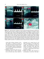
Figure 1 Transverse section of the chest. Diaphragm.
1 – breastbone;
2 – parietal pleura;
3 – intercostal muscles;
4 – aorta;
5 – vertebral body;
6 – costal part of diaphragm ;
7 – tendinous center of diaphragm;
8 – pericardium;;
9 – esophagus;
10 – costomediastinal sinus;
11 – inferior vena cava;
12 – ribs.
6
7
5
3
4
5
9
11
8
"he sintopia of the chest cavity organs is clearly visible on
the computer tomogram: the inferior vena cava (11) and
the esophagus (9) 0re located in front of backbone, to the
right of which the aorta (4) is located, to which the heart
with the pericardium (8) are attached.
12
12
2
12
3
The Chest
3
4
Atlas of the Chest, Abdomen, Lumbar Region, and Retroperitoneal Space
1
1
3
2
2
3
4
6
4
7
9
8
6
5
5
9
7
8
Figure 2 Lung segments.
Lobus superior: 1 – seg. apicale; 2 – seg. posterius; 3 – seg. anterius. Lobus medius: 4 – seg. laterale (правого
легкого) et seg. lingulare superius (left lung); 5 – seg. mediale (right lung) et seg. lingulare inferius (left lung).
Lobus inferior: 6 – seg. apicale; 7 – seg. basale anterius; 8 – seg. basale laterale; 9 – seg. basale posterius
of the 2nd rib on the right with the upper edge of the 3rd rib 1 cm to the
right of the breast bone; then it continues in the form of the arch from
the 3rd to the 5th ribs, as a bulge, heading to the right, at a distance of
1.5 cm from the right edge of the breast bone. The lower margin starts
from the place where the 5th rib is attached on the right, through the
metasternum base to the fifth intercostal space on the left, stopping
short 1.5 cm from the midclavicular line.
The left margin is a line connecting the lower edge of the 1st rib on the
left and the upper edge of the 2nd rib where they are attached to the
breast bone, at the level of the 2nd intercostal space 2.5 cm to the left
of the breast bone edge, then up to the point, placed 1.5-2 cm inwards
the midclavicular line. The apex of the heart is projected on the left in
the 5th intercostal space lower the 5th costal cartilage junction. An atrial
The Chest
4
5
1
1
6
2
3
4
3
2
5
1 –lobus dexter gl. thyroideae;
therocervicalis;
2 –lobus dexter thymi;
thoracica interna;
Blood supply of thymus gland
1 –a. thyroidea inferior; 2 –truncus
3 –a. subclavia sinistra; 4 –a.
3 –lobus sinister thymi;
6 –a. thyroidea ima
5 –rr. thymici a. thoracicae internae;
4 –istmus gl. thyroideae (lobus piramidalis)
Figure 3 Thymus gland and its connection with the thyroid gland.
and ventrical borderline goes between the attachment points of the 3rd
left and 6th right costal cartilages to the breast bone.
Thoracic aorta projection (Figure 6). The ascending aorta
starts from the left ventricle at the level of the 3rd intercostal space behind the breast bone. It turns left and back,
passing into the aortic arch at the level of the 2nd right sternocostal articulation.
Pulmonary trunk projection. The pulmonary artery starts
from the right ventricle, left to the ascending aorta, in the
2nd intercostal space on the left.
Superior vena cava projection. The superior vena cava is
formed by the confluence of two brachiocephalic veins
at the level of the first costal cartilage attachment to the
breast bone. It falls into the right atrium at the level of the
3rd costal cartilage.
6
Atlas of the Chest, Abdomen, Lumbar Region, and Retroperitoneal Space
(a)
(b)
(C)
Figure 4 Differences in the shape and number of thymus glands.
а – two lobes, b – three lobes, c – four lobes.
1
2
15
14
13
3
12
4
5
6
11
10
7
9
8
Figure 5 Topography of blood vessels, heart, right lung.
1 – larynx; 2 – gl. thyroidea; 3 – arcus aortae; 4 – truncus arteriosus; 5 – truncus pulmonalis; 6 – auricula sinistra;
7 – ventriculus sinister; 8 – apex cordis; 9 – ventriculus dexter; 10 – atrium dextrum; 11 – pulmo dextrum; 12 – v.
cava superior; 13 – v. subclavia dextra; 14 – v. jugularis interna; 15 – a. carotis communis dextra.
The Chest
1 – n. recurrensdexter;
2 – n. vagus;
3 – a. carotiscommunis;
4 – a. subclavia;
5 – truncusbrachiocephalicus;
6 – arcusaortae;
7 – bifurcatiotracheae;
8 – nodilimphaticitracheobronchiales;
9 – n. recurrens sinister;
10 – bronchus sinister;
11 – bronchusdexter;
12 – oesophagus
7
Топографияаорты
1 – общаясоннаяартерия;
2 – подключичная артер
3 – плечеголовной ствол;
4 – дугааорты
Figure 6 The relationship of the trachea, esophagus and aorta. Topography of the
aorta.
Esophagus projection. The thoracic esophagus stretches
from the superior thoracic aperture at the level of the 2nd
thoracic vertebra; then at the level of the 2nd–4th thoracic
vertebra, it lies to the right of the median line. Below the
thoracic esophagus it crosses the median line again, and at
the level of the 10th thoracic vertebra it enters through the
esophageal opening, positioning itself 2.5 cm to the left of
the median line.
Chest wall layers (Figure 7):
1. Skin - derma;
2. Subcutaneous fat - panniculus adiposus;
8
Atlas of the Chest, Abdomen, Lumbar Region, and Retroperitoneal Space
1
3
2
6
4
9
11
8
5
10
12
7
Figure 7 Layers of the front upper region of the sternum.
1 – clavicula; 2 – m. subclavicularis; 3 – v. subclavia; 4 – paniculus subdermalis; 5 – fascia superficialis; 6 – fascia propria; 7 – m. pectoralis major; 8 – spatium subpectorale superficialis; 9 – fascia coracoclaidocostalis; 10 – m. pectoralis
minor; 11 – spatium subpectorale profundum; 12 – mm. intercostales
3.
4.
5.
6.
7.
Subcutaneous veins
Suphenous nerves
Superficial pectoral fascia - fascia pectoralis superficialis;
Pectoral fascia - fascia pectoralis propria;
Major and minor pectoral muscles - mm. рectoralis
major et minor;
8. Coracoclavicular pectoral fascia - fascia
coracoclavipectoralis;
9. Endothoracic fascia - fascia endothoracica.
Vessels and nerves of the thoracic wall (Figures 8, 9) are divided
into superficial and deep ones. Cutaneous branches of intercostal
The Chest
9
6
6
7
7
7
6
6
7
5
5
3
2
3
2
2
2
1
1
4
1
1
4
4
4
Figure 8 Vessels and nerves of the chest wall.
1 – n. vagus; 2 – vv. pulmonales; 3 – a. pulmonalis; 4 – cor; 5 – aorta; 6 – mm. intercostalis internii; 7 – a. intercostalis posterior et n. intercostalis
1
5
1
5
4
4
3
7
7
3
2
2
2
2
6
6
7
7
7
7
Figure 9 Vessels and nerves of the chest wall.
1 – n. vagus sinistra; 2 – vv. pulmonales; 3 – a. pulmonalis; 4 – cor; 5 – aorta; 6 – mm. intercostales; 7 – fasciculus
vasus intercostalis
10
Atlas of the Chest, Abdomen, Lumbar Region, and Retroperitoneal Space
arteries—a. thoracica interna, a. thoracica lateralis—belong to superficial vessels. Superficial nerves branch from intercostal nerves, forming
anterior and lateral cutaneous branches.
Arteries, including a.thoracoacromialis, a. thoracica lateralis, a. thoracodorsalis, aa. Intercostales and their branches belong to deep vessels. Deep layer nerves are presented by intercostal nerves producing
muscular branches to intercostal muscles. The long thoracic nerve (n.
thoracicus longus) goes down along the lateral surface of the serratus
аnterior. Anterior thoracic nerves come out of the infraclavicular triangle, previously perforating fascia coracoclavipectoralis.
There is also a fusiform cellular space around the esophagus. Intercostal
spaces are spaces between exterior and interior intercostal muscles
with v. intercostalis posterior, a. intercostalis posterior, and n. intercostalis placed downwards inside the intermucsular cell space of the intercostal space (Figure 10).
2
1
3
12
4
11
5
13
6
7
14
8
9
10
Figure 10 Intercostal space.
1 – costa; 2 – paniculus prepleuralis; 3 – fascia intercostalis; 4 – pleura parietalis; 5 – cavitas pleuralis;
6 – pleura visceralis; 7 – pulmo; 8 – v., a., n. intercostalis; 9 – m. intercostalis externus; 10 – m. intercostalis
internus; 11 – m. pectoralis major; 12 – fascia sternalis; 13 – paniculus subdermalis; 14 – derma.
The Chest
11
Mammary gland (Figures 11, 12). The female breast (mamma muliebris) differs depending on the age and individual anatomy.
It is placed on the anterior thoracic wall at the level from the 3rd to the
6th ribs.
The mammary gland reaches the breast bone medially; and laterally it
goes down from the major pectoral muscle to the lateral chest wall, covering m. serratus anterior. There is an areola (areola mammae) in the middle part of the gland prominence, which has a nipple (papilla mammae)
in its centre. The mammary gland is divided into four quadrants – upper
outer, upper inner, lower outer, and lower inner. Between both mammary
glands there is a deepening that is called a sinus (sinus mammarum).
The principal part of the breast (corpus mammae) consists of
15–20 lobes (lobi mammae); each of them has an excretory lactiferous
12
4
2
1
3
10
6
8
9
5
11
14
7
13
Figure 11 Mammary gland.
1 – sinus lactiferus; 2 – lobi gl. mammariae; 3 – ductus lactiferous; 4 – papilla; 5 – fatty tissue and interlobular
connective tissue; 6 – cellular tissue between the superficial and own fascia; 7 – fascia pectoralis propria;
8 – m. pectoralis major; 9 – intercostal spaces; 10 – fascia intrathoracica; 11 – costa; 12 – fascia superficialis;
13 – pleura; 14 – pulmo.
12
Atlas of the Chest, Abdomen, Lumbar Region, and Retroperitoneal Space
4
1
2
MPT
3
5
5
4
1
2
KT
3
5
5
Figure 12 MRI and CT of the breast.
1 – milky sinus; 2 – lobules of the gland; 3 – milk ducts; 4 – thoracic nipple; 5 – thoracic fascia
duct (ductus lactiferous). Every 2–3 ducts merge and open with a lactiferous duct opening (porus lactiferous). A nipple may be of three
different forms – cylindric, bulb-shaped, and conic.
Lactiferous ducts are opened directly on the tip of the nipple or
inside the nipple. The common lactiferous sinus (sinus lactiferous
communis) is formed from several merging lactiferous sinuses (sinus
lactiferous).
The Chest
13
The skin of the nipples and areola contains oil glands (glandulae sebaceae), perspiratory glands, (glandulae sudorifera), and special rudimentary lacteal glands (glandulae areolares).
Mammary gland blood supply is carried out from three sources:
1) internal thoracic artery (a. thoracica interna) that sends perforating branches (rami perforantes ) to the 3rd – 5th intercostal spaces,
which penetrate into mammary gland materia by the greater pectoral muscle perforation. 2) lateral thoracic artery (a. thoracica
lateralis) that goes down along m. serratus anterior and springs
frontwards the branches supplying external parts of the mammary gland with blood. 3) intercostal arteries (aа. intercostales) ––
3–7 intercostal arteries spring branches for mammary gland blood
supply. All arteries inosculate with each other and surround lobes
and ducts. The venous outflow is carried out by the same-name veins
accordingly.
Mammary gland lymphatic system consists of lymphatic vessels positioned in three levels:
1) subpapillar lymphatic plexus (plexus lymphaticus subpapillaris);
2) superficial areol plexus (plexus areolaris superficialis); 3) profound
areol plexus (plexus areolaris profundus). The main way for lymph outflow from the mammary gland is an axillary one. According to topoanatomic character, the axillary lymph nodes can be divided into five
groups: 1) Lateral axillary nodes; 2) Central axillary nodes; 3) Medial
or pectoral; 4) Subscapular or dorsal; and 5) Apical lymph nodes.
Innervation. There are genuine gland nerves and cutaneous gland
nerves. Innervation is performed by anterior branches of 2nd – 7th intercostal nerves. Anterior nerve branches at the posterior surface form a
plexus, which springs nerves, comprising its own plexus.
Diaphragm (Figure 13). The midriff or diaphragm separates the thoracic cavity from the abdominal cavity. It is divided into two parts:
muscular (pars muscularis diaphragmaticus) and the central tendon of
diaphragm (centrum tendineum).
14
Atlas of the Chest, Abdomen, Lumbar Region, and Retroperitoneal Space
5
11
7
9
4
12
6
3
8
10
2
1
Figure 13 Diaphragm.
1 – sternum; 2 – pleura parietalis; 3 – mm. intercostales; 4 – aorta; 5 – corpus vertebrae; 6 – pars phrenicocostalis;
7 – centrum tendineum; 8 – pericardium; 9 – oesophagus; 10 – recessus costomediastinalis; 11 – v. cava inferior;
12 – costa.
The central tendon of diaphragm consists of the anterior folio (folium
anterius) and lateral folios, (folium dexter and folium sinister) placed
in the horizontal plane, and its muscular part— in vertical plane. The
heart is placed at the anterior folio of the central tendon, and the lungs
are placed at the lateral folios.
Depending on the attachment points, the muscular part of the diaphragm is divided into sternal part (pars sternalis), costal part (pars
costalis), and lumbar part (pars lumbalis).
Diaphragm cruses of the lumbar part are the following:
1. Crus mediale – medial crus that starts from lig. longitudinale anterius and the body of the 3rd or 4th lumbar vertebra
on the right; one vertebra higher on the left. Both cruses
merge at the level of the 1st lumbar vertebra, restricting
aortic hiatus for aorta and thoracic duct.
The Chest
15
2. Crus intermedius – intermediate crus that starts from the
lateral surface of the 2nd lumbar vertebra body and higher
it passes into the muscular part of diaphragm.
3. Crus laterale – lateral crus that starts from the lateral surface of the 2nd lumbar vertebra forming two tendon arches
and represents thickening fascia endoabnominalis.
Arcus lumbocostalis medialis—medial lumbocostal arch that starts from
the 2nd lumbar vertebra body, bestrides m. psoas major and attaches to
the transverse process of the 1st lumbar vertebra.
Arcus lumbo costalis lateralis – lateral lumbocostal arch starts from the
transverse process of 1st lumbar vertebra, bestrides m. quadratus lumborum and attaches to the 12th rib.
Trigonum lumbocostale – lumbocostal triangle is placed between the
lumbar and costal parts of diaphragm. It is based on the lower margin
of the 12th rib. At the thoracic cavity side the triangle bottom is covered
with pleura, adherent with thin fascial leaves, which are adjacent to
kidneys and the posterior surface of adrenal capsule, surrounded with
adipose capsule.
Trigonum sternocostale – sternocostal triangle is placed between sternal and costal parts of the diaphragm. Its height is from 1.8 to 2.7 cm;
its base is from 2.5 to 3 cm.
The diaphragm has the following large apertures:
1. Hiatus aorticus — aortic hiatus — placed between medial
cruses of diaphragm and their tendon part. Aorta and thoracic duct pass through the hiatus at the level of the 12th
thoracic vertebra.
2. Hiatus oesophageus — esophageal hiatus; the medial crus
of the diaphragm that twists after formation of the aortic
hiatus, and then diverging again, forms the second hiatus
16
Atlas of the Chest, Abdomen, Lumbar Region, and Retroperitoneal Space
in the muscle part (hiatus oesophageus). The esophagus
and the vagus nerve pass through it. The lumbar part of
the diaphragm has two eight-shaped openings: a lower one
– aortic (tendon) and upper one – esophageal (muscular).
3. Foramen quadrilaterum s. venae cavae inferiori—inferior
vena cava quadrilateral foramen is placed in the right folio
of the central tendon of diaphragm, where the inferior
vena cava passes through.
Diaphragm blood supply (Figure 14). The anterior parts of the diaphragm periphery are supplied with blood by аа. Intercostales and
its anterior part by аа. pericardiacophrenicae and аа. intercostales.
Therefore, the system of descending aorta branches, thoracic and
abdominal sections and subclavian artery take part in collateral circulation of diaphragm.
19
1
3
18
2
16
17
4
5
14
7 6
15
13
12
9
8
11
10
Figure 14 Diaphragm. Vessels.
1 – pars sternalis; 2 – v. phrenica inferior sinistra; 3 – pars costalis; 4 – r. anterior a. phrenica inferior sinistra;
5 – esophagus; 6 – a. phrenica inferior sinistra; 7 – aorta; 8 – r. lumbalis v. phrenica inferior sinistra; 9 – v. hemiazygos; 10 – trigonum lumbocostale; 11 – mm. psoas major et minor; 12 – v. azygos; 13 – truncus thoracicus; 14 – a.
phrenica inferior dexera; 15 – rr. posteriors aa. phrenica inferior 16 – v. phrenica inferior dextra; 17 – v. cava inferior; 18 – centrum centrum tendineum; 19 – trigonum sternocostale
The Chest
17
А. phrenica superior supplies the lumbar part of diaphragm as well as
its pleura with blood from the thoracic cavity side. А. phrenica inferior
supplies the diaphragm and abdominal membrane with blood; besides
the left artery supplies terminal esophagus part, and the right one —
the inferior vena cava wall, inferior diaphragm veins,
vv. phrenicae inferioris falls into the inferior vena cava, adhering by two
to the same name artery.
Diaphragm innervation. Phrenic nerve (N. Phrenicus) outgoes from
the anterior branches of the 3rd -4th cervical nerves. The left phrenic
nerve penetrates the diaphragm and branches on its inferior surface,
the right one ends with its branches on the superior surface of diaphragm. Six inferior intercostal nerves take part in the innervation of
the posterior part of the diaphragm.
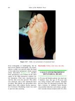
Surgical Anatomy of Thoracic Wall Congenital Malformation
Hollowed chest is a congenital malformation caused by hyperplasia of
the inferior pair of ribs and their cartilages. The midsternum is retroposed and forms a deepening. The deformation is often asymmetric;
the degree of manifestation varies heavily.
Keeled chest is a protruding deformation of the breast bone, which
occurs more rarely than the hollowed chest.
Amastia is a fetal pathology resulting in the birth of a child without
one or both mammary glands. In this case, breast feeding is impossible. Malformation may come with deficiencies of the ovary or other
systems, which results in developmental disorder of the whole reproductive system. Such a woman has neither breast tissue nor nipple.
Polymastia is the presence of additional, multiple glands and nipples, appearing as developed or underdeveloped glands, with clear
nipples, placed along “the milk line”, which passes from the axillary
cavity to the femoroinguinal area. Thereby, additional glands may
swell and lactate while service period.
18
Atlas of the Chest, Abdomen, Lumbar Region, and Retroperitoneal Space
Gynekomastia is mammary gland enlargement in men with glands
and fatty tissue hypertrophy. Painful asymmetric breast lumpiness often appears and then disappears spontaneously. The size of
lumpiness may be different. Physiologic gynecomastia occurs in
newborns, puberty and old men. There is also a pathologic form of
gynecomastia.
Diaphragmatic hernia is an outpunching of the esophagus, stomach
or small bowel through the diaphragm into the thoracic cavity. The
work of the esophageal sphincter which closes the pass between esophagus and stomach is thereby disrupted.
Diaphragm aplasia is an abnormal development of the diaphragm,
when a part of the diaphragm or a section of a part is absent. Newborns
may have a congenitally missing diaphragm, which is fatal. One can
distinguish between unilateral and total diaphragm aplasia. Unilateral
aplasia may be full or partial.
Relaxation of diaphragm is cupula relaxation and high-position of
the diaphragm, based on paralysis, drastic thinning and constant displacement of diaphragm into the thoracic cage together with adjacent
abdominal cavity organs. The congenital elevation of the diaphragm is
connected with aplasia or underdevelopment of its muscular part, and
also with fetal trauma or aplasia of phrenic nerve. Acquired elevation
appears because of secondary atrophy, phrenic nerve injury or diaphragm injury: trauma or tumor.
Thoracic Cavity
Pleura is the serosa of lungs divided into two layers: parietal pleura
(pleura parietalis) and visceral pleura (pleura viscerali). The latter layer
covers the pulmonary surface and forms the pulmonary ligament (lig.
pulmonale) in the root of the lung area while passing into the parietal folium. It is placed under pulmonary veins and stretches vertically
down almost to the inferior margin of the lung. A narrow stripe of the
lung between pulmonary ligament layers is not covered with the visceral layer of pleura.