Ebook Color atlas & synopsis of clinical ophthalmology pediatric ophthalmology (2/E): Part 2
Bạn đang xem bản rút gọn của tài liệu. Xem và tải ngay bản đầy đủ của tài liệu tại đây (15.38 MB, 171 trang )
CHAPTER
7
Congenital Abnormalities of
the Optic Nerve
Aldo Vagge and Leonard B. Nelson
■
OPTIC NERVE HYPOPLASIA
Optic nerve hypoplasia (ONH) is a congenital, nonprogressive developmental
abnormality in which the optic nerve is smaller than usual because of reduced numbers
of retinal ganglion cells. It is frequently associated with other central nervous system
(CNS) abnormalities.
ONH may be unilateral or bilateral (80%) and may be asymmetric.
Most common congenital optic disc anomaly
Optic nerve aplasia is rare. No pupillary light reflex and absence of the optic disc,
nerve fiber layer, and retinal blood vessels on examination.
Etiology
Not completely understood
Parental drug and alcohol abuse contributes to an increasing prevalence of ONH.
Drug associations include exposure to carbemazepine, isotretinoin, phenytoin, quinine,
and valproic acid. Young maternal age and maternal insulin-dependent diabetes have
also been implicated in some cases (associated with subtype—superior segmental optic
hypoplasia).
Genetics
Most cases are sporadic.
Bilateral ONH is inherited in an autosomal-dominant pattern based on the few
families reported. Mutation in the PAX6 (11q13) gene is responsible.
Mutation in the HESX1 gene has been identified in sporadic septo-opto dysplasia
and pituitary disease.
Mutation in the TUBA8 gene is associated with polymicrogyria and ONH.
Symptoms
Decreased vision in one or both eyes
Strabismus may be associated with unilateral ONH.
Signs
Range of visual acuity is 20/20 to no light perception since vision is determined
primarily by the integrity of the papillomacular nerve fibers more than the overall size
of the disc.
Amblyopia as a result of accompanying strabismus and anisometropia
Nystagmus: often develops at 1 to 3 months of age in bilateral cases
Strabismus may be associated with unilateral ONH.
Afferent pupil defect in asymmetric or unilateral cases
Visual fields (VFs) often have localized defects as well as general constriction.
Abnormally small optic nerve head, often gray or pale in color with “double-ring
sign” (scleral canal surrounds a small optic nerve) (Fig. 7-1)
Superior segmental hypoplasia of the optic nerve is a segmental form of ONH
occurring in some children of insulin-dependent diabetic mother.
Retinal vascular tortuosity is common.
Associated Conditions
Septa-optic dysplasia: combination of small anterior visual pathways, absence of the
septum pellucid, and thinning or agenesis of the corpus callosum
Endocrine dysfunction: pituitary gland abnormalities in approximately 15% of
patients with ONH. Patients are at risk for hypothalamic and pituitary dysfunction such
as growth hormone deficiency (most common), hypothyroidism, hyperprolactinemia,
panhypopituitarism, and diabetes insipidus.
Cerebral anomalies such as error in hemispheric migration (schizencephaly, cortical
heterotopias) or hemispheric injury (periventricular leukomalacia [PVL],
encephalomalacia). PVL can be associated with another form of ONH characterized by
large optic cups and a thin neuroretinal rim contained within normal-sized optic discs.
This occurs secondary to trans-synaptic degeneration of optic axons caused by bilateral
lesions in the optic radiations.
Developmental delay more common in patients with bilateral ONH, highly
correlated with corpus callosum hypoplasia and hypothyroidism
Diagnostic Evaluation
Magnetic resonance imaging (MRI) to rule out CNS malformations
Refer to a pediatric endocrinologist if patients show clinical signs of endocrine
dysfunction or pituitary abnormalities on MRI. Pediatrician should follow growth chart
for endocrine changes. Undiagnosed endocrine deficiencies are at risk for impaired
growth, hypoglycemia, seizures, and death.
Automated VF testing may be useful but children are often too young to cooperate.
Differential Diagnosis
Optic atrophy
Optic nerve coloboma
Ocular albinism
Treatment
No treatment available to improve the vision in ONH
Correction of refractive errors
Treatment for superimposed amblyopia
Surgery for concurrent strabismus or nystagmus may be considered.
Consider polycarbonate eye glasses for protection of the better-seeing eye.
Prognosis
Visual acuity is generally nonprogressive. Complications are in general related to
endocrinopathies and CNS malformations.
FIGURE 7-1. Optic nerve hypoplasia. Note the double-ring sign.
MORNING GLORY DISC ANOMALY
Morning glory disc anomaly (MGDA) is a rare, congenital, usually unilateral funnellike excavation of the posterior fundus that incorporates the optic disc.
The name derives from the similarity to the morning glory flower.
More common in female and rare in African Americans.
Etiology
The embryologic basic of MGDA is unclear. A defect in fetal fissure closure or a
primary mesenchymal abnormality has been hypothesized as embryonic origins of
morning glory anomaly.
Symptoms
Decreased vision most common in the involved eye
Color vision defect
Signs
Visual acuity can range from normal vision to no light perception but in general is
approximately 20/100 to 20/200.
Strabismus
Leucokoria
Amblyopia
Myopia
Afferent pupil defect
VF defects, commonly enlarged blind spots
The optic disc is markedly enlarged, orange or pink in color, with a surrounding
annular ring of pigmented uveal tissues. Retinal vessels increased in number emanate
radially from the disc, a central white tuft of glial tissue. Macula may be incorporated
into the excavation (macular capture) (Fig. 7-2).
Serous retinal detachment (RD) in one-third of patients
Associated Conditions
Trans-sphenoidal basal encephalocele associated with midfacial anomalies
(hypertelorism, flat nasal bridge, midline notch in the upper lip, and sometimes a
midline cleft in the soft palate).
Midline or other brain abnormalities (e.g., agenesis of the corpus callosum, pituitary
abnormalities)
Ipsilateral abnormalities of the carotid circulation such as stenosis or aplasia of the
carotid arteries with or without Moyamoya syndrome (progressive stenosis of the
terminal portion of the internal carotid artery and its main branches)
Associated with ipsilateral orofacial hemangioma—this association may fall within
the spectrum of the PHACE syndrome (posterior fossa malformation, large facial
hemangioma, arterial anomalies, cardiac anomalies and aortic coarctation, and eye
anomalies)
Associated with neurofibromatosis type 2
MGDA has been described as part of the spectrum of renal coloboma syndrome.
Diagnostic Evaluation
MRI and magnetic resonance angiography should be obtained to rule out brain and
vascular abnormalities.
Rule out endocrine dysfunction (thyroid-stimulating hormone and growth hormone
levels) and kidney involvement (basic metabolic panel and urinalysis)
Differential Diagnosis
Optic nerve coloboma
Peripapillary staphyloma
Treatment
No treatment available to improve the vision in MGDA
Correction of refractive errors
Treatment for amblyopia if associated
RD is usually addressed with pars plana vitrectomy and long-standing gas
tamponade.
Prognosis
Vision is usually stable unless RD occurs. Optic neuritis and progressive optic
atrophy have been documented.
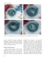
FIGURE 7-2. Morning glory disc. Morning glory disc anomaly showing an enlarged excavation,
abnormal retinal vascular pattern, annular pigmentation surrounding the nerve head, and central glial tuft
and peripapillary changes. (Courtesy of Alex Levin, MD.)
OPTIC DISC COLOBOMA
Clearly demarcated bowl-shaped excavation of the optic disc, which is typically
decentered and deeper inferiorly.
Unilateral and bilateral optic disc coloboma occur with similar frequencies.
Occasionally involvement of the entire disc occurs.
Other types of uveal coloboma can coexist.
They may be isolated or part of a systemic syndrome.
Etiology
Thought to result from incomplete or abnormal fusion of the two sides of the
proximal end of the embryonic fissure
Most cases are sporadic but may be autosomal dominant, autosomal recessive, or Xlinked recessive.
A wide variety of mutations have been documented in patients with coloboma
—CHD7 mutation is associated with 60% of cases of CHARGE syndrome.
Symptoms
Visual acuity may be mildly or severely decreased in one or both eyes—the degree
of foveal involvement by the coloboma is the only feature that relates to visual outcome.
Signs
White, bowl-shaped excavation that occurs in an enlarged optic disc. The excavation
is decentered inferiorly and the superior neuroretinal rim is relatively spared. In case of
complete excavation of the entire disc, the excavation is deeper inferiorly.
Chorioretinal coloboma can be associated—if so, microphthalmia is frequently
present
Iris and ciliary body colobomas often coexist.
Rhegmatogenous or serous RD may develop in some patients—rhegmatogenous are
often associated when chorioretinal coloboma, whereas serous detachment is more
common in case of isolated optic nerve coloboma.
Associated Conditions
CHARGE association: coloboma, choanal atresia, congenital heart disease, and
multiple other abnormalities
Walker-Warburg syndrome
Goltz focal dermal hypoplasia
Aicardi syndrome
Goldenhar sequence
Linear sebaceous nevus syndrome
Dandy-Walker malformation
Renal coloboma syndrome—with a mutation of PAX2 transcription
Microphthalmia—in case of chorioretinal involvement
Diagnostic Evaluation
Optical coherence tomography (OCT) has been useful in observing the intercalary
membrane that covers the chorioretinal defect and is continuous with the neural retina.
A complete systemic evaluation is important to rule out other associated anomalies.
Differential Diagnosis
MGDA
Peripapillary staphyloma
Treatment
No specific treatment is available for optic disc coloboma
Treatment for amblyopia, if associated
Optimal refractive correction may be indicated.
RD surgery as indicated
Prognosis
Vision is usually stable unless RD occurs
OPTIC DISC PITS
Optic disc pit (ODP) is an oval or round excavation of variable color, depth, and
location in the optic disc.
The temporal optic disc side is the most commonly involved.
Often unilateral, although bilateral cases have been reported in 15% of the cases.
They are rare, with an estimated incidence of 1 in 11,000 people.
Etiology
It is not entirely clear—they are thought to result from an imperfect closure of the
superior edge of the embryonic fissure.
Histologically, an ODP is a herniation of dysplastic retina into a collagen-rich
excavation that extends into the subarachnoid space through a defect in the lamina
cribrosa.
The pathogenesis of the macular changes is still controversial—the fluid may be
vitreous fluid, cerebrospinal fluid, leakage from blood vessels at the base of the pit, or
leakage from the choroid. Also, serous macular detachment is caused by direct
communication between the optic pit and the subretinal space or from the optic pit and
retina. In the latter case, fluid may move into the retina, causing a schisis-like separation
of the inner and outer layers, with the neurosensory serous RD occurring secondary to
this schisis. In addition, vitreous traction appears to be an important factor in the
pathogenesis of optic pit–related macular detachment.
ODPs are generally sporadic, although familial occurrence has been reported as a
dominant trait.
Symptoms
Optic pits are asymptomatic unless there is subretinal fluid—approximately 45% of
the eyes develop serous macular detachment usually in the second or third decades of
life. When subretinal fluid is present, visual acuity decreases to 20/40–20/60.
Signs
ODPs are usually seen as single, oval-shaped depressions at the optic disc. They are
most commonly found at the inferotemporal aspect of the optic disc, but may also be
found elsewhere, including centrally. Usually they are gray, white, or yellowish in color
(Fig. 7-3).
The signs associated with ODP maculopathy include intraretinal and subretinal fluid
accumulation, and retinal pigment changes.
Amblyopia in children especially in eye with serous macular detachment
Associated Conditions
Rarely associated with basal encephalocele
Diagnostic Evaluation
OCT to evaluate the subretinal fluid—typically OCT may show a schisis-like
separation between the inner and outer retina.
VF—arcuate scotoma is most common
Intravenous fluorescein angiography (IVFA) is helpful in the differential diagnosis of
serous detachment.
Amsler grid can be used to monitor the macular involvement.
Differential Diagnosis
Optic disc anomalies such as choroidal and scleral crescent
Tilted disc syndrome (TDS)
Circumpapillary staphyloma
Hypoplastic disc
Glaucomatous optic neuropathy
Central serous retinopathy and subretinal neovascular membranes for serous macular
detachment
Treatment
No treatment is required for an isolated optic pit.
Laser photocoagulation (between the area of the serous RD and the optic disc),
macular buckling, and a combination of posterior vitrectomy, photocoagulation, and gas
tamponade are the commonly used procedures for the treatment of ODP maculopathy.
Less-invasive treatments like laser photocoagulation should be tried initially.
Prognosis
Isolated ODP has usually an excellent prognosis.
Associated retinal complications such as serous macular detachment can be
progressive and decrease significantly visual acuity—advice patients about the
importance of regular comprehensive eye exams and the use of Amsler grid testing.
Posterior macular reattachment can occur in rare instances.
FIGURE 7-3. Optic disc pit. Congenital pit of the optic nerve head.
TILTED DISC SYNDROME
TDS is a congenital, nonhereditary bilateral condition where the optic nerve appears
to enter the eye in an oblique angle.
Elevated superior pole of the optic disc with posterior displacement of the inferior
nasal disc resulting in an oval appearance of the optic nerve head
Often accompanied by:
Scleral crescent located inferiorly and inferonasally (Fig. 7-4)
Situs inversus of retinal vessel
Posterior ectasia of the inferonasal retina and choroid
Typically associated with myopic astigmatism because of the fundus abnormalities
(posterior ectasia)
Etiology
Unknown, but may have some pathogenic relationship with colobomatous defect.
Symptoms
Best corrected visual acuity (BCVA) may be reduced.
Signs
Myopic astigmatism
Tilted disc with associated features as previously described
VF defects are often associated with complete bitemporal hemianopia that does not
respect the midline. Repeat perimetry after addition of a −4.00 lens often eliminates the
VF abnormalities (refractive nature of the defects). In some cases, VF defects persist
despite refractive correction due to an abnormal inferonasal ectasia. However, TDS has
been reported with true bilateral hemianopsia and congenital suprasellar brain tumor.
Amblyopia
Associated Conditions
Suprasellar brain tumors
Tilted disc without retinal ectasia occurs in patients with trans-sphenoidal
encephalocele.
Craniofacial anomalies such as hypertelorism, Crouzon syndrome, and Alport
syndrome have been observed in association with tilted disc.
Other conditions reported with tilted disc are:
Ehler-Danlos type III
Hemifacial atrophy
Congenital horizontal gaze palsy
Familial dextrocardia
Exotropia
Diagnostic Evaluation
Refraction and dilated fundus exam—diagnosis can be made based on the
fundoscopic appearance of the optic disc.
VF often shows complete bitemporal hemianopia that does not respect the midline
(unlike chasmal lesions).
MRI brain in any patient with TDS and VF defects to rule out suprasellar tumors
Differential Diagnosis
ONH
Optic nerve coloboma
Peripapillary staphyloma
Papilledema
Treatment
No medical treatment for the primary disorder
Appropriate refractive error correction
Amblyopia therapy as indicated may improve nonorganic visual loss.
Prognosis
Broad range of BCVA
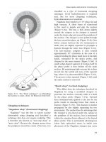
FIGURE 7-4. Tilted disc. Note the temporal scleral crescent.
PERIPAPILLARY STAPHYLOMA
Peripapillary staphyloma is a generally sporadic, rare, usually unilateral optic disc
anomaly characterized by a deep excavation of the area of the fundus surrounding the
optic disc.
Optic disc head sits at the base of the posterior pole excavation.
Not associated with glial or vascular abnormalities of the disc, uveal coloboma, and
progression
Affected eyes typically are emmetropic or slightly myopic, although high myopia has
been reported.
Etiology
The etiology is unknown. It appears to arise as incomplete differentiation of sclera.
Staphyloma may be the consequence of the development of normal intraocular pressure
causing scleral herniation.
Symptoms
Visual acuity is usually mildly or severely reduced.
Signs
Peripapillary staphyloma is usually associated with a relatively normal appearance
of the optic disc.
Centrocecal scotomas commonly occur in eyes with decreased vision.
Associated Conditions
Usually absence of associated systemic abnormalities or intracranial diseases
Peripapillary staphyloma has been reported to be associated with basal
encephalocele in patients with midfacial abnormalities.
PHACE syndrome, linear nevus sebaceous syndrome, and 18q-syndrome have been
observed in association with peripapillary staphyloma.
Nystagmus, strabismus, and head turn
Increased risk of RD
Diagnostic Evaluation
Ocular ultrasound and electroretinogram (ERG) can help the diagnosis especially in
pediatric patients.
OCT can be used to evaluate a peripapillary staphyloma.
VF can show a centrocecal scotoma especially in eyes with decreased vision.
MRI of the brain is indicated for children with midfacial abnormalities.
Differential Diagnosis
MGDA
Optic disc coloboma
TDS
Treatment
No medical treatment for the primary disorder
Amblyopia therapy and strabismus surgery as needed
RD surgery as indicated
Consider polycarbonate eye glasses.
Prognosis
Risk of RD
OPTIC DISC DRUSEN (PSEUDOPAPILLEDEMA)
Optic disc drusen are acellular calcific deposits located within the optic nerve head.
Optic drusen are typically buried in the optic disc early in life and become more
superficial later.
Often bilaterally
Most common form of pseudopapilledema—anomalous elevation of the optic disc
unrelated to increased intracranial pressure
Etiology
The etiology is unknown—they are thought to result by a disturbance in axonal
metabolism with slowed axoplasmic flow, congenitally dysplastic discs with a
propensity for drusen formation, or a small scleral canal that physically compresses the
optic nerve, causing ganglion cell death, with extrusion and calcification of
mitochondria.
Optic drusen may be transmitted as an irregular dominant trait—they are frequently
familial.
Symptoms
Usually asymptomatic with no visual complaints
Rarely (especially in children) transient visual obscuration—probably secondary to
transient disc ischemia
Signs
Disc is often elevated and its margins are blurred and obscured.
Disc vessel is clearly visible, without hyperemia, dilated capillaries, or venous
congestion.
Absence of exudates and cotton wool spots
Retinal vasculature of eyes is frequently anomalous—higher frequency of cilioretinal
arteries
Afferent pupillary defect and acquired dyschromatopsia may be present and they are
signs of an optic neuropathy.
VF can show peripheral defects that tend to increase in frequency with increasing
age. The progression is generally slow. The most common VF defects are nasal defect,
concentric constriction, and enlarged blind spot.
Peripapillary or disc hemorrhage, choroidal neovascular membrane (CNVM),
nonarteritic anterior ischemic optic neuropathy (NAION), and retinal artery or vein
occlusion can be complications of optic disc drusen.
Associated Conditions
Optic disc drusen have been reported in association with many ocular and systemic
disorders.
Space-occupying lesions have been harbored with optic disc drusen and progressive
visual loss.
The most commonly associated conditions are:
Retinitis pigmentosa
Pseudoxanthoma elasticum and angioid streaks
Alagille syndrome
Diagnostic Evaluation
B scan ultrasonography: drusen appear with high reflectivity and posterior
shadowing
Fundus autofluorescence: drusen display autofluorescence (poor reliability in buried
drusen)
Fluorescein angiography: drusen stain in late stage. Helpful to distinguish between
optic disc drusen and true optic disc edema
OCT: focal hyper reflective mass posterior to the outer plexiform and outer nuclear
layers, with loss of the inner and outer segment photoreceptor junction (poor reliability
at distinguishing buried drusen vs. true optic disc edema)
VF testing should be performed as soon as children can do so reliably.
Orbital CT—calcification in optic disc
MRI brain in any patient with associated progressive visual loss
Differential Diagnosis
Optic neuritis
Posterior scleritis
Toxoplasmosis
Idiopathic intracranial hypertension
Ischemic optic neuropathy
Compressive optic neuropathy
Optic nerve infiltrates
Papilledema
Sarcoidosis
Optic nerve tumors
Leber hereditary optic neuropathy
Treatment
Drusen alone need no medical therapy.
CNVMs may require laser photocoagulation or intravitreal anti-VEGF.
Ischemic complications (NAION and retinal vascular occlusions) managed in the
absence of drusen
Prognosis
Usually very good but complications can occur
VF defects are identified in up to 51% of children and become more common with
increasing age.
Hemorrhagic complications—peripapillary or disc hemorrhage
CNVM
NAION
Retinal artery or vein occlusion
REFERENCES
Dutton GN. Congenital disorders of the optic nerve: excavations and hypoplasia [review]. Eye (Lond).
2004;18(11):1038–1048.
Hoyt C, Taylor D. Pediatric Ophthalmology and Strabismus. 4th ed. St. Louis, MO: Elsevier Saunders; 2013:543–560.
Maguire JI, Murchison AP, Jaeger EA. Wills Eye Hospital 5-Minute Ophthalmology Consult. Philadelphia, PA:
Lippincott Williams & Wilkins; 2012.
CHAPTER
8
Retinal Anomalies
BEST DISEASE
Barry N. Wasserman
■
Etiology
An autosomal dominant disorder also called vitelliform macular dystrophy, Best
disease leads to retinal pigment epithelium (RPE) degeneration and secondary loss of
photoreceptors in the macula. Mutations lead to abnormal bestrophin, a Ca2+ sensitive
Cl− channel protein. Lipofuscin accumulates in the RPE cells yielding a characteristic
“egg yolk” appearance (hence the name vitelliform) in the fovea and macula. Multifocal
lesions may also occur. Several genes have been identified, including the BEST1 and
PRPH2, which is associated with an adult form. In families negative for these genes,
IMPG1 and IMPG2 genes have been identified as causal.
Symptoms
Early in the disease process, patients may be asymptomatic, but later metamorphopsia
and decreased visual acuity occur. Symptoms are variable and may be asymmetric, with
significant vision loss in adulthood.
Signs
Stages are described on the basis of phenotypic appearance in the macula, which may
be asymmetric between the eyes. Early stage may only reveal subtle RPE mottling or
may be normal. Later, yellow-orange material collects in the macula as a 0.5-to-5.0mm-diameter lesion, yielding the classic “egg yolk” appearance. Clear fluid slowly
builds around the lesion in the subretinal space, with resultant cystic appearance with
fluid level (Fig. 8-1A). As this fluid dissipates, the lesion again changes to a more
“scrambled egg” (vitelliruptive) stage, associated with pigment clumping (Fig. 8-1B).
Later stages include atrophic changes with fibrosis, macular degeneration, and
significant vision loss, followed sometimes with choroidal neovascularization.
Differential Diagnosis
Stargardt disease
Sorsby macular dystrophy
Pattern dystrophy
North Carolina macular dystrophy
Solar retinopathy
Coalescence of basal laminar drusen
Central serous retinopathy with fibrinous exudate
Pigment epithelial detachment of age-related macular degeneration
Adult foveomacular dystrophy
Age-related macular degeneration
Diagnostic Evaluation
Diagnosis is based on clinical findings, along with electrophysiologic studies revealing
normal electroretinogram and abnormal electrooculogram. In addition, there is high
autofluorescence. Optical coherence tomography (OCT) detects subretinal deposits and
fluid. Genetic studies may reveal mutation in the bestrophin gene.
Treatment
No treatment for early disease, but late choroidal neovascularization has been treated
with intravitreal injection of bevacizumab. Genetic counseling and examination of
family members are recommended, as are low vision and occupational consultations.
Prognosis
Some patients maintain good vision (20/40) throughout life. In others, good vision
(20/20 to 20/50) usually persists through the early stages but decreases to 20/200 in the
fifth and sixth decades as the atrophic and cicatricial stages progress.
REFERENCES
Goodwin P. Hereditary retinal disease. Curr Opin Ophthalmol. 2008;19:255–262.
Leu J, Schrage NF, Degenring RF. Choroidal neovascularisation secondary to Best’s disease in a 13-year-old boy
treated by intravitreal bevacizumab. Graefes Arch Clin Exp Ophthalmol. 2007;245(11):1723–1725.
Meunier I, Manes G, Bocquet B, et al. Frequency and clinical pattern of vitelliform macular dystrophy caused by
mutations of interphotoreceptor matrix IMPG1 and IMPG2 genes. Ophthalmology. 2014;121:2406–2414.
Spaide RF, Noble K, Morgan A, et al. Vitelliform macular dystrophy. Ophthalmology. 2006;113:1392–1400.
FIGURE 8-1. Best disease. A. Best disease with early macular “egg yolk” appearance. B. Best
disease with later scrambled-egg appearance.
CHOROIDEREMIA
Barry N. Wasserman
■
Etiology
An X-linked recessive disease, choroideremia is a progressive retinal degeneration.
Males carriers show loss of choriocapillaris and RPE. Female carriers can show mild
signs of the disease, with patchy retinal abnormalities and corresponding visual field
(VF) defects due to Lyonization, but are generally asymptomatic. Mutation in the CHM
gene affects production of the Rab escort protein 1 (REP-1). Degeneration begins in the
midperiphery and progresses centrally both toward the macula and toward the
periphery.
Symptoms
Patients present in the first two decades of life with early loss of night vision and
peripheral vision. Central vision may be maintained until around the fifth decade but is
eventually lost.
Signs
Retinal examination reveals loss of the RPE and choriocapillaris, with exposure of the
larger choroidal vessels (Fig. 8-2). Remaining RPE may have a salt-and-pepper
appearance. Later in the disease, large areas of exposed sclera may be seen on
fundoscopy. Posterior subcapsular cataracts may be associated.
Differential Diagnosis
Advanced retinitis pigmentosa
Gyrate atrophy
Pathologic myopia
Chorioretinitis (e.g., acute retinal necrosis, following cytomegalovirus [CMV]
retinitis)
Diagnostic Evaluation
X-linked inheritance with typical fundus appearance is seen in male patients. VF testing
demonstrates constriction. Fluorescein angiography reveals large choroidal vessels due
to loss of overlying RPE and choriocapillaris. Electroretinogram may be consistent with
a rod–cone dystrophy early in the disease but eventually becomes extinguished. Genetic
analysis to assess mutation in the CHM gene may be performed, and analysis of
peripheral blood is available to demonstrate absence of the Rab escort protein 1.
Female carriers may demonstrate patches of irregular pigmentation in the RPE. SDOCT may demonstrate reduced subfoveal choroidal thickness and increased foveal
thickness as the disease progresses, despite unchanged visual acuity.
Treatment
Genetic counseling for family members should be suggested. Low vision evaluation and
treatment may be helpful. Gene therapy has shown some success in animal models.
Prognosis
Patients progressively lose night and peripheral vision. Central vision is often
maintained into adulthood but is ultimately lost.
REFERENCES
Kamron KN, Islam F, Moore AT, et al. Clinical and genetic features of choroideremia in childhood. Ophthalmology.
2016;123(10):2158–2165.
Lee TK, McTaggart KE, Sieving PA, et al. Clinical diagnoses that overlap with choroideremia. Can J Ophthalmol.
2003;38(5):364–372.
MacDonald IM, Russell L, Chan CC. Choroideremia: new findings from ocular pathology and review of recent
literature. Surv Ophthalmol. 2009;54(3):401–407.
MacDonald IM, Smaoui N, Seabra MC. Choroideremia. In: Pagon RA, Bird TC, Dolan CR, Stephens K, eds.
GeneReviews [Online]. Seattle: University of Washington; 2010.
FIGURE 8-2. Choroideremia. Choroideremia at advanced stage, with complete loss of retinal pigment
epithelium and choriocapillaris.
GYRATE ATROPHY
Barry N. Wasserman
■
Etiology
Gyrate atrophy is an autosomal recessive disease caused by the deficiency of the
mitochondrial enzyme, ornithine aminotransferase. Elevated ornithine levels are toxic to
the RPE, causing gradual loss of peripheral vision and night vision. Mutations of the
ornithine aminotransferase gene (10q26) have been identified.
Symptoms
Nyctalopia and loss of visual field may begin in the first two decades but may not be
manifest until the fifth decade. The disease affects both eyes symmetrically. Central
vision is spared usually until the fourth or fifth decade but then declines. Symptoms and
signs may vary widely in patients in all age groups.
Signs
Fundus examination early in the disease shows scalloped areas of geographic atrophic
RPE and choriocapillaris (Fig. 8-3). The macula is relatively spared until late in the
disease, though epiretinal membranes and cystoid macular edema can occur. Patients
may have cataracts and myopia.
Differential Diagnosis
Choroideremia
Retinitis pigmentosa
Choroidal atrophy
Chorioretinitis (e.g., acute retinal necrosis, following CMV retinitis)
Diagnostic Evaluation
Ocular examination may reveal mildly decreased visual acuity, and refractive error is
commonly myopic. Cataracts may be seen on slit-lamp evaluation. Fundoscopy shows
midperipheral and peripheral scalloped geographic areas of RPE and choriocapillaris.
Areas of intact RPE may have increased pigmentation. The macula is usually spared
early in the disease, but cystoid macular edema may be demonstrated with OCT. Optic
atrophy and attenuated retinal vessels are seen later in the disease. VFs are markedly
constricted, and electroretinogram reveals absent photopic and scotopic responses. SDOCT may demonstrate intraretinal cystic spaces and hyperreflective deposits in the
ganglion cell layer. Outer retinal tubulations are round tubular, rosette-like structures
found in the outer nuclear layer of some patients with advanced disease. Plasma
ornithine levels are markedly elevated.
Treatment
Genetic counseling is suggested. A diet restricting arginine will lower the plasma
ornithine and may slow the loss of visual function. Low vision services should be