Retrocecal mucocele of the appendix: Case report and review of literature
Bạn đang xem bản rút gọn của tài liệu. Xem và tải ngay bản đầy đủ của tài liệu tại đây (381.42 KB, 4 trang )
Journal of military pharmaco-medicine no9-2018
RETROCECAL MUCOCELE OF THE APPENDIX:
CASE REPORT AND REVIEW OF LITERATURE
Ngo Tuan Minh1; Nguyen Thi Ha1; Le Vu Duy1
Phung Anh Tuan1; Tran Quang Vinh2; Ho Van Thanh3
SUMMARY
Appendiceal mucocele was described for the first time by Rokitansky in 1842 [9]. Its incidence
was 0.2 - 0.4% of all apendectomies performed, as it is observed predominantly in women with
the ratio of 4/1 versus men and most frequently at the age of 50. We reported a 78-year-old
woman, who was admitted to the hospital because of mild pain in the right lower quadrant.
Appendiceal mucocele with a retrocecal location, which is a rare position, was determined
intraoperatively. The formation has been dissected and appendectomy was performed without
mucocele integrity being compromised. The permanent histological specimen revealed mucinous
cystadenoma with clear resection lines, without any data on the appendiceal base being affected.
The patient was discharged on the third day after surgery without any complications, and further
follow-up was scheduled in 6 months.
* Keywords: Retrocecal mucocele of appendix.
INTRODUCTION
Appendiceal mucocele (AM) is a rare
entity that can present with a variety of
clinical symptoms or occur as an incidental
surgical finding. The incidence is 0.2 - 0.4%
of all appendectomied specimens [6]. AM
is a progressive dilatation of the appendix
from the intraluminal accumulation of the
mucoid substance [4, 6]. It may be a benign
or malignant process. There are four
histological types, which lead to individualized
surgical treatment and course in each
case [6].
Preoperative diagnosis that distinguishes
AM from acute appendicitis (AA) is essential
for the best choice of surgical approach
(open vs. laparoscopy) to prevent peritoneal
dissemination and perform the appropriate
surgery [6]. Herein, we reported a retrocecal
AM case, which is a rare location, discuss
the process of diagnosis and review the
relevant literature.
CASE PRESENTATION
A 78-year-old woman was admitted to
the hospital because of pain in the right
lower quadrant of the abdomen for 2 days.
When palpating the lower right quadrant
of the abdomen, the patient felt mild pain,
muscles were not rigid, and rebound
tenderness was not noted.
Body temperature was 36.8°C.
Leukocytosis (8.5 x 10⁹/L) was notable
from laboratory test results.
1. 103 Military Hospital
2. 110 Military Hospital
3. Vocational School of Military Medicine
Corresponding author: Ngo Tuan Minh ()
Date received: 17/08/2018
Date accepted: 21/11/2018
194
Journal of military pharmaco-medicine no9-2018
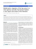
Ultrasonography (USG) showed a cystic mass (29.9 x 16.9 mm) with thin, calcified
wall located posteriorly and originated from the ceacum. There was no finding of
inflammation in the iliac fossa.
Fig.1: USG showed a cystic mass with thin, calcified wall.
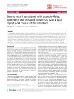
Computed tomography (CT) revealed a well-circumscribed, low-attenuation, tubular,
curvilinear and mural calcified mass contiguous with the base of the caecum.
Preoperative diagnosis was retrocecal AM.
Fig.2: CT demonstrated a cystic mass in the retrocecal position.
The patient underwent total appendectomy and the appendiceal mucocele with
retrocecal location was determined intraoperatively.
Histopathological diagnosis was benign mucinous cystadenoma. Postoperative course
was unremarkable and she was discharged home on the 3rd postoperative day.
195
Journal of military pharmaco-medicine no9-2018
DISCUSSION
Preoperative diagnosis of appendicular
mucocele is very important for the
selection of an adequate surgical method
to prevent peritoneal dissemination, to
prevent intraoperative and postoperative
complications, and repeated surgery [10].
USG, CT, and colonoscopy are used for
diagnostics.
called ‘onion skin’, is an important sonographic
marker of AM [1]. Visualisation of appendix
and its diameter exceeding 15 mm is
considered as specific appendiceal mucocele
index with sensitivity of 83% and specificity
of 92% [5]. Discontinuity of the appendiceal
wall with leakage of the internal contents
into surrounding tissues indicates rupture
of the mucinous tumor [11]. In cases of
developed pseudomyxoma peritonei (PMP),
typical ultrasound findings are non-mobile,
sonogenic ascites with multiple semi solid
masses and scalloping of the hepatic and
splenic margins [12]. In case of CT, it is
important to assess formation interrelation
with surrounding organs, which may make
diagnostication easier. CT is considered
to be the most informative imaging method,
although accurate diagnostication is often
not possible. In most of the cases, a wellcapsulated cystic formation with calcificates
in the wall is visualised, which is considered
a specific index, as well [7]. Another
important CT indication is the presence of
appendiceal lumen exceeding 1.3 cm [2].
When the mucinous tumor is ruptured and
pseudomyxoma peritonei is developed,
the most common findings on a CT-scan
are a large volume of mucinous ascites
with the density of fat that displace the
small bowel and the normal mesenteric
fat [12]. Other characteristic findings are
omental thickening, multiseptate lesions,
scalloping of organs, and curvilinear
calcifications [12].
Radiographically, a mucocele is seen
as a soft tissue mass, possibly with peripheral
curvilinear calcification. The typical image,
At first, the patient was performed
USG. Ultrasonography showed a cystic
mass (29.9 x 16.9 mm) with thin, calcified
Mucocele of the appendix was first
described by Rokitansky [9]. This disease
is characterized by dilatation of a lumen
as a result of an accumulation of a large
amount of mucus. The appendix is lined
by epithelium containing more goblet cells
than the colon. As a result, most appendiceal
epithelial tumors are mucinous and start
as mucoceles [10]. It falls under the
category of rare diseases. Its incidence
ranges between 0.2% and 0.7% of all
excised appendixes [6]. This condition can
have benign as well as malignant processes.
According to modern classification, there
are 4 histologic types: Retention cyst,
mucosal hyperplasia, mucinous cystadenoma,
and mucinous cystadenocarcinoma [3].
The clinical flow of the disease does
not have a specific picture. It often flows
asymptomatically. In about 50% of cases
it is discovered accidentally during radiologic
and endoscopic examinations or at surgery.
A patient’s clinical symptoms may include
pain in the right lower quadrant of the
abdomen, palpable abdominal mass, nausea,
vomiting, weight loss, gastrointestinal
bleeding, and signs of intussusception of
the intestines [5, 10].
196
Journal of military pharmaco-medicine no9-2018
walls located posteriorly and originated
from the ceacum. There was no finding of
inflamation in the iliac fossa, therefore it
might not have been appendicitis. So, we
could not make an appropriate diagnosis,
and CT was requested. CT revealed a
well-circumscribed, low-attenuation, tubular,
curvilinear and mural calcified mass
contiguous with the base of the ceacum.
Preoperative diagnosis was retrocecal AM.
Surgery is the only treatment with curative
potential. Surgical treatment depends on
the dimensions and histology of the
mucinous neoplasm, as well as the clinical
presentation [11]. During the operation we
must be cautious while handling the
mucocele to avoid rupture and dispersion
of mucus or epithelial cells into the
peritoneal cavity as this is associated with
a poorer prognosis [11]. Conventional surgery
is preferred rather than laparoscopic
approaches for the treatment [5]. Laparoscopic
approach has an increased risk of rupture
and subsequent pseudomyxoma peritonei
formation.
CONCLUSION
Appendiceal mucocele is a rare
disease and has a clinical picture that
resembles acute appendicitis. A correct
diagnosis before surgery is very important
for the selection of surgical technique to
avoid severe intraoperative and postoperative
complications. USG, particularly CT, should
be used extensively for this purpose.
In our opinion, every patient more than
50 years old who arrives at the emergency
department with clinical symptoms of
acute appendicitis must undergo CT and
open surgery should be favored against
laparoscopic surgery
REFERENCES
1. Caspi B et al. The onion skin sign:
A specific sonographic marker of appendiceal
mucocele. J Ultrasound Med. 2004, 23 (1),
pp.117-21, quiz 122-123.
2. Demetrashvili Z. Mucocele of the appendix:
Case report and review of literature. 2012, 97 (3),
pp.66-69.
3. Higa E et al. Mucosal hyperplasia,
mucinous cystadenoma, and mucinous
cystadenocarcinoma of the appendix. A reevaluation of appendiceal "mucocele". Cancer.
1973, 32 (6), pp.1525-1541.
4. Jaffe B.M, Berger D.H. The appendix,
Schwartz’s principles of surgery. F Brunicardi
et al, McGraw Hill Companies Inc. 2005,
pp.1119-1137.
5. Karakaya K et al. Appendiceal mucocele:
Case reports and review of current literature. World
J Gastroenterol. 2008, 14 (14), pp.2280-2283.
6. Lien W.C et al. Appendiceal outer
diameter as an indicator for differentiating
appendiceal mucocele from appendicitis.
Am J Emerg Med. 2006, 24 (7), pp.801-805.
7. Puvaneswary M, Proietto A. Mucocele of
the appendix with magnetic resonance imaging
findings. Australas Radiol. 2006, 50 (1), pp.71-74.
8. Rampone B et al. Giant appendiceal
mucocele: Report of a case and brief review. World
J Gastroenterol. 2005, 11 (30), pp.4761-4763.
9. Rokitansky C.F. A manual of pathological
anatomy. Philadelphia: Blanchard & Lea. 1855.
10. Sugarbaker P.H. Epithelial appendiceal
neoplasms. Cancer J. 2009, 15 (3), pp.225-235.
11. Tirumani S.H et al. Mucinous neoplasms
of the appendix: A current comprehensive
clinicopathologic and imaging review. Cancer
Imaging. 2013, 13, pp.14-25.
12. Zhong Y et al. Pseudomyxoma
peritonei as an intractable disease and its
preoperative assessment to help improve
prognosis after surgery: A review of the
literature. Intractable Rare Dis Res. 2012, 1 (3),
pp.115-121.
197