Ebook Text and atlas of wound diagnosis and treatment: Part 1
Bạn đang xem bản rút gọn của tài liệu. Xem và tải ngay bản đầy đủ của tài liệu tại đây (18.12 MB, 245 trang )
Text and Atlas of Wound
Diagnosis and Treatment
Hamm_FM_i-xviii.indd i
18/12/14 11:13 AM
NOTICE
Medicine is an ever-changing science. As new research and clinical experience broaden
our knowledge, changes in treatment and drug therapy are required. The authors and
the publisher of this work have checked with sources believed to be reliable in their
efforts to provide information that is complete and generally in accord with the standards
accepted at the time of publication. However, in view of the possibility of human error
or changes in medical sciences, neither the authors nor the publisher nor any other party
who has been involved in the preparation or publication of this work warrants that the
information contained herein is in every respect accurate or complete, and they disclaim
all responsibility for any errors or omissions or for the results obtained from use of the
information contained in this work. Readers are encouraged to confirm the information
contained herein with other sources. For example and in particular, readers are advised
to check the product information sheet included in the package of each drug they plan
to administer to be certain that the information contained in this work is accurate and
that changes have not been made in the recommended dose or in the contraindications
for administration. This recommendation is of particular importance in connection with
new or infrequently used drugs.
Hamm_FM_i-xviii.indd ii
18/12/14 11:13 AM
Text and Atlas of Wound
Diagnosis and Treatment
Edited by
Rose L. Hamm, PT, DPT, CWS, FACCWS
Assistant Professor of Clinical Physical Therapy
Division of Biokinesiology and Physical Therapy
Ostrow School of Dentistry
University of Southern California
Los Angeles, California
New York Chicago San Francisco Athens London Madrid
Mexico City Milan New Delhi Singapore Sydney Toronto
Hamm_FM_i-xviii.indd iii
18/12/14 11:13 AM
Copyright © 2015 by McGraw-Hill Education. All rights reserved. Except as permitted under the United States Copyright Act of 1976, no part of this publication may be
reproduced or distributed in any form or by any means, or stored in a database or retrieval system, without the prior written permission of the publisher, with the exception that
the program listings may be entered, stored, and executed in a computer system, but they may not be reproduced for publication.
ISBN: 978-0-07-180724-1
MHID: 0-07-180724-1
The material in this eBook also appears in the print version of this title: ISBN: 978-0-07-180721-0,
MHID: 0-07-180721-7.
eBook conversion by codeMantra
Version 1.0
All trademarks are trademarks of their respective owners. Rather than put a trademark symbol after every occurrence of a trademarked name, we use names in an editorial
fashion only, and to the benefit of the trademark owner, with no intention of infringement of the trademark. Where such designations appear in this book, they have been printed
with initial caps.
McGraw-Hill Education eBooks are available at special quantity discounts to use as premiums and sales promotions or for use in corporate training programs. To contact a
representative, please visit the Contact Us page at www.mhprofessional.com.
TERMS OF USE
This is a copyrighted work and McGraw-Hill Education and its licensors reserve all rights in and to the work. Use of this work is subject to these terms. Except as permitted
under the Copyright Act of 1976 and the right to store and retrieve one copy of the work, you may not decompile, disassemble, reverse engineer, reproduce, modify, create
derivative works based upon, transmit, distribute, disseminate, sell, publish or sublicense the work or any part of it without McGraw-Hill Education’s prior consent. You may
use the work for your own noncommercial and personal use; any other use of the work is strictly prohibited. Your right to use the work may be terminated if you fail to comply
with these terms.
THE WORK IS PROVIDED “AS IS.” McGRAW-HILL EDUCATION AND ITS LICENSORS MAKE NO GUARANTEES OR WARRANTIES AS TO THE ACCURACY,
ADEQUACY OR COMPLETENESS OF OR RESULTS TO BE OBTAINED FROM USING THE WORK, INCLUDING ANY INFORMATION THAT CAN BE ACCESSED
THROUGH THE WORK VIA HYPERLINK OR OTHERWISE, AND EXPRESSLY DISCLAIM ANY WARRANTY, EXPRESS OR IMPLIED, INCLUDING BUT NOT
LIMITED TO IMPLIED WARRANTIES OF MERCHANTABILITY OR FITNESS FOR A PARTICULAR PURPOSE. McGraw-Hill Education and its licensors do not
warrant or guarantee that the functions contained in the work will meet your requirements or that its operation will be uninterrupted or error free. Neither McGraw-Hill
Education nor its licensors shall be liable to you or anyone else for any inaccuracy, error or omission, regardless of cause, in the work or for any damages resulting therefrom.
McGraw-Hill Education has no responsibility for the content of any information accessed through the work. Under no circumstances shall McGraw-Hill Education and/or its
licensors be liable for any indirect, incidental, special, punitive, consequential or similar damages that result from the use of or inability to use the work, even if any of them
has been advised of the possibility of such damages. This limitation of liability shall apply to any claim or cause whatsoever whether such claim or cause arises in contract,
tort or otherwise.
This book is dedicated to my late husband,
Dr. David Hamm, Jr., who inspired me with
his instinctive and well-recognized ability
to diagnose subtle and often rare disorders,
not by reading the chart but by listening to and
looking at the patient. He loved medicine,
was devoted to his patients, and cared
for them with compassion and authenticity.
Whenever a professional opportunity was presented to me,
Dave supported me with a hearty “Go for it!”
He would be pleased with this effort and his spirit
encouraged me every step of the way.
Hamm_FM_i-xviii.indd v
18/12/14 11:13 AM
This page intentionally left blank
Reviewers
Jaimee Haan, PT, CWS
Donald E. Mrdjenovich, DPM, CWS, FACCWS
Team Leader
Physical Therapy Wound Management
Indiana University Health
Indianapolis, Indiana
Central PA Podiatry Associates, PC
Altoona, Pennsylvania
Sharon Lucich, PT, CWS
Indiana University Health Methodist Hospital
Methodist Wound Center
Adjunct Faculty
Indiana University School of Health
and Rehabilitation Sciences
Department of Physical Therapy
Indianapolis, Indiana
Laurie M. Rappl, PT, DPT, CWS
Medical Science Liaison
Cytomedix, Inc.
Gaithersburg, Maryland
vii
Hamm_FM_i-xviii.indd vii
18/12/14 11:13 AM
This page intentionally left blank
Contents
Contributors
Foreword
Preface
Acknowledgments
xi
xiii
xv
xvii
10. Burn Wound Management
11. Factors That Impede Wound Healing
PA R T O N E
Integumentary Basics
PA R T T H R E E
3
Wound Bed Preparation
12. Wound Debridement
2. Healing Response in Acute
and Chronic Wounds
Tammy Luttrell, PT, PhD, CWS, FACCWS
15
3. Evaluation of the Patient with a Wound
67
PA R T T W O
97
4. Vascular Wounds
99
Michael Sigman, MD
Christian Ochoa, MD
Vincent L. Rowe, MD, FACS
13. Wound Dressings
143
165
Aimée D. Garcia, MD, CWS, FACCWS
Stephen Sprigle, PhD, PT
193
Pamela Scarborough, PT, DPT, CDE,
CWS, CEEAA
James McGuire, DPM, PT, CPed, FAPWHc
8. Atypical Wounds
Nicolas D. Hamelin, MD, DMV,
MBA, FRCSC
Alex K. Wong, MD, FACS
Biophysical Technologies
14. Electrical Stimulation
15. Negative Pressure Wound Therapy
385
387
401
Karen A. Gibbs, PT, PhD, DPT, CWS
Rose L. Hamm, PT, DPT, CWS, FACCWS
16. Ultrasound
423
Karen A. Gibbs, PT, PhD, DPT, CWS
Rose L. Hamm, PT, DPT, CWS, FACCWS
17. Pulsed Lavage with Suction
439
Karen A. Gibbs, PT, PhD, DPT, CWS
Rose L. Hamm, PT, DPT, CWS, FACCWS
18. Hyperbaric Oxygen Therapy
451
Lee C. Ruotsi, MD, CWS, UHM
227
Jayesh B. Shah, MD, CWSP, FACCWS,
FAPWCA, FUHM, FAHM
Rose L. Hamm, PT, DPT, CWS, FACCWS
9. Flaps and Skin Grafts
343
Dot Weir, RN, CWON, CWS
C. Tod Brindle, MSN, RN, ET, CWOCN
Karen A. Gibbs, PT, PhD, DPT, CWS
Rose L. Hamm, PT, DPT, CWS, FACCWS
Marisa Perdomo, PT, DPT, CLT-Foldi, CES
Rose L. Hamm, PT, DPT, CWS, FACCWS
7. Diabetes and the Diabetic Foot
319
PA R T F O U R
Wound Diagnosis
6. Pressure Ulcers
317
Dot Weir, RN, CWON, CWS
Pamela Scarborough, PT, DPT, CDE, CWS, CEEAA
Rose L. Hamm, PT, DPT, CWS, FACCWS
5. Lymphedema
297
Rose L. Hamm, PT, DPT, CWS, FACCWS
Tammy Luttrell, PT, PhD, CWS, FACCWS
1
1. Anatomy and Physiology of the
Integumentary System
Rose L. Hamm, PT, DPT, CWS, FACCWS
281
Gabrielle B. Davis, MD, MS
Joseph N. Carey, MD, FACS
Alex K. Wong, MD, FACS
19. Ultraviolet C
475
Jaimee Haan, PT, CWS
Sharon Lucich, PT, CWS
255
20. Low-Level Laser Therapy
483
Jaimee Haan, PT, CWS
Sharon Lucich, PT, CWS
Index
489
ix
Hamm_FM_i-xviii.indd ix
18/12/14 11:13 AM
This page intentionally left blank
Contributors
C. Tod Brindle, MSN, RN, ET, CWOCN
Wound and Ostomy Consultant
Virginia Commonwealth University Medical Center
Wound Care Team
Richmond, Virginia
Joseph N. Carey, MD, FACS
Assistant Professor of Surgery
Division of Plastic and Reconstructive Surgery
Keck School of Medicine
University of Southern California
Los Angeles, California
Gabrielle B. Davis, MD, MS
General Surgeon
Division of Plastic & Reconstructive Surgery
Keck School of Medicine of
University of Southern California
Los Angeles, California
Aimée D. Garcia, MD, CWS, FACCWS
Associate Professor, Department of Medicine,
Geriatrics Section
Baylor College of Medicine
Houston, Texas
Karen A. Gibbs, PT, PhD, DPT, CWS
Associate Professor
Texas State University
Department of Physical Therapy
San Marcos, Texas
Jaimee Haan, PT, CWS
Program Manager
Physical Therapy Wound Management
Rehabilitation Services
Indiana University Health
Indianapolis, Indiana
Nicolas D. Hamelin, MD, DMV, MBA, FRCSC
Microsurgery Fellow
Division of Plastic and Reconstructive Surgery
Keck School of Medicine
University of Southern California
Los Angeles, California
Rose L. Hamm, PT, DPT, CWS, FACCWS
Assistant Professor of Clinical Physical Therapy
Division of Biokinesiology and Physical Therapy
Ostrow School of Dentistry
University of Southern California
Los Angeles, California
Sharon Lucich, PT, CWS
Physical Therapist
Methodist Wound Center
North Senate Avenue
Indiana University Health
Indianapolis, Indiana
Tammy Luttrell, PT, PhD, CWS, FACCWS
Director of Level 1 Trauma Center and
Lions Burn Unit
University Medical Center
Las Vegas, Nevada
James McGuire, DPM, PT, CPed, FAPWHc
Director of Leonard Abrams Center for
Advanced Wound Healing
Temple University
School of Podiatric Medicine
Philadelphia, Pennsylvania
Christian Ochoa, MD
Assistant Professor of Surgery
Division of Vascular Surgery and
Endovascular Therapy
Keck School of Medicine at USC
Los Angeles, California
Marisa Perdomo, PT, DPT, CLT-Foldi, CES
Assistant Professor of Clinical Physical Therapy
Division of Biokinesiology and Physical Therapy
Ostrow School of Dentistry
University of Southern California
Los Angeles, California
xi
Hamm_FM_i-xviii.indd xi
18/12/14 11:13 AM
xii
Contributors
Vincent L. Rowe, MD, FACS
Professor of Surgery
Program Director Vascular Surgery Residency
Chief, Vascular Surgery Services LAC+USC Medical Center
Division of Vascular Surgery and Endovascular Therapy
Keck School of Medicine at USC
Los Angeles, California
Lee C. Ruotsi, MD, CWS, UHM
Medical Director
Catholic Health Advanced Wound Healing Centers
Cheektowaga, New York
Pamela Scarborough, PT, DPT, CDE, CWS, CEEAA
Director Public Policy and Education
American Medical Technologies
Cartwright Road
Irvine, California
Jayesh B. Shah, MD, CWSP, FACCWS,
FAPWCA, FUHM, FAHM
Medical Director
NE Baptist Wound Healing Center President
South Texas Wound Associates, Pennsylvania
Hamm_FM_i-xviii.indd xii
Michael Sigman, MD
General Surgery Resident
Loyola University Health System
Maywood, Illinois
Stephen Sprigle, PhD, PT
Professor of Applied Physiology
Bioengineering & Industrial Design
Georgia Institute of Technology
Atlanta, Georgia
Dot Weir, RN, CWON, CWS
Clinical Wound Staff
The Wound Healing Center of Osceola Regional Medical Center
Kissimmee, Florida
Alex K. Wong, MD, FACS
Assistant Professor of Surgery
Member, Institute for Genetic Medicine
Associate Director, Microsurgery Fellowship
Division of Plastic and Reconstructive Surgery
Keck School of Medicine
University of Southern California
Los Angeles, California
18/12/14 11:13 AM
Foreword
Wounds, in particular chronic wounds, present a challenge to
patients and healthcare providers worldwide. In the United
States alone, chronic wounds affect more than 6 million
patients annually, costing the health care system an estimated
$20–25 billion. Patient care is often envisioned to be driven by
discoveries in basic, translational, and clinical research, and in
fact, wound healing research has been quite productive despite
significant underfunding from federal sources in the United
States. However, patient care is more often driven by professional education. While wound care has improved, practice
gaps exist and chronic wounds will become a more significant
public health concern as the US population ages and the incidence of risk factors for chronic wounds (such as diabetes)
continues to rise. To combat the increasing number of patients
with wounds and wound healing problems, more and better
trained clinicians are needed.
Wound healing has a long history, extending some thousands of years, in both oral and written traditions. Few editors
are better suited to prepare clinicians for the complex wound
problems they are likely to encounter than Rose Hamm. With
an all-star cast of chapter authors, Rose set out to create a textbook for all medical professionals entering wound care to help
them acquire the needed knowledge about wound healing and
chronic wound pathophysiology, and to also help them appreciate the cadre and the varied backgrounds of clinicians needed
to help care for patients with wounds. Rose succeeded by transcending professional differences and focusing on the common
goal of healing for patients.
The reader of this book will find an enormous range of
facts and concepts, some of which developed during the last
two or three decades. Significantly, these topics have been
recognized as worthy of workshops, seminars, international
congresses, and in some cases, inclusion in the curricula of
the schools of medicine and allied health professionals. This
attention reflects a better understanding of the basic research
underpinning of care as well as applied research into dressings
and medical devices.
Many will encounter this book as “beginners,” and it is possible the reader may find the range of topics covered somewhat
overwhelming. Unfortunately many never receive any wound
care education prior to entering into practice. For example, in
US Medical Schools little didactic or clinical time is devoted to
wound care education in most academic medical centers. As a
result, no single discipline is expected to absorb all of the information contained herein. Indeed, while any one individual may
not apply all of the information contained within to their daily
practice, the information presented will and should be used at
all levels of health care, and at each level, some of the information contained will be selected and some may be shelved.
However, despite the volume and complexity of the information, one element that transforms the Text and Atlas of Wound
Diagnosis and Treatment is Rose’s passion for patient care and
for teaching the science and art of wound care to students and
residents in the university hospital setting. Her choice of an
atlas rather than a traditional textbook allows the material to
be much more approachable than often a traditional textbook
will allow.
As the reader gains a greater appreciation about wound
pathophysiology, patient evaluation, the variety of wound types,
and the host of management approaches, each chapter builds
on the next, all aimed at helping the reader to become more facile with caring for patients with wounds. Therein lies the magic
of Text and Atlas of Wound Diagnosis and Treatment, taking
new and complex information and making it real for the reader
by relating it to patients the reader will or might encounter. As a
result, practice gaps are narrowed, an opportunity for improved
care for individual patients can be achieved, and improved public health of our nation remains an achievable promise.
Robert S. Kirsner, MD, PhD
Professor, Vice Chairman & Stiefel Laboratories Chair
Department of Dermatology & Cutaneous Surgery
Chief of Dermatology, University of Miami Hospital
University of Miami Miller School of Medicine
xiii
Hamm_FM_i-xviii.indd xiii
18/12/14 11:13 AM
This page intentionally left blank
Preface
When Michael Weitz first approached me about writing a textbook on wound care for physical therapists, I said, “No way!
There are excellent text books and they are written by my
friends and mentors.” I had lugged my stack of wound care
books to the meeting with the well-known authors of Carrie
Sussman, Barbara Bates-Jensen, Luther Kloth, Joe McCulloch,
Glenn Irion, Diane Krasner, and Caroline Fife, to name just a
few. As we talked and brainstormed about how best to teach
entry-level students, Michael recognized my passion for caring for patients with wounds and for teaching the science and
art of wound care to students and residents in the university
hospital setting. When he suggested an atlas rather than a traditional textbook, I was hooked.
My mission in editing Text and Atlas of Wound Diagnosis
and Treatment was to create a textbook for entry-level students in all of the medical professions (doctors, podiatrists,
physician assistants, nurses, physical therapists, occupational
therapists), so that upon entering the clinical setting everyone would: (1) have the same knowledge about wound healing and chronic wound pathophysiology, and (2) understand
the role that each of the disciplines has in caring for patients
with wounds. I believe the book has achieved that purpose.
The chapters have a transparency that transcends professional
differences and focuses on the common goals for healing and
return to function for these challenging and often misunderstood patients.
I am deeply grateful to each of the authors who shared my
vision for how wound care should be taught and who dedicated many, many hours to transferring their clinical knowledge and experiences to paper and picture. Their commitment
to the project, in addition to their full and busy professional
lives, was the driving force that kept everyone focused on the
finished product. Text and Atlas of Wound Diagnosis and Treatment collectively belongs to all of the contributing authors.
The editors at McGraw Hill – Michael Weitz, Karen G.
Edmonson, and Ritu Joon have been incredible mentors
throughout this entire process. They have taught, guided,
reminded, and encouraged me. They too shared my vision.
Their professionalism has been exemplary and I am indeed
fortunate to have had the opportunity to work with them.
Lastly, I am deeply indebted to each and every patient, my
own and those of the other authors, who so willingly agreed to
have their lives be a part of this learning and teaching experience. The patient’s ability to educate students through their
disability, pain, impairment, and uncertainty is something we
can never take for granted. During their last clinical rotations,
I tell my students that they have entered the professional environment where the patients are their most important teachers,
not their professors. So I thank our patients for trusting, teaching, and sharing with all of you, the readers. Let us learn from
them so that we may be better, more effective providers for all
patients with wounds.
Rose L. Hamm, PT, DPT, CWS, FACCWS
Assistant Professor of Clinical Physical Therapy
Division of Biokinesiology and Physical Therapy
Ostrow School of Dentistry
University of Southern California
Los Angeles, California
xv
Hamm_FM_i-xviii.indd xv
18/12/14 11:13 AM
This page intentionally left blank
Acknowledgments
The support and love of my family and friends have been
amazing. They have arranged family dinners, tennis dates,
ski trips, and sleepovers to accommodate my work commitments, always with encouragement and understanding. Many
of them have declared a desire to have a signed copy, but I
remind them that this is not a coffee table book or an autobiography! I am grateful as well to Dr. James Gordon, Chair of
the Division of Biokinesiology and Physical Therapy at USC,
for his encouragement and mentorship throughout this project, especially when the deadlines seemed ominous and the
schedule unmanageable.
Throughout this process, I was constantly amazed at
the patience, support, and encouragement of the McGraw
Hill crew, especially Michael Weitz and Karen G. Edmonson. They were incredible mentors to me through this entire
process. Every request from me was thoughtfully considered
and wisely granted or denied. Otherwise the finished product
would be twice as long and at least a year late! The art department at McGraw Hill, led by Armen Osvepyan, was creative
and accommodating, no matter how complex the illustrations
needed to be. Their awesome creations are what make this atlas
able to transfer complex concepts into simple but effective
learning tools. Ritu Joon and her staff at Thomson Digital were
terrific at formatting the text and illustrations in order to make
the book flow smoothly for the reader. I am deeply indebted
to everyone whose minds and hands help bring the project to
completion.
Thanks are also extended to my colleagues at Keck Hospital at USC who were cooperative in gathering information,
making suggestions, and taking on extra patients when I was
working on the book. They are an exceptional group of clinicians who constantly challenge me to be a more complete
professional.
xvii
Hamm_FM_i-xviii.indd xvii
18/12/14 11:13 AM
This page intentionally left blank
PART ONE
Integumentary Basics
Hamm_Ch01_001-014.indd 1
18/12/14 10:42 AM
This page intentionally left blank
C H A P T E R O N E
Anatomy and Physiology of
the Integumentary System
Rose L. Hamm, PT, DPT, CWS, FACCWS
CHAPTER OBJECTIVES
At the end of this chapter, the learner will be able to:
1. Identify each layer of the skin and its components
and discuss their function.
2. Relate the function of each cell type to the overall
function of the integumentary system.
3. Recognize the role of non-cellular components
of skin in maintaining a healing integumentary
system.
4. Diagnose tissue injury based on the depth of
skin loss.
The layers of the skin are organized into the outermost
epidermis and the underlying dermis. Beneath the dermis
is a structure called the hypodermis or subcutaneous layer,
although it is not a true part of the skin. (FIGURE 11) The junction of the epidermis and dermis is reticular, with an individualized pattern that forms dermatoglyphs, or the fingerprints
and footprints, of the hands and feet.1 The reticular structure
allows the skin to withstand the repeated friction and shear
forces that occur with activities of daily living; however, as the
skin ages the ridges flatten out and the skin is more susceptible
to frictional tears and blistering. Between the epidermis and
dermis is a laminar adhesive layer termed the basement membrane that binds the two layers of the skin.
SKIN
Epidermis
Skin is an important part of one’s personality and character;
a lot can be learned by observing an individual’s skin and its
abnormalities. Wrinkles are an indication of one’s mood, age,
social habits, or overexposure to the sun. The color reflects
one’s ethnicity as a result of the melanin content; the texture,
of one’s life occupation from repeated mechanical forces or
weather exposure. Skin reflects one’s emotions as it moves fluidly with the underlying muscles and connective tissue. Skin
abnormalities can be a response to a disease process, injury,
allergy, or medication. But what does the skin have to do with
wound healing? In order to be considered closed, a wound has
to have full re-epithelialization, defined as new skin growth,
and no drainage or weeping from the pores. An appreciation
for the anatomy and physiology of the integumentary system
and the skin’s role in healing is needed to understand wound
closure, complete with optimal aesthetics and function.
The layers of the epidermis are, from innermost to the surface, stratum basale, stratum spinosum, stratum granulosum,
stratum lucidum, and stratum corneum; in totality the layers
are 50–150 μm in thin skin, 400–1400 μm in thick skin.1,2
(FIGURE 12) The primary cells composing the epidermal
layers are keratinocytes, with melanocytes, Langerhans cells,
and Merkel cells embedded in layers. The keratinocytes are
mitotically active in the stratum basale, but through a process
defined as stratification, they migrate outward to the avascular
stratum spinosum and begin to flatten out and become less
active. When they reach the outer stratum corneum, the keratinocytes are termed corneocytes, dead flat cells that form the
outer protective layer of the skin.
The keratinocytes are composed of keratin protein filaments, present in greater concentrations as the cells migrate
toward the stratum corneum. In the stratum basale, the keratinocytes are bound to the basal lamina by hemidesmosomes;
and in all the epidermal layers, to each other by desmosomes.
These cell-to-cell adherent discs are composed of transmembrane glycoproteins, termed cadherins, and include four desmoglein proteins FIGURE 13.3
As the keratinocytes move into the stratum spinosum,
they become active in keratin or protein synthesis. The keratin forms filament bundles called tonofibrils that converge
on the hemidesmosomes and desmosomes to give the skin
strength to withstand friction or shear force. As the keratinocytes migrate into the stratum granulosum, filaggrin (derived
from “filament-aggregating protein”) binds to the tonofibrils,
ANATOMY OF THE SKIN
The skin is a complex, dynamic, multilayered organ that covers the body, making it the largest single organ. It comprises
15–20% of the total body weight; if laid out flat, the skin would
cover a surface of 1.5–2 m2.1 Embedded in the layers are a
plethora of cells, vessels, nerve endings, hair follicles, glands,
and collagen matrixes, each performing a specific task that as
a whole enables the skin to protect and preserve the rest of
the body. Both the cellular and non-cellular components of the
epidermis and dermis are described in TABLES 11 and 12.
3
Hamm_Ch01_001-014.indd 3
18/12/14 10:42 AM
4
Chapter 1 Anatomy and Physiology of the Integumentary System
Stem cell bulge
Hair shaft
Sweat pore
Epidermis
Epidermal ridge
Dermal papilla
Papillary
layer
Arrector pili muscle
Sebaceous (oil) gland
Dermis
Sweat gland duct
Reticular
layer
Merocrine sweat gland
Vein
Artery
Subcutaneous
layer
Adipose connective tissue
Areolar
Hair follicle Sensory
Sensory
receptors connective tissue nerve fiber
FIGURE 11 Anatomy of the skin
Dead keratinocytes
Stratum corneum
Stratum lucidum
Stratum
granulosum
Living keratinocyte
Stratum spinosum
Melanocyte
Epidermal
dendritic cell
Basement membrane
Tactile cell
Sensory nerve ending
Stratum basale
Dermis
A
B
FIGURE 12 Layers of the epidermis
Stratum basale—composed of a single layer of cuboid cells,
attached to the underlying dermis by the basement membrane. The
stratum basale is constantly producing epidermal cells (keratinocytes)
from stem cells located in both the basal layer and in the bulge of the
hair follicles in the dermis.
Stratum spinosum—composed of slightly flattened cells that
are responsible for protein synthesis, primarily keratin that forms
bundles called tonofibrils. This is the thickest layer of the epidermis.
Stratum granulosum—composed of flattened cells that are
undergoing terminal differentiation as they approach the outermost
Hamm_Ch01_001-014.indd 4
layer of skin. The intercellular spaces are filled with a lipid-rich
material that forms a sheet or envelope around the cells, thereby
making skin a barrier to both water loss and extrinsic foreign material.
Stratum lucidum—composed of 3–5 layers of flattened
eosinophilic cells, creating a clear or translucent layer located only
in the soles of the feet and palms of the hands. Cells contain densely
packed keratin and are connected by desmosomes. Provides
thickness and strength to withstand friction to the soles and palms.
Stratum corneum—composed of 15–20 layers of dead keratinized
cells that are continuously being shed in a process called desquamation.
18/12/14 10:42 AM
5
Anatomy of the Skin
Desmosome
Hemidesmosome
FIGURE 13 Cell adherence with desmosomes and
hemidesmosomes Desmosomes are adherent glycoprotein
discs that bind keratinocytes to each other. Hemidesmosomes
are adherent glycoprotein half-discs that bind keratinocytes
to the basement membrane between the stratum basale and
the dermis.
thereby forming an insoluble keratin matrix that “acts as a protein scaffold for the attachment of cornified-envelope proteins
and lipids that together form the stratum corneum.”4 Also in
the stratum granulosum, lamellar granules containing many
lamellae of lipids undergo exocytosis, releasing a lipid-rich
material into the intercellular spaces and forming envelopes
around the protein-filled cells that are undergoing keratinization.1 This combination of tightly adhered filaments and lipidrich envelopes is what gives the skin its ability to serve as both
a barrier to loss of water from the body and protection from
extrinsic foreign material.
The stratum lucidum is present primarily in the thick,
hairless skin of the palms and soles (termed glabrous skin) and
consists of dead, clear keratinocytes, thus the term “clear layer.”
The stratum lucidum is between the stratum granulosum
and the stratum corneum and provides the palms and soles
more protection from friction and serves as a greater moisture
barrier.
When the keratinocytes enter the stratum corneum, they
are flat and keratinized with modified adhesive desmosomes,
termed corneodesmosomes.5 Filled with proteins encased in
plasma membranes, they are called squames, hence the term
desquamation, meaning they are continually sloughed or shed.
Over a period of 30 days, the entire process of migration and
desquamation is completed and the epidermis is renewed.
Hamm_Ch01_001-014.indd 5
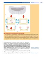
FIGURE 14 Dermal/epidermal junction The epidermal/dermal
junction is composed of dermal papillae and epidermal pegs that
interdigitate to create a bond that will withstand friction and shear
forces on the skin. The junction flattens with age, making geriatric
skin more susceptible to skin tears.
Dermis
The dermis is composed of connective tissue and binds the epidermis to the hypodermis or subcutaneous tissue. The extracellular matrix of the dermis is composed of collagen (mostly
Type I), elastic fibers, and ground substances such as glycosaminoglycans (GAGs) and proteoglycans. The uppermost
surface of the dermis is reticular and interdigitates with the
ridges of the epidermis; the structures are termed epidermal
pegs and dermal papillae. (FIGURE 14) Between the dermis and
epidermis is the basement membrane, consisting of the basal
lamina and the reticular lamina. Besides holding the two layers
together, the basement membrane allows the nutrients from the
dermal vasculature to pass through to the avascular epidermis.
The acellular dermal components are the extracellular
matrix, anchor fibrils of Type VII collagen linking the dermal
papillae and the basal lamina, and the elastic fibers that are
18/12/14 10:42 AM
6
Chapter 1 Anatomy and Physiology of the Integumentary System
TABLE 11
Cellular Components of Skin
Cell Name
Description
Location
Function
Keratinocytes—dead
Flat, elongated, nonnuclear
Stratum corneum of epidermis
Provide mechanical strength2
Squames
Horny, cornified cells
Surface of stratum corneum
Sloughing layer of epidermis
Keratinocytes—living
Polyhedral, slightly flattened
Stratum spinosum of epidermis
Synthesize keratin filaments;
phagocytose the tips of
melanocytes to release melanin
Keratinocytes—living
Flattened polygonal
Stratum granulosum
Keratinization at terminal
differentiation of the epithelial cells
Eosinophilic cells
Flat, no nuclei or organelles; densely
packed keratin filaments embedded
in a dense matrix
Stratum lucidum (only in soles of
the feet and palms of the hands)
Provide dense, thick layer of skin
Melanocytes
Round cell bodies with long
irregular dendritic extensions
Between the stratum basal and
stratum spinosum; in hair follicles1
Produce melanin, the pigment that
gives color to the skin
Langerhans cells (dendritic
cells)
Round cell bodies with long
dendritic extensions into
intercellular spaces
Stratum spinosum with cytoplasmic
processes extending between the
keratinocytes of all the epidermal
layers
Bind, process, and present antigens
to the T-lymphocytes
Lymphocytes
Dendritic
Epidermis
Recognize epitopes; produce
cytokines
Merkel cells
Dendritic; have contact with
unmyelinated sensory fibers in basal
lamina
Stratum basale of highly sensitive
areas; base of hair follicles
Mechanoreceptors for touch
Basal cells
Cuboid or columnar, contain keratin
in progressively increasing amounts
as the cells migrate toward the
stratum corneum
Stratum basale
Continuous production of
epidermal cells
Stratum basale, bulge
of the hair follicle
Production of keratinocytes
EPIDERMIS
DERMIS
Stem cells
Fibroblasts
Elongated, irregular shape with
large, ovoid nucleus
In the connective tissue of the
papillary layer of dermis
Synthesize collagen, elastin, GAGs,
proteoglycans, and glycoproteins
Mast cells
Large, oval or round; filled with
basophilic secretory granules
Near the capillaries in the dermis
Produce histamine and heparin
Macrophages
Large cells with off-center, large
nuclei
In the connective tissue of the
papillary layer of dermis; become
Langerhans cells in the epidermis
Phagocytosis, produce enzymes
and cytokines that facilitate wound
healing; immune processes
Leukocytes
Spherical white blood cells
Papillary layer of dermis
Phagocytose foreign material and
dead cells
Free nerve cell endings
Unencapsulated receptors
Papillary layer of dermis into
lower epidermal layers
Detect temperature changes, pain,
itching, light touch
Tactile discs
Unencapsulated receptors
Papillary layer of dermis
Receptors for light touch
Root hair plexus
Web of sensory fibers
Reticular layer of dermis
Detect movement of the hairs
Meissner corpuscles (tactile
corpuscles)
Elliptical encapsulated nerve
endings
Reticular layer of dermis
Detect light touch
Pacinian corpuscles
(lamellated)
Large oval nerve endings with outer
capsule and concentric lamellae
of flat Schwann-type cells and
collagen around an unmyelinated
axon
Reticular layer of the dermis,
hypodermis
Detect coarse touch, pressure,
vibration
Adipocytes
Globular cells containing fat
molecules
Hypodermis
Produce and store lipids that can be
used for energy, provide insulation,
produce cytokines for cell-to-cell
communication (restin, leptin,
adiponectin)
Data from Mescher AL. eds. Junqueira’s Basic Histology: Text & Atlas. 12th ed. New York, NY: McGraw Hill; 2010.
Hamm_Ch01_001-014.indd 6
18/12/14 10:42 AM


