Ebook Color atlas and textbook of human anatomy Vol.3 - Nervous system and sensory organs (5th edition): Part 2
Bạn đang xem bản rút gọn của tài liệu. Xem và tải ngay bản đầy đủ của tài liệu tại đây (17.27 MB, 203 trang )
Diencephalon
204
Diencephalon: Hypothalamus and Hypophysis
Neuroendocrine System
(continued)
nevertheless certainly influence the release
of the hormones.
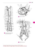
Hypothalamohypophysial System (A – D)
Presumably the regulation of neurosecretion is
achieved not only via synaptic contacts but
also via the bloodstream. The exceptionally
rich vascularization of hypothalamic nuclei
and the existence of endocellular capillaries
support this hypothesis. This arrangement
provides a pathway for humoral feedback
and forms a regulatory circuit for controlling the production and secretion of hormones, consisting of a neural limb (supraopticohypophysial tract) and a humoral
limb (circulation).
The hypothalamohypophysial tract (D) consists
of the supraopticohypophysical tract and the
paraventriculohypophysial tract which originate in the supraoptic nucleus (D1) and in
the paraventricular nucleus (D2), respectively. The fibers run through the hypophysial stalk into the hypophysial posterior lobe
where they terminate at the capillaries. The
hormones produced by the neurons of both
hypothalamic nuclei migrate along this
pathway to the axon terminals and enter
from here into the bloodstream. Electrical
stimulation of the supraoptic nucleus (C3)
leads to an increased secretion of vasopressin (antidiuretic hormone), while stimulation of the paraventricular nucleus (C4)
leads to an increased secretion of oxytocin.
In this system, the neurons do not release
stimulating substances that affect the secretion of a hormone by an endocrine gland
(such as the glandotropic hormones or releasing factors of the tuberoinfundibular
system), but they themselves produce hormones that have a direct effect on the target
organs (effector hormones). The carrier substances to which the hormones are bound
during their migration in the axons can be
demonstrated histologically. These Gomoripositive substances often cause swellings of
the axons (Herring bodies) (B5).
CD8 Optic chiasm.
CD9 Mamillary body.
The neurosecretory substances in axons and
swellings appear in the electron-microscopic image as granules that are much
larger than synaptic vesicles. At the capillaries of the neurohypophysis, the axons form
club-shaped endings (AD6) containing
small, clear synaptic vesicles in addition to
the large granules. At the sites of contact
with axon terminals, the capillary walls lack
the glial covering layer that, in the central
nervous system, forms the boundary between ectodermal and mesodermal tissues
and envelops all vessels (p. 44). It is here
that the neurosecretory product enters the
bloodstream. At the terminal bulbs of the
neurosecretory cells, there are also synapses (A7) of unknown origin, which
Kahle, Color Atlas of Human Anatomy, Vol. 3 © 2003 Thieme
All rights reserved. Usage subject to terms and conditions of license.
Hypothalamohypophysial System
205
5
4
B Herring bodies
(according to Hild)
3
8
C Regions where stimulation triggers the secretion of hypophysial
hormones (according to Harris)
7
2
1
6
A Supraopticohypophysial tract,
electron-microscopic diagram
(according to Bargmann)
Diencephalon
9
8
9
6
D Hypothalamohypophysial
tract
Kahle, Color Atlas of Human Anatomy, Vol. 3 © 2003 Thieme
All rights reserved. Usage subject to terms and conditions of license.
Kahle, Color Atlas of Human Anatomy, Vol. 3 © 2003 Thieme
All rights reserved. Usage subject to terms and conditions of license.
Telencephalon
Overview 208
Sections Through the
Telencephalon 214
Paleocortex and Amygdaloid
Body 224
Archicortex 230
Neostriatum 236
Insula 238
Neocortex 240
Imaging Procedures 264
Kahle, Color Atlas of Human Anatomy, Vol. 3 © 2003 Thieme
All rights reserved. Usage subject to terms and conditions of license.
208
Telencephalon
Overview
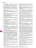
Rotation of the Hemisphere (C – F)
Subdivision of the Hemisphere
(A, B)
The hemispheric vesicle does not expand
evenly in all directions during its development but widens primarily in caudal and
basal directions. The temporal lobe is
formed in this way, and it finally turns rostrally in a circular movement (C); to a lesser
degree, such a rotation can also be observed
with the frontal lobe. The axis around which
the hemispheric vesicle rotates is the insular region; like the putamen (E6) lying
beneath it, the insula does not participate in
the movement. Other structures of the
hemisphere, however, follow the rotation
and end up having an arched shape in the
mature brain. The lateral ventricle (D7)
forms such an arch with its anterior and inferior horns. The lateral portion of the striatum, the caudate nucleus (E8), participates
in the rotation as well and follows precisely
the arched shape of the lateral ventricle. The
main part of the archipallium, the hippocampus (F9), moves from its original dorsal
position in basal direction and comes to lie
in the temporal lobe. The remnants of the
archipallium on the dorsal aspect of the corpus callosum, the indusium griseum (F10),
and the fornix (F11) reflect the arched expansion of the archipallium. The corpus callosum (F12) also expands in caudal direction
but follows the rotation only partially as it
develops only late toward the end of this
process.
Telencephalon
The embryonic hemispheric vesicle (A)
clearly shows the subdivision of the telencephalon into four parts, some of which
develop early (phylogenetically old portions), while others develop late (phylogenetically new portions). The four parts are
the paleopallium, the striatum, the neopallium, and the archipallium.
The hemispheric wall is called the pallium,
or brain mantle, because it covers the diencephalon and brain stem and envelops them
like a mantle.
The paleopallium (blue) (AB1) is the oldest
portion of the hemisphere. It forms the floor
of the hemisphere and corresponds, with
the olfactory bulb (A2) and adjacent paleocortex (p. 224 ff), to the olfactory brain, or
rhinencephalon, in the narrower sense. The
neostriatum (deep yellow) (AB3) (p. 236)
develops above the paleopallium; it, too, is
part of the hemispheric wall, although it
does not appear on the outer aspect of the
hemisphere.
The largest area is made up by the neopallium (light yellow). Its outer aspect, the neocortex (p. 240 ff) (AB4), develops very late
and encircles ventrally a transitional area to
the paleocortex that lies over the striatum;
this is the insula (p. 238) (B14).
D13 Third ventricle.
The medial hemispheric wall is formed by
the archipallium (red) (AB5), an old portion of the brain; its cortical band, the archicortex (p. 230 ff), later curls up to form the
hippocampus (Ammon’s horn).
The relationships in the mature brain are
determined by the massive expansion of the
neocortex, which pushes the paleocortex
and the transitional cortex of the insula into
the deeper parts of the brain. The archicortex becomes displaced caudally and appears
on the surface of the corpus callosum only
as a thin layer (B5, F10).
Kahle, Color Atlas of Human Anatomy, Vol. 3 © 2003 Thieme
All rights reserved. Usage subject to terms and conditions of license.
Subdivision and Rotation of the Hemisphere
209
A, B Subdivision of the hemispheres
4
5
5
3
14
1
1
2
B Adult brain
Telencephalon
A Embryonic brain
7
C Rotation of the hemisphere
(according to Jacob and Spatz)
13
8
D Ventricles
6
E Caudate nucleus and putamen
12
10
11
9
F Hippocampus (archipallium)
Kahle, Color Atlas of Human Anatomy, Vol. 3 © 2003 Thieme
All rights reserved. Usage subject to terms and conditions of license.
210
Telencephalon: Overview
Evolution (A – D)
Telencephalon
During primate evolution, the telencephalon has undergone changes similar to
those taking place during human embryonic development; it developed late and
then overgrew the other parts of the brain.
Thus, the cerebellum (A1) is still completely
exposed in the brain of primitive mammals
(hedgehog), while it becomes more and
more covered by the hemispheres of the telencephalon during primate evolution.
The paleopallium (rhinencephalon) (blue)
(A – C2) with olfactory bulb (A – C3) and piriform lobe (A – C4) forms the largest part of
the hemisphere in the primitive mammalian brain (A), and the archipallium (red)
(A – D5) still has its original dorsal position
above the diencephalon. These two old
components of the hemisphere then become overgrown by the neopallium (yellow)
(A – D6) during the course of evolution. The
paleopallium of prosimians (C) is still of
considerable size. In humans (D), however,
it becomes displaced deep into the base of
the brain and no longer appears in the
lateral view of the brain. The archipallium
(hippocampus), which lies above the diencephalon in the hedgehog (A5), appears as a
part of the temporal lobe at the base of the
brain in humans (D5). Only a narrow remnant remains above the corpus callosum
(indusium griseum).
The positional changes largely correspond
to the rotation of the hemisphere during
embryonic development; they also lead to
the formation of the temporal lobe (B – D7).
While still absent from the brain of the
hedgehog (A), the temporal lobe is already
recognized as a ventrally directed projection in the brain of the tree shrew (Tupaia),
the most primitive of primates (B). In the
prosimian brain (C), a caudally directed
temporal lobe has developed that finally
turns rostrally in the human brain (D). In addition, sulci and gyri develop in the region
of the neopallium. Whereas the neopallium
of primitive mammals is smooth (lissencephalic brains), a relief of convolutions
develops only in higher mammals (gyrencephalic brains). The development of sulci
and gyri considerably enlarges the surface
of the cerebral cortex. In humans, only onethird of the cortical surface lies at the surface of the hemispheres, two-thirds lie deep
in the sulci.
Two types of cortical areas can be distinguished on the neocortex: the primary areas
of origin (light red) and termination areas
(green) of long pathways, and between
them the secondary association areas (yellow).
The area of origin of motor pathways, the
motor cortex (A – D8), constitutes the entire
frontal lobe in the hedgehog. An association
area (B – D9) appears for the first time in
primitive primates (Tupaia) and achieves
extraordinary expansion in the human
brain. The termination area of sensory pathways, the sensory cortex (A – D10), borders
caudally on the motor cortex. Owing to the
enlargement of the adjacent association
area, most of the termination area of the
visual pathway, the visual cortex (A – D11),
becomes displaced to the medial
hemispheric surface in humans. The termination area of the acoustic pathway, the
auditory cortex (CD12), becomes displaced
deep into the lateral sulcus (fissure of Sylvius) by the expansion of the temporal association areas. Thus, the association areas expand much more during evolution than the
primary areas; they represent the largest
part of the neocortex in humans.
Kahle, Color Atlas of Human Anatomy, Vol. 3 © 2003 Thieme
All rights reserved. Usage subject to terms and conditions of license.
Evolution
8
10
11
6
211
5
3
6
1
4
2
8
3
2
A Hedgehog
10
11
6
9
6
Telencephalon
1
5
2
4
8
10
B Tupaia
7
12
2
11
6
9
3
6
2
7
4
8
5
1
2
C Lemur
10
9
6
6
11
5
1
7
12
D Homo sapiens
A – D Evolution of the telencephalon (modified after Edinger, Elliot Smith, and Le Gros-Clark)
Kahle, Color Atlas of Human Anatomy, Vol. 3 © 2003 Thieme
All rights reserved. Usage subject to terms and conditions of license.
212
Telencephalon: Overview
Cerebral Lobes (A – C)
The hemisphere is divided into four cerebral lobes:
Telencephalon
ț
ț
ț
ț
The frontal lobe (red) (p. 246)
The parietal lobe (light blue) (p. 250)
The temporal lobe (dark blue) (p. 252)
The occipital lobe (purple) (p. 254)
The hemispheric surface consists of grooves,
or sulci, and convolutions, or gyri. We distinguish primary, secondary, and tertiary sulci.
The primary sulci appear first and are
equally well developed in all human brains
(central sulcus, calcarine sulcus). The secondary sulci are variable. The tertiary sulci
appear last, being irregular and different in
each brain. Thus, each brain has its own surface relief as an expression of individuality,
like the features of the face.
The frontal lobe extends from the frontal
pole (AC1) to the central sulcus (AB2), which
together with the precentral sulcus (A3) defines the precentral gyrus (A4). The latter is
grouped with the postcentral gyrus (A5) to
form the central region, which spreads beyond the edge of the hemisphere (AB6) to the
paracentral gyrus (B7). Furthermore, the
frontal lobe exhibits three major convolutions: the superior frontal gyrus (A8), the
middle frontal gyrus (A9), and the inferior
frontal gyrus (A10 ); they are separated by
the superior frontal sulcus (A11) and the inferior frontal sulcus (A12). Three parts are
distinguished at the inferior frontal gyrus
that define the lateral sulcus (sulcus of Sylvius) (AC13): the opercular part (A14), the
triangular part (A15), and the orbital part
(A16).
The parietal lobe adjoins the frontal lobe
with the postcentral gyrus (A5) which is defined caudally by the postcentral sulcus
(A17). This is followed by the superior
parietal lobule (A18) and the inferior parietal
lobule (A19), which are separated by the intraparietal sulcus (A20). The end of the
lateral sulcus is surrounded by the supramarginal gyrus (A21); the angular gyrus
(A22) lies ventrally to it. The medial surface
of the parietal lobe is formed by the precuneus (B23).
The temporal lobe includes the temporal
pole (AC24) and three major convolutions:
the superior temporal gyrus (A25), the
middle temporal gyrus (A26), and the inferior
temporal gyrus (AC27), which are separated
by the superior temporal sulcus (A28) and
the inferior temporal sulcus (A29). The transverse temporal gyri (Heschl ’s convolutions)
of the dorsal aspect of the temporal lobe lie
in the depth of the lateral sulcus (p. 252, C).
On the medial surface is the parahippocampal gyrus (BC30) which merges rostrally into
the uncus (BC31) and caudally into the lingual gyrus (BC32). It is separated by the collateral sulcus (BC33) from the middle occipitotemporal gyrus (BC34). Ventrally lies the
lateral occipitotemporal gyrus (BC35),
delimited by the occipitotemporal sulcus
(BC36).
The occipital lobe includes the occipital
pole (A – C37) and is crossed by the transverse occipital sulcus (A38) and the deep calcarine sulcus (B39). Together with the
parieto-occipital sulcus (B40), the latter defines the cuneus (B41).
The cingulate gyrus (limbic gyrus) (green)
(B42) extends around the corpus callosum
(B43). Caudally, it is separated by the hippocampal sulcus (B44) from the dentate gyrus
(dentate band) (B45) and tapers rostrally
into the paraterminal gyrus (B46) and into
the subcallosal area (parolfactory area)
(B47). Isthmus of cingulate gyrus (B48).
Base of the brain. The basal aspect of the
frontal lobe is covered by the orbital gyri
(C49). Along the edge of the hemisphere
runs the gyrus rectus (C50), laterally defined
by the olfactory sulcus (C51) into which the
olfactory bulb (C52) and the olfactory tract
are embedded. The olfactory tract splits into
the two olfactory striae which embrace the
anterior perforated substance (olfactory
area) (C53).
C54 Hippocampal sulcus.
C55 Longitudinal cerebral fissure.
Kahle, Color Atlas of Human Anatomy, Vol. 3 © 2003 Thieme
All rights reserved. Usage subject to terms and conditions of license.
Cerebral Lobes
3
11
2
213
17
20
4
8
12
21
5
9
19
10
22
14
1
25
15
26
13
16
27
29
24
28
2
38
37
A Lateral view of the hemisphere
6
7
23
42
40
41
43
47
46
31
44
39
32
45
37
30
34
35
36
33 48
13
B Median view of the hemisphere
27
49
35
36
34
33
30
51
31
50
37
32
55
1
52
53
54
24
C Basal view of the two hemispheres
Kahle, Color Atlas of Human Anatomy, Vol. 3 © 2003 Thieme
All rights reserved. Usage subject to terms and conditions of license.
Telencephalon
6
18
214
Telencephalon
Sections Through the
Telencephalon
Frontal Sections
The posterior cut surface is shown for each
brain section.
Telencephalon
Section at the Level of the Exit of the
Olfactory Tract (A)
The cut surface shows the two hemispheres
separated by the cerebral longitudinal fissure
(AB1); the gray matter (cortex and nuclei) is
easily distinguished from the white matter
(myelinated fiber masses). The corpus callosum (AB2) connects the two hemispheres.
The section shows the cingulate gyrus (AB3)
above the corpus callosum.
The lateral field of the section shows the
deep lateral sulcus (AB4). Dorsally to it lies
the frontal lobe with the superior frontal
gyrus (AB5), the middle frontal gyrus (AB6),
and the inferior frontal gyrus (AB7). They are
separated by the superior frontal sulcus
(AB8) and the inferior frontal sulcus (AB9).
Ventrally to the lateral sulcus lies the temporal lobe with the superior temporal gyrus
(AB10), the middle temporal gyrus (AB11),
and the inferior temporal gyrus (AB12). The
temporal gyri are separated by the superior
temporal sulcus (AB13) and inferior temporal
sulcus (AB14). The lateral sulcus expands
deep into the lateral fossa (fossa of Sylvius)
(AB15), on the inner surface of which is the
insula. The insular cortex extends basally almost to the exit of the olfactory tract (A16).
It represents a transitional area between
paleocortex and neocortex.
In the depth of the hemisphere lies the neostriatum which is divided by the internal
capsule (AB17) into the caudate nucleus
(AB18) and the putamen (AB19). The section
shows the anterior horn (AB20) of the lateral
ventricle. The lateral wall of the ventricle is
formed by the caudate nucleus, while its
medial wall is formed by the septum pellucidum (AB21) containing the cavity of the
septum pellucidum (AB22). At the lateral
aspect of the putamen lies a narrow, cupshaped layer of gray matter, the claustrum
(AB23). It is separated from the putamen by
the external capsule (AB24) and from the insular cortex by the extreme capsule (AB25).
Section at the Level of the Anterior
Commissure (B)
At this level, the section shows the central
regions of the frontal lobe and the temporal
lobe. The lateral fossa is closed, and the insula is covered by the frontal operculum
(AB26) and the temporal operculum (AB27).
The ventral regions of both hemispheres are
connected by the anterior commissure (B28)
where fibers of the paleocortex and the
temporal neocortex cross. Above the commissure appears the globus pallidus (B29)
(part of the diencephalon), and close to the
midline lies the septum pellucidum (AB21),
or more specifically, its wide ventral segment containing the septal nuclei (also
known as peduncle of the septum pellucidum). The mediobasal aspect of the
hemisphere is covered by the paleocortex,
the olfactory cortex (B30).
Claustrum. In the past, the claustrum
(AB23) was either grouped together with
the striatum to form the so-called basal ganglia or was assigned to the insular cortex as
an additional cortical layer. Developmental
studies and comparative anatomical investigations, however, suggest that it consists
of cell clusters of the paleocortex which have
become displaced during development. The
claustrum merges with its wide base into
paleocortical regions (namely, the prepiriform cortex and the lateral nucleus of the
amygdaloid body). Unmyelinated fibers
from the cortices of parietal, temporal, and
occipital lobes are thought to terminate in
the claustrum in a topical arrangement. The
function of the claustrum is largely unknown.
B31 Optic chiasm.
A B
Kahle, Color Atlas of Human Anatomy, Vol. 3 © 2003 Thieme
All rights reserved. Usage subject to terms and conditions of license.
Planes of sections
Frontal Sections
215
1
8
5
6
9
3
21 22
7
17
20
2
18
26
19
13
4
25
24
15
23
1
11
15
27
12
14
16
A Frontal section at the exit of the olfactory tract
1
8
5
9
6
3
22
17
20
2
7
18
26
19
27
29
15
10
13
4
21
28
30
25
23
11
12
14
31
B Frontal section at the level of the anterior commissure
Kahle, Color Atlas of Human Anatomy, Vol. 3 © 2003 Thieme
All rights reserved. Usage subject to terms and conditions of license.
24
Telencephalon
10
216
Telencephalon: Sections
Frontal Sections (continued)
Telencephalon
Section at the Level of the Amygdaloid
Body (A)
At this level the central sulcus (AB1), which
runs obliquely from dorsocaudal to ventrorostal, has been cut in the more rostral
part; the frontal lobe, which is dorsal to it,
therefore occupies a far larger part of the
section than the parietal lobe, which is ventral to it. The convolution above the central
sulcus is the precentral gyrus (AB2); the convolution below it is the postcentral gyrus
(AB3). Deep in the temporal lobe appears
the amygdaloid body (amygdala) (A4). It
reaches the surface at the medial aspect of
the temporal lobe and might therefore be
regarded partly as cortex, partly as nucleus,
or rather as a transition between the two
structures. Since not only the surrounding
periamygdalar cortex but also its corticomedial half belong to the primary olfactory
centers, the amygdaloid body can be assigned to the paleocortex, despite its nuclear features. The claustrum (AB5) ends
above this region with a wide base.
inner surface of the temporal operculum exhibits prominent convolutions; these are
the obliquely cut transverse temporal gyri
(B19), or Heschl’s convolutions, representing the auditory cortex. In the ventral
region of the diencephalon lie the subthalamic body (B20), the mamillary body
(B21), and the substantia nigra (B22), which
is a part of the midbrain.
Basal Gaglia. The gray nuclear complexes
deep in the hemisphere are collectively
known as basal ganglia. Some authors use
the term only for the striatum and the pallidum, while others include the amygdaloid
body and the claustrum, some even the
thalamus. As this term is vague and ill-defined, it is not used in the present description. Earlier anatomists viewed the pallidum and the putamen as parts of the lentiform nucleus (a concept still surviving as
lenticular ansa and lenticular fasciculus), a
term that is no longer used.
Between the hemispheres lies the diencephalon with thalamus (AB6), globus pallidus (AB7), and hypothalamus (A8). Laterally to the diencephalic nuclei border the
neostriatum with putamen (AB9) and caudate nucleus (AB10). Below the corpus callosum (AB11) lies a strong fiber bundle, the
fornix (AB12). Also seen are the longitudinal
cerebral fissure (AB13), the lateral cerebral
sulcus (AB14), the lateral fossa (AB15), the
optic tract (A16), and the infundibulum
(A17).
Section at the Level of the Hippocampus
(B)
Once the more caudally cut sections no
longer show the amygdaloid body, the hippocampus (B18) appears in the medial area
of the temporal lobe. This most important
portion of the archicortex is a cortical formation that has curled up and projects
against the inferior horn of the lateral ventricle (B23). The section also shows the
caudal part of the lateral fossa (B15). The
A B
Kahle, Color Atlas of Human Anatomy, Vol. 3 © 2003 Thieme
All rights reserved. Usage subject to terms and conditions of license.
Planes of sections
Frontal Sections
217
13
1
2
2
3
10
11 12
3
6
9
1
14
15
7
7
Telencephalon
16
5
4
8 17
A Frontal section at the level of the amygdaloid body
13
2
10
9
7
1
11 12
3
6
14
20
22
15
19
5
23
18
21
B Frontal section at the level of the hippocampus
Kahle, Color Atlas of Human Anatomy, Vol. 3 © 2003 Thieme
All rights reserved. Usage subject to terms and conditions of license.
218
Telencephalon: Sections
Frontal Sections (continued)
Telencephalon
Section at the Level of Midbrain and Pons
(A)
The caudal portion of the lateral fossa (A1) is
open to the lateral aspect of the hemisphere. Dorsally to the lateral sulcus (A2)
lies the parietal lobe, ventrally the temporal
lobe. The dorsal convolutions of the latter,
which lie deep in the lateral sulcus and represent the transverse temporal gyri (A3)
(p. 252, C1), are cut obliquely. At the bottom
of the lateral fossa lies the insular cortex,
which rests on the caudal extensions of
claustrum (A4) and putamen (A5). The caudate nucleus (A6) appears at the lateral wall
of the lateral ventricle (A7). At the medial
aspect of the temporal lobe, concealed by
the parahippocampal gyrus (A8), the cortex
curls up to form the hippocampus (Ammon’s
horn) (A9). Corpus callosum (A10) and fornix (A11) are seen above the choroid plexus.
The field between the hemispheres represents the transition between diencephalon
and midbrain. The section shows the caudal
nuclear regions of the thalamus (A12). Separated from the main complex lies the lateral
geniculate body (A13), and medially to the
ventricular wall lies the habenular nucleus
(A14). The plane of section has been
oriented according to Forel’s axis (p. 4, B),
thus showing telencephalon and diencephalon in frontal section, while the structures of midbrain and pons (Meynert’s axis;
p. 4, B) have been cut obliquely. Ventral to
the aqueduct (A15) lies the decussation of
the superior cerebellar peduncle (A16). A narrow strip of dark cells, the substantia nigra
(A17), extends ventrally on both sides. The
cerebral peduncles (A18) are seen laterally to
it; the course of their fiber masses can be
traced from the internal capsule to the pons
(A19).
parietal lobe and temporal lobe lies in the
region of the angulate gyrus (B20). The
lateral sulcus and the lateral fossa are no
longer present in the section. The cut surface of the corpus callosum is particularly
wide at the level of the splenium (B21)
(p. 220, A6; p. 260, E14). Dorsally and ventrally to it lies the cingulate gyrus (B22),
which encircles the splenium in an arch. The
parahippocampal gyrus (B23) adjoins ventrally. Neither the hippocampus nor the calcarine sulcus are present in the section;
hence, the section lies behind the hippocampus but in front of the calcarine sulcus.
The two lateral ventricles are remarkably
wide, each representing the most anterior
part of the posterior horn at the transition
into inferior horn and central part (see
p. 281, BC7 – 9).
The lower aspects of the hemispheres
border on the cerebellum. The medulla oblongata appears in the middle, the oblique
section shows the fourth ventricle (B24),
the olives (B25), and the pyramids (B26).
Section at the Level of the Splenium of
the Corpus Callosum (B)
In this section, the dorsal part of the hemisphere belongs to the parietal lobe and the
ventral part to the temporal lobe; at this
plane of section, the latter is merging into
the occipital lobe. The boundary between
Planes of sections
A B
Kahle, Color Atlas of Human Anatomy, Vol. 3 © 2003 Thieme
All rights reserved. Usage subject to terms and conditions of license.
Frontal Sections
7
6
11
1
2
219
10
12
3
5
13
14
17
9
15
Telencephalon
4
18
16
8
19
A Frontal section at the level
of midbrain and pons
20
22
21
22
23
24
25
26
B Frontal section at the level
of the splenium of the
corpus callosum
Kahle, Color Atlas of Human Anatomy, Vol. 3 © 2003 Thieme
All rights reserved. Usage subject to terms and conditions of license.
220
Telencephalon: Sections
Horizontal Sections
Telencephalon
Superior Aspect of Corpus Callosum and
Lateral Ventricles (A)
The horizontal section through the brain
has been cut above the corpus callosum, and
the superior aspect of the corpus callosum
and the lateral ventricles have been exposed
by removal of deeper portions of white matter. The section shows the frontal lobes (A1)
at the top, the temporal lobes (A2) on both
sides, and the occipital lobes (A3) at the
bottom. The superior surface of the corpus
callosum (A4) belongs to the free brain surface lined by the pia mater and arachnoidea.
Lying deep in the brain, it is covered by the
convolutions of the medial walls of the
hemispheres. Rostrally, the superior surface
of the corpus callosum turns in ventral
direction and forms the genu of the corpus
callosum (A5) (p. 260, E11); caudally, it
forms the splenium of the corpus callosum
(A6) (p. 260, E14). On the superior aspect of
the corpus callosum extend four myelinated
fiber ridges: one lateral longitudinal stria
(A7) and one medial longitudinal stria of
Lancisi (A8) run along each half of the corpus callosum (see p. 230). Their fiber tracts
extend from the hippocampus to the subcallosal area. Between the two longitudinal
striae lies a thin layer of gray matter consisting of a narrow layer of neurons, the indusium griseum. This is a cortical portion of
the archicortex that regressed as a result of
the extensive development of the corpus
callosum (p. 7, E) and subsequent displacement of the archicortex into the inferior
horn of the lateral ventricle (see p. 209, F).
The anterior horns (A9) of the lateral ventricles (p. 280, A1) are opened in the area of
the frontal lobes, and the posterior horns
(A10) in the area of the occipital lobes. The
protruding hippocampus (A11) forms the
floor of the inferior horn. The central part
and the inferior horn of the lateral ventricle
contain the choroid plexus (A12) (p. 282).
Exposure of the Roof of the Diencephalon
(B)
This is an oblique horizontal section below
the corpus callosum, which has been
completely removed. Upon opening the two
lateral ventricles, the dorsal aspect of the
caudate nucleus (B13) and, bordering medially, the dorsal aspect of the thalamus (B14)
become visible. Parts of the diencephalon
become exposed as well, namely, the pineal
gland (B15) and both habenulae (B16) which
are connected to it. The two fornices (B17)
between the heads of the two caudate nuclei have been cut in their rostral part
(columns of fornix). The septum pellucidum
(B18) extends from there to the corpus callosum.
The lateral wall of the hemisphere contains
a particularly wide medullary layer between the cortex and the ventricle, the
semioval center (B19). The central sulcus
(B20) cuts into it and separates the frontal
lobe (at the top of the figure) from the
parietal lobe (bottom). Starting from the
central sulcus, the precentral gyrus (B21)
and the postcentral gyrus (B22) can be located.
Caudally in the longitudinal cerebral fissure
(AB23), the cerebellum (B24) is visible. The
caudal portion of the hemisphere is formed
by the occipital lobe. The striate area (B25),
the visual cortex, lies in this region and occupies primarily the calcarine sulcus (B26) at
the medial aspect of the occipital lobe,
while extending only a short distance onto
the occipital pole. It can be distinguished
even by the naked eye from the rest of the
cortex through a white streak, the line of
Gennari (B27), which divides the cortex into
two gray bands. Gennari’s line is a wide
band of myelinated nerve fibers corresponding to the slightly narrower external
band of Baillarger in the other areas of the
neocortex (see p. 240, A16; p. 254).
B28 Mesencephalic tectum.
Kahle, Color Atlas of Human Anatomy, Vol. 3 © 2003 Thieme
All rights reserved. Usage subject to terms and conditions of license.
Horizontal Sections
221
1
5
8
9
7
4
11
Telencephalon
2
12
6
10
3
23
23
A Horizontal section with
superior surface of the
corpus callosum
18
13
17
19
21
20
22
14
16
15
28
24
25
26
B Horizontal section exposing
the roof of the diencephalon
27
Kahle, Color Atlas of Human Anatomy, Vol. 3 © 2003 Thieme
All rights reserved. Usage subject to terms and conditions of license.
222
Telencephalon: Sections
Horizontal Sections (continued)
Telencephalon
Horizontal Section through the
Neostriatum (A)
At this level, the lateral cerebral fossa (AB1)
is exposed in its longitudinal expansion. The
lateral sulcus (A2) is found more rostrally,
with the frontal operculum (AB3) in front of
it and the elongated temporal operculum
(AB4) caudally to it. The longitudinal expansion is also apparent in the deep structures
of the telencephalon, the claustrum (AB5)
and the putamen (AB6). The arched structures have been cut twice; the corpus callosum (A7) appears rostrally with its anterior
part, the genu of the corpus callosum, and
caudally with its end, the splenium. The caudate nucleus has been cut twice as well; the
head of the caudate nucleus (AB8) is seen
rostrally and the tail of the caudate nucleus
(AB9) caudolaterally to the thalamus
(AB10). The thalamus is separated from the
globus pallidus (AB11) by the internal capsule which, in horizontal sections, exhibits
the shape of a hook made up of the anterior
limb (AB12) and the posterior limb (AB13).
Also the lateral ventricle has been exposed
twice. Its anterior horn (A14) has been cut in
the area of the frontal lobe and, caudally, in
the transition to the posterior horn (A15).
The two anterior horns are separated by the
septum pellucidum (A16), which spans between corpus callosum and fornix (A17).
anterior commissure (B24) connecting the
two hemispheres. The two columns of the
fornix (B25), lying close together in the previous section, are separated at the level of
the anterior commissure. While the posterior limb of the internal capsule (AB13) retains its usual width, the anterior limb
(AB12) is only indicated by some fiber
bundles. As a result, the head of the caudate
nucleus (AB8) is no longer separated from
the putamen (AB6), and the striatum is seen
as uniform nuclear complex. In the area of
the temporal lobe, the curled-up cortical
band of the hippocampus (Ammon’s horn)
(B26) is almost covered by the parahippocampal gyrus (B27).
B28 Mesencephalic tectum.
The section also shows the frontal lobes
(AB18), the temporal lobes (AB19), the
occipital lobes (A20), the longitudinal cerebral fissure (AB21), and the striate area
(visual cortex) (A22).
Horizontal Section at the Level of the
Anterior Commissure (B)
While the section still shows the entire
frontal lobe and temporal lobe, the occipital
lobe has only been cut in its anterior part at
the transition to the temporal lobe. Between
the two hemispheres appears the coneshaped dorsal aspect of the cerebellum
(B23). The anterior horn of the lateral ventricle and the corpus callosum are no longer
seen in this section. Instead there is the
A
B
Planes of sections
Kahle, Color Atlas of Human Anatomy, Vol. 3 © 2003 Thieme
All rights reserved. Usage subject to terms and conditions of license.
Horizontal Sections
223
21
18
3
14
2
1
7
8
4
17
5
11
13
10
19
9
7
Telencephalon
16
12
6
A Horizontal section at the
level of the neostriatum
15
20
21
21 22
18
3
1
25
24 8
12
6
4
11
5
13
10
28
9
26
27
19
19
B Horizontal section at the
level of the anterior commissure
23
Kahle, Color Atlas of Human Anatomy, Vol. 3 © 2003 Thieme
All rights reserved. Usage subject to terms and conditions of license.
224
Telencephalon
Paleocortex and Amygdaloid
Body
Paleocortex
Telencephalon
Subdivision (A, B)
The paleocortex (blue) is the oldest cortical
area of the telencephalon. Together with the
olfactory bulb and the olfactory tract it
forms the olfactory brain, or rhinencephalon.
In primitive mammals (hedgehog) (A), this
is the largest part of the telencephalon. The
large, compact olfactory bulb (A1) lies rostrally and, adjacent to it, the olfactory
tubercle (A2), or olfactory cortex. The rest of
the base of the brain is occupied by the piriform lobe (A3) with the uncus (A4). The piriform lobe contains various cortical areas,
namely, laterally the prepiriform area (A5),
medially the diagonal band of Broca (bandeletta diagonalis) (A6), and caudally the
periamygdalar area (A7). The caudal part of
the piriform lobe is occupied by the entorhinal area (A8), a transitional area (orange)
between archicortex (red) and neocortex.
Medially appears a portion of the hippocampal formation, the uncus with the superficial dentate gyrus (dentate band) (A9).
The enormous expansion of the neocortex
in humans (B) has displaced the paleocortex
into the depth where it represents only a
small part of the base of the brain. The
slender olfactory bulb (B10) is connected by
the olfactory tract (B11) with the olfactory
cortex. The fibers of the tract divide at the
olfactory trigonum (B12) into two (but often
into three or more) bundles: the medial olfactory stria (B13) and the lateral olfactory
stria (B14). They enclose the olfactory
tubercle which, in humans, has sunk into the
depth as anterior perforated substance (B15).
It is delimited caudally by the diagonal band
of Broca (B16) which contains afferent fibers
for the olfactory bulb.
The rotation of the hemisphere in humans
has displaced the other parts of the piriform
lobe mainly to the medial aspect of the temporal lobe, where they form the ambient
gyrus (B17) and the semilunar gyrus (B18).
The ambient gyrus is occupied by the pre-
piriform cortex (B19), and the semilunar
gyrus by the periamygdalar cortex (B20).
Ventrocaudally to it the uncus (B21) bulges
with the superficial end of the dentate gyrus
known as Giacomini’s band. It merges into
the parahippocampal gyrus (B22) which is
covered by the entorhinal cortex (B23).
Olfactory Bulb (C)
The olfactory bulb has regressed in humans,
who belong to the microsmatic mammals.
Mammals with a highly developed sense of
smell (macrosmatic mammals) possess a
large olfactory bulb of complex structure
(p. 211, AB3). In the human olfactory bulb
we distinguish a glomerular layer (C24), a
mitral layer (C25), and a granular layer
(C26). The mitral cells of the glomerular
layer form synaptic contacts with the terminals of the olfactory nerves (p. 228, A). The
axons of the mitral cells run through the olfactory tract to the primary olfactory centers. The olfactory tract contains a discontinuous aggregation of medium-sized neurons along its entire length, the anterior olfactory nucleus. Their axons join the fibers of
the olfactory tract and partly cross to the
contralateral olfactory bulb.
Anterior Perforated Substance (D)
The anterior perforated substance, which is
characterized by numerous vascular perforations (D27), is covered externally by an
irregular layer of small pyramidal cells, the
pyramidal layer (D28), and internally by the
loose multiform layer (D29) with individual
clusters of dark cells, the islands of Calleja
(D30). Olfactory bulb, olfactory tract, and
anterior perforated substance contain large
numbers of peptidergic neurons (corticoliberin, enkephalin, and other peptides).
D31 Nucleus of the diagonal band.
D32 Longitudinal cerebral fissure.
D33 Lateral ventricle.
D34 Paraterminal gyrus.
Kahle, Color Atlas of Human Anatomy, Vol. 3 © 2003 Thieme
All rights reserved. Usage subject to terms and conditions of license.
Paleocortex
225
1
2
5
6
9
7
4
4
3
Telencephalon
8
A Base of the brain
in the hedgehog
(according to Stephan)
10
11
14
13
12
15
18
19
16
17
20
21
21
23
22
B Base of the human brain
33
C Olfactory bulb,
frontal section
30
31
26
25
30
24
34
32
29
28
D Anterior perforated substance, olfactory
cortex (according to Crosby and Humphrey)
27
Kahle, Color Atlas of Human Anatomy, Vol. 3 © 2003 Thieme
All rights reserved. Usage subject to terms and conditions of license.
226
Telencephalon: Paleocortex and Amygdaloid Body
Amygdaloid Body
The amygdaloid body (amygdala) lies at the
medial aspect of the temporal lobe (B). It
consists of a cortical part, the cortical nucleus, and a nuclear part lying in the depth;
hence, it must be viewed as a transition between cortex and nucleus. The nuclear complex is covered by the periamygdalar cortex
(A1).
Telencephalon
Subnuclei (A – D)
The complex is divided into several subnuclei, namely, the superficial cortical nucleus
(ACD2), the central nucleus (ACD3), the
basal nucleus (CD4) consisting of a parvocellular medial part (A5) and a magnocellular lateral part (A6), and the lateral nucleus
(ACD7). The assignment of the medial nucleus (A8) to the amygdala complex is
questionable. The amygdaloid body is rich
in
peptidergic
neurons.
Primarily
enkephalin and corticoliberin can be demonstrated in the central nucleus and VIP in
the lateral nucleus.
The subnuclei form two groups: the phylogenetically old corticomedial group (cortical
nucleus, central nucleus) and the phylogenetically younger basolateral group (basal
nucleus, lateral nucleus). The corticomedial
group receives fibers of the olfactory bulb
and is the area of origin of the stria terminalis. The basolateral group has fiber connections with the prepiriform area and the
entorhinal area. Electrophysiological recordings have demonstrated that only the
corticomedial group receives olfactory impulses, while the basolateral group receives
optic and acoustic impulses.
tions of alertness associated with turning
the head. Stimulation may induce chewing
(ć), licking (b), or salivation (̆) (D). It may
also result in food uptake, secretion of gastric juice, and increased intestinal motility
or bulimia. Hypersexuality may occur as a
result of stimulation but may also be produced by lesions to the basolateral group of
nuclei. Urination (̅) or defecation may be
induced as well.
The stimulation responses are difficult to
arrange topically; many fibers run through
the nuclear complex, and the stimulation
responses may originate not only from the
site of stimulation but also from affected
fiber bundles of other nuclei. The medial
part of the basal nucleus has been assigned
to the corticomedial group of nuclei, and an
attempt has been made to correlate the two
nuclear groups with the different responses; the corticomedial group (E9) is
thought to promote aggressive behavior,
sexual drive, and appetite, while the lateral
group (E10) has an inhibitory effect.
Clinical Note: Stimulation of the amygdaloid
body in humans (a diagnostic measure in the
treatment of severe epilepsy) may trigger anger
or anxiety, but also a feeling of tranquillity and relaxation. The patients may feel “transformed” or
“in a different world”. The response will essentially be influenced by the emotional state at the
onset of the stimulation.
A – E11 Optic tract.
A12
Hypothalamus.
A13
Claustrum.
Functional Organization (C – E)
Electrical stimulation of the amygdala and
its surroundings induces autonomic and
emotional responses. Anger (í) or flight reaction () with the corresponding autonomic phenomena (dilatation of pupils, rise
in blood pressure, increase in cardiac and
respiratory rates) can be triggered by stimulation of the collecting area of the stria terminalis fibers (C). Other sites produce reac-
Kahle, Color Atlas of Human Anatomy, Vol. 3 © 2003 Thieme
All rights reserved. Usage subject to terms and conditions of license.
Amygdala
227
8
13
11
3
12
2
7
5
Telencephalon
6
1
A Subdivision of the amygdaloid body, frontal section, semi-diagram
3
11
7
4
B Location of section in A
2
C Fight-or-flight reaction, stimulation experiment
in the cat (according to de Molina and Hunsperger)
9
3
11
7
10
11
10
9
2
4
D Autonomic reactions, stimulation experiments
in the cat (according to Ursin and Kaada)
E Functional organization
(according to Koikegami)
Kahle, Color Atlas of Human Anatomy, Vol. 3 © 2003 Thieme
All rights reserved. Usage subject to terms and conditions of license.
9
228
Telencephalon: Paleocortex
Fiber Connections
Telencephalon
Olfactory Bulb (A)
The bundled axons of the olfactory cells
(A1) (p. 331, C) pass as olfactory nerves (1st
neuron) through the openings of the cribriform lamina (A2) into the olfactory bulb
(A3). Here they terminate on the dendrites
of the mitral cells (A4) with which they form
glomeruli (A5). In this glomerular system,
one mitral cell is in contact with numerous
sensory cells. Other cell types, such as
granule cells, periglomerular cells, and tufted
cells, belong to the integration center of the
olfactory bulb. The axons of the mitral cells
(2nd neuron) pass through the olfactory tract
(A6) to the primary olfactory centers. Medium-sized neurons are scattered along the
olfactory tract; they constitute the anterior
olfactory nucleus (AC7). The axons, or their
collaterals, of the mitral cells terminate
here. The neuronal processes partly cross
through the anterior commissure to the
contralateral olfactory bulb, where they
form the medial olfactory stria (B8).
Lateral Olfactory Stria (B)
All fibers of the mitral cells extend in the
lateral olfactory stria to the primary olfactory centers, namely, the anterior perforated substance (olfactory area) (BC9), the
prepiriform area (B10), and the periamygdalar area (B11) including the cortical nucleus of the amygdaloid body. The prepiriform area and the periamygdalar area are
thought to be the olfactory cortex proper for
the conscious perception of olfactory
stimuli. The medial olfactory stria is
thought to receive exclusively fibers running from the olfactory cortex to the olfactory bulb.
Fiber systems extend from the olfactory cortex (olfactory impulses for the search for
food, food uptake, and sexual behavior) to
the entorhinal area (B12), to the basolateral
nuclear group of the amygdaloid body
(BC13), to the anterior and lateral portions
of the hypothalamus (B14), and to the magnocellular nucleus of the medial thalamic
nuclei (B15). A connection to the centers of
the brain stem is established through fibers
running to the habenular nuclei (B16)
(p. 176, A). These association pathways do
not directly belong to the olfactory system.
Amygdaloid Body (B)
The basolateral nuclear group receives
fibers from the premotor, prefrontal, and
temporal cortices; from the magnocellular
nucleus of the medial thalamic nuclei; and
from nonspecific thalamic nuclei. The most
important efferent fiber system of the
amygdaloid body is the stria terminalis
(BC17). It arches in the sulcus between caudate nucleus and thalamus and runs below
the thalamostriate vein (p. 171, C14; p. 175,
AB2) as far as the anterior commissure. Its
fibers terminate in the septal nuclei (B18), in
the preoptic area (B19), and in the nuclei of
the hypothalamus. Fiber bundles cross from
the stria terminalis into the medullary stria
(B20) and extend to the habenular nuclei.
Other efferent bundles from the basolateral
portion of the amygdaloid body extend as
ventral amygdalofugal fibers (B21) to the entorhinal area, to the hypothalamus, and to
the medial thalamic nuclei, from where additional connections lead to the frontal lobe.
The stria terminalis is rich in peptidergic
fibers.
Anterior Commissure (C)
In the anterior part of the anterior commissure, fibers of the olfactory tract (anterior
olfactory nucleus) (AC7) and fibers of the olfactory cortex (BC9) cross to the contralateral side. The anterior part is poorly
developed in humans. The main part is
formed by the posterior part, where fibers of
the temporal cortex (C22) cross; they are
primarily from the cortex of the medial
temporal gyrus. Furthermore, the posterior
part contains crossing fibers from the amygdaloid bodies (BC13) and the striae (terminales) (BC17).
B23 Optic chiasm.
Kahle, Color Atlas of Human Anatomy, Vol. 3 © 2003 Thieme
All rights reserved. Usage subject to terms and conditions of license.