Pocket Atlas of Human Anatomy 4th edition - part 2 pot
Bạn đang xem bản rút gọn của tài liệu. Xem và tải ngay bản đầy đủ của tài liệu tại đây (1.38 MB, 51 trang )
43
1
2
3
4
5
6
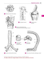
7
8
9
10
11
12
13
14
15
16
17
18
19
20
21
22
23
24
25
A
aa
19
15
13
20
24
21
14
11
4
30 23
29
31
22
10
10
44.1
44.8
15
17
26
13
25
19
18
16
20
22
23
27
45.5
45.4
11
21
6
97
8
4
28
10
5
12
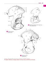
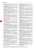
Right hip bone,
lateral view
A
Epiphyseal plates in the hip bone
of an adolescent
B
Right hip bone,
medial view
C
Bones
Feneis, Pocket Atlas of Human Anatomy © 2000 Thieme
All rights reserved. Usage subject to terms and conditions of license.
44
1
2
3
4
5
6
7
8
9
10
11
12
13
14
15
16
17
18
19
20
21
22
23
24
25
1 Ischium. Os ischii. Bone which forms the poste-
rior and inferior boundary of the obturator
foramen. A B
2 Body of ischium. Corpus ossis ischii. The por-
tion of the ischium situated behind the obtura-
tor foramen. A B
3 Ramus of ischium. Ramus ossis ischii. The por-
tion of the ischium situated below the obtura-
tor foramen. The anterior end unites with the
inferior ramus of the pubis. A B
4
Ischial tuberosity. Tuber ischiadicum
(ischiale). Ischial process at the lower end of
the lesser sciatic notch. A B
5 Ischial spine. Spina ischiadica (ischialis). Bony
prominence between the greater and lesser sci-
atic notches. B
6 Greater sciatic notch. Incisura ischiadica
(ischialis) major. Large notch between the post-
erior inferior iliac spine and the ischial spine. B
7 Lesser sciatic notch. Incisura ischiadica
(ischialis) minor. Notch between the ischial
spine and the ischial tuberosity. B
8 Pubis. Os pubis. Bone which forms the anterior
and inferior borders of the obturator foramen.
AB
9 Body of the pubis. Corpus ossis pubis. A B
10
Pubic tubercle. Tuberculum pubicum. Pro-
tuberance located anterolateral to the symphy-
sis. A B
11
Symphyseal sur face. Facies symphysialis. The
median surface of the symphysis facing the
contralateral pubis. B
12
Pubic crest. Crista pubica. Ridge extending
medially from the pubic tubercle to the sym-
physis. Attachment site of the rectus abdominis
muscle. A B
13 Superior ramus of the pubis. Ramus superior
ossis pubis. The part of the pubis situated above
the obturator foramen. A B
14
Iliopubic (iliopectineal) eminence. Eminen-
tia iliopubica [e. iliopectinea]. Flat prominence
at the proximal portion of the pubis. A B
15
Pecten (pectineal line) of the pubis. Pecten
ossis pubis. Sharp, bony ridge which passes to
the pubic tubercle as a continuation of the ar-
cuate line. Origin of the pectineus muscle. A B
16
Obturator crest. Crista obturatoria. It extends
from the pubic tubercle to the acetabulum.
Origin of the pubofemoral ligament. A
17
Obturator groove. Sulcus obturatorius. Sulcus
above the obturator foramen. A B
18
Anterior obturator tubercle. Tuberculum ob-
turatorium anterius. Small protuberance ante-
rior to the obturator groove. A B
19
Posterior obturator tubercle. [Tuberculum
obturatorium posterius]. Prominence occasion-
ally present behind the obturator groove. A B
20 Inferior ramus of pubis. Ramus inferior ossis
pubis. The portion of the pubis located anter-
oinferior the obturator foramen between the
symphysis and the suture line with the
ischium. A B
21 PELVIS. Portion of the body located between the
stomach and lower extremities, i.e., a bony ring
comprised of the sacrum, ilium, pubis and
ischium. C D E F
21 a Pelvic cavity. Cavitas pelvis (pelvica).
22 Pubic arch. Arcus pubis. The arch below the
symphysis formed by the right and left pubic
bones. D
23
Subpubic angle. Angulus subpubicus. The
angle between the right and left inferior ramus
of the pubis (average of 75° in men and 90°−
100° in women). C
24 Greater pelvis. Pelvis major. The space be-
tween the two alae of the ilium above the linea
terminalis.
25 Lesser pelvis. Pelvis minor. The space below
the linea terminalis.
26 Terminal line. Linea terminalis. Line extending
along the arcuate line from the promontory to
the upper margin of the symphysis. It marks
the boundary between the greater pelvis and
lesser pelvis as well as the plane of the pelvic
inlet. C D E
27 Upper pelvic aperture (pelvic inlet). Apertura
pelvis (pelvica) superior. Upper opening of the
lesser pelvis in the plane of the linea terminalis.
D
28 Lower pelvic aperture (pelvic outlet). Aper-
tura pelvis (pelvica) inferior. Lower opening of
the lesser pelvis between the coccyx, pubic
arch and sacrotuberous ligaments. F
29 Pelvic axis. Axis pelvis. Imaginary line passing
through all median connecting lines between
the symphysis and the anterior surface of the
sacrum. The fetal head follows its course during
birth. F
30 Conjugate diameter. Diameter conjugata. An-
teroposterior diameter of the pelvis, measured
from the sacral promontory to the posterior
surface of the symphysis (about 11 cm). E F
31 Transverse diameter of pelvis. Diameter trans-
versa. Widest part of the inlet (ca. 13 cm). E
32 Oblique diameter. Diameter obliqua. It is
measured from the iliosacral joint obliquely
forward to the iliopubic eminence of the op-
posite side (ca. 12.5 cm). E
33 Pelvic inclination. Inclinatio pelvis. The angle
between the plane of the pelvic inlet and the
horizontal plane. F
Bones
Feneis, Pocket Atlas of Human Anatomy © 2000 Thieme
All rights reserved. Usage subject to terms and conditions of license.
45
1
2
3
4
5
6
7
8
9
10
11
12
13
14
15
16
17
18
19
20
21
22
23
24
25
A
aa
29
30
28
33
32
30
31
26
26
22
27
26
23
14
12
11
20
18
3
4
5
2
19
1
6
13
9
7
1
8
17
15
10
14
15
10
9
20
3
2
4
1
19
16
18
13
1
8
12
17
Lower half of right hip bone,
external surface
A Lower half of right hip bone,
internal surface
B
Male pelvis, anterior viewC Female pelvis, anterior viewD
Pelvis, superior view
E Pelvis, medial viewF
Bones
Feneis, Pocket Atlas of Human Anatomy © 2000 Thieme
All rights reserved. Usage subject to terms and conditions of license.
46
1
2
3
4
5
6
7
8
9
10
11
12
13
14
15
16
17
18
19
20
21
22
23
24
25
1 FREE LOWER LIMB. Parsliberamembriinferioris.
2 Femur. Thigh bone. (Os femoris). A B
3 Head of femur. Caput femoris. A B
4
Pit (fovea) in the head of the femur. Fovea
capitis femoris. Depression for attachment of
the ligament of the head of the femur. A B
5 Neck of femur. Collum femoris. Portion of the
femur between the femoral head and greater
trochanter. A B
6 Greater trochanter. Trochanter major. Large
prominence on the superolateral aspect of the
femur shaft for attachment of the gluteus me-
dius, gluteus minimus, and piriformis muscles.
AB
7
Trochanteric fossa. Fossa trochanterica. De-
pression medial to the root of the greater tro-
chanter. Origin of the internal obturator and
gemelli muscles. A B
8 Lesser trochanter. Trochanter minor. Small
prominence on the posteromedial aspect of the
proximal femur shaft for attachment of the il-
iopsoas muscle. A B
9 /Trochanter tertius}. Process occasionally pre-
sent posteriorly at the lateral end of the linea
aspera at the level of the lesser trochanter for
attachment of a part of the gluteus maximus. B
10 Intertrochanteric line. Linea intertrochanter-
ica. Rough anterior line between the shaft and
neck of the femur, extending from the greater
to the lesser trochanter. A
10 a Quadrate tubercle. Tuberculum quadratum.
Rounded elevation on the intertrochanteric
crest. B
11 Intertrochanteric crest. Crista intertrochanter-
ica. Posterior bony ridge between the shaft and
neck of the femur, running from the greater to
the lesser trochanter. B
12 Shaft of femur. Corpus femoris. A B
13
Linea aspera. Rough double line on the poste-
rior aspect of the femur for attachment of two
vasti muscles and the short head of the biceps.
Insertion of the adductors, gluteus maximus,
and pectineus muscles. B
14
Lateral lip of the linea aspera. Labium laterale. B
15
Medial lip of the linea aspera. Labium mediale. B
16
Pectineal line. Linea pectinea. Bony ridge ex-
tending downward from the lesser trochanter,
nearly reaching the linea aspera. Gives attach-
ment to the pectineus muscle. B
17
Gluteal tuberosity. Tuberositas glutaealis.
Rough, oblong field continuous with the linea
aspera superolaterally. Insertion of the gluteus
maximus. B
18 Intercondylar fossa. Fossa intercondylaris.
Posterior notchbetweenthe femoral condyles. B
19 Intercondylar line. Linea intercondylaris. Post-
erior ridge between the roots of the condyles. B
20
Popliteal sur face. Facies poplitea. Triangular
field on the posterior aspect of the femur be-
tween the intercondylar line and the diverging
lips (supracondylar lines) of the linea aspera. B
20 a
Medial supracondylar line. Linea supracon-
dylaris medialis. Continuation of the medial lip
of the linea aspera toward the medial condyle.
B
20 b
Lateral supracondylar line. Linea supracon-
dylaris lateralis. Continuation of the lateral lip
of the linea aspera toward the lateral condyle. B
21 Medial condyle. Condylus medialis. Medial
knee joint surface of the femur. A B
22
Medial epicondyle. Epicondylus medialis.
Bony elevation on the medial aspect of the me-
dial condyle. A B
23
Adductor tubercle. Tuberculum adductorium.
Small process situated above the medial epi-
condyle for attachment of the adductor magnus
muscle. A B
24 Lateral condyle. Condylus lateralis. Articular
surface of the femur on the lateral aspect of the
knee joint. A B
25
Lateral epicondyle. Epicondylus lateralis.
Bony elevation on the lateral aspect of the
lateral condyle. A B
25 a
Groove for popliteus. Sulcus popliteus.
Groove between the lateral condyle and the
lateral epicondyle. B
26 Patellar surface. Facies patellaris. Surface that
articulates with the patella. A
27 Tibia. CD
28 Superior articular surface. Facies articularis
superior. Tibial articular surface of the knee
joint. C D
29 Medial condyle. Condylus medialis. Medial ex-
pansion at the proximal end of the tibia. C D
30 Lateral condyle. Condylus lateralis. Lateral ex-
pansion at the proximal end of the tibia. C D
31
Articular facet for the fibula. Facies articu-
laris fibularis. Articular surface for the head of
the fibula on the posterolateral aspect of the
lateral condyle. C D
32 Anterior intercondylar area. Area intercondy-
laris anterior. Area between the knee joint sur-
faces of the tibia and in front of the intercondy-
lar eminence. C D
33 Posterior intercondylar area. Area intercondy-
laris posterior. The region between the knee
joint surfaces of the tibia and behind the inter-
condylar eminence. D
34 Intercondylar eminence. Eminentia intercon-
dylaris. Bony elevation between the articular
surfaces of the tibia, for attachment of the
cruciate ligaments and menisci. C D
35
Medial intercondylar tubercle. Tuberculum
intercondylare mediale. Elevation of the medial
articular surface at the margin facing the inter-
condylar eminence. C D
36
Tuberculum intercondylare laterale. Eleva-
tion of the lateral articular surface at the mar-
gin facing the intercondylar eminence. C D
Bones
Feneis, Pocket Atlas of Human Anatomy © 2000 Thieme
All rights reserved. Usage subject to terms and conditions of license.
47
1
2
3
4
5
6
7
8
9
10
11
12
13
14
15
16
17
18
19
20
21
22
23
24
25
A
aa
31
30
28
33
29
32
34
35
36
31
36
34
35
32
30
29
28
67
3
4
3
76
5
10a
10
8
11
8
16
9
17
12 12
14
15
13
20a 20b
20
23
22
25
24 26 21 21 18 24
25a
25
19
Head of right tibia,
anterior view
C
Right femur,
anterior view
A
Right femur,
posterior view
B
Head of right tibia,
superior view
D
Bones
Feneis, Pocket Atlas of Human Anatomy © 2000 Thieme
All rights reserved. Usage subject to terms and conditions of license.
48
1
2
3
4
5
6
7
8
9
10
11
12
13
14
15
16
17
18
19
20
21
22
23
24
25
1 Shaft of tibia. Corpus tibiae (tibiale). A B D
2
Tibial tuberosity. Tuberositas tibiae. Rough-
ened area on the upper end of the anterior mar-
gin of the tibia. Attachment site of the patellar
ligament. A
3
Medial sur face. Facies medialis. Surface of
tibia directed anteromedially. A D
4
Posterior sur face of tibia. Facies posterior. B
D
5
Soleal line of tibia. Linea musculi solei. Line ex-
tending obliquely from the upper, lateral part of
the tibia, downward and across to the medial
part, giving attachment to the soleus muscle. B
6
Facies lateralis. Lateral surface of tibia facing
anterolaterally. A D
7
Medial (inner) margin. Margo medialis. A B D
8
Anterior margin. Margo anterior. A D
9
Interosseous margin. Margo interosseus.
Border facing the fibula and providing attach-
ment to the interosseus membrane along most
of its margin. A B D
10 Medial malleolus. Malleolus medialis. A B
11
Malleolar groove. Sulcus malleolaris. Small
groove on the posterior aspect of the medial
malleolus for the tendon of the tibialis poste-
rior muscle. B
12
Articular sur face of malleolus. Facies articu-
laris malleoli. Lateral surface of the medial mal-
leolus facing the talus. A B
13 Fibular notch. Incisura fibularis. Depression on
the lateral surface of the distal end of the tibia.
Articulates with the fibula. B
14 Inferior articular surface. Facies articularis in-
ferior. Inferior joint surface facing the talus. A B
15 Fibula. ABD
16 Head of fibula. Caput fibulae (fibulare). The
proximal end of the fibula. A B
17
Facies articularis capitis f ibulae. Articular
surface facing the tibia at the proximal end of
the fibula. A B
18
Apex (styloid process) of head of fibula.
Apex capitis fibulae. Upward pointing process
on the head of the fibula. A B
19 Neck of fibula. Collum fibulae. A
20 Shaft of fibula. Corpus fibulae. A
21
Facies lateralis. Lateral surface that faces
slightly upward. A D
22
Facies medialis. Medial surface b etween the
anterior and interosseous margins. It faces the
tibia. A B D
23
Facies posterior. Posterior surface between
the posterior and interosseous margins. B D
24 Medial crest. Crista medialis. Bony ridge on the
posterior surface at the border between the
origins of the tibialis posterior and flexor hal-
lucis longus muscles. B D
25
Anterior margin. Margo anterior. A D
26
Interosseous margin. Margo interosseus. Os-
seous ridge located between the anterior mar-
gin and the medial crest for attachment of a
portion of the interosseous membrane. A B D
27
Margin posterior. Margo posterior, directed
posterolaterally. B D
28 Lateral malleolus. Malleolus lateralis. A B
29
Articular sur face of malleolus. Facies articu-
laris malleoli. Articular surface on the lateral
malleolus facing the talus. A B
30
Lateral malleolar fossa. Fossa malleoli later-
alis. Depression on the posteromedial aspect of
the lateral malleolus for attachment of the
posterior talofibular ligament. B
30 a
Sulcus malleolaris. Groove lateral to the
malleolar fossa.
31 Patella. The knee cap, which is embedded in
the quadriceps tendon. C
32
Base of patella. Basis patellae. Broad, superior
border of the patella. C
33 Apex of patella. Apex patellae. Inferior, pointed
border of the patella. C
34 Facies articularis. Cartilage-covered articular
surface of the patella facing the femur.
35 Facies anterior. Anterior surface of the patella.
C
Bones
Feneis, Pocket Atlas of Human Anatomy © 2000 Thieme
All rights reserved. Usage subject to terms and conditions of license.
49
1
2
3
4
5
6
7
8
9
10
11
12
13
14
15
16
17
18
19
20
21
22
23
24
25
A
aa
3
7
14
6
9
26 22
25
15
21
2723
24
23
8
32
35
33
31
18
16
17
22
5
26
9
7
423
24
24
27
13
28
11
10
12 14
15
30
29
17
18
16
19
2
3
8
7
6
1
9
21
25
22
20
28
29
14 12
10
26
Patella, anterior viewC
Right tibia and fibula,
anterior view
A
Right tibia and fibula
in cross section
D
Right tibia and fibula,
posterior view
B
Bones
Feneis, Pocket Atlas of Human Anatomy © 2000 Thieme
All rights reserved. Usage subject to terms and conditions of license.
50
1
2
3
4
5
6
7
8
9
10
11
12
13
14
15
16
17
18
19
20
21
22
23
24
25
OSSA PEDIS. Bones of the foot.
1 TARSUS. The region of articulation extending
from the heel to the metatarsals. E
2 TARSAL BONES. Ossa tarsi (tarsalia). The seven
bones of the ankle, including the talus, cal-
caneus, os cuboideum, and three cuneiform
bones. E
3 Talus. The ankle bone, which is located be-
tween the tibia, calcaneus, navicular bone, and
fibula. A B E
4 Head of talus. Caput tali (talare). It articulates
with the navicular bone. A B
5 Neck of talus. Collum tali. Proximal tapering
part of the head of the talus. A B
6 Body of talus. Corpus tali. B
7 Trochlea tali (talare). Cylindrical surface of the
talus that articulates with the tibia and fibula. A
8
Superior surface. Facies superior. Upper sur-
face of the talus that articulates with the infe-
rior articular surface of the tibia. A
9 Medial malleolar surface. Facies malleolaris
medialis. Almost sagittaly oriented surface of
the talus that articulates with the medial
malleolus. A
10 Lateral malleolar surface. Facies malleolaris
lateralis. Surface on the lateral part of the talus
that articulates with the lateral malleolus. A
11
Lateral process of the talus. Processus later-
alis tali. Bony projection below the lateral
malleolar surface. A
12 Posterior calcanean facet. Facies articularis
calcanea posterior. Posteroinferior surface that
articulates with the calcaneus. B
13
Sulcus of talus. Sulcus tali. A groove between
the middle and posterior articular facets for the
calcaneus. B
14 Middle calcanean facet. Facies articularis cal-
canea media. Middle articular surface of the
calcaneus. B
15 Anterior calcanean facet. Facies articularis cal-
canea anterior. Anterior articular surface of the
calcaneus below the head of the talus. B
16
Facies articularis navicularis. Surface on the
anterior part of the head of the talus that ar-
ticulates with the navicular bone. A B
17 Posterior process of talus. Processus posterior
tali. Broad process below the posterior margin
of the trochlea. It bears the medial and lateral
tubercles with the groove for the tendon of the
flexor hallucis longus between them. A B
18
Sulcus tendinis m. flex. hall. longi. Groove
for the flexor hallucis longus tendon post-
eromedial to the posterior process of the talus.
AB
19
Medial tubercle. Tuberculum mediale. Bony
process anteromedial to the groove for the
flexor hallucis longus tendon. A B
20
Lateral tubercle. Tuberculum laterale. Bony
process lateral to the groove for the flexor hal-
lucis longus tendon. A
21 [Os trigonum]. Independent bone occasionally
formed by the lateral tubercle of the posterior
process of the talus due to a separate ossific
center. E
22 Calcaneus. Heel bone. C D E
23 Tuber calcanei. Tuberosity on the posterior
aspect of the calcaneus. C D
24 Medial process of calcaneus. Processus me-
dialis tuberis calcanei. Weak process anterior,
medial and inferior to the tuberosity of the cal-
caneus. D
25
Lateral process of calcaneus. Processus later-
alis tuberis calcanei. Weak process inferolateral
to the tuberosity of the calcaneus. C
26 Anterior tubercle of calcaneus. Tuberculum
calcanei. Eminence on the anterior aspect of
the inferior surface of the calcaneus. Attach-
ment site of the plantar calcaneocuboid liga-
ment. C
27 Sustentaculum tali. Medial prolongation of the
calcaneus bearing the medial posterior facet of
the calcaneus. D E
28
Sulcus tendinis m. flex. hall. longi. Bony
groove for the flexor hallucis longus tendon lo-
cated below the sustentaculum tali. D
29 Sulcus calcanei. Groove between the middle
and posterior articular facets. C D
30 Sinus tarsi. Laterally opening, funnel-shaped
space forming a continuation of the calcaneal
sulcus and the sulcus of the talus. The inferior
ankle joint is palpable here. B C. See also
pp. 53C, 71 A C
Bones
Feneis, Pocket Atlas of Human Anatomy © 2000 Thieme
All rights reserved. Usage subject to terms and conditions of license.
51
1
2
3
4
5
6
7
8
9
10
11
12
13
14
15
16
17
18
19
20
21
22
23
24
25
A
aa
29
2827 24
23
23
26
30
29
25
5
15
14
4; 16
30
12
13
19
18
6
17
4
19
16
5
9
7; 8
10
11
20
18
17
52.16
52.10
52.9 52.7 52.12 27
1
22
21
3
Right talus, superior viewA Right talus, inferior view
B
Right calcaneus, lateral view
C
Right calcaneus, medial view
D
Right foot, medial view
E
Bones
Feneis, Pocket Atlas of Human Anatomy © 2000 Thieme
All rights reserved. Usage subject to terms and conditions of license.
52
1
2
3
4
5
6
7
8
9
10
11
12
13
14
15
16
17
18
19
20
21
22
23
24
25
1 Anterior facet for the talus. Facies articularis
talaris anterior. Small anterior articular surface
for the head of the talus. A B
2 Middle facet for the talus. Facies articularis
talaris media. Middle articular surface for the
talus separated from the posterior facet by the
sulcus calcanei. A B
3 Posterior facet for the talus. Facies articularis
talaris posterior. Large posterior surface for ar-
ticulation with the talus. A B
4 Sulcus tendinis m. peronei (fibularis) longi.
Groove for the tendon of the peroneus longus
muscle on the lateral aspect of the calcaneus
below the peroneal trochlea. B
5 Peroneal trochlea. Trochlea peronealis (fibu-
laris). Bony eminence above the groove for the
tendon of the peroneus longus. It functions like
a pulley for this muscle and attaches a part of
the peroneal retinaculum. The peroneal brevis
runs cranial to the trochlea. B
6 Facies articularis cuboidea. Cuboid articular
surface forming the anterior aspect of the cal-
caneus. A B
7 Navicular bone. Os naviculare. Bone medial to
the head of the talus and the three cuneiform
bones. C D
8 Tuberosity of navicular bone. Tuberositas
ossis navicularis. Rough area on the inferome-
dial aspect of the navicular bone, for attach-
ment of the tibialis posterior muscle. It is pal-
pable through the skin. D
9 Medial cuneiform. Os cuneiforme mediale.
Most medial of the cuneiform bones, located
between the navicular and the 1
st
metatarsal
bones. Its wedge-shaped base is directed
downward. C D
10 Intermediate cuneiform. Os cuneiforme inter-
medium. Middle cuneiform bone located be-
tween the navicular and 2
nd
metatarsal bones.
Its wedge-shaped base is directed upward. C D
11 Lateral cuneiform. Os cuneiforme laterale.
Most lateral cuneiform bone located between
the navicular and 3
rd
metatarsal bones. Its
wedge-shaped base is directed upward. C D
12 Cuboid bone. Os cuboideum. Bone found be-
tween the calcaneus and the fourth and fifth
metatarsals. C D
13 Groove for tendon of peroneus longus. Sulcus
tendinis musculi peronei (fibularis) longi.
Groove on the inferolateral aspect of the cuboid
that serves as a guide for the tendon. D
14 Tuberosity of cuboid. Tuberositas ossis
cuboidei. Bony elevation on the inferior aspect
of the cuboid bone proximal to the groove for
the peroneus longus. D
15 Calcanean process. Processus calcaneus. Plan-
tar process of the cuboid bone. The inferior seg-
ment of the proximal articular surface projects
upwardly and obliquely to support the cal-
caneus. D
16 METATARSUS. The part of the foot situated be-
tween the tarsus and the toes. It comprises five
metatarsal bones. C D
17 METATARSAL BONES. Ossa metatarsi (metatar-
salia) [I−V]. The five metatarsal bones. D
18 Base of metatarsal bone. Basis metatarsalis.
The thickened proximal end of the metatarsal
bones. D
19 Shaft of metatarsal bone. Corpus metatarsale.
D
20 Head of metatarsal bone. Caput metatarsale. C
D
21 Tuberosity of first metatarsal. Tuberositas
ossis metatarsalis primi (I). Protuberance pro-
jecting downward and laterally from the proxi-
mal part of the first metatarsal bone. D
22 Tuberosity of fifth metatarsal. Tuberositas
ossis metatarsalis quinti (V). Protuberance pro-
jecting laterally from the proximal part of the
fifth metatarsal bone. Attachment site of the
peroneus brevis muscle. C D
23 PHAL ANGES OF TOES. Ossa digitorum pedis. C
D
24 PHALANGES. Osseous segments or bones that
comprise the toes. C D
25 Proximal phalanx. Phalanx proximalis. First or
proximal phalanx of the toes. D
26 Middle phalanx. Phalanx me dia. Middle seg-
ment of the toes. D
27 Distal phalanx. Phalanx distalis. Distal or ter-
minal nail-bearing bone of the toe. D
28
Distal tuberosity of toes. Tuberositas phalan-
gis distalis. Roughened area located on the
plantar aspect of the distal end of the distal
phalanx for attachment of the tactile pads. D
29 Base of phalanx. Basis phalangis. Proximal end
of each phalanx with an acetabular articular
surface. D
30 Shaft of phalanx. Corpus phalangis. D
31 Head of phalanx. Caput phalangis. Distal, artic-
ular end of the phalanx. D
32 Sesamoid bones. Ossa sesamoidea. Wormian
bones embedded in tendons or ligaments. They
regularly occur below the head of the first
metatarsal on both sides of the tendon of the
flexor hallucis longus muscle. D
Bones
Feneis, Pocket Atlas of Human Anatomy © 2000 Thieme
All rights reserved. Usage subject to terms and conditions of license.
53
1
2
3
4
5
6
7
8
9
10
11
12
13
14
15
16
17
18
19
20
21
22
23
24
25
A
aa
31
30
29
20
22
14
11
10
9
12
15
7
8
50.27
18
18
21
17
17
17
17
17
19
32
25
27
28
20
23
16
26
18
18
18
13
20
23
16
9
10
11
12
22
7
50.7; 8
50.30
45
6
2
3
1
2
3
6
Right calcaneus,
superior view
A Right calcaneus,
lateral view
B
Skeleton of right foot,
superior view
C
Skeleton of right
foot, inferior view
D
Bones
Feneis, Pocket Atlas of Human Anatomy © 2000 Thieme
All rights reserved. Usage subject to terms and conditions of license.
54
1
2
3
4
5
6
7
8
9
10
11
12
13
14
15
16
17
18
19
20
21
22
23
24
25
ARTICULAR SYSTEM
1 SUTURES OF THE SKULL. Suturae cranii
(craniales).
2 Coronal suture. Sutura coronalis. It lies be-
tween the frontal bone and the two parietal
bones. A C D
3 Sagittal suture. Sutura sagittalis. The median
suture situated b etween the right and left
parietal bones. C
4 Lambdoidal suture. Sutura lamboidea. It is lo-
cated between the occipital bone and the two
parietal bones. A D
5 Occipitomastoid suture. Sutura occipitomas-
toidea. Continuation of the lambdoidal suture
that extends to the base of the skull. A D
6 Sphenofrontal suture. Sutura sphenofrontalis.
Smooth suture that extends flatly upward and
backward lateral to the skull to join the greater
wing of the sphenoid bone and the frontal
bone. In the skull, it joins the frontal bone and
the lesser wing of the sphenoid bone. A B D
7 Sphenoethmoidal suture. Sutura sphenoeth-
moidalis. Short line in front of the jugum sphe-
noidale that connects the body of the sphenoid
and the ethmoid. D
8 Sphenosquamosal suture. Sutura sphenosqua-
mosa. Line of junction between the squamous
portion of the temporal bone and the greater
wing of the sphenoid. A C D
9 Sphenoparietal suture. Sutura spheno-
parietalis. Line of junction between the greater
wing of the sphenoid andtheparietalbone.ACD
10 Squamous suture. Sutura squamosa. Line of
junction between the squamous temporal and
parietal bones. A C D
11 Frontal (metopic) suture. [Sutura frontalis (su-
tura metopical)]. Suture connecting the right
and left halves of the frontal bone. It generally
fuses within 2 to 3 years after birth, but persists
in 7−8% of all Central Europeans. C
12 Parietomastoid suture. Sutura parietomas-
toidea. Posterior suture connecting the parietal
bone and the mastoid process of the temporal
bone. A
13 Squamosomastoid suture. [Sutura squamo-
somastoidea]. Line of junction between the
squamous and mastoid portions of the temporal
bone that generally fuses early in life. A
14 Frontonasal suture. Sutura frontonasalis. Ante-
rior line of junction between the frontal and
nasal bones. C
15 Frontoethmoidal suture. Sutura frontoeth-
moidalis. Internal line of junction between the
ethmoid and frontal bones. B D
16 Frontomaxillary suture. Sutura frontomaxil-
laris. Suture lateral to the nasal bone that con-
nects the nasal portion of the frontal bone and
the frontal process of the maxilla. A B C
17 Frontolacrimal suture. Sutura frontolacrimalis.
Line ofjunctionbetweenthefrontal andlacrimal
bones. A B C
18 Frontozygomatic suture. Sutura frontozygo-
matica. Suture at the lateral margin of the orbit
between the frontal and zygomatic bones. A B C
19 Zygomaticomaxillary suture. Sutura zygomat-
icomaxillaris. Suture in the floor of the orbit con-
necting the zygomatic bone and the maxilla. A B
C
20 Ethmoidomaxillary suture. Sutura ethmoi-
domaxillaris. Suture in the me dial wall of the
orbit connecting theorbital plate of the ethmoid
bone and the maxilla. B C
21 Ethmoidolacrimal suture. Sutura eth-
moidolacrimalis. Sutureinthemedialwall of the
orbit betweentheethmoidandlacrimalbones.B
22 Sphenovomerine suture.Suturasphenovomer-
iana. Suture at the nasal septum connecting the
sphenoid bone and the vomer.
23 Sphenozygomatic suture. Sutura sphenozygo-
matica. Suture inthelateral walloftheorbitcon-
necting the greater wing ofthesphenoid and zy-
gomatic bone. B C
24 Sphenomaxillary suture. Sutura sphenomaxil-
laris. Inconstant suture connecting the ptery-
goid process and the maxillae lateral to the
palatine bone. A
25 Temporozygomatic suture. Sutura temporozy-
gomatica. Suture connecting the zygomatic
process of the temporal bone and the zygomatic
bone on the lateral aspect of the zygomatic arch.
A
26 Internasal suture. Sutura internasalis. Suture
connecting the right and left nasal bones. C
27 Nasomaxillary suture. Sutura nasomaxillaris.
Suture connecting the nasal bone and the frontal
process of the maxilla. A C
28 Lacrimomaxillary suture. Sutura lacrimomax-
illaris. Suture connecting the anterior margin of
the lacrimal bone and the maxilla. A B C
29 Lacrimoconchal suture. Sutura lacrimo-
conchalis. Suture within the nasal cavity con-
necting the lacrimal bone and the inferior nasal
concha.
30 Intermaxillary suture. Sutura intermaxillaris.
Medial lineofjunctionbetweentherightand left
maxillary bones, located just below the anterior
nasal spine. C
31 Palatomaxillary suture. Sutura palatomaxil-
laris. Line of junction between the palatine bone
and the maxilla situated posteriorly in the orbit
and on the lateral wall of the nasal cavity. B
32 Palatoethmoidal suture. Sutura palatoeth-
moidalis. Suture in the back of the orbit connect-
ing the palatine and ethmoid bones. B
33 Median palatine suture. Sutura palatina medi-
ana. Suture within the oral cavity connecting
both halves of the palatine bone. E
34 Transverse palatine suture. Sutura palatina
transversa.Lineof junctionbetweenthepalatine
process of the maxilla and the palatine bone. E
Sutures, joints and ligaments
Feneis, Pocket Atlas of Human Anatomy © 2000 Thieme
All rights reserved. Usage subject to terms and conditions of license.
55
1
2
3
4
5
6
7
8
9
10
11
12
13
14
15
16
17
18
19
20
21
22
23
24
25
a
aa
33
34
2
9
15
2
66
8
7
9
10
4
5
32
2
10
11
9
8
28
30
19
23
18
17 14 16
26
27
20
18
17
16
28
21
20
15
32
23
6
19
19
31
17 18
27
24
25
8
9
6
13
12
5
4
10
2
28
16
19
Base of skull, superior viewD
Hard palate,
inferior view
E
Skull from leftA Right orbit, anterior viewB
Skull, anterior view
C
Sutures, joints and ligaments
Feneis, Pocket Atlas of Human Anatomy © 2000 Thieme
All rights reserved. Usage subject to terms and conditions of license.
56
1
2
3
4
5
6
7
8
9
10
11
12
13
14
15
16
17
18
19
20
21
22
23
24
25
1 CRANIAL SYNCHONDROSES. Synchondroses
cranii (craniales). Cartilaginous joints between
skull bones. Most are temporary and become
ossified.
2 Spheno-occipital synchondrosis. Synchondro-
sis spheno-occipitalis. Developmental car-
tilaginous joint postero-inferior to the sella tur-
cicabetweenthesphenoidand occipitalbones.A
3 Sphenopetrosal synchondrosis. Synchondrosis
sphenopetrosa. Cartilaginous union between
the sphenoid and petrous bones in the lateral
continuation of the foramen lacerum, for trans-
mission of the lesser petrosal nerve. A
4 Petro-occipital synchondrosis. Synchondrosis
petro-occipitalis. Anteromedial cartilaginous
continuation of the jugular foramen. A
4a Intraoccipital synchondroses. Synchondroses
intraoccipitalis. Cartilaginous joints between
developmental parts of the occipital bone.
5 Posterior intraoccipital synchondrosis. [Syn-
chondrosisintra-occipitalisposterior]. Develop-
mental synchondrosis between the posterior
and lateral ossific centers of the occipital bone. It
usually disappears within 1−2 years after birth.
A
6 Anterior intraoccipital synchondrosis. [Syn-
chondrosis intra-occipitalis anterior]. Develop-
mental cartilaginous joint between the anterior
and lateral ossific centers of the occipital bone
beginning at the anterior circumference of the
foramen magnum. Disappears during the 6
th
year of life. A
7 Sphenoethmoidal synchondrosis. Synchon-
drosis spheno-ethmoidalis. Cartilaginous pre-
cursor of the spheno-ethmoidal suture. See
page 54.7.
8 JOINTS OF VERTEBRAL COLUMN. THORAX
AND SKULL. Articulationes columnae verte-
bralis. Thoracis et cranii. The connections of the
vertebral column, thorax and skull.
9 Intervertebral symphysis. Symphysis inter-
vertebralis. Union between adjacent vertebral
bodies.
10 Intervertebral disc. Discus intervertebralis. An
elastic plate consisting of ring-shaped fibrous
lamellae, fibrocartilage, and a central gelatinous
nucleus located on either side of a vertebral
body, between the adjacent vertebrae. B C
11
Anulus fibrosus. Annular fibrous connection
between adjacent vertebral bodies consisting of
obliquely oriented connective tissue fibers ar-
ranged in alternating directions. B
12
Nucleus pulposus. Gelatinous, semifluid mass
forming the central core of an intervertebral
disc. B
13 Ligamenta flava. Yellow ligaments. Elastic net-
works of roughly parallel fibers between the
vertebral arches. B
14 Zygapophysial joints. Articulationes zy-
gapophysiales. Joints between articular
processes of vertebrae. C
15 Intertransverse ligaments. Ligg. intertransver-
saria. Narrow ligaments between transverse
processes of vertebrae. C
16 Interspinal ligaments. Ligg. interspinalia. Broad
ligaments between adjacent spinous processes.
B
17 Supraspinal ligaments. Ligg. supraspinalia.
Longitudinal ligaments connecting the tips of
the spinous processes. C
18 Ligamentum nuchae. Sagittal extension of the
supraspinalligamentsin theupperneckregion.B
19 Anterior longitudinal ligament. Lig. longitudi-
nale anterius. Longitudinal ligament attached to
the ventral surface of the vertebral bodies. B
20 Posterior longitudinal ligament.Lig.longitudi-
nale posterius. Longitudinal ligament connect-
ing the intervertebral discs. It is attached to the
dorsal surface of the vertebral bodies and thus
lies on the anterior wall of the vertebral canal. It
fuses with the tectorial membrane from the 3
rd
cervical vertebrae upward. B
21 Sacrococcygeal joint. Articulatio sacrococcy-
gea. Connection between the sacrum and coc-
cyx; it is frequently a true joint, but often occurs
as a synchondrosis. D
22 Superficial dorsal sacrococcygeal ligament.
Lig. sacrococcygeum posterius(dorsale)superfi-
ciale. D
23 Deep dorsal sacrococcygeal ligament. Lig.
sacrococcygeum posterius (dorsale) profun-
dum. D
24 Ventral sacrococcygeal ligament. Lig. sacro-
coccygeum anterius (ventrale).
25 Lateral sacrococcygeal ligament. Lig. sacro-
coccygeum laterale. D
26 Atlanto-occipital joint. Articulatio atlanto-
occipitalis.Jointbetweentheatlasand the occip-
ital bone. See page 59 A B
27 Anterior atlanto-occipital membrane. Mem-
brana atlanto-occipitalis anterior. Membranous
connection between the arch of the atlasandthe
occipital bone. It lies in front of the apical liga-
ment of the dens. B
28
Anterior atlanto-occipital ligament. [Lig.
atlanto-occipitale anterius]. Thickened portion
of the atlanto-occipital membrane emanating
from the anterior tubercle.
29 Posterior atlanto-occipit al membrane. Mem-
brana atlanto-occipitalis posterior. Connection
between the arch of the atlas and the occipital
bone situated in the posterior wall of the verte-
bral canal. B
30 Lateral atlanto-occipital membrane. Lig.
atlanto-occipitalelaterale.Obliquetractoffibers
extending from the transverse process of the
atlas to the jugular process of the occipital bone.
Sutures, joints and ligaments
Feneis, Pocket Atlas of Human Anatomy © 2000 Thieme
All rights reserved. Usage subject to terms and conditions of license.
57
1
2
3
4
5
6
7
8
9
10
11
12
13
14
15
16
17
18
19
20
21
22
23
24
25
a
aa
25
22
23
21
17
14
15
10
15
58.6
29
8.22
20
19
12
11
10
27
10
13
13
16
18
18
58.8
3
4
6
5
2
Skull of newborn,
inferior view
A
Ligaments of cervical vertebral column,
medial view
B
Ligaments of vertebral column
and ribs, lateral view
C
Coccygeal ligaments, posterior view
D
Sutures, joints and ligaments
Feneis, Pocket Atlas of Human Anatomy © 2000 Thieme
All rights reserved. Usage subject to terms and conditions of license.
58
1
2
3
4
5
6
7
8
9
10
11
12
13
14
15
16
17
18
19
20
21
22
23
24
25
1 Lateral atlanto-axial joint. Articulatio atlanto-
axialislateralis.Joint betweentheinferiorarticu-
lar facet of the atlas and the superior articular
facet of the axis. A B
2 Median atlanto-axial joint. Articulatio atlanto-
axialis mediana. Articulation b etween the atlas
and the dens of the axis. C
3 Alar ligaments.Ligg.alaria. Pairedligamentsex-
tending from the dens of the axis to the lateral
margin of the foramen magnum. A B
4 Apical ligament of the dens. Lig. apicis dentis.
Unpaired ligament extending from the apex of
the dens to the anterior margin of the foramen
magnum. A C
5 Cruciform ligament of atlas. Lig. cruciforme
atlantis. Cruciate ligament consisting of the two
following ligamentous bands (6, 7) located be-
tween the dens and the tectorial membrane. B
6
Longitudinal fasciculi of cruciform liga-
ment.
Fasciculi longitudinales. Connective
tissuetractsfromthebodyof the axistotheante-
riormarginoftheforamenmagnum.Theyaresit-
uatedbehindthedens anditsapicalligament. BC
7
Transverse ligament of atlas. Lig. transver-
sum atlantis. Part of the cruciform ligament of
the atlas passing behind the dens and extending
transversely from one side of the atlas to the
other. It holds the dens in position. B C
8 Tectorial membrane. Membrana tectoria.
Bilayered continuation of the posterior longi-
tudinal ligament. It passes from the axis to the
anterior margin of the foramen magnum and is
continuous with the dura-periosteal layer of the
skull base. C
9 JOINTS OF THORAX.Articulationesthoracis. Ar-
ticular connections ofthe skeleton of the thorax.
10 COSTOVERTEBRAL JOINTS. Articulationes cos-
tovertebrales. Joints between theribs and verte-
brae. D
11 Joints of rib heads. Articulatio capitis costae
(costalis). Articular unions that connect the
headsofthe ribswiththevertebralbodiesandin-
tervertebral discs. D
12 Radiate ligament of head of rib. Lig. capitis
costae radiatum. Ligament radiating predomi-
nantlyfromthe anteriorside oftheheadofaribto
the adjacent vertebral body and intervertebral
disc. D E
13 Intra-articular ligament of head of rib. Lig.
capitis costae intra-articulare. Ligament extend-
ing from the crest of the head of the rib to the in-
tervertebraldisc. Itliesbetweenthetwoarticular
facets of the head of the rib. E
14 Costotransverse joint. Articulatio costotrans-
versaria. Joint between the articular surface of
the tubercle of the rib and the transverse process
of the corresponding vertebra. D
15 Costotransverse ligament. Lig. costotransver-
sarium. Ligament between the neck of a rib and
the transverse process of the corresponding
vertebra. D
16 Superior costotransverse ligament. Lig. co-
stotransversarium superius. Ligament extend-
ing from a rib to the next higher transverse
process. E
17 Lateral costotransverse ligament. Lig. costotr-
ansversarium laterale.Ligament extendingfrom
the end of a transverse process to the corre-
sponding rib. D
18 Lumbocostal ligament. Lig. lumbocostale. Deep
layer of the thoracolumbar fascia. Fibrous con-
nectionbetweenthecostal processof thelumbar
vertebrae,thetwelfthrib,and theedgeofthe pel-
vis.
19 Costotransverse foramen.Foramencostotrans-
versarium.Opening forthe intercostalnervesbe-
tween the superior costotransverse ligament
and the neck of the rib. E
20 Sternocostal joint. Articulationes sterno-
costales. Articulations between the costal car-
tilage and sternum. F
21 Intra-articular sternocostal ligament. Lig. ster-
nocostale intra-articulare. Ligament within the
articular cavity between the costal cartilage and
sternum, especially pronounced at the 2
nd
rib. F
22 Radiate sternocostal ligaments. Ligg. sterno-
costaliaradiata.Fibertractslocatedinfrontofthe
sternocostal joint and radiating from the end of
the costal cartilage to the sternum. F
23 Sternal membrane. Membrana sterni. Mem-
branous covering of the anterior surface of the
sternum formed by the fibers of the radiate ster-
nocostal ligaments. F
24 Costoxiphoid ligaments. Ligg. costoxiphoidea.
Fiber tracts extending downward from the 7
th
costal cartilage to the xiphoid process.
25 External intercostal membrane. Membrana in-
tercostalis externa. Continuation of the external
intercostal muscles at the sternal end of the in-
tercostal space. F
26 Internal intercostal membrane. Membrana in-
tercostalis interna. Continuation of the internal
intercostal muscles near the vertebral end of the
intercostal space. E
26 a Sternocostal synchondrosis of the first rib.
Synchondrosis sternocostalis costae primae.
27 Interchondral joints. Articulationes interchon-
drales. Articulations between the costal car-
tilages, usually between those of ribs 6−9. See
page 7 D
28 Costochondral joints. Articulationes costoch-
ondrales. Unions between the bony and car-
tilaginous parts of ribs without an articular cav-
ity.
Sutures, joints and ligaments
Feneis, Pocket Atlas of Human Anatomy © 2000 Thieme
All rights reserved. Usage subject to terms and conditions of license.
59
1
2
3
4
5
6
7
8
9
10
11
12
13
14
15
16
17
18
19
20
21
22
23
24
25
a
aa
25
23
22
20
20
21
16
12
19
19
26
13
17 15 15 14
10
11
12
4
2
6
8
7
6
53
6
7
6
1
34 3
1
Ligaments between atlas,
axis and occipital bone
C Ligaments of vertebral column and ribs,
on right in cross section
D
Ligaments of vertebral column and ribs
E Sternocostal articulationsF
Dens of axis with ligaments,
posterior view
A Atlanto-occipital articulation,
posterior view
B
Sutures, joints and ligaments
Feneis, Pocket Atlas of Human Anatomy © 2000 Thieme
All rights reserved. Usage subject to terms and conditions of license.
60
1
2
3
4
5
6
7
8
9
10
11
12
13
14
15
16
17
18
19
20
21
22
23
24
25
1 SYNOVIAL JOINTS OF THE SKULL. Articulationes
synoviales cranii. Cf. pp. 56.26, 58.1, 58.2
2 Temporomandibular joint. Articulatio tem-
poromandibularis. A B C
3 Articular disc. Discus articularis. Biconcave disc
of fibrous tissue and fibrocartilage positioned
betweenthe headofthemandibleandthearticu-
lar fossa. Since it is connected with the articular
capsule on all sides, it divides the joint into two
compartments, both of which are functional
units of the discocapsular system. C
4 Lateral (temporomandibular) ligament. Lig.
laterale. A strong fibrous band occasionally pre-
sent on the lateral surface of the joint. It passes
obliquely upward and forward from the neck of
the mandible. A
5 Medial ligament.Lig.mediale.Reinforcementof
the medial wall of the capsule. B
6 Superior synovial membrane. Membrana syn-
ovialis superior. Synovial lining of the superior
articular cavity. C
7 Inferior synovial membrane. Membrana syn-
ovialis inferior. Synovial lining of the inferior ar-
ticular cavity. C
8 Sphenomandibular ligament. Lig. spheno-
mandibulare.Flat ligamentontheinneraspectof
the mandibular ramus extending from the man-
dibular foramen to the spine of the sphenoid
bone lateral to the foramen spinosum. B
9 Stylomandibular ligament. Lig. stylomandibu-
lare. Ligament passing from the anterior surface
of the styloid process to the angle of the
mandible. A B
10 Pterygospinal ligament. Lig. pterygospinale.
Broadconnectivetissue bandextendingfrom the
upper part of the lateral plate of the pterygoid
process to the spine of the sphenoid. B
11 Stylohyoid ligament. Lig. stylohyoideum. Liga-
ment running between the styloid process and
the lesser horn of the hyoid bone. Vestige of the
second pharyngeal arch. B
12 JOINTS OF THE SHOULDER GIRDLE. Articula-
tiones cinguli pectoralis. D E F G
13 Coracoacromial ligament. Lig. cora-
coacromiale. Strong band extending from the
coracoid process to the acromion. It forms the
roof of the shoulder joint. D
14 Superior transverse scapular ligament. Lig.
transversum scapulae superius. Ligament lying
medial to the coracoid process and bridging the
scapular notch. D
15 Inferior transverse scapular ligament. [Lig.
transversum scapulae inferius]. Weak fibrous
band passing from the root of the spine of the
scapula to the posterior margin of the glenoid
cavity. F
16 Acromioclavicular joint. Articulatio acromio-
clavicularis. Joint between the acromion and the
clavicle. D
17 Acromioclavicular ligament. Lig. acromio-
claviculare. Strong fibrous band within and
abovethe articularcapsuleservingtoprotectand
hold together the clavicle and acromion. D
18 Articular disc. Discus articularis. Fibrocar-
tilaginous interarticular disc. D
19 Coracoclavicular ligament. Lig. coracoclavicu-
lare. Two-part band connecting the coracoid
process and the clavicle. D
20
Trapezoid ligament. Lig. trapezoideum. The
portion of the coracoclavicular ligament taking
an upward and lateral course from the coracoid
process to the clavicle. It lies between the conoid
and coraco-acromial ligaments. D
21
Conoid ligament. Lig. conoideum. The portion
of the coraco-clavicular ligament medial to the
trapezoid ligament. It arises from the root of the
coracoid process. D
22 Sternoclavicular joint. Articulatio sternoclavic-
ularis. Two-chambered joint between the ster-
num and clavicle. G
23 Articular disc. Discus articularis. Interarticular
disc anchored below to the first rib and above to
the clavicle. G
24 Anterior sternoclavicular ligament. Lig. ster-
noclaviculare anterius. Band that reinforces the
anterior wall of the joint capsule. G
25 Posterior sternoclavicular ligament. Lig. ster-
noclaviculareposterius. Band that reinforces the
posterior wall of the joint capsule.
26 Costoclavicular ligament. Lig. costoclaviculare.
Ligamentous union between the first rib and the
clavicle lateral to the sternoclavicular joint. G
27 Interclavicular ligament. Lig. interclaviculare.
Ligament passing across the suprasternal notch
and uniting both clavicles. G
28 JOINTS OF THE FREE UPPER LIMB.Articulationes
membri superioris liberi.
29 Shoulder (glenohumeral) joint. Articulatio
humeri (glenohumeralis). D E F
30 Glenoid lip. Labrum glenoidale. The fibrocar-
tilaginous margin of the bony glenoid cavity. E
31 Coracohumeral ligament. Lig.coracohumerale.
Thickened portion of the capsule passing from
therootofthe coracoidprocesstotheupper mar-
gin of the greater and lesser tubercles. D E
32 Glenohumeral ligaments. Ligg. glenohumer-
alia.Threethickenedbands(superior,middle,in-
ferior)withintheanterior wallofthe capsule. DE
Sutures, joints and ligaments
Feneis, Pocket Atlas of Human Anatomy © 2000 Thieme
All rights reserved. Usage subject to terms and conditions of license.
61
1
2
3
4
5
6
7
8
9
10
11
12
13
14
15
16
17
18
19
20
21
22
23
24
25
a
aa
23
27
24
26
15
34.24
31 32 34.10
32
32
30
16
17
18
19
21
20
14
32
32
31
13
7
36
1
10
5
8
9
11
4
9
Temporomandibular joint,
lateral viewl
A
Temporomandibular
joint, medial view
B
Temporomandibular joint,
sagittal section
C
Lateral ligaments of shoulder, anterior view
D Shoulder joint, disarticulated,
lateral view
E
Shoulder joint, posterior view
F Sternoclavicular jointsG
Sutures, joints and ligaments
Feneis, Pocket Atlas of Human Anatomy © 2000 Thieme
All rights reserved. Usage subject to terms and conditions of license.
62
1
2
3
4
5
6
7
8
9
10
11
12
13
14
15
16
17
18
19
20
21
22
23
24
25
1 Elbow joint. Articulatio cubiti (cubitalis). Artic-
ular connection between the upper arm and
forearm. A
2 Humeroulnar joint. Articulatio humero-ul-
naris. Joint between the humerus and ulna.
3 Humeroradial joint. Articulatio humero-
radialis. Joint between the humerus and radius.
4 Proximal radioulnar joint. Articulatioradio-ul-
naris proximalis. Joint formed by the articular
circumference of the radius and the radial notch
of the ulna.
5 Ulnar collateral ligament. Lig. collaterale ul-
nare. Collateral ligament on the medial part of
the arm between the ulna and humerus. A
6 Radial collateral ligament. Lig. collaterale
radiale. Ligament which spreads from the lateral
epicondyle to the annular ligament of the radius
and the ulna. A
7 Annular ligament of the radius. Lig. anulare
radii. Circularband embracingapart of the artic-
ular circumference of the radius. It isattached to
the anterior and posterior margins of the radial
notch of the ulna. A
8 Quadrate ligament. Lig. quadratum. Thin band
of fibers passing from the distal margin of the
radial notch of the ulna to the neck of the radius.
9 Radioulnar syndesmosis (joint). Syndesmosis
[articulatio] radioulnaris. Fibrous joint between
the radius and ulna.
10 Interosseous membrane of forearm. Mem-
brana interossea antebrachii. Membranous
sheet which spreads between the interosseous
margins of the radius and ulna. A
11 Oblique cord. Chorda obliqua. Ligamentous
band extending obliquely downward from the
ulnar tuberosity to the radius. It runs in an op-
posite direction to most fibers of the interos-
seous membrane. A
12 Distal radioulnar joint. Articulatio radioulnaris
distalis. B
13 Articular disc. Discus articularis. Interarticular
discbetweentheulnaandcarpus. Itisattachedat
the radius and styloid process of the ulna and, as
an intra-articular ligament, it connects the
radius and ulna. B
14 Recessus sacciformis. Proximalextension of the
flaccid articular capsule. B
15 Radiocarpal joint. Articulatio radiocarpalis.
Proximal wrist joint between the proximal row
of carpal bones and the radius including the ar-
ticular disc. B
15 a Carpal joints. Articulationes carpi.
16 Intercarpal joints. Articulationes intercarpales.
Joints between the carpal bones permitting only
slight movement. B
17 Midcarpal joint. Articulatio mediocarpalis. The
distalwristjointbetweentheproximalanddistal
rows of carpal bones. B
18 Dorsal radiocarpal ligament. Lig. radiocarpale
dorsale. Ligament on the dorsumof the wrist ex-
tendingfromtheradiustothetriquetrumbone. C
19 Palmar radiocarpal ligament. Lig. radiocarpale
palmare. Ligament on the flexor side radiating
from the radius to the lunate and capitate bones.
D
20 Palmar ulnocarpal ligament. Lig. ulnocarpale
palmare. Ligament extending from the flexor
sideofthe headofthe ulnarchieflytothecapitate
bone. It often unites with fibers of the palmar
radiocarpal ligament. D
21 Radiate carpal ligament. Lig. carpi radiatum.
Groups of fibers radiating to both sides of the
wrist mainly from the head of the capitate bone.
D
22 Ulnar carpal collateral ligament. Lig. col-
lateralecarpiulnare.Collateral ligamentextend-
ing from the styloid process of the ulna to the
triquetrum and pisiform bones. C D
23 Radial carpal collateral ligament. Lig. col-
laterale carpi radiale. External collateral liga-
ment passing from the styloid process of the
radius to the scaphoid bone. C D
24 Dorsal intercarpal ligaments. Ligg. intercar-
palia dorsalia. Ligamentous bands extending be-
tween the proximal and distal rows of carpal
bones on the dorsum of the wrist. C
25 Palmar intercarpal ligaments. Ligg. intercar-
palia palmaria. Groupsofligamentsbetween the
carpal bones on the palmar aspect below the
radiate carpal ligament. D
Sutures, joints and ligaments
Feneis, Pocket Atlas of Human Anatomy © 2000 Thieme
All rights reserved. Usage subject to terms and conditions of license.
63
1
2
3
4
5
6
7
8
9
10
11
12
13
14
15
16
17
18
19
20
21
22
23
24
25
a
aa
13
12
14
15
17
16
16
88.12
5
11
6
7
10
22
24
23
18
20
19
23
25
21
Elbow joint, anterior viewA Carpal joints in horizontal
section
B
Carpal ligaments, dorsal view
C Carpal ligaments, palmar viewD
Sutures, joints and ligaments
Feneis, Pocket Atlas of Human Anatomy © 2000 Thieme
All rights reserved. Usage subject to terms and conditions of license.
64
1
2
3
4
5
6
7
8
9
10
11
12
13
14
15
16
17
18
19
20
21
22
23
24
25
1 JOINTS OF THE HAND. Articulationes manus. A B
C
2 Interosseous intercarpal ligaments. Ligg. in-
tercarpalia interossea. Ligaments penetrating
directlythrough the joint clefts between the car-
pal bones within a row. A
3 Pisotriquetral joint. Articulatioossispisiformis.
Articulation b etween the pisiform and
triquetrum bones. A
4 Pisohamate ligament. Lig. pisohamatum. Me-
dialcontinuationofthetendon oftheflexorcarpi
ulnaris to the hook of the hamate bone. B
5 Pisometacarpal ligament. Lig. pisometacar-
pale. Lateral continuation of the tendon of the
flexor carpi ulnaris to the base of the fifth meta-
carpal. B
6 Carpal canal or tunnel. Canalis carpi (carpalis).
Palmar canal located between the tubercles of
the scaphoid and trapezium on the one side and
thepisiformbone andthe hookof thehamulus on
the otherside.Itis bridged overbytheflexor reti-
naculum (93.26). B
7 Metacarpal articulations. Articulationes car-
pometacarpales. Slightly movable joints be-
tween the distal carpal bones and the metacar-
pals. A
8 Dorsal carpometacarpal ligaments. Ligg. car-
pometacarpalia dorsalia. Rigid ligaments on the
dorsum of the hand between the distal carpal
bones and the metacarpal bones. C
9 Palmar carpometacarpal ligaments. Ligg. car-
pometacarpalia palmaria. Ligaments on the pal-
mar side of the hand between the distal carpal
bones and the metacarpal bones. B
10 Carpometacarpal joint of the thumb. Articula-
tio carpometacarpalis pollicis. Saddle joint be-
tweenthefirstmetacarpalandthetrapezium.AB
11 Intermetacarpal joints. Articulationes inter-
metacarpales. Joints between the bases of the
metacarpal bones. A
12 Dorsal metacarpal ligaments. Ligg. metacar-
palia dorsalia. Ligaments between the proximal
ends of the metacarpals on the extensor side. C
13 Palmar metacarpal ligaments. Ligg. metacar-
palia palmaria. Ligaments between the bases of
the metacarpal bones on the palmar side. B
14 Interosseous metacarpal ligaments. Ligg.
metacarpalia interossea. Short, tense ligaments
at the bases of the metacarpal bones. They lie in
the intracapsular spaces between the dorsal and
palmar metacarpal ligaments. A
15 Interosseous spaces of metacarpus. Spatia in-
terossea metacarpi. Spaces between the meta-
carpal bones. A C
16 Metacarpophalangeal joints. Articulationes
metacarpophalangeales. Joints between the
heads of the metacarpal bones and the bases of
the proximal phalanges. B
17 Collateral ligaments. Ligg. collateralia. Col-
lateral ligaments of the metacarpophalangeal
joints. They slacken during extension of the fin-
gers and become tense when making a close d
first. B
18 Palmar ligaments. Ligg. palmaria. Fibers in the
floor of the tendon sheaths extending from the
root of the collateral ligaments to the palmar
side.Theyshouldnot be confusedwiththeannu-
larpartsofthefibroussheaths.See page 92.28.B
19 Deep transverse metacarpal ligament. Lig.
metacarpale transversum profundum. Trans-
verselyorientedfibroustractsonthe palmarside
of the heads of the metacarpal bones at the level
of the joint spaces. They hold the distal parts of
the metacarpus together. B
20 Interphalangeal joints. Articulationes inter-
phalangealesmanus.Middle anddistaljoints be-
tween the phalanges. B
21 Collateral ligaments of the interphalangeal
joints. Ligg. collateralia. B
22 Palmar ligaments. Ligg. palmaria. Fibers which
pass into the floor of the tendon sheaths above
the interphalangeal joints. B
Sutures, joints and ligaments
Feneis, Pocket Atlas of Human Anatomy © 2000 Thieme
All rights reserved. Usage subject to terms and conditions of license.
65
1
2
3
4
5
6
7
8
9
10
11
12
13
14
15
16
17
18
19
20
21
22
23
24
25
a
aa
15 15
15
12
8
8
8
8
12
21
21
18
17
9
4
5
6
10
20
20
22
19 19 19
13
13
13
16
9
14
15
15 15
14
10
11
7
7
7
7
7
11
3
2
Carpal joints in horizontal
section
A
Carpal joints of right hand,
dorsal view
C
Carpal joints, palmar view
B
Sutures, joints and ligaments
Feneis, Pocket Atlas of Human Anatomy © 2000 Thieme
All rights reserved. Usage subject to terms and conditions of license.
66
1
2
3
4
5
6
7
8
9
10
11
12
13
14
15
16
17
18
19
20
21
22
23
24
25
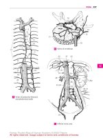
1 JOINTS OF THE PELVIC GIRDLE. Articulationes
cinguli pelvici.
2 Obturator membrane. Membrana obturatoria.
Membrane which closes off the obturator fora-
men except for the obturator canal. It is covered
by the obturator externus and internus muscles.
ABCD
3 Obturator canal. Canalis obturatorius. Opening
in the supralateral part of the obturator mem-
brane.Itcorrespondsto the obturatorgroovebe-
tween the two obturator tubercles and is
traversedbythe obturatorartery,vein andnerve.
ACD
4 Lumbosacral joint. Articulatio lumbosacralis.
Articulation between the sacrum and lumbar
vertebra 5 (4). A
5 Iliolumbar ligament. Lig. iliolumbale. Strong
ligamentthatpasses totheilium mainlyfromthe
transverse processes of L4 and 5. A B
6 Sacrotuberous ligament. Lig. sacrotuberale.
Strong ligament that extends from the medial
margin of the ischial tuberosity to the sacrum
and ilium. B D
7
Falciform process. Processus falciformis.
Slender extension of fibers from the sacro-
tuberous ligament to the inner aspect of the
ischium. B D
8 Sacrospinous ligament. Lig. sacrospinale.
Fibrous band medial to the sacrotuberous liga-
ment. It passes from the ischial spine to the
sacrum and coccyx and separates the greater
from the lesser sciatic foramen. B D
9 Greater sciatic foramen. Foramen sciaticum
(ischiadicum) majus. Foramen between the
greater sciatic notch, sacrum, sacrospinous liga-
ment and the upper part of the sacrotuberous
ligament.Itistraversedbythepiriformismuscle,
superior and inferior gluteal arteries, veins and
nerves, the internal pudendal vein, pudendal
nerve, sciatic nerve and posterior femoral cu-
taneous nerve. A B D
10 Lesser sciatic foramen. Foramen sciaticum
(ischiadicum) minus. Foramen between the
lesser sciatic notch and the sacrospinous and
sacrotuberous ligaments. It transmits the obtu-
rator internus muscle as well as the internal pu-
dendalarteryandveinandthepudendal nerveto
the ischiorectal fossa. B D
11 Sacroiliac joint. Articulatio sacroiliaca. Joint
connected by fibers that permits little motion
[[amphiarthrosis]]. It is located between the
sacrum and the ilium and may become syn-
osteotic. A
12 Ventral sacroiliac ligaments. Ligg. sacroiliaca
anteriora (ventralia). Thin but broad fibrous
bandsthatextendfromtheanteriorsurfaceofthe
first and second sacral vertebraeto the ilium. A D
13 Interosseous sacroiliac ligaments. Ligg.
sacroiliaca interossea. Dorsal mass of ligaments
thatpassfromthetub erosity ofthesacrumto the
tuberosity of the ilium. B
14 Dorsal sacroiliac ligaments. Ligg. sacroiliaca
posteriora (dorsalia). Superficial bundle of liga-
ments attached dorsally to the interosseous
sacroiliac ligaments between the sacrum and
ilium. B
15 Pubic symphsis. Symphysis pubica. Synchon-
drosis that articulateswiththeinterpubicdisc.A
16 Superior pubic ligament. Lig. pubicum su-
perius. Fibrous connection between the two
halvesofthesymphysisemanatingfrom thepec-
ten ossis pubis on either side. A
17 Arcuate pubic ligament. Lig. arcuatum pubis.
Strong, curved ligament below the symphysis. A
18 Interpubic disc. Discus interpubicus. Fibrocar-
tilage plate with asynovia-filled median groove,
located between the articular surfaces made of
hyaline cartilage on the right and left pubic
bones. A
19 JOINTS OF THE FREE LOWER LIMB. Articula-
tiones membri inferioris liberi.
20 HIP JOINT.Articulatiocoxae (iliofemoralis).Joint
formed by the acetabulum and the head of the
femur. A B C
21 Joint capsule. Capsula articularis. It is attached
anteriorly to the intertrochanteric line, posteri-
orly above to the intertrochanteric crest. A frac-
ture of the neck of thefemur can therefore be in-
tracapsular when in the anterior region or extra-
capsular when in the posterior region. A
22 Orbicular zone. Zona orbicularis. Ligamentous
fibers encircling the neck of the femur. B
23 Iliofemoral ligament. Lig. iliofemorale. Strong
anteriorligamentofthehipjointcapsuleextend-
ing fromtheiliumto the intertrochantericline.A
B
24 Ischiofemoral ligament. Lig. ischiofemorale. It
radiates into the orbicular zone from the poste-
rior margin of the acetabulum and is also at-
tached to the anterior margin of the greater tro-
chanter and to the intertrochanteric line. B
25 Pubofemoral ligament. Lig. pubofemorale.
Ligamentthatarisesmediallyfromthejointcap-
sule of the pubicbone and extends to the orbicu-
lar zone and to the part of the femur proximal to
the lesser trochanter. A
26 Acetabular lip. Labrum acetabulare. A ring of fi-
brocartilage and connective tissue that
completes and deepens the bony acetabulum. C
27
Transverse acetabular ligament. Lig. trans-
versumacetabuli.Itbridgestheacetabularnotch.
C
28 Ligament of head of femur.Lig.capitisfemoris.
A smooth ligament extending from the acetabu-
lar notch to the pit on the head of the femur. It
transmits blood vessels and has no direct me-
chanical action. C
Sutures, joints and ligaments
Feneis, Pocket Atlas of Human Anatomy © 2000 Thieme
All rights reserved. Usage subject to terms and conditions of license.
67
1
2
3
4
5
6
7
8
9
10
11
12
13
14
15
16
17
18
19
20
21
22
23
24
25
a
aa
12
3
2
10
9
6
8
20
26
28
27
3
2
13 5
20; 21
22
107
6
8
9
14
23
24
2
11
5
4
12
86.10
20; 21
9
163
21
15
18
17
23
23
25
Pelvic ligaments, anterior viewA
Pelvic ligaments,
posterior view
B
Hip joint, opened
C
Pelvic ligaments,
medial view
D
Sutures, joints and ligaments
Feneis, Pocket Atlas of Human Anatomy © 2000 Thieme
All rights reserved. Usage subject to terms and conditions of license.