Ebook A history of surgery (3E): Part 2
Bạn đang xem bản rút gọn của tài liệu. Xem và tải ngay bản đầy đủ của tài liệu tại đây (45.5 MB, 142 trang )
9
The surgery of warfare
Mankind has always been subject to injury; the
earliest surgeons were no doubt those men and
women who were particularly skilled in binding up the contusions, lacerations, fractures,
perforations and eviscerations of their fellows
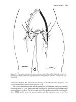
(Figure 9.1). Since man is undoubtedly the most
vicious and aggressive of all animals, much of
this trauma was inflicted in battle, and warfare
has therefore played an important part in the
development of wound management. Indeed, it
has been said that the only thing to benefit from
war is surgery.
Figure 9.1 Achilles bandages the arm of
Patroclus during the Trojan Wars 1200 bc.
(From a painting on an ancient Greek vase.)
Until the introduction of gunpowder into warfare in the 14th century, war wounds were inflicted
mainly by knives, swords, spears, arrows and various blunt weapons such as the mace and cudgel.
The sharp weapons would produce p
enetrating
and lacerating injuries, and the blunt instruments
would produce severe contusions. The early surgeons well recognised that some injuries were going
to prove almost invariably fatal. These comprised
penetration of a vital structure, such as a perforating wound of the skull, chest or abdomen, or
haemorrhage from a major blood vessel. However,
if the victim survived the initial injury, he was very
likely to live. This was because these lacerated and
contused wounds produced little tissue destruction and thus allowed the natural powers of the
body’s healing to cure the victim. So the surgeon
became skilled at dressing and bandaging wounds
and splinting fractures. The various ointments
employed, although probably usually ineffective,
at least did little harm. Haemorrhage would be
treated by pressure on the wound or the use of the
cautery. The technique of tying the bleeding artery,
a device introduced by the Alexandrian surgeons
around 250 bc and described by the Roman writer
Celsus in the 1st century An, appeared to have
been forgotten.
The medieval surgical textbooks often carried an illustration of a ‘wound man’ that showed
the various injuries the surgeons of the Middle
Ages might be called upon to treat; we can guess
quite accurately which would prove successful and which would be almost certainly lethal
(Figure 9.2).
125
126 The surgery of warfare
gangrene of a type not previously seen were encountered by surgeons treating these war wounds. Now
this, of course, was centuries before our knowledge
of the bacterial causation of wound infection. It
was not unreasonable, therefore, for military surgeons to conclude that these awful complications
were due to the poisonous nature of the gunpowder itself. The solution was obviously to destroy the
poison, and this was done by means of a red-hot
cautery or by the use of boiling oil poured into the
wound. The great popularity of the latter method
was undoubtedly due to the writings of the Italian
surgeon Giovanni da Vigo (1460–1525), whose surgical treatise titled A Compendious Practice of the
Art of Surgery was first published in Rome in 1514
and went through more than 40 editions in many
languages; it greatly influenced the surgical thinking of his time. Of course, we now know that this
practice had the opposite effect to the one desired.
The red-hot cautery (Figure 9.3) and the boiling oil
in fact destroyed more tissue than the missile itself
and aggravated an already serious situation, as well
Figure 9.2 A ‘wound man’. (From Hans Gersdorff:
Feldbusch der Wundarztney. Strasburg, 1517.
Courtesy of J Kirkup, Fellow of the Royal College
of Surgeons [FRCS].)
THE INVENTION OF GUNPOWDER
Gunpowder appears to have been invented in China
and was used in the manufacture of fireworks and,
probably, also in cannons. It first appeared in Europe
in the 14th century, and it is well documented that
cannons were employed in the Battle of Crécy in
1346 when Philip VI of France was defeated by
Edward III and his longbowmen. The introduction
of firearms completely changed the pathology of
war wounds. The gross tissue destruction produced
by the musket ball and cannon provided a wonderful medium for the growth of bacteria, especially anaerobic microbes, those that thrive in the
absence of oxygen and grow on dead tissues. These
include the organisms that produce tetanus and
gas gangrene. Thus, dreadful wound infection and
Figure 9.3 Cauterisation of a wound of the thigh.
The invention of gunpowder 127
as inflicting untold torture upon the poor soldier
victim.
We now come to one of those great landmarks
that punctuate surgical history; a surgeon who,
through his example and writings, greatly influenced progress in the management of wounds.
Ambroise Paré (1510–1590) was born in the little
town of Laval in the Province of Maine (Figure 9.4).
His father was probably a valet de chambre and
barber to the local squire, and he may thus have
obtained some interest in the work of barbersurgeons. Paré’s sister married a barber-surgeon
who practised in Paris, and his elder brother was
a master barber-surgeon in Vitré. Paré may have
begun the study of surgery with his brother, and it
is certain that he did work with a barber-surgeon
in the provinces before coming to Paris at the age
of 22 as an apprentice barber-surgeon. He was soon
appointed compagnon-chirurgeon, roughly equivalent to house surgeon today, at the Hôtel Dieu,
that immense medieval hospital and the only one
in Paris at the time, where he worked for the next
Figure 9.4 Ambroise Paré, aged 45. (From
Geoffrey Keynes: Apologie and Treatise of
Ambroise Paré. London, Falcon, 1951.)
3 or 4 years and must have gained a great experience
in that repository of pathology.
Perhaps because he could not afford to pay
the fees for admission to the ranks of the barbersurgeons, Paré started his career at the age of 26
as a military surgeon. In those days, there was no
organised medical care for the humble private soldiers of armies in the field. Surgeons were attached
to individual generals and to other important personages, and might, if they wished, give what aid
they could to the common soldiers in their spare
time. Otherwise, the troops had to rely on the
rough and ready help of their companions or of
a motley crowd of horse doctors, farriers, quacks,
mountebanks and camp followers.
Paré was appointed surgeon to the Mareschal
de Montejan, who was colonel-general of the
French infantry. This, his first of many campaigns,
took him to Turin, and it was here in 1537 that he
made his fundamental observations on the treatment of gunshot wounds. He soon realised that
the accepted method of treating these injuries with
boiling oil did more harm than good and substituted a more humane and less destructive dressing.
Here is his description of what today might well be
called one of the earliest controlled surgical experiments. How many of us have carried out some new
untried treatment and have shared Paré’s experience of being unable to sleep and have come into
the ward to see how a patient is before anyone else
is around, with pulse racing, to see whether the
treatment we have carried out has been a brilliant
success or a disastrous failure?
I was at that time a fresh-water surgeon,
since I had not yet seen and treated
wounds made by firearms. It is true I had
read in Jean de Vigo in his first book
of Wounds in General Chapter 8, that
wounds made by firearms are poisoned
because of the powder. For their cure
he advised their cauterisation with oil of
elders mixed with a little theriac. To not
fail, this oil must be applied boiling even
though this would cause the wounded
extreme pain. I wished to know first how
to apply it, how the other surgeons did
their first dressings, which was to apply
the oil as boiling as possible. So I took
128 The surgery of warfare
heart to do as they did. Finally, my oil
was exhausted and I was forced instead
to apply a digestive made of egg yolk,
rose oil and turpentine. That night I
could not sleep easily, thinking that by
failure of cauterising, I would find the
wounded in whom I had failed to put
the oil dead of poisoning. This made
me get up early in the morning to visit
them. There, beyond my hopes, I found
those on whom I had used the digestive medication feeling little pain in
their wounds, without inflammation and
swelling, having rested well through the
night. The others on whom I had used
the oil I found feverish, with great pain,
swelling and inflammation around their
wounds. Then I resolved never again to
so cruelly burn the poor wounded by
gunshot.
one of my servants, to teach him and to
embolden him in such works, and there
he readily tied the vessels to stay the
bleeding without application of hot
irons (Figure 9.5). He was well cured,
God be praised, and is returned home
to his house with a wooden leg.
So here was Paré at the age of 73 passing down
his skill and experience to his apprentices, a tradition we still see today as surgeons teach their residents in the operating theatre.
Paré went from fame to fame and dominated
the history of surgery in the 16th century. He was
a veteran of no less than 17 military campaigns
and surgeon to four successive kings of France.
However, his practice continued to embrace the
Paré also went on to show that bleeding after
amputation of a limb should be arrested not by
the terrible method of the red-hot cautery but by
simply tying the divided blood vessels. Ligation
of blood vessels was known to the ancients, and
Paré’s only claim, as he makes quite clear in his
own writings, was that he was the first to apply
this technique in performing amputations. He first
employed the ligature in amputation of the leg in
1552 at the siege of Danvillier but did not publish
his technique until 1564 when he wrote: ‘wherefore I must earnestly entreat all surgeons that leaving this old and too cruel way of healing they will
embrace this new, which I think was taught me by
the special favour of the sacred Deity, for I learned
it not of my masters nor of any other, neither have
I at any time found it used by any’.
A description by Paré of one such case is worth
repeating here:
In the year 1583, the tenth day of
December, Toussaint Posson, having his
leg all ulcered and all the bones carried
and rotten, prayed me for the honour of
God to cut off his leg by reason of the
great pain which he could no [sic] longer
endure. After his body was prepared
I caused his leg to be cut off four fingers below the patella by Daniel Poullet,
Figure 9.5 A below-knee amputation in the
16th century. Note the patient in the background
who has had his left hand amputated. (From
Hans von Gersdorff: Feldbuch der Wundartzney.
Strasburg, 1517.)
The invention of gunpowder 129
humblest soldier as well. He died at the age of 80 in
Paris as he had always lived: a simple, humble man.
In his very first campaign, he ended his description
of the treatment of a gunshot wound of the ankle
with perhaps his most famous phrase, ‘I dressed
the wound and God healed him’.
The most notable English surgeon of the 16th
century was Thomas Gale (1507–1587), whose
long life corresponded closely to that of Ambroise
Paré and indeed is known as ‘the English Paré’.
He combined his military career with his civilian practice in London and eventually succeeded
Thomas Vicary (see Figure 5.2) as Master of the
Company of Barber-Surgeons. He served in the
army of Henry VIII and was present at the siege of
Montreuil in 1544. Later, he was serjeant surgeon
to Elizabeth I. Gale was a prolific author who published in English; his most famous publication was
his Certaine Workes of Chirurgerie (1563) that contained a section on ‘wounds made with gunshot’
in which he denied the traditional misconception
that gunpowder was itself poisonous. He decried
the poor quality of men pretending to be surgeons
in the military; these included tinkers, cobblers
and sowgelders, who treated wounds with grease
used to lubricate horse’s hooves, shoemaker’s wax
and the rust of old kettles.
Over the next two and a half centuries, until
the revolution was affected by anaesthesia and
antisepsis (see Chapter 7), there was essentially
little change in the surgery of warfare. Many surgeons gained much practical experience on the
battlefield, some later achieving great fame. For
example, John Hunter (1728–1793) served at Belle
Isle and Portugal during the Seven Years’ War, and
Sir Charles Bell (1774–1842) attended the wounded
after Waterloo.
A number of surgeons made their careers in
military or naval service and rendered important
contributions by their experience and writings.
Among the most colourful of the military surgeons was Richard Wiseman (?1621–1676), whose
life reads more like a novel than the biography of
a distinguished surgeon (Figure 9.6). We do not
even know the exact date or place of his birth and
know nothing of his parentage, which indicates
that he was probably illegitimate. In 1637, he was
apprenticed to Richard Smith, a surgeon, and following this, he may have served in the Dutch Navy.
Figure 9.6 Richard Wiseman. Royal College of
Surgeons of England.
At the beginning of the Civil War in 1645 between
the Cavaliers of Charles I and the Roundheads of
Oliver Cromwell, Wiseman was appointed surgeon
to a Royalist battalion and was present at the battles of Taunton and Truro. With the defeat of his
troops, Wiseman escaped and worked in exile in
France and the Low Countries as a surgeon.
The year 1649 saw the trial and execution by
decapitation of Charles I. The following year, his
son, now Charles II, left Holland and landed with
his followers in Scotland. He was accompanied
by Richard Wiseman, who acted as a surgeon
at several bloody battles, including the battle of
Dunbar, but the Royalists were finally defeated in
1651 at the battle of Worcester. Charles, after many
adventures, managed to escape to the continent
but many of his followers, including Wiseman,
were captured and spent many months in prison
at Chester. On his release, Wiseman practised as a
surgeon in London but was imprisoned again for
some months. In 1654, his practice in ruins, he left
for Spain and served in the Spanish navy. On the
restoration of Charles II in 1660, Wiseman was
appointed as his surgeon. Five years later, he was
elected master of the Company of Barber-surgeons,
130 The surgery of warfare
and in 1672, he was appointed as serjeant surgeon
to the king. He was a sick man, probably from pulmonary tuberculosis, but in 1676, the year of his
death, he published his major work by which he is
remembered to this day. The Several Chirurgical
Treatises recalls Wiseman’s wide surgical experience afloat and ashore in both military and civilian
practice. He quotes no less than 600 cases from his
personal experience. The work is logically arranged
and is particularly detailed in the sections devoted
to injuries. He stressed that the decision to amputate a limb should be made promptly, when the
patient would be less sensitive to pain. He wrote:
‘In the heat of fight, whether it be at sea or land, the
chirurgeon ought to consider at the first dressing,
what possibility there is of preserving the wounded
member; accordingly if there would be no hope of
saving it, to make his amputation at that instant,
while the patient is free of fever’.
Typical of Wiseman’s vivid writings is this case
report in his section on wounds on the brain:
At the siege of Melcombe-Regis, a footsoldier of Lieutenant-Colonel Ballard’s
by the grazing of a cannon-shot, had a
great part of his forehead carried off,
and the skull fractured into many pieces
and some of it driven with the hairy
scalp into the brain. The man fell down
as dead, but after a while moved and an
hour or two after, his fellow soldiers seeing him endeavour to rise, fetched me
to him. I pulled out the pieces of bone
and lacerated flesh from amongst the
brain in which they were entangled, and
dressed him up with soft folded linen
dipped in a Cephalick Balsam, and with
plaster [sic] and bandage, bound him
up supposing I should never dress him
anymore [sic]. Yet he lived 17 days and
the 15th day walked from that great
corner fort over against Portland by the
bridge which separates Weymouth from
Melcombe-Regis only led by the hand of
someone of his fellow soldiers. The second day after he fell into a spasmus, and
died, howling like a dog as most of those
do who have been so wounded.
Presumably he died of tetanus.
THE NAPOLEONIC WARS
The Napoleonic Wars produced two outstanding
French surgeons, Percy and Larrey. Pierre François
Percy (1754–1825) served as a surgeon in chief
with the French army in Spain. He was the first
to introduce into any army a trained corps of field
stretcher bearers for the skilled transportation of
wounded to surgical aid. His system was universally adopted by the French army in 1813.
Although vast numbers of surgeons, from every
European country, were engaged in dealing with
the carnage of the Napoleonic Wars (1792–1815),
one stood out as the greatest military surgeon
since Ambroise Paré; he was another Frenchman,
Dominique Jean Larrey (1766–1842) (Figure 9.7).
At the tender age of 13, he became apprenticed to his
brother, a surgeon in Toulouse. On qualification,
he joined the French navy in 1787 and served as a
ship’s surgeon along the coast of Newfoundland.
He returned to France a few months before the revolution of 1789. In 1792, Larrey was posted to the
Figure 9.7 Dominique Jean Larrey, portrait
attributed to Mme. Benoit. (From Dible JH:
Napoleon’s Surgeon. London, Heinemann, 1970.)
The Napoleonic Wars 131
Army of the Rhine, and from then on was engaged
in almost continuous active military duties until
Waterloo in 1815, where he was seriously wounded.
He served all over Europe, in Egypt, Syria and
Russia, in a total of 25 campaigns and 60 battles.
He was a chief surgeon to the Imperial Guard, surgeon in chief to the Imperial Army and a professor
of surgery at the army medical school at Val-deGrâce in Paris. After the Napoleonic War, Larrey
became a surgeon inspector to the army and a chief
surgeon at the Invalides, continuing to serve military medicine in his care of the army veterans until
his retirement at the age of 72.
Larrey’s contributions to military surgery were
primarily his organisational skills. He insisted on
getting his special surgical teams near the front
line to ensure early surgery for the wounded and
stressed the rapid evacuation of wounded men by
means of his specially designed light horse-drawn
vehicles, which he named his ‘flying ambulances’
(Figure 9.8). He laid emphasis on the desirability
of immediate amputation for seriously damaged
limbs. His work constituted the foundation of the
present concepts of military surgery.
It should be noted that the word ‘ambulance’
in French has a different connotation and means
a field hospital attached to the army, and moving
with it, not the conveyance used for transportation
of the wounded.
In the midst of Larrey’s wartime duties, he published his massive Memoirs of Military Surgery,
which was promptly translated into English! In it,
he writes
When a limb is so much injured by a
gunshot wound that it cannot be saved,
it should be amputated immediately.
Figure 9.8 Larrey’s light ambulance. (From Dible
JH: Napoleon’s Surgeon. London, Heinemann,
1970.)
The first 24 hours is the only period during which the system remains tranquil,
and we should hasten during this time,
as in all dangerous diseases, to adopt
the necessary remedy. In the army many
circumstances force the necessity of
primitive amputation: first the inconvenience which attends the transportation
of the wounded from the field of battle
to the military hospitals on badly constructed carriages; the jarring of these
wagons produces such disorder in the
wounds, and in all the nerves, that the
greater part of the wounded perish on
the way, especially if it be long, and the
heat or cold of the weather be extreme.
Secondly, the danger of remaining long
in the hospital. This risk is much diminished by amputation. It converts a gunshot wound into one which is capable
of being speedily healed, and obviates
the causes that produce the hospital
fever and gangrene. Thirdly, in case the
wounded are of necessity abandoned
on the field of battle, it is then important
that amputation be performed, because
when it is completed, they may remain
several days without being dressed and
the subsequent dressings are more easily accomplished. Moreover, it often
happens, that these unfortunate persons do not find surgeons sufficiently
skilful to operate, as we have seen
among some nations whose military
hospitals were not organised like ours.
Not only did Larrey have great organisational
and teaching skills he was also a brave soldier
and a skillful and rapid surgeon. At the battle of
Alexandria in 1801, he operated on General Sylly
in the field, then hoisted him onto his back and ran
with him to escape the advancing enemy. In recalling this incident 40 years later, Larrey wrote
General Sylly had his left leg almost
completely shot away at the knee joint,
the limb being attached only by a few
strands of ligaments and tendons. He
was carried behind the line of battle to
the ambulance of the centre but did not
132 The surgery of warfare
realise the seriousness of his wound on
account of his state of extreme collapse
from loss of blood… I performed the
amputation in three minutes amidst the
fighting, had just finished when we were
charged by a body of English cavalry.
I had barely time to hoist the patient
onto my shoulders and carry him as
quickly as I could towards our army,
which had begun to retreat. I crossed a
series of holes or ditches used for cultivation of capers, which saved us, since
the cavalry could not follow over broken ground and I was fortunate enough
to gain our rearguard ahead of the
English dragoons. I ultimately reached
Alexandria with my patient on my shoulders and effected his cure there. The
General has been living in France in
retirement for many years.
Larrey was wounded and left for dead at the
battle of Waterloo, captured by the Prussians and
sentenced to be shot. Just before the time of his execution, he was fortunately recognised by a German
surgeon who had attended his lectures and who
interceded for him. He was brought before the
Prussian Commander, Marshall Blücher, whose
son had been wounded, captured by the French
and treated successfully by Larrey. Not surprisingly, Blücher cancelled the death sentence.
At the battle of Borodino in the Russian campaign of 1812, Larrey performed no less than 200
amputations in a 24-hour period. He described
his own technique for the rapid disarticulation of
the arm at the shoulder joint (Figure 9.9). Here is
a typical case report of Larrey from his memoirs:
At the latter engagement [the battle of
Wagram 1809] the first who was brought
to my ambulance was General Daboville,
then Colonel of light artillery. A large
ball had carried away part of his right
shoulder and fractured the scapulohumeral articulation. A large portion
of the pectoralis major, the deltoid and
latissimus dorsi muscles were torn away
and the acromion and extremity of the
clavicle were fractured. The head of the
humerus was broken into three pieces
Figure 9.9 Larrey’s method of amputation at the
shoulder. (From Dible JH: Napoleon’s Surgeon.
London, Heinemann, 1970.)
and driven into the axilla. One of them
was wedged into the brachial plexus,
and several of its nerves broken. The
axillary artery was much distended and
ready to break. His pulse was scarcely
perceptible and he appeared to be in
articulo mortis. Indeed, death seemed
to approach so rapidly that I hesitated
under the supposition that he could not
live under the operation. But I resolved
to go through with it, more with an
expectation of relieving his pain than of
seeing him survive. The operation was
performed in a few minutes and to my
great surprise succeeded completely.
Had it been delayed in this case a few
minutes longer, he never would have
gathered the laurels which he deserved.
He was placed on a miserable bed of
straw, where he lay very quietly until he
was sent to Vienna. During this period,
The Crimean War 133
he several times fell into syncope, and
I was apprehensive he could not support the fatigue of this short journey
and he was therefore removed among
the last…. His wound was very large
but he continued calm and spoke with a
more audible voice. The dressings were
simple, and were performed under my
own inspection. The Colonel’s strength
gradually returned and in a short time
he could use light food and was cured
perfectly in three months.
of the war, Guthrie published his Gunshot Wounds,
in which, like Larrey, he advised early amputation, where this was indicated, certainly within
the first 24 hours of wounding. He served on the
staff of Westminster Hospital, founded the Royal
Westminster Ophthalmic Hospital and wrote
The Operative Surgery of the Eye (1823), where he
advised extraction of the lens in cataract surgery
rather than ‘couching’ (i.e. displacing) it.
This quotation from Guthrie’s Treatise on
Gunshot Wounds gives an example of his pithy
writing, based on his considerable experience:
On the British side, one surgeon distinguished himself sufficiently to earn the title of ‘the
British Larrey’. This was George James Guthrie
(1785–1856) (Figure 9.10). At the age of 16, he
entered the army as a hospital mate, but soon after
this, it became compulsory for such men to become
medically qualified, so Guthrie sat and passed the
Membership of the Royal College of Surgeons
(MRCS) exam. This was followed by 5 years of
military surgery in Canada and then 6 years as
surgeon in the peninsular campaign. Guthrie
returned from civilian life to help deal with the
wounded at Waterloo. He was present at numerous
battles, for example, he cared for 3,000 wounded
after the Battle of Talavera in Spain and even captured a French cannon single-handed. At the end
A wound from a cannon-shot injuring
the bones of the elbow joint demands
immediate amputation, as the neighbouring parts are also generally injured.
The operation being necessary, the
patient should be placed upon a chair…
if the surgeon has the slightest confidence in himself, and the assistants are
good, no tourniquet should be applied,
but the artery be compressed against
the bone by two fore-fingers. For my
own part, I never apply a tourniquet; and
I believe if by any accident this assistant
should fail, the operator can without
difficulty compress the artery himself,
so as to prevent any evil consequence,
and not interrupt the operation; and in
the first case in which I tried the operation on the arm, I had to compress the
artery against the head of the humerus
with the left hand, whilst I sawed the
bone with the right.
THE CRIMEAN WAR
Figure 9.10 George James Guthrie. Royal
College of Surgeons of England.
The Crimean War (1854–1855) was the first major
campaign in which anaesthesia was employed.
Apart from this, the war was a story of an illplanned catastrophe on the part of the British
Medical Services. The French, due no doubt to
the lessons of Larrey, had the advantages of light
ambulances to transport their wounded. The miserable sufferings of the British sick and wounded
caused an outcry at home. Florence Nightingale
(1820–1910) (Figure 9.11), a lady of good birth
and education, who had trained in Germany and
134 The surgery of warfare
had set up a nursing home in London, organised
a staff of women nurses for service at the military
hospital at Scutari. The first things she requisitioned on her arrival were 300 scrubbing brushes.
Figure 9.11 Florence Nightingale. Signed and
dated photograph, 18 July 1861. (Reproduced
by courtesy of the Florence Nightingale Museum
Trust, London.)
Returning to England after the war, she established
the Nightingale School at St Thomas’ Hospital and
remained superintendent of the school for the
following 27 years. She is rightly regarded today
as one of the founders of the nursing profession
(Figures 9.12 and 9.13).
The greatest Russian military surgeon of the
time was Nikolai Pirogoff (1810–1881), who was
trained in Moscow and became a professor of
surgery in St Petersburg. He served in many campaigns and, in particular, was a surgeon in chief in
Crimea. Here, he did equivalent work to Florence
Nightingale, introducing skilled female nurses into
his hospitals and emphasising the need for proper
medical equipment for the wounded. He was early
to adopt anaesthesia and devised a conservative
amputation of the foot, which still bears his name.
He insisted that surgeons required a high standard
of anatomical knowledge and published a remarkable atlas of anatomy in five volumes between 1852
and 1859. This contained a series of 200 plates
depicting transverse sections through the body,
obtained from cadavers, which he froze in the
snow!
A few years after the Crimean War, a young
Swiss banker, JH Dunant, witnessed the bloody
battle of Solferino between the French and the
Austrians in 1859. His description of the battle and
the horrors of the neglected wounded, published
in 1862, inspired the formation of the Red Cross.
Figure 9.12 Watercolour by captain Hedley Vicars of a scene from the Crimean War; wounded being
transported after the Battle of Inkerman. Vicars served in the 97th regiment of infantry; he was killed
during an assault on the Russian trenches near Sebastopol on 22 March 1855. (Reproduced by courtesy
of the Florence Nightingale Museum Trust, London.)
The American Civil War 135
Figure 9.13 Watercolour by General Edward Wray of the burial ground at the General Hospital, Scutari,
in April 1855. There were two British Army Hospitals at Scutari during the Crimean War, the Barrack
Hospital and the smaller General Hospital. Scutari (the anglicised version of Uskudar) was a suburb on
the Asian side of Constantinople. Major (later Lieutenant General) Edward Wray (1823–1892), a British
artillery officer, was attached to the Turkish Army during the Crimean War. (Reproduced by courtesy of
the Florence Nightingale Museum Trust, London.)
THE AMERICAN CIVIL WAR
The American Civil War (1861–1865) saw the widespread use of anaesthesia; this was usually chloroform (because of the convenience of the small
amount that needed to be employed), less often
ether or a mixture of the two. William Morton himself, the dentist who introduced the use of ether (see
Chapter 7), served as a civilian anaesthetist in the
Union Army. He wrote in a letter to a friend in 1864:
When there is any heavy firing heard
the ambulance corps, with its attendants, stationed close to the scene of
the action, starts for the wounded. The
ambulances are halted nearby, and
the attendants go with stretchers and
bring out the wounded. The rebels do
not generally fire upon those wearing
ambulance badges. Upon the arrival of
a train of ambulances at a field hospital,
the wounds are hastily examined and
those who can bear the journey are sent
at once to Fredericksburg. The nature of
the operations to be performed on the
others is then decided upon and noted
upon a bit of paper pinned to the pillow
or roll of blanket under each patient’s
head. When this has been done I prepare the patient for the knife, producing perfect anaesthesia in the average
time of three minutes, and the operators follow, performing their operations
with dexterous skill, while the dressers
in their turn bound up the stumps.
Although the agonies of the surgeon’s knife
were relieved, mortality remained high, principally because of post-operative wound infection,
with pyaemia, burrowing abscesses and secondary
haemorrhage as infected ligatures around blood
vessels loosened. The mortality for amputation of
the lower limbs was 33.2%; at the thigh, it rose to
54.2% and at the hip reached a fearful 83.3%.
136 The surgery of warfare
It should be remembered that the deaths from
battle were matched, indeed exceeded, in this war,
as in all others up to well into the 20th century,
by deaths from the medical diseases of crowding
and of poor sanitation. Thus, the Union forces in
the American Civil War lost 96,000 in battle but
183,000 from diseases, of which dysentery featured
highest on the list.
THE FRANCO-PRUSSIAN WAR
The Franco-Prussian War (1870–1871) was the
first major conflict after the publication of Lister’s
papers on the antiseptic treatment of wounds in
1867 (see Chapter 7). Although this was recognised by the German surgeons to be an important advance – more so than by their French and,
indeed, their British counterparts at this time –
Lister’s technique for the most part was put into
effect rather casually, wounds tending to be packed
with whatever dressing was available. Lister himself published a short paper in the British Medical
Journal in 1870, which gave excellent advice on
the management of war wounds. This comprised
meticulous cleansing of the wound by irrigation
with carbolic acid, extraction of foreign material,
spicules of bone, etc., ligation of blood vessels with
sterilised catgut and then leaving the wound open,
meticulously protected with a large antiseptic
dressing. Towards the end of the war, the British
supplied both sides with the necessary material for
Lister’s method to be used. Although the experience of a number of hospitals that did use the antiseptic method helped to convince surgeons of the
value of this technique, mostly it was ignored, and
the death rate for penetrating wounds remained
high, even worse in fact in many series, than those
published from the American Civil War. For
example, at the battle of Metz, the German mortality for upper extremity wounds was 41% and for
lower extremity wounds was 50%, while penetrating injuries of the knee joint carried a 77% mortality. In most cases, it was the old story of sepsis.
THE BOER WAR
The Boer War (1899–1902), once again, placed a far
greater burden on the physicians than on the surgeons. Enteric fever alone accounted for twice as
many deaths among the British (over 8,000) than
occurred from Boer shot and shell. Sir Almroth
Wright (1861–1947) produced a vaccine against
the enteric fever organisms – typhoid, paratyphoid
A and paratyphoid B – which was shown to be
highly effective. For example, during the siege of
Ladysmith, the incidence of typhoid fever among
1,705 inoculated soldiers was 2%, whereas among
10,529 uninoculated men, the incidence was 14%.
(In the First World War, 90% of the troops were
inoculated; the incidence of typhoid fever per
1,000 strength was 2.35 cases compared with 105
cases in the Boer War.)
To the surgeon, the results of treatment of the
wounded seemed highly satisfactory. Most wounds
were caused by Mauser rifle bullets fired at considerable range, which produced relatively ‘clean’
wounds. Furthermore, the campaign took place
over a terrain of sunbaked rock and sand, on which
the risk of infection from dangerous soil and faecal
organisms was minimal. Such injuries responded
extremely well to basic Listerian antiseptic treatment. William McCormack (1836–1901), a surgeon
at St Thomas’, who had practical battle experience
in the Franco-Prussian and the Russo-Turkish
wars, was appointed a consultant surgeon to the
South African Field Force. As a result of his observations, he advised strictly conservative treatment
for gunshot wounds of the abdomen, advice that,
as we shall see, had disastrous consequences in
the early days of the Great War a few years later.
His advice was no doubt based on the result of seeing patients at the base hospitals who had survived
the immediate injury to the abdomen and subsequent several days of evacuation to the rear. Such
patients, if still alive, had obviously sealed off their
injury by this time and certainly would not have
benefited from meddlesome surgical interference
at this stage.
THE RUSSO-JAPANESE WAR
During the Russo-Japanese war of 1904, excellent
results were obtained by a pioneer woman surgeon,
results that were to be largely ignored by the outside world. Princess Vera Gedroitz was a Russian
surgeon who had studied medicine in Germany.
She brought a well-equipped ambulance train
close to the front line and was able to operate on
The First World War 137
battle casualties within a short time of wounding.
Her policy of early surgery for penetrating wounds
of the abdomen produced statistics far better than
had previously been obtained. Although a princess, Gedroitz survived the Revolution and became
professor of surgery in Kiev in the 1920s.
THE FIRST WORLD WAR
In the early days of ‘The Great War’ (1914–1918), as
it was called until the next world catastrophe, surgeons in the Royal Army Medical Corps (RAMC)
in Flanders were amazed and horrified at the
wounds they were called upon to treat. These surgeons were experienced men: the regular soldiers
were often veterans of South Africa, the Territorials
had extensive experience of major industrial accidents at home, and they were therefore familiar
with the good results to be expected from routine
antiseptic treatment of such wounds. Now they
were seeing a different pathology, the effects of high
explosive, high velocity missiles – machine-gun
bullets, shell fragments, shrapnel – at close range
on human tissues. Moreover, these wounds were
heavily contaminated with the fertile and fertilised
soil of Belgium and Northern France (Figure 9.14)
and teemed with the anaerobic clostridial organisms of gas gangrene and tetanus, which found an
ideal culture medium in devascularised soft tissues. Gas gangrene was more common than in any
war previously documented (Figure 9.15), and tetanus complicated 8.8 per 1,000 wounds. Pyaemia
and erysipelas were common, and secondary
haemorrhage was a feared complication as ligatures sloughed off blood vessels in septic wounds.
A compound fracture of the femur carried with it
an 80% mortality.
Strenuous attempts were made to improve the
situation; antiseptic infusions were not found to be
the answer, but over the next year or so, it became
obvious that best results were obtained by early
surgery at which excision of all dead and devitalised tissues from the wound could be carried out,
together with removal of any foreign matter such as
Figure 9.15 Multiple shell wounds of the leg,
leading to gas gangrene. Illustrations of war surgery. (From British Journal of Surgery 1916; 4, 55.)
Figure 9.14 The primitive conditions at the Western Front. (a) A regimental aid post; first aid is given
by the regimental medical officer. (b) A horse-drawn ambulance of the RAMC. (Permission of trustees,
Imperial War Museum, London.)
138 The surgery of warfare
Figure 9.16 Stages of delayed primary suture. (a) Explosive exit wound in arm caused by rifle bullet
13 hours after infliction. Comminuted fracture of the humerus. (b) Wound after excision of damaged
muscle and cleansing of the fracture. Deep sutures of silk in position. (c) Closure of the wound 7 days
later. The wound healed by first intention. (Pictures and text from Fraser F: Primary and Delayed Primary
Suture of Gunshot Wounds. A Report of Research Work at a CCS, 27 December 1917–1 March 1918.)
pieces of uniform. The wound was not closed, but
the skin approximated by a few loose stitches over
a sterile dressing. Four or five days later, with the
patient by now at a base hospital, the wound was
inspected and, if healthy, the skin could be sutured.
This technique, called delayed primary suture, was
perhaps the greatest advance made in military surgery during the war and was a lesson that had to be
re-learned in subsequent conflicts (Figure 9.16).
The need for early surgery was met by establishing advanced surgical units, manned by surgeons
and anaesthetists and nursing sisters (the nearest women were to get to the front line during the
war), termed Casualty Clearing Stations (CCS)
(Figure 9.17). These were situated six to nine miles
Figure 9.17 Operating theatre in a CCS, behind
the line at the Battle of the Somme 1916. Note
the ‘QA’, the Queen Alexandra’s Nursing Service,
sister; this is the closest to the front line that
women reached in the Great War. (Permission of
Trustees, Imperial War Museum, London.)
from the front line and were designed to admit
between 150 and 300 casualties at a time. The
problem of the high death rate from compound
fractures of the femur was addressed by Sir Robert
Jones (1857–1933), an orthopaedic surgeon from
Liverpool who had had considerable experience
organising the casualty services in the construction
of the Manchester Ship Canal. As director general
of military orthopaedics, he introduced the use of
the Thomas Splint, invented by his uncle, Hugh
Owen Thomas (1834–1891) to the Western Front
(see Figures 9.18 and 10.2). Stretcher bearers were
taught how to apply the splint blindfolded, so that
they could immobilise the leg of a wounded soldier
on the battlefield in the dark. (I have attempted to
do this myself, and I can confirm that it is very difficult!) Special wards were established to deal with
Figure 9.18 The Thomas splint used to treat a
compound fracture of the femur. (From Max
Page C, Le Mesurier AB: The early treatment of
gunshot fractures of the thigh. British Journal of
Surgery 1918; 5, 66.)
The First World War 139
Figure 9.19 A ward dedicated to fractures of the
femur. (From Hurley V, Weedon SH: Treatment
of cases of fractured femur at a base hospital in
France. British Journal of Surgery 1919; 6, 351.)
Figure 9.20 Lacerated bullet wound of spleen.
(From Cuthbert Wallace: A study of 1200 cases of
gunshot wounds of the abdomen. British Journal
of Surgery 1917; 4, 679.)
this injury (Figure 9.19), and there was a satisfactory drop in mortality by the end of 1915.
Wound excision combined with tetanus prophylaxis given at the field ambulance reduced the
incidence of tetanus to the region of 0.2 per 1,000.
Gas gangrene, however, was still encountered when
there was a delay in the wounded soldier receiving
definitive surgery.
In the early days of the war, surgeons were
directed to treat penetrating abdominal injuries
conservatively, in line with the South African
experiences. It soon became evident to the frontline surgeons that the results of such management
were disastrous. At the base hospitals, the mortality for abdominal injuries was in the region of
80% and, of course, many more deaths had already
occurred in the lines of evacuation. This is hardly
surprising because of the devastating effects of
high explosive missiles on the abdomen (Figures
9.20 and 9.21). Impressed by these awful results,
a group of young British surgeons, operating at
Figure 9.21 Multiple wounds of the small intestine as the result of a rifle bullet. The bowel was
resected, but the patient died a few hours later
at the CCS. (From Illustrations of war surgery.
British Journal of Surgery 1916; 4, 63.)
140 The surgery of warfare
wounds of the bladder, which were closed with
catheter or suprapubic drainage.
One of the young British surgeons working at
the CCSs was Major Gordon Taylor (1878–1960)
(Figure 9.23) of the Middlesex Hospital, London.
His speed and skill, particularly with the surgery of
abdominal injuries, became a legend. He ended the
war as consultant surgeon to the Fourth Army and
in the Second World War joined the Naval Medical
Service as a rear admiral. At the outbreak of the
Second World War, he published a small book on
abdominal wounds based on his war experience;
this extract gives a striking example of the wartime
surgery of penetrating wounds of the abdomen:
Figure 9.22 Portion of the small intestine showing 20 wounds produced by a fragment of shell.
The piece of bowel, which is 6 feet in length,
was successfully excised by Owen Richards on
18 March 1915. This was the first successful case
of bowel injury treated on the British front. The
patient walked back with his intestines outside
his abdomen because ‘he wanted to die in his
own lines’. (Text and illustration from Gordon
Taylor G: Abdominal Injuries in Warfare. Bristol,
John Wright, 1939.)
the CCSs close behind the front line, were able to
show that early intervention gave the patients with
wounds of the belly their only reasonable chance
of survival. The first notable success was that of
Owen Richards, a professor of surgery who had
been made a temporary captain in the British
Expeditionary Force. Early in 1915, he performed
two successful resections for gunshot wounds of
the small intestine (Figure 9.22). It was soon evident that early surgery was the only hope for such
cases, and even then, of course, in the absence of
antibiotics and effective fluid replacement and paucity of blood transfusions, the mortality remained
high: for the small intestine in the region of 65%
and for the colon in the region of 59%. Perforations
of the small bowel were sutured with drainage or
resected if extensive. Perforations of the colon were
sutured if small but otherwise usually exteriorised.
Wounds of the stomach were sutured, as were
Private T. was admitted into a Casualty
Clearing Station on September 18th
1918, with a severe wound of the abdomen. He came to operation eight and a
half hours after being hit, and was found
to have a hernia of shattered, strangled
small intestine through a wound in the
right hypochondrium; about 18 inches
of bowel was thus prolapsed. The missile had then passed down between
the internal oblique and transversalis
Figure 9.23 Sir Gordon Gordon Taylor as a
major in the RAMC in the First World War. Royal
College of Surgeons of England.
The First World War 141
muscles of the abdominal wall on the
right side, and had struck against and
shattered the anterior part of the crest
of the ilium. Thence its course was
deflected again into the peritoneal
cavity, and it had become impacted in
the posterior surface of the right pubic
bone, transfixing the bladder and impaling a coil of ileum against that bone.
With such force had the projective been
driven into the os pubis, that a considerable pull was required to dislodge it.
The patient, when placed on the operating table, had a surprisingly good
pulse of 96; but immediately the wound
of entry was enlarged and the constriction of the neck of the prolapsed
bowel thereby released, the pulse-rate
rose to 130. The wound was filthy, and
parietes and bowel alike were covered
with grease and dirt. Four feet of badly
damaged and perforated jejunum were
resected, and other coils of jejunum and
upper ileum were assiduously cleansed
of grease and clothing. The coil of lower
ileum impaled against the pubic bone
was gangrenous and stinking, and a
second resection of 2½ ft was performed. The posterior wall of the bladder was sutured and a glove drain was
passed down into the cave of Retzius
towards the wound on the anterior vesical surface. Very wide excision of the
damaged abdominal muscles was performed, after the peritoneum had been
closed; a defect in the latter was filled
in by a graft of fascia obtained from the
anterior layer of the sheath of the rectus. The anterior end of the crest of the
ilium was widely exsected, the wound
was packed with gauze soaked in flavine,
and frequent instillations with flavine
through Carrel’s tubes were enjoined. A
transfusion of 900 cc of blood was given
and the patient was treated by the usual
resuscitatory measures. The gauze and
Carrel’s tubes were removed on the
fifth day and skin was resutured. The
patient was evacuated to the Base on
the fourteenth day, and subsequently
to England, February 7th 1919. Nearly
21 years later he is in good health.
Compound skull injuries were common, as
men peered over the parapet of the trenches
(Figure 9.24). Many lives were undoubtedly saved
by the introduction of steel helmets to the armies
confronting each other on the Western Front
(Figure 9.25). Important work was carried out by
Harvey Cushing (1869–1939) on the management of
penetrating injuries of the brain. Cushing was one
of the founding fathers of American neurosurgery,
first in Baltimore and then in Boston (Figure 8.27).
He taught the importance of meticulous excision
Figure 9.24 Severe orbito-frontal perforating
wound from a rifle bullet. Patient died from gas
encephalitis. (From Harvey Cushing: A study of
a series of wounds involving the brain and its
enveloping structures. British Journal of Surgery
1918; 5, 558.)
Figure 9.25 A British ‘Tommy’s’ helmet. The subtitle reads: ‘Showing seriously damaged helmet
of patient with but lightly scored cranium’. (From
Harvey Cushing: A study of a series of wounds
involving the brain and its enveloping structures.
British Journal of Surgery 1918; 5, 558.)
142 The surgery of warfare
Figure 9.26 Cushing’s technique of suction
debridement of a cerebral wound track.
of the wound and showed how a glass sucker could
be used to debride pulped brain (Figure 9.26).
Removal of the missile from the wound track was
important, and this was helped by the availability
of X-rays at the CCSs. Cushing also pioneered the
use of the electromagnet to remove metallic foreign bodies from the brain. Because of its excellent
blood supply, the scalp wound could be closed by
primary suture, but if there was extensive skin loss,
Cushing introduced his rotation flap for closure of
the scalp defect.
Most of Cushing’s experience came from his
periods of intensive military surgery, first in the
spring of 1915 with an American unit dealing
mainly with French casualties. On his return to
the United States, perhaps realising that American
intervention in the war was inevitable, he set about
organising a Base Hospital in Boston. He was sent
to France again in May 1917 attached to the British
Expeditionary Force (Figure 9.27). Throughout this
period of military service, Cushing kept a meticulous, almost daily diary, which he edited into a single volume (now long out of print). Today, his case
reports read with great poignancy and illustrate,
perhaps as well as any written account by any other
surgical author, the horrors and futility of war:
Wednesday 15 August 1917
We nearly ‘busted’ on six cases in the
twenty four hours since yesterday’s note.
We began at 8 p.m. on ‘L/Cpl. Wiseman
Figure 9.27 Harvey Cushing and his team at a
CCS in 1917. Cushing sits in the front row on the
left. (From Cushing H: From a Surgeon’s Journal
1915–1918. London, Constable, 1936.)
392332; 1/9 Londons S.W. Frac. Skull’,
which interpreted means that a lance
corporal of the 9th Londons had a shell
wound. It went through his helmet in
the parietal region, with indriven fragments to the ventricle. These cases take
a long time if done carefully enough to
forestall infection, and it was eleven
o’clock before we got to ‘Sgt. Chave,
C.25912, M.G.C. 167-S.W. head and
backpenet’ according to his fieldambulance card. This sergeant of the
Machine Gunners had almost the whole
of his right frontal lobe blown out, with
a lodged piece of shell almost an inch
square, and extensive radiating fractures, which mean taking off most of
his frontal bone, including the frontal
sinuses – an enormous operation done
under local anaesthesia. We crawled
home for some eggs in the mess and
to bed at 2.30 a.m. – six hours for these
two cases.
Friday 17 August 1917
We beat our record today with eight
cases – all serious ones. A prompt
start at 9 a.m. with two cases always in
waiting – notes made, X-rays taken, and
heads shaved. It’s amusing to think that
at home I used to regard a single major
The First World War 143
cranial operation as a day’s work. These
eight averaged two hours apiece – one
or two very interesting ones. One in
particular – a sergeant, unconscious,
with a small wound of entrance in the
vertex and a foreign body just beside
the sella turcica. We have learned a new
way of doing these things – viz., to encircle the penetrating wound in the skull
with Montenovesi forceps, and to take
the fractured area with the depressed
bone fragments out in one piece – then
to catheterize the tract and to wash it
out with a Carrel syringe through the
tube. In doing so the suction of the bulb
is enough occasionally to bring out a
small bone fragment clinging to the eye
of the catheter. Indeed, one can usually detect fragments by the feel of the
catheter; they are often driven in two or
three inches.
In this particular man, however, after
the tract was washed clear of blood and
disorganized brain, the nail was inserted
its full 6 inches, and I tried twice unsuccessfully to draw out the fragment with
the magnet. On the third attempt, I
found to my disgust that the current
was switched off. There was nothing
to do but make the best of it, and a
small stomach tube was procured, cut
off, boiled, inserted in the 6-inch tract,
suction put on, and a deformed shrapnel ball (not the expected piece of steel
shell) was removed on the first trial – of
course, a non-magnetisable object.
Tonight while operating on a Boche
prisoner with a ‘G.S.W. head’ about 11
p.m. – our seventh case – some Fritz
planes came over on a bombing raid, as
they do almost every night nowadays –
nowanights (which is it?). Of course all
our lights were switched off, and we
had to finish with candles. If we didn’t
do a very good job, it was Fritz’s fault,
not entirely ours.
The Boche prisoner, I may add, was
a big fellow with a square head, badly
punctured though it was. The case in
waiting was a little 18-year-old Tommy
from East London – scared, peaked,
underfed, underdeveloped. He had
been in training for 6 months and was in
the trenches for the first time during the
present show – just 10 minutes when he
was hit.
Cushing’s slow and meticulous neurosurgical technique came in for considerable criticism both from
his British and American colleagues. It is true that
during major battles many cases of head wounds
died before they could be operated on. However,
Cushing insisted that unless adequate surgery was
carried out, the patient was probably better left
untouched.
In spite of the pioneer work of Carrel (see Figure
15.4), who had shown how to suture blood vessels
in the experimental laboratory, arterial reconstruction surgery was virtually unknown. Major arteries, if torn, were ligated, and this led, especially in
the presence of an associated fracture, to amputation in most cases – a finding made again in the
Second World War. It was not, indeed, until the
Korean War that arterial reconstruction became a
possibility in military surgery.
A particularly serious problem was wound
infection. After much experimentation, irrigation
of the wound with hypochloride solution through
multiple tubes (the Carrel–Dakin technique) was
in common use. Its value probably lay more in the
fact that careful drainage of the wound was performed rather than any effect of the irrigating solution itself (Figure 9.28).
Many fatalities of war were due to, or compounded by, severe blood loss. Sir Christopher
Wren (1632–1723), the celebrated English architect, experimented with intravenous injections
of various fluids in animals. Richard Lower
(1631–1691) first transfused blood from one animal
into the vein of another and later transfused blood
from a sheep into a man, having been preceded in
this experiment by a few months in 1667 by Jean
Baptiste Denys (1625–1704). The first successful
human blood transfusions for specific therapeutic purposes were carried out by James Blundell
(1790–1877) (Figure 9.29). He trained at the United
Hospitals of Guy’s’ and St Thomas’s and continued his medical education in Edinburgh, where
144 The surgery of warfare
Figure 9.28 Diagram of the Carrel–Dakin method
for irrigation of a massive penetrating wound of
the thigh.
Figure 9.29 James Blundell, pioneer of human
blood transfusion. Gordon Museum, Guy’s
Hospital.
he graduated with an MD in 1813. He returned to
Guy’s to teach midwifery and became a professor
of physiology and obstetrics in 1823. He practised
and taught the importance of artificial respiration
in the apparently stillborn baby and described a
tracheal pipe, which he inserted by sliding the tube
along his forefinger passed over the baby’s tongue
and down to the entry of the larynx.
Blundell first carried out numerous experiments
in blood transfusion in dogs. His first human experiment was in 1818. This was in a man ‘dying from
inanition induced by malignant disease of the pylorus’. He improved after the transfusion, but ‘died
of exhausation’ 56 hours later. Of the remaining
nine cases documented, five were successful. The
first of these was a woman dying of post-partum
haemorrhage, who recovered after receiving a
transfusion from her husband. His other successes
were three further cases of post-partum bleeding
and a boy in shock after amputation of the leg. The
amounts transfused ranged from 4 to 14 ounces,
and the donors were either the patient’s husband
or the attending doctor. Blundell’s equipment varied as the studies continued. One example, the
‘gravitator’, is shown in Figure 9.30.
The problem of clotting of the donor blood was
solved in 1914, when it was found that sodium
citrate was an effective anti-coagulant. A major
complication of transfusion was encountered
frequently when the transfused blood was rapidly destroyed in the recipient’s circulation, often
accompanied by shock and even death. This was
shown by Karl Landsteiner (1868–1943) in 1900
Figure 9.30 John Blundell’s method of blood
transfusion, 1829.
The First World War 145
to be due to the presence of two complex agglutinating substances, A and B. This enabled him to
divide subjects into four main groups (A, B, AB
and O) and enabled the transfusion of matched
blood to be made. Landsteiner was awarded the
Nobel Prize in 1930.
By 1914, transfusion of blood was well recognised, but it was a tedious procedure and difficult
to carry out under the wartime conditions of the
CCSs, although transfusion with saline and with a
solution of gum acacia in normal saline was often
used.
Sir Geoffrey Keynes (1887–1982), surgeon at
St Bartholomew’s Hospital and a CCS surgeon in
Flanders, was an enthusiast in the use of blood
transfusion. Donors were chosen by preliminary
blood grouping of both patient and prospective
donor, and donors were chosen from among the
lightly wounded men. The inducement was an
extra fortnight’s leave. Keynes writes in his autobiography The Gates of Memory:
Transfusion naturally provided an
incomparable extension of the possibilities of life-saving surgery. Trained
anaesthetists were scarce, and often
I dispensed with their services. A preliminary transfusion followed by a spinal analgesic enabled me to do a major
amputation single-handed. A second
transfusion then established the patient
so firmly on the road to recovery that
he could be dismissed to the ward
without further anxiety. At other times
I was greatly distressed by the state of
affairs in one large tent known as ‘the
moribund ward’. This contained all the
patients regarded by a responsible officer as being probably past surgical aid,
since it was our duty to operate where
there was reasonable hope of recovery,
rather than to waste effort where there
seemed to be none. The possibility of
blood transfusion now raised hopes
where formerly there had not been any,
and I made it my business during any lull
in the work to steal into the moribund
ward, choose a patient who was still
breathing and had a perceptible pulse,
transfuse him and carry out the necessary operation. Most of them were suffering primarily from shock and loss of
blood, and in this way I had the satisfaction of pulling many men back from the
jaws of death.
The specialty of plastic surgery was created
during the First World War. At first, little could
be done for the dreadful deformities of face and
jaw that resulted from high-velocity missiles
(Figure 9.31). A young New Zealander in the
RAMC, Harold Delf Gillies (1882–1960), an ENT
surgeon, set up a special unit at the Cambridge
Hospital, Aldershot, and later established a major
hospital for this work at Queen Mary’s Hospital,
eveloped a team of surgeons and
Sidcup. Here, he d
dental surgeons from all over the Dominions and,
starting from scratch, invented techniques such as
the tubed pedicle flap, usually taken from the chest
or the neck, to replace missing facial tissue. Bone
grafts, usually from the iliac crest, were used to
reconstruct shattered jaws.
Figure 9.31 High-velocity compound fracture
of the jaw. (From Kazanjian VH, Burrows H: The
treatment of haemorrhage caused by gunshot
wounds of the face and jaws. British Journal of
Surgery 1918; 5, 126.)
146 The surgery of warfare
The anaesthetists encountered two problems;
how to anaesthetise a patient with a smashed face
and how to keep the equipment away from the
surgeon. Two young doctors, Stanley Rowbotham
(1890–1979) and Ivan Magill (1888–1986), who
were to become leaders in the field, developed
the technique of using a tube passed along the
nose into the trachea (naso-tracheal intubation),
through which the anaesthetic could be administered, a method which is now standard practice.
It is therefore easy, though amazing, to appreciate that, in four terrible years, enormous advances
were made in orthopaedic, traumatic, abdominal,
neurological and plastic surgery, and in resuscitation and anaesthesia.
THE SPANISH CIVIL WAR
The Spanish Civil War (1936–1939) was the first
time in the Western world that massive civilian casualties were to be sustained from aerial
Figure 9.32 Joseph Trueta. (Photograph provided by Mr John Goodfellow, FRCS.)
bombardment, a foretaste of the horrors of the
Second World War. Joseph Trueta (1897–1977),
a professor of surgery in Barcelona (Figure 9.32),
preached the importance of thorough wound excision, then dressing the wound with gauze and
immobilising the limb in plaster of Paris. This
obviated the need for frequent dressings, a great
advantage in the crowded hospitals with lack
of skilled surgeons. Although the plaster casts
smelled to high heaven, the patients remained well
and comfortable, and there were very few cases of
gas gangrene or tetanus, since the wounds had an
excellent blood supply and devitalised tissue had
been removed. The disadvantage of this method
was the slow healing of the wound, although this
could be speeded up by skin grafting (Figure 9.33).
The wound was left untouched between 4 and
6 weeks, and the plasters were changed every couple of months until the wound healed. In his own
hands, Trueta’s method gave excellent results. By
the end of the war, he and his team had treated
nearly 20,000 casualties with only four amputations and fewer than 100 deaths, although other,
less experienced, surgeons had much less satisfactory
results.
Towards the end of the war, when it was obvious that Franco’s Nationalists were winning and
that the future of people on the Government
side, even eminent surgeons, would be in jeopardy, Trueta left Spain. He was put on the staff of
the Wingfield–Morris Orthopaedic Hospital in
Oxford, made great contributions to the training of allied s urgeons in the Second World War
and became a professor of orthopaedic surgery in
Figure 9.33 The Trueta technique, Spain 1936. (a) Photograph at 6 days. Wounds of shoulder and femur
produced in an air raid. Note that the plaster is bloodstained. The patient is comfortable. (b) Photograph
taken after removal of the plaster on the 70th day.
The Second World War (1939–1945) 147
Oxford. In 1955, he was the examiner for my master of surgery thesis – and passed me!
THE SECOND WORLD WAR
(1939–1945)
Whereas surgery in the First World War produced
important innovations, surgery in the Second
World War consisted of consolidation and confirmation of the lessons of 1914–1918: the value
of rapid evacuation, surgical units as near to the
battle front as possible, early excision of wounds,
delayed primary suture, effective immobilisation
of injured limbs, early surgery of abdominal and
chest wounds, meticulous care of head injuries
and specialised units for plastic surgery. A surgeon from a CCS at Somme in 1916 would have
felt very much at home in a Field Surgical Unit in
Normandy in 1944.
It was in the ancillary aspects of the care of the
wounded that enormous advances were made, in
particular, in blood transfusion and in the introduction of sulphonamides and, especially, of penicillin in combating wound infection.
By the end of the First World War, citrated
blood was stored before major battles. By 1939,
the Red Cross had organised a register of blood
donors, and it was well recognised that refrigerated citrated blood could be stored safely for up
to a couple of weeks. Thanks to the organising
genius Brigadier Sir Lionel Whitby (1895–1956),
and the RAMC entered the war with a fully
operational plan. This enabled large quantities
of stored blood and dried plasma to be available
to both military and civilian casualties (Figures
9.34 and 9.35). Whitby himself had served as an
officer, had been seriously wounded in 1918 and
had received a blood transfusion before having
a leg amputated through the thigh by Gordon
Taylor (see Figure 9.23), who then aided his
patient’s admission to his medical school, the
Middlesex, as a student.
Since the work of Louis Pasteur on the bacterial
basis of wound infection and of Joseph Lister on the
antiseptic treatment of wounds, in which chemical
agents were used to kill the contaminating bacteria, medical scientists dreamed of the possibility
of an agent that would destroy invading microbes
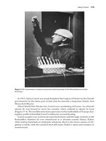
Figure 9.34 The army blood bank at Bristol
shortly after the D-Day landings in France,
June 1944. (From Cope Z, ed.: History of the
Second World War Medical Series – Surgery,
1953. Crown copyright; reproduced with
permission of the Controller of Her Majesty’s
Stationery Office.)
Figure 9.35 A blood transfusion taking place in
a tented CCS, Normandy 1944. (From Cope Z,
ed.: History of the Second World War Medical
Series – Surgery, 1953. Crown copyright;
reproduced with permission of the Controller
of Her Majesty’s Stationery Office.)
without damage to the patient’s healthy tissues.
Paul Ehrlich (1854–1915) of Frankfurt-on-Maine,
Germany, synthesised the arsenical compound
Salvarsan, which was used clinically in 1911 as
the first really effective drug against syphilis. It
was Ehrlich who coined the term ‘magic bullet’ to
mean a chemical bullet that would kill the organism but not the patient. Salvarsan was hardly the
148 The surgery of warfare
perfect bullet since it is a toxic drug with unpleasant side effects.
The next major landmark in chemotherapy
again came from Germany. Gerhardt Domagk
(1895–1964) showed that the aniline dye Prontosil
Rubra was highly effective against the muchdreaded spreading infections produced by streptococci, in spite of the disadvantage that the drug
stained the patient, fortunately temporarily, a
bright red colour. These important findings were
published in 1935. Within weeks of this paper
appearing, workers at the Pasteur Institute in
Paris showed that it was the sulphanilamide moiety of the Prontosil molecule that was the active
agent. The next few years saw a flurry of activity,
both by the synthetic chemists and clinicians,
in the development of new sulphonamide drugs.
The effectiveness of these agents against many
infections, such as pneumonia and puerperal
fever (sepsis following childbirth), seemed almost
miraculous. Sulphonamides were used during
the Spanish Civil War and also in the Second
World War in the treatment of major wounds and
certainly reduced the risk of wound infections.
However, they had the serious disadvantage of
being ineffective in the presence of pus, i.e. once
wound infection was established, and were also
valueless in the treatment of gas gangrene and
tetanus.
But what of the antimicrobial agents derived
from fungi and bacteria, the antibiotics? Most
people believe that the story begins with the
description of penicillin by Alexander Fleming
in 1928. In fact, the story goes back much further than this. In 1870, John Burdon Sanderson
(1828–1905), while working as a medical officer of health in Paddington (he subsequently
became the professor of medicine in Oxford), in
numerous experiments showed that bacteria did
not grow in a culture fluid that contained visible mould. The publication of Sanderson’s report
stimulated Joseph Lister himself to begin a series
of experiments in which he showed that urine that
had a heavy growth of mould showed abnormal
degenerate bacteria or the complete absence of
micro-organisms and that the urine under these
circumstances usually remained sweet smelling.
Aided by his brother Arthur, an expert mycologist,
Lister identified the fungus as Penicillium glaucum. In 1884, Lister treated a nurse named Ellen
Jones at King’s College Hospital, London, who
had a deep buttock abscess that was healing very
slowly with an extract of a culture of this fungus.
Unfortunately, Lister did not publish his methods
or the results of using what was presumably crude
penicillin. Numerous other reports appeared over
the years, including one from Louis Pasteur himself in 1877, in which he reported that anthrax
bacilli were inhibited in culture by unspecified
bacteria and postulated that this might prove to be
of clinical value.
Now to Alexander Fleming (1881–1955) and
his place in the history of antibiosis. While working as a bacteriologist at St Mary’s Hospital,
London, in 1928, he made the observation that
a culture plate of Staphylococcus aureus, a common cause of boils, abscesses and many other
serious infections, contaminated by spores of a
Penicillium mould showed lysis around the contaminating fungi. He made a detailed study of
this phenomenon, named the agent produced by
the mould ‘penicillin’, showed that a crude extract
from the mould was remarkably active against a
whole range of bacteria and published a report
on this phenomenon in 1929. However, efforts by
Fleming and his colleagues failed to concentrate
and purify penicillin.
Ten years passed before Howard Florey
(1898–1968), a professor of pathology at the
University of Oxford, and a young German Jewish
refugee biochemist, Ernst Chain (1906–1979),
determined to carry out a systematic study of
the known naturally occurring antibacterial substances. A review of previous publications in this
field naturally included Fleming’s paper of 1929
and, with the assistance of a team of dedicated
young scientists, the difficult task of extracting
penicillin from the mould of Penicillium notatum
was carried out. In May 1940, enough penicillin
was available for a crucial animal experiment,
which showed that the dry, stable brown powder
prepared by a process of freeze-drying was highly
effective in protecting mice given a lethal injection of Staphylococcus aureus. By the beginning of
1941, Florey had enough material to begin his first
trial on human beings, and, again, the results in
The Second World War (1939–1945) 149
patients with overwhelming bacterial infections
were most encouraging.
It was obvious that penicillin was a potentially powerful weapon in both the treatment and
prevention of infection in war wounds. Superhuman efforts were made to increase the yield of
penicillin in the ‘factory’ set up in the Pathology
Department at Oxford. In 1941, with the United
States in the war, production of penicillin was
undertaken by a number of major American pharmaceutical companies. By the Sicily landings in
1943 (Figure 9.36), enough penicillin was available
for extensive clinical trials, both as local treatment
in the wound and by intramuscular injection; the
results were excellent. It was soon shown that the
clostridia group of bacteria (those responsible for
gas gangrene and tetanus) was highly sensitive to
the drug. By the D-Day landings in Normandy in
1944, there was enough penicillin to allow its use
for all casualties. The antibiotic era had well and
truly commenced.
Subsequent wars have reinforced the lessons
of the two Great Wars, lessons learned from the
sufferings of countless millions of injured men
and women. Significant advances continued to
be made; for example, the development of sophisticated vascular surgery in the 1950s, using vein
Figure 9.36 A tented CCS and field surgical unit at the Sicily landings (1943). Penicillin
was now available for local but not systemic
treatment of wounds in the Services. (From
Cope Z, ed.: History of the Second World
War Medical Series – Surgery, 1953. Crown
copyright; reproduced with permission of
the Controller of Her Majesty’s Stationery
Office.)
and synthetic grafts, enabled many extremities to
be saved in the Korean and subsequent wars that
would previously have required amputation.
These principles of treatment, of course,
have been applied to the surgery of civilian
trauma. The dreadful vascular injuries produced
by ‘kneecapping’ carried out by terrorists in
Northern Ireland, were treated along wartime
principles, the damaged vessels repaired by grafts
and limbs rarely lost. I was involved in treating
casualties from four major terrorist ‘incidents’
at Westminster Hospital, London. Wound excision, immobilisation, antibiotics and delayed
primary suture were carried out in every case
and without a single example of wound infection
(Figure 9.37). The only thing to benefit from war
is surgery.
Figure 9.37 A victim of the Harrods bombing
by the Irish Republic Army (IRA) 1984; multiple
injuries including t raumatic amputation of the
right leg at mid-thigh. Treated by wound excision and delayed primary closure. (Photographic
Department, Westminster Hospital, London.)