A History of Vascular Surgery - part 8 ppsx
Bạn đang xem bản rút gọn của tài liệu. Xem và tải ngay bản đầy đủ của tài liệu tại đây (242.86 KB, 24 trang )
extremities before the International Society of Cardiovascular Surgery. The
long-term results were excellent (Figure 13.9).
It was only a matter of time before prosthetic grafts made their way into the
venous system. In 1979, Rosenthal used a prosthetic interposition graft for a case
of portal hypertension. Thirteen years later, Gloviczki reported his results with
three PTFE grafts used for reconstruction of the superior vena cava. Two
Venous surgery 155
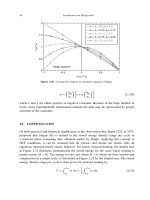
Figure 13.7 The saphenous–femoral venous crossover graft of Palma (from Palma E, Esperon R.
Vein transplants and grafts in the surgical treatment of postphlebitic syndrome. J Cardiovasc Surg
1960; 1:94).
required early thrombectomy, and two were patent after 2 and 5 years respec-
tively. The median patency rate of eleven inferior vena cava PTFE grafts was 9
months and an atrial–caval Dacron graft remained patent for 3 years. In 1997,
Alimi also reported favorable results with prosthetic reconstruction of iliac
veins.
In recognition of the different etiologies and locations of lower extremity
venous disease, the CEAP classification was devised in 1994. Under the auspices
of the American Venous Forum, this classification defined the clinical class
156 Chapter 13
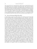
Figure 13.8 Psathakis’ “substitute valve” (from Psathakis N. Has the “substitute valve” at the
popliteal vein solved the problem of venous insufficiency of the lower extremity? J Cardiovasc Surg
1968; 9:64).
(C), the etiology (E), the anatomic (A) distribution, and the pathologic (P)
mechanism of the venous disease. Seven classes were designated according
to the clinical signs, and severity and disability rating scales were also
devised.
In 1996, Gloviczki reported preliminary results with endoscopic subfacial
division of perforating veins. A mean of 4.4 veins were divided in each of 11
extremities, and ulcer improvement or healing was noted in 10. In 1999, the
North American Subfacial Endoscopic Perforator Surgery Registry reported
results with 146 patients followed for a mean of 2 years. Perforator interrup-
tion combined with superficial reflux ablation was effective in healing ulcers. In
Venous surgery 157
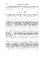
Figure 13.9 Kistner’s technique of venous valvular repair (from Kistner R. Surgical repair of a
venous valve. Straub Clin Proc 1968; 34:41).
patients with post-thrombotic limbs, however, recurrent or new ulcer develop-
ment remained a problem.
Most venous disorders are treated without surgery, and the mainstay of
treatment was developed by an engineer, not a surgeon. Conrad Jobst designed
brush-making machines and eventually obtained more than 40 patents. Jobst
suffered from varicose veins for most of his life, and began the first of many scle-
rotherapy sessions at the Henry Ford Hospital in 1930. He eventually recog-
nized that venous insufficiency resulted from excessive hydrostatic pressure,
and designed the first ambulatory gradient compression stockings for the treat-
ment of venous insufficiency. Half a century later, Jobst’s innovation remains the
most important therapy for this disorder.
The first use of intravenous sclerotherapy was reported by Pravaz in 1840; he
used absolute alcohol and eventually resorted to ferric chloride.
In 1910, Scharf reported his results with injection of sublimate into his own
varicose veins, and into the veins of 90 patients. In 1916, Linser recommended
perchloride of mercury and ambulatory treatments. In the first half of the 20th
century, many other substances were used for sclerotherapy including grape
sugar and sodium citrate; they were all abandoned, however, owing to allergic
reactions, skin sloughing, pain, and death in several cases.
In 1939, McAusland reported his successful treatment with sclerotherapy of
10000 patients. He advocated injection into empty veins, postsclerotherapy
compression, and minimal concentrations of sodium morrhuate to limit
complications. Two years later, Brunstein reiterated the value of McAusland’s
techniques, and sclerotherapy became an accepted treatment for venous
insufficiency.
Bibliography
AbuRahma AF, Robinson PA, Boland JP. Clinical hemodynamic and anatomic predictors
of long-term outcome of lower extremity veno-venous bypasses. J Vasc Surg 1991; 14:
635.
Alimi YS, DiMauro P, Fabre D, Juhan C. Iliac vein reconstructions to treat acute and chronic
venous occlusive disease. J Vasc Surg 1997; 25:673.
Anning ST. The historical aspects. In: Dodd H, Cockett FB, eds. The Pathology and Surgery of the
Veins of the Lower Limb. London: Churchill, Livingstone, 1976.
Barber RF, Shatara FI. The varicose disease. NY State Med J 1925; 25:162.
Bauer G. The etiology of leg ulcers and their treatment by resection of the popliteal vein. J Int
Chir 1948; 8:937.
Bazy L. Thrombose de la veine axillaire droite (thrombophlebite dite “par effort”). Phlébotomie
ablation des caillots. Suture de la veine. Bull Soc Nation Chir (Paris) 1926; 52:529.
Beberich J, Hirsch S. Die roentgenologische darstellung der arterien und venen in lebenden
menschen. Klin Wschr 1923; 49:222b.
Bhishagratna KL. An English Translation of the Sushruta Samhita. Varanasi: Chowkhamba
Sanskrit Series Office, 1963.
Brunstein IA. Prevention of discomfort and disability in the treatment of varicose veins. Am J
Surg 1941; 54:362.
158 Chapter 13
Carrel A, Guthrie CC. Uniterminal and biterminal venous transplantation. Surg Gynecol Obstet
1906; 2:266.
Cerino M, McGraw JY, Luke JC. Autogenous vein graft replacement of thrombosed deep veins.
Experimental approach to the treatment of the postphlebitic syndrome. Surgery 1964; 55:
123.
Clowes W. Extra-anatomical bypass of iliac vein obstruction: Use of a synthetic (expanded
polytetrafluoroethylene [Goretex] graft). Arch Surg 1980; 115:767.
Coar T. The Aphorisms of Hippocrates with a Translation into Latin and English. 1822. Birmingham:
Gryphon Editions, Ltd, 1982.
Dale WA, Scott HW Jr. Grafts of the venous system. Surgery 1963; 53:52.
Dale WA, Harris J, Terry RB. Polytetrafluoroethylene reconstruction of the inferior vena cava.
Surgery 1984; 95:625.
Dos Santos JC. La phlebographic direct. J Int Chir 1938; 3:625.
Fiore AC, Cromartie RS, Peigh PS, et al. Prosthetic replacement for the thoracic vena cava.
J Thorac Cardiovasc Surg 1982; 84:560.
Gay J. On varicose disease of the lower extremities. The Lettsomian Lectures of 1867. London:
Churchill, 1868.
Gloviczki P, Pairolero PC, Toomey BJ, et al. Reconstruction of large veins for nonmalignant
venous occlusive disease. J Vasc Surg 1992; 16:750.
Gloviczki P, Cambria RA, Rhee RY, et al. Surgical technique and preliminary results of endo-
scopic subfascial division of perforating veins. J Vasc Surg 1996; 23:517.
Gloviczki P, Bergan JJ, Rhodes JM, et al. North American Study Group: mid-term results of
endoscopic perforator vein interruption for chronic venous insufficiency: lessons learned
from the North American Subfascial Endoscopic Perforator Surgery (NASEPS) registry.
J Vasc Surg 1999; 29:489.
Homans J. The operative treatment of varicose veins and ulcers, based upon a classification of
these lesions. Surg Gynecol Obstet 1916; 22:143.
Homans J. The etiology and treatment of varicose ulcer of the leg. Surg Gynecol Obstet 1917;
24:300.
Howard-Jones N. Acritical study of the origins and early development of hypodermic medica-
tion. J Hist Med1947; 2:201.
Husni EA. In situ saphenopopliteal bypass graft for incompetence of the femoral and popliteal
veins. Surg Gynecol Obstet 1970; 2:279.
Ijima H, Sakurai J, Mori M, et al. Temporary arteriovenous fistula for venous reconstruction
using a synthetic graft: Clinical and experimental evaluation. J Cardiovasc Surg 1981; 222:
480.
Kistner R. Surgical repair of a venous valve. Straub Clin Proc 1968; 34:41.
Kistner R. Surgical repair of the incompetent femoral vein valve. Arch Surg 1975; 110:1336.
Kunlin J. The reestablishment of venous circulation with grafts in cases of obliteration from
trauma or thrombophlebitis. Mem Acad Clin 1953; 79:109.
Laewen A. Weitere erfahrungen ueber operative thrombenentfernung bei venenthrombose.
Arch Klin Chir 1938; 193:723.
Linser F. Uber die Konservative Behandlung der Varicen. Med Klin 1916; 12:897.
Linton RR. The communicating veins of the lower leg and the operative technic for their liga-
tion. Ann Surg 1938; 107:582.
Linton RR. Modern concepts in the treatment of the postphlebitic syndrome with ulcerations of
the lower extremity. Angiology 1952; 3:431.
Linton RR, Harry IB Jr. Postthrombotic syndrome of the lower extremity. Surgery 1948; 24:452.
Linton RR, Keeley JK. The postphlebitic varicose ulcer. Am Heart J 1939; 17:27.
Venous surgery 159
McAusland S. The modern treatment of varicose veins. Med Press 1939; 201:404.
Moore TC, Young NK. Experimental replacement and bypass of large veins. Bull Soc Int Chir
1964; 23:274.
O’Donnell TF, Fredricks R. Venous obstruction: an analysis of one hundred thirty-seven cases
with hemodynamic, venographic, and clinical correlations. J Vasc Surg 1991; 14:305.
O’Donnell TF, Mackey WC, Shepard AD, Callow AD. Clinical hemodynamic and anatomic
follow-up of direct venous reconstruction. Arch Surg 1987; 122:474.
Palma E, Esperon R. Vein transplants and grafts in the surgical treatment of postphlebitic
syndrome. J Cardiovasc Surg 1960; 1:94.
Psathakis N. Has the “substitute valve” at the popliteal vein solved the problem of venous
insufficiency of the lower extremity? J Cardiovasc Surg 1968; 9:64.
Raju S. Venous insufficiency of the lower limb and stasis ulceration. Changing concepts and
management. Ann Surg 1983; 197:688.
Rhodes JM, Gloviczki P, Canton LG, et al. Factors affecting clinical outcome following endo-
scopic perforator vein ablation. Am J Surg 1998; 176:162.
Rhodes JM, Gloviczki P, Canton LG, et al. Endoscopic perforator vein division with ablation of
superficial reflux improves venous hemodynamics. J Vasc Surg 1998; 28:839.
Rogoff SM, DeWeese JA. Phlebography of the lower extremity. JAMA 1960; 172:1599.
Rosenthal D, Deterling RA, O’Donnell TF, et al. Interposition grafting with expanded polyte-
trafluoroethylene for portal hypertension. Surg Gynecol Obstet 1979; 148:378.
Scharf P. Ein neues Verfahren der intravenosen Behandlung der Varicositaten der Unterex-
tremitaten. Berliner Klin Wochenschr 1910; 13:582.
Smirk FM. Observations on the causes of oedema in congestive heart failure. Clin Sci 1936;
2:317.
Steinman C, Alpert J, Haimovici H. Inferior vena cava bypass grafts: An experimental evalua-
tion of a temporary arteriovenous fistula on their long-term patency. Arch Surg 1966; 93:747.
Taheri SA, Lazar L, Elias S, et al. Surgical treatment of postphlebitic syndrome with vein valve
transplant. Am J Surg 1982; 144:221.
Toledo-Pereyra LH. Galen’s contribution to surgery. J Hist Med 1973; Oct, 357.
Trendelenburg F. Ueber die unterbindung der vena saphena magna bei unterschenkelvaricen.
Beit Klin Chir 1890; 7:195.
Unna PG. Ueber paraplaste: Eine neue form medikamentoser pflaster. Wien Med Wschr 1896;
46:1854.
Warren R, Thayer TR. Transplantation of the saphenous vein for postphlebitic stasis. Surgery
1954; 35:867.
160 Chapter 13
CHAPTER 14
Extra-anatomic bypass
161
The shortest route is not the most direct one, but rather the one where the most favorable winds
swell our sails.
(Friedrich Nietzsche)
It was not long after Jacques Oudot’s original aorta-iliac bypass that reconstruc-
tion of these vessels was recognized as an effective method of treating lower
extremity ischemia. The necessity of a laparotomy and retroperitoneal dissec-
tion, however, made direct reconstruction of diseased aorta–iliac segments
too hazardous for some patients. The possibility of an indirect, less invasive
procedure was first conceived by Norman Freeman in 1952. In a paper describ-
ing recent advances in operations on large arteries, Freeman reported a case of
left iliofemoral endarterectomy in which a right iliac artery aneurysm was
noted. Cellophane was wrapped about the aneurysm, resulting in its subse-
quent thrombosis, and in gangrene of the right fifth toe 6 weeks later. At reoper-
ation, Freeman divided the chronically occluded left superficial femoral artery
at the adductor tendon, performed an endarterectomy, and then tunneled it into
the right groin via a subcutaneous route, where an end-to-end anastomosis was
performed to the divided right superficial femoral artery (Figure 14.1). The
patient recovered well, with the circulation to the right foot intact. Freeman
concluded:
It is fully recognized that operative intervention does not solve the main problem –
arteriosclerosis – since this condition is generally widespread and operation is limited
to the particular vessel involved. However, it does give promise of relief of some of the
complications when the disease is limited to a single vessel.
In 1958, McCaughan and Kahn reported two cases of iliac-to-contralateral
popliteal crossover grafts for limb-threatening ischemia, with good results. In
the first case, an anastomosis was also performed from the Dacron prosthesis to
the profunda femoris of the ischemic extremity, one of the earliest uses of the
sequential bypass technique. McCaughan and Kahn concluded that the
procedure was safer than the usual graft from the aorta to the popliteal artery.
In 1960, Vetto attempted to render the procedure of McCaughan and Kahn
safer when he used the common femoral artery, rather than the external iliac, as
a donor vessel for a bypass to the contralateral extremity. In 1962, he reported a
series of 10 femoral–femoral bypasses with follow-up to 16 months. Nine of the
cases were successful. By 1966, Vetto had accumulated 39 cases, with continued
good results, leading him to consider use of this procedure in good-risk patients
as well.
Cecil Lewis of Australia developed the concept of using an upper extremity
artery to supply circulation to the lower extremities. In 1959, he used a nylon
prosthesis to construct a bypass from the subclavian artery to an aorta–iliac
homograft in a case of ruptured abdominal aortic aneurysm. The patient sur-
vived and eventually returned to his occupation of greenkeeper (Figure 14.2).
The first axillary–femoral artery bypass was performed by Blaisdell in 1962,
following an abdominal aortic aneurysmectomy in an elderly man who had
undergone left above-knee amputation 8 years previously. On the third post-
operative day, the aortic graft thrombosed, placing the right lower extremity in
jeopardy. The patient was returned to the operating room and suffered cardiac
arrest upon induction of anesthesia. Resuscitation was successful but because of
the patient’s fragile state an abdominal procedure was considered too danger-
ous. Blaisdell constructed a bypass from the right axillary artery to the common
162 Chapter 14
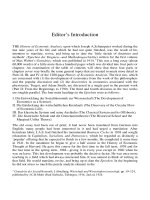
Figure 14.1 The first femoral–femoral crossover graft (from Freeman NE, Leeds FH. Operations on
large arteries. Application of recent advances. Cal Med 1952; 77:229).
femoral artery under local anesthesia, resulting in salvage of the patient’s ex-
tremity. The Dacron prosthesis was still patent 8 months later (Figure 14.3).
Less than 1 month after Blaisdell’s operation, J.H. Louw performed the iden-
tical procedure in a 52-year-old South African man with gangrenous toes.
In 1963, Blaisdell reported his use of axillary–femoral bypass in seven
patients with good immediate results. Three years later, Sauvage introduced the
addition of a crossover graft to the axillary–femoral for bilateral lower extrem-
ity ischemia.
Extra-anatomic bypasses were also recognized as effective alternatives to
intrathoracic or mediastinal procedures, in the treatment of occlusive disease of
the aortic arch and its branches. The first extrathoracic bypass was performed by
Lyons and Galbraith in 1956. They used a nylon prosthesis to construct a subcla-
vian–carotid bypass in a 67-year-old man who had internal carotid artery steno-
sis and transient ischemic attacks. The patient was asymptomatic 7 months after
surgery. Variations of this procedure include subclavian–subclavian bypass,
first performed by Ehrenfeld in 1965; and axillary–axillary bypass, introduced
by Myers in 1971. Additional experiences with these procedures soon followed.
Extra-anatomic bypass 163
Figure 14.2 Lower extremity blood supply derived from the subclavian artery (from Lewis CD. A
subclavian artery as the means of blood-supply to the lower half of the body. Br J Surg 1961;
48:574).
164 Chapter 14
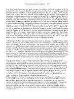
Figure 14.3 The first axillary–femoral graft (from Blaisdell FW, Hall AD. Axillary–femoral artery
bypass for lower extremity ischaemia. Surgery 1963; 54:563).
Dietrich reported 125 cases of subclavian–carotid bypass in 1967. In 1972 Fin-
klestein reported 15 cases of subclavian–subclavian bypass for the subclavian
steal syndrome and, by 1979, Myers had performed 18 axillary–axillary
bypasses. For cases in which a cervical arterial source was unavailable, Sproul
suggested femoral–axillary bypass in 1971.
The original indications for extra-anatomic bypasses were complications
of aortic reconstructions, and impending limb loss in ill patients. In 1970,
Parsonnet suggested that the indications for these procedures should be
broadened, since they often worked well. He reported good results with 38
femoral–femoral, 11 axillary–femoral, and 10 carotid–subclavian grafts; and
assuaged fears of a steal syndrome. Two years later, Parsonnet’s group reported
an 85 percent 5-year patency rate in 66 femoral–femoral grafts. In 1980, they
reported 73 percent 5-year and 64 percent 10-year patency rates in 133
femoral–femoral grafts.
In 1977, Logerfo reported the results of 66 axillary–bifemoral and 64 axil-
lary–femoral grafts in 120 patients. The 5-year patency rate for the former was 74
percent (20 thrombectomies in 15 grafts), versus 37 percent (25 thrombectomies
in 22 grafts) for unilateral grafts. The authors concluded that axillary–bifemoral
grafts had similar 5-year patency rates to aorta–iliac grafts, and were preferable
to unilateral grafts owing to their superior patency rate.
In the same issue of the Annals of Surgery bearing Logerfo’s study, more sober-
ing results with these bypasses were reported by Eugene. One-half of his 59
axillary–femoral bypasses thrombosed within 2 years, and 47 percent of his 33
femoral–femoral bypasses closed within 4 years. He counseled that subcuta-
neous grafts should be performed only when an intra-abdominal procedure
was contraindicated or the life expectancy was limited.
The use of “extended” extra-anatomic bypasses was reported by Veith in
1978. Twelve out of 14 axillary–popliteal bypasses were patent after 14 months.
Six years later Connolly reported his results with 13 axillary–popliteal, and
three axillary–tibial bypasses. Two of the former were patent after 3 years,
and one of the latter was open after 18 months. In 1989, Ascer summarized the
Montefiore experience, with 55 axillary–popliteal grafts performed over 12
years; the 5-year patency rate was 40 percent.
Several reports in the early 1990s renewed the debate about broadening the
indications for axillary–femoral bypass. Harris found a primary patency rate of
85 percent for 76 axillary–bifemoral grafts followed for nearly 2.5 years, and
concluded that more patients could be helped by this procedure.
In 1992 Schneider compared the results of 34 axillary–bifemoral and
unifemoral grafts, with those of 107 aorta–femoral grafts performed synchro-
nously. He concluded that extra-anatomic bypasses were acceptable, but hemo-
dynamically inferior alternatives to direct reconstruction, and should be
reserved for properly selected high-risk patients. One year later, El-Massry re-
ported a primary patency rate of 73 percent for 79 axillary–femoral bypasses
after 7 years, and recommended their use for incapacitating claudication as well
as limb salvage.
Extra-anatomic bypass 165
By the millennium, most reports favored a limited role for extra-anatomic
bypasses, reserving them for critically ill patients unable to tolerate direct aortic
reconstructions. Advances in anesthesiology, cardiology, and critical care medi-
cine have significantly reduced the number of these patients.
Bibliography
Alpert J, Brief DK, Parsonnet V. Vascular restoration for aortoiliac occlusion and an alternative
approach to the poor risk patient. J Newark Beth Israel Hosp 1967; 18:4.
Ascer E, Veith FJ, Gupta S. Axillofemoral bypass grafting: indications, late results, and deter-
minants of long-term patency. J Vasc Surg 1989; 10:285.
Blaisdell FW, Hall AD. Axillary-femoral artery bypass for lower extremity ischemia. Surgery
1963; 54:563.
Brief DK, Alpert J, Parsonnet V. Crossover femorofemoral grafts: compromise or preference: A
reappraisal. Arch Surg 1972; 105:889.
Brief DK, Brener BJ, Alpert J, et al. Crossover femorofemoral grafts followed up five years or
more. Arch Surg 1975; 110:1294.
Connolly JE, Kwaan JHM, Brownell D, et al. Newer developments of extraanatomic bypass.
Surg Gynecol Obstet 1984; 158:415.
Criado E, Burnham SJ, Tinsley EAJr., et al. Femorofemoral bypass graft: analysis of patency and
factors influencing long term outcome. J Vasc Surg 1993; 18:495.
Dick LS, Brief DK, Alpert J, et al. A12 year experience with femorofemoral crossover grafts. Arch
Surg 1980; 115:1359.
Diethrich EB, Garrett HE, Ameriso J, et al. Occlusive disease of the common carotid and subcla-
vian arteries treated by carotid-subclavian bypass. Analysis of 125 cases. Am J Surg 1967;
114:800.
Donaldson MC, Louras JC, Bucknam CA. Axillofemoral bypass: A tool with a limited role.
J Vasc Surg 1986; 3:757.
Ehrenfeld WK, Levin SM, Wylie EJ. Venous crossover bypass grafts for arterial insufficiency.
Ann Surg 1968; 167:287.
El-Massry S, Saad E, Sauvage LR, et al. Axillofemoral bypass using externally-supported,
knitted Dacron grafts: a follow-up through twelve years. J Vasc Surg 1993; 17:107.
Eugene J, Goldstone J, Moore WS. Fifteen-year experience with subcutaneous bypass grafts for
lower extremity ischemia. Ann Surg 1976; 186:177.
Finkelstein NM, Byer A, Rush BF Jr. Subclavian-subclavian bypass for the subclavian steal
syndrome. Surgery 1972; 71:142.
Freeman NE, Leeds FH. Operations on large arteries. Application of recent advances. Cal Med
1952; 77:229.
Harris EJ, Taylor LM, McConnell DB, et al. Clinical results of axillobifemoral bypass using
externally supported polytetrafluoroethylene. J Vasc Surg 1990; 12:416.
Illuminati G, Calio PG, Mangialardi N, et al. Results of axillofemoral by-passes for aorto-iliac
occlusive disease. Langenbecks Arch Surg 1996; 381:212.
Johnson WC, LoGerfo FW, Vollman RW. Is axillobilateral femoral graft an effective substitute
for aortobilateral iliac femoral graft? Ann Surg 1976; 186:123.
Keller MP, Hoch JR, Harding AD, et al. Axillopopliteal bypass for limb salvage. J VascSurg 1992;
15:817.
Lewis CD. Asubclavian artery as the means of blood-supply to the lower half of the body. Br J
Surg 1961; 48:574.
166 Chapter 14
LoGerfo FW, Johnson WC, Corson JD, et al. Acomparison of the late patency rates of axillobilat-
eral femoral and axillounilateral femoral grafts. Surgery 1977; 81:33.
Louw JH. Splenic-to-femoral and axillary-to-femoral bypass grafts in diffuse atherosclerotic
occlusive disease. Lancet 1963; 1:1401.
Lyons C, Galbraith G. Surgical treatment of atherosclerotic occlusion of the internal carotid
artery. Ann Surg 1957; 146:487.
McCaughan JJ Jr., Kahn SF. Cross-over graft for unilateral occlusive disease of the iliofemoral
arteries. Ann Surg 1960; 151:26.
Mannick JA, Williams LE, Nabseth DC. The late results of axillofemoral grafts. Surgery 1970;
68:1038.
Myers WO, Lawton BR, Sautter RD. Axillo-axillary bypass graft. JAMA 1971; 217:826.
Myers WO, Lawton BR, Ray JF III, et al. Axillo-axillary bypass for subclavian steal syndrome.
Arch Surg 1979; 114:394.
Parsonnet V, Alpert J, Brief DK. Femorofemoral and axillofemoral grafts: compromise or
preference? Surgery 1970; 67:26.
Passman MA, Taylor LM, Moneta GL, et al. Comparison of axillofemoral and aortofemoral
bypass for aortoiliac occlusive disease. J Vasc Surg 1996; 23:263.
Plecha FR, Plecha FM. Femorofemoral bypass grafts: Ten-year experience. J Vasc Surg 1984;
1:555.
Posner MP, Riles TS, Ramirez AA, et al. Axilloaxillary bypass for symptomatic stenosis of the
subclavian artery. Am J Surg 1983; 145:644.
Rutherford RB, Patt A, Pearce WH. Extra-anatomic bypass: a closer view. J Vasc Surg 1987; 5:437.
Sauvage LR, Wood SJ. Unilateral axillary bilateral femoral bifurcation graft: A procedure for
the poor risk patient with aortoiliac disease. Surgery 1966; 60:573.
Schanzer H, Chung-Loy H, Kotok M, et al. Evaluation of axillo-axillary artery bypass for the
treatment of subclavian or innominate artery occlusive disease. J Cardiovasc Surg 1987;
28:258.
Schneider JR, McDaniel MD, Walsh DB, et al. Axillofemoral bypass: outcome and hemody-
namic results in high-risk patients. J Vasc Surg 1992; 15:952.
Veith FJ, Moss CM, Daly V, et al. New approaches to limb salvage by extended extra-anatomic
bypasses and prosthetic reconstructions to foot arteries. Surgery 1978; 84:764.
Vetto RM. The treatment of unilateral iliac artery obstruction with a transabdominal, subcuta-
neous, femorofemoral graft. Surgery 1962; 52:342.
Vetto RM. The femorofemoral shunt. An appraisal. Am J Surg 1966; 112:162.
Vetto RM, Dunphy JE. Recent revisions in the operative treatment of vascular disease. Surg
Gynecol Obstet 1964; 119:1026.
Ziomek S, Quinones-Baldrich WJ, Busuttil RW, et al. The superiority of synthetic arterial grafts
over autogenous veins in carotid-subclavian bypass. J Vasc Surg 1986; 3:140.
Extra-anatomic bypass 167
The French connection
Mathieu Jaboulay
171
It is common sense to take a method and try it. If it fails, admit it frankly and try another. But
above all, try something.
(Franklin D. Roosevelt)
Mathieu Jaboulay was born in France in 1860 and was the first in a succession of
French surgeons that limned most of the basic concepts in vascular surgery. As a
surgeon in Lyon, Jaboulay was fascinated by the report of arterial suturing by
Jassinowsky, in 1891, and several years later by Heidenhain. These prompted
him to begin the first experiments in France, on arterial suturing. He was
assisted by his intern, Eugèbe Briau.
In 1892, Jaboulay was named Head Surgeon at the Hotel-Dieu. Four years
later, Briau and Jaboulay published the first French article on vascular surgery in
Lyon Médicale. They described their results with circular anastomoses and
carotid interposition grafts in dogs. All of the arteries thrombosed within 4 days.
Undaunted, Jaboulay revised his technique by everting the arterial edges.
With better results he concluded:
The arterial graft will give us the means to combat gangrene of arterial origin against
which we are helpless. The treatment of aneurysms and arterial contusions will be
transformed.
Jaboulay also correctly predicted the use of this technique in the venous sys-
tem. He speculated about placing venous autografts into the arterial system and
predicted that this would replace the ligature as a treatment for arterial injuries.
In 1901, Jaboulay advised Carrel and Morel to attempt carotid–jugular anas-
tomoses in dogs, as a means of improving cerebral circulation. Carrel obtained
good results, with beating subcutaneous jugular veins after 3 weeks. These
results were also reported in Lyon Médicale, in 1902.
In 1906, Mathieu Jaboulay carried out the first attempts at human kidney
transplantation. On January 22, he transplanted a porcine kidney to the brachial
vessels of a woman suffering from nephrotic syndrome. Three months later, he
repeated this treatment in a different patient, with a goat kidney. Neither of the
xenografts lasted more than several hours and both had to be excised. Jaboulay
was unfazed by these failures and concluded:
If these grafts become feasible, no area of the body will know better how to employ it
than the bend of the elbow for ease and mildness of operating maneuvers.
The pinnacle of Jaboulay’s career was reached in 1902, when he became
Chairman of the Surgical Clinic at the Hotel-Dieu. Jaboulay held this post until
his death in 1913. While traveling to Paris to examine applicants for ophthal-
mology positions at a local university, he died in a train accident in Melun.
Bibliography
Bouchet A. Les pionniers Lyonnais de la chirurgie vasculaire: M. Jaboulay, A. Carrel, E. Villard
et R Leriche. Hist Sci Med 1994; 28:223.
Jaboulay M. Le traitement de quelques troubles trophiques du pied et de la jambe par la
denudation de l’artère fémorale et la distension des nerfs vasculaires. Lyon Méd 1899; 91:467.
Jaboulay M. Chirurgie des artères. Semin Méd 1902: 405.
Jaboulay M. Greffe du rein au pli du coude par soudure artérielle et veineuse. Lyon Méd 1906;
107:575.
Jaboulay M, Briau E. Recherches expérimentales sur la suture et la greffe artérielle. Lyon Méd
1896; 81:97.
172 Chapter 15
CHAPTER 16
Eugène Villard
173
Curiosity is, in great and generous minds, the first passion and the last.
(Samuel Johnson)
Eugène Villard was born in France in 1868. Inspired by the work of Carrel in the
United States, and by a slew of Lyonese theses dedicated to vascular surgery
(Louis Bérard, 1909; Pierre Charnois, 1909; Emile Perrin, 1911) Villard began
experimenting with vascular and renal grafts in 1910. He collaborated with
fellow surgeons Louis Tavernier and Emile Perrin, and relied upon Delachanal
and Dubreuil for histologic examination of specimens. After 4 years of experi-
mental surgery in Lyon, their results were presented at the New York Interna-
tional Convention of Surgery, in 1914.
Regarding autogenous carotid arterial grafts in dogs Villard wrote: “The ar-
teries implanted in the same animals formed scar tissue without modification.”
In another series of experiments, canine iliac arteries were grafted onto the
carotid arteries of other dogs. The grafts were harvested 12 days later and exam-
ined microscopically by Dubreuil, who concluded: “The histologic structure is
so perfectly preserved that it is impossible to distinguish where the graft was
cut.”
Villard and his colleagues also performed numerous autogenous venous
grafts in dogs with continuous failures in the early period. Eventually, however,
they succeeded in replacing a carotid artery with external jugular vein. The graft
was examined after nearly 4 months and found to be in perfect condition. Villard
offered an early description of neointimal fibrous hyperplasia:
The vascular wall thickening, which shows up especially in the middle membrane, is
made up for the most part of neoformations of a smooth muscular type. To synthesize
in a word these histologic modifications, one could say that the venous graft implanted
on an artery truly makes itself an artery.
Villard also experimented with grafts preserved by freezing, but most of his
grafts thrombosed. Among the few successes were three carotid homografts,
and a human saphenous vein graft implanted into a feline abdominal aorta.
Villard was pessimistic about the prospects for preserved grafts, unless the
technique for preservation could be improved.
Villard distinguished himself as a great teacher and in 1921 became Chair-
man of Operating Medicine, in Lyon. From 1925 to 1927 he was also the Chair-
man of the Gynecology Clinic.
Eugène Villard died in 1953.
Bibliography
Bouchet A. Les pionniers Lyonnais de la chirurgie vasculaire: M. Jaboulay, A. Carrel, E. Villard
et R Leriche. Hist Sci Med 1994; 28:223.
Villard E. Greffes vasculaires. XIVe Congrès international de Chirurgie, New York, April 1914.
Villard E, Perrin E. Greffes vasculaires. Lyon Chir 1912; 8:267.
Villard E, Perrin E. Traitement des obliterations vasculaires. Lyon Chir 1913; 9:4.
Villard E, Tavernier L, Perrin E. Recherches expérimentales sur les greffes vasculaires. Lyon Chir
1911; 6:144.
174 Chapter 16
CHAPTER 17
Alexis Carrel
175
To yield to every whim of curiosity, and to allow our passion for inquiry to be restrained by noth-
ing but the limits of our ability, this shows an eagerness of mind not unbecoming to scholarship.
But it is wisdom that has the merit of selecting from among the innumerable problems which
present themselves, those whose solution is important to mankind.
(Immanuel Kant)
Historians of every field seek an individual upon whom to fix the epithet
“Father.” In vascular surgery, the search ends upon review of the life and
contributions of Alexis Carrel, whose extraordinary imagination and foresight
suggest a parallel to the vision of his more celebrated countryman, Jules Verne
(Figure 17.1). Many decades prior to their invention, Verne accurately predicted
the use of airplanes, submarines, television, guided missiles, and space satel-
lites. His well-known tales have carried readers under, above, and around the
earth. Carrel foresaw the routine suturing of blood vessels and use of vein by-
pass grafts; reimplantation of severed limbs; the preservation and transplanta-
tion of kidneys, thyroid, heart, and lung; and cardiac valvular reconstruction
and extracorporeal circulation. Unlike Verne’s imaginings, however, Carrel’s
were realized in his own lifetime.
Carrel was born in Lyon, France, in 1873. When Carrel was 5 years old, his
father died, and the responsibility of helping to care for a younger brother and
sister had an early maturing effect. Alexis was a very quiet, serious child and
attended St. Joseph’s Day School, an institution administered by Jesuit priests.
Carrel showed little interest in music and art and spent most of his free time
reading. In 1889, he received a Baccalaureate in Letters, and one in Science the
following year. After graduation, Carrel enrolled in the medical school at the
University of Lyon. Following 3 years there, he became an extern at the Red
Cross Hospital and the Hôpital Antiguaille.
In 1895, Carrel fulfilled 1 year of military service with the French mountain
troops and spent the next 5 years completing his internship in several hospitals
throughout Lyon.
At this time there was little work being done in the field of vascular surgery.
In 1896, Mathieu Jaboulay, a teacher of Carrel during his internship, published
one of the first papers describing end-to-end anastomosis of blood vessels. In
the United States, John Murphy would soon describe his repair of a lacerated
femoral artery and in Germany Edwin Payr was conducting preliminary
experiments substituting magnesium tubes for arterial segments. Vascular sur-
gery was, therefore, barely in its infancy when an event occurred that altered the
life of Carrel and hastened the age of routine operation on the heart and blood
vessels.
In 1894, the President of the French Republic was Sadi Carnot. While in Lyon,
he suffered a stab wound to the abdomen at the hands of an Italian anarchist. The
blade severed the portal vein and, in accordance with the prevailing notions of
the day, the best surgeons in France threw up their hands in frustration, con-
vinced that nothing could be done to save their President.
Carrel was deeply moved by the death of Carnot and could not accept the
helplessness of Carnot’s surgeons. Carrel was emphatic in his belief that if sur-
geons were able to repair blood vessels as they could skin and other tissues,
Carnot would have been saved.
In 1899, mindful of Jaboulay’s attempts at uniting blood vessels, Carrel began
his first experiments in the laboratory of Mariel Soulier, a professor of therapeu-
176 Chapter 17
Figure 17.1 Alexis Carrel (courtesy of the Rockefeller University Archives).
tics. Most of these involved construction of arterial–venous fistulas in canine
necks, between the external jugular vein and carotid artery. Carrel developed
new sutures and needles for this work and he also received embroidery lessons
to which he later ascribed his manual dexterity.
At the turn of the century, it was necessary to pass a difficult clinical examina-
tion to gain a surgical faculty position in Lyon. Most students required several
attempts to pass and by 1903, Carrel had failed twice. That same year Carrel ac-
companied a pilgrimage to Lourdes, where miraculous cures were said to occur.
There he encountered a young girl dying of tuberculous peritonitis. Uncon-
scious and deemed too ill to undergo the usual immersion in the curative pool,
she was sprinkled with a few of its drops. The girl regained consciousness with-
in a few hours and went on to make a miraculous recovery. She became a nun
and lived for 34 more years. Carrel was mystified by these events and chose to
credit the power of suggestion as the only rational explanation. He nonetheless
faithfully reported what he had witnessed, and was attacked by clergy and
medical colleagues alike upon his return to Lyon: by the one contingent for his
skepticism and by the other for his gullibility. Informed that he now had no
chance of passing his surgical examination, he contemplated leaving France and
medicine altogether.
In May 1904, Carrel left France for Montreal. Several months later he pre-
sented a paper on vascular anastomosis to the Second Medical Congress of
the French Language of North America. It was well received by the audience,
of which Karl Beck, a respected Chicago surgeon, was a member. Beck ap-
proached Carrel with the possibility of working in the United States and, in
August 1904, Carrel began a 2-month trek west across Canada, south through
California, then east to Chicago. He eventually accepted a position at the Uni-
versity of Chicago in the Physiology Department under the chairmanship of
Dr George Stuart.
Carrel was assigned to work with Charles Claude Guthrie, a young physiolo-
gist who had graduated from medical school 4 years earlier. Between November
1904 and August 1906, the two shared one of the most productive relationships in
the history of medicine. During these 21 months, 9 of which Guthrie spent at the
University of Missouri on sabbatical, they wrote 28 papers together. Carrel
added five more of his own and Guthrie two. Their experimental work included
perfection of vascular anastomoses and the use of vein grafts in the arterial sys-
tem; development of tissue preservation techniques; reimplantation of limbs and
transplantation of kidneys, ovaries, thyroids, and hearts.
Carrel’s vision would be realized in the first routine use of saphenous vein
bypasses in 1948, the first successful human renal transplant in 1955, and the
performance of the first human limb reimplantation in 1962. Christian Barnard
would perform the first human heart transplant in 1967, 62 years after Carrel’s
description.
The collaboration of these two great men ended in 1906, when Guthrie ac-
cepted a position as Professor of Physiology and Pharmacology at Washington
University in St. Louis. Carrel was disappointed by the lack of financial support
Alexis Carrel 177
for his research so he moved to the Rockefeller Institute in New York (Figure
17.2).
Carrel began his work in the Experimental Surgical Department of the Rock-
efeller Institute by continuing his investigations of preserved vascular homo-
grafts to replace segments of cat abdominal aortas. During the next 4 years, he
improved preservation techniques for transplantation of carotid arteries from
one dog to another. Carrel performed experiments on the thoracic aorta, inter-
posing vena cava grafts and using paraffin tubes as shunts to prevent spinal
cord ischemia.
In a paper presented to the American Surgical Association in 1910, Carrel de-
scribed mitral valvulotomy and annuloplasty, ventricular aneurysmectomy,
and coronary artery bypass. His fame was also growing as a result of his contri-
butions to the field of tissue culture (Figure 17.3). Carrel’s meticulous applica-
tion of aseptic techniques and his fine dexterity were responsible for his
successes in this field, just as they had been in vascular surgery.
For his hitherto unparalleled accomplishments in vascular surgery and
organ transplantation, Alexis Carrel was awarded the Nobel Prize for Physiolo-
gy and Medicine in October 1912. It is alleged that Carrel learned of the award
while browsing through a New York morning paper. Carrel was the youngest
scientist, as well as the first United States scientist, to earn this prize. At a cere-
mony in his honor, President William Taft pronounced:
The names of Harvey Pasteur, Walter Reed, Koch, are great names which share the
progress toward a superior knowledge of the human and of medicine, and from now
on, Dr. Carrel will take his place among them.
178 Chapter 17
Figure 17.2 Carrel’s operating room at the Rockefeller Institute (courtesy of the Rockefeller
University Archives).