Ebook Medical parasitology: Part 2
Bạn đang xem bản rút gọn của tài liệu. Xem và tải ngay bản đầy đủ của tài liệu tại đây (4.15 MB, 161 trang )
SECTION III
Cestodes
CHAPTER 20
Taeniasis and Cyticercosis
Hannah Cummings, Luis I. Terrazas and Abhay R. Satoskar
Background
Taeniasis and cysticercosis are diseases resulting from infection with parasitic
tapeworms belonging to Taenia species. Approximately 45 species of Taenia have
been identified; however, the two most commonly responsible for human infection are the pork tapeworm Taenia solium and the beef tapeworm Taenia saginata.
Parasitic tapeworm infections occur worldwide, causing sickness, malnutrition and
often resulting in the death of their host. Infection with adult tapeworms of either
T. solium or T. saginata cause taeniasis in humans. The metacestode, or larval stage,
of Taenia solium causes the tissue infection, cysticercosis. Clinical manifestations
associated with the tapeworm infection can vary greatly and may range from mild
forms where patients exhibit little to no symptoms, to severe life-threatening forms
which are often fatal.
Geographic Distribution and Transmission
Taenia infections are estimated to affect 100 million people worldwide, with
major endemic areas located primarily in the developing countries of South
America, Africa, India, China and Southeast Asia. The ingestion of cysticerci
from raw or undercooked meat facilitates the transmission of T. solium from
pigs to humans and is presumably responsible for the high prevalence of human cysticerosis in these regions. It is estimated that anywhere between 5-40%
of individuals carrying the adult tapeworm will develop cysticercosis. Taenia
infections are less common in North America; however neurocysticercosis has
been recognized as an important health problem in California. Although this
disease is mainly seen in migrant workers from Latin American, it has also been
reported in US residents who have not traveled to endemic countries.
Life-Cycle
The complete life-cycle of Taenia solium involves two hosts: the pig and
the human, whereas that of Taenia saginata involves the cow and the human
(Fig. 20.1). Humans act as the definitive host and harbor the adult tapeworm in
the small intestine. Infection is acquired either through the accidental ingestion
of embryonated eggs passed in the feces of an individual infected with the adult
tapeworm, or through the consumption of raw or poorly cooked meat containing
cysticerci. The cysticerca develops into an adult worm in the gut; these worms
can survive up to 25 years. Depending on the species of Taenia, an adult worm
can reach lengths between 2-25 meters and may produce as many as 300,000 eggs
Medical Parasitology, edited by Abhay R. Satoskar, Gary L. Simon, Peter J. Hotez
and Moriya Tsuji. ©2009 Landes Bioscience.
Taeniasis and Cyticercosis
139
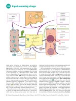
Figure 20.1. Life cycles of the beef tapeworm, Taenia saginata and the pork
tapeworm, T. solium. Reproduced from: Nappi AJ, Vass E, eds. Parasites of
Medical Importance. Austin: Landes Bioscience, 2002:61.
per day. The morphology of the adult worm consists of a scolex and a strombila.
The scolex acts as the organ of attachment and consists of four suckers equipped
with hooklets. The strombila consists of several segments (proglottids) with the
gravid or egg-carrying proglottids located toward the posterior end of the worm
(Fig. 20.2). Individual proglottids may contain as many as 40,000 eggs in T. solium
or as many as 100,000 eggs in T. saginata.
Both the proglottids and the eggs are released with the feces of infected
individuals and serve as a source of infection for pigs and cattle, which act as
intermediate hosts for these parasites. Following the ingestion of eggs, mature
larvae (onchospheres) are released in the gut. These onchospheres enter the blood
stream by penetrating the small intestine and migrate to skeletal and cardiac
muscles where they develop into cysticerci. Cysticerci may survive in the host
tissues for several years causing cysticercosis (Fig. 20.3). The consumption of
raw or undercooked meat containing cysticerci facilitates the spread of infection from pigs to humans. In humans, cysticerci transform into adult tapeworms
which persist in the small intestines for years causing taeniasis. The time between
initial infection and the development of the adult worm occurs over a period of
approximately 2 months. In some instances, an infected individual harboring
the adult worm can become auto-infected through the accidental ingestion of
eggs released in the feces.
20
Medical Parasitology
140
Figure 20.2. Morphology of Taenia saginata and T. solium. Reproduced
from: Nappi AJ, Vass E, eds. Parasites of Medical Importance. Austin: Landes
Bioscience, 2002:62.
Immunobiology
Infection with the adult tapeworm occurs in the small intestine of the human
20 host and has been shown to induce a Th2-type immune response characterized
by high levels of IL-4 and IL-10 expression and an increase in immunoglobulin
production, primarily IgG. Antibodies produced in response to parasite antigens
appear to be somewhat effective in the destruction of the early larval form, but
offer little to no protection against cysticerci present within the tissues.
Viable cysticerci produce little to no inflammation within the surrounding
tissues and their ability to suppress the host inflammatory response undoubtedly plays a major role in their ability to survive within the host for extended
periods of time. In contrast, the death or destruction of cysticerci within host
tissues has been shown to induce a strong Th1-type cell-mediated inflammatory
response, characterized by high levels of interferon-gamma and the formation
of granulomas containing lymphocytes, eosinophils, granulocytes and plasma
cells. Experimental data using a mouse model suggest that the development of
a Th1 cell-mediated inflammatory response controls parasite growth, whereas a
Th2-type response increases levels of susceptibility to chronic infection.
These parasites have developed numerous methods for evading the host immune
response. Although the ingested oncospheres which are capable of penetrating the
intestinal mucosa are susceptible to destruction by host compliment and antibody
responses, the time required to generate these antibodies allows the oncosphere
to transform into the highly resistant metacestode form. The metacestode form,
resistant to complement-mediated destruction, produces a variety of molecules
effective in evading the host immune response. The serine-threonine protease
Taeniasis and Cyticercosis
141
20
Figure 20.3. Development of cysticercosis in humans. Reproduced from:
Nappi AJ, Vass E, eds. Parasites of Medical Importance. Austin: Landes
Bioscience, 2002:63.
inhibitor, Taeniastatin, inhibits complement activation, blocks cytokine production and interferes with neutrophil function. Paramyosin renders parasite killing
by the host complement cascade ineffective, primarily through inhibiting the
activity of C1q. Activated complement is directed away from the parasite by the
production of sulfated polysaccharides. Antibodies produced by the host bind the
metacestode form through Fc receptors and are degraded, possibly functioning as
142
Medical Parasitology
a source of amino acids for the parasite. Glutathione S-transferase and other small
molecules produced by the cyst form are involved in the detoxification of toxic
oxygen intermediates and the suppression of host inflammation.
Signs and Symptoms
Taeniasis
Taeniasis is an infection with the adult tapeworm which usually remains confined to the small intestine. Most often, such infection results in minor gastrointestinal irritation and is frequently accompanied by nausea, diarrhea, constipation,
hunger pains, chronic indigestion and passage of proglottids in the feces. Although
these symptoms are usually milder when the infection is caused by T. solium, the
risk of developing cysticercosis remains high.
Cysticercosis
Cysticercosis refers to the tissue infection caused by the metacestode, or
larval stage, of Taenia solium and is acquired by the accidental ingestion of eggs.
The clinical manifestations associated with cysticercosis are a direct result of
the inflammatory response induced to control parasite growth and may occur
months to years after initial infection. Manifestations of disease are dependent
upon a variety of factors including the site of infection as well as the number of
cysticerci present within the tissues, which most often localize to sites within the
eyes, skeletal muscles and brain. Cysticercosis is the most common intra-orbital
parasitic infection and is observed in 13-46% of infected individuals. Infection
may involve the sub-retinal space (intra-ocular) or the extraocular muscles, eyelid
and/or lachrymal glands (extra-ocular) surrounding the eye(s). Patients suffering
20
from ocular infection frequently experience pain in the eyes accompanied by blurriness and partial or complete loss of vision. In extreme cases, infection may cause
complete detachment of the retina.
Patients infected with cysticerci in the skeletal muscles and/or subcutaneous
tissues are usually asymptomatic. In most cases, multiple cysts are present within
the tissues, although solitary cysts may also be detected. Cysts range from 10-15
mm in length and arrange themselves in the same orientation as the muscle fibers.
Leakage of fluid into the tissues, or death of the parasite, can trigger a strong
inflammatory response, resulting in sterile abscess formation accompanied by
localized pain and swelling.
Neurocysticercosis
Neurocysticercosis is the most common parasitic infection of the human
central nervous system and is observed in 60-90% of infected patients. Cysts
localized within the brain may range anywhere from 4-20 mm in length, but most
commonly average between 8-10 mm. As with cysts localized in skeletal muscles
and subcutaneous tissues, the destruction of parasites induces an inflammatory
response, granulomas and fibrosis which may result in a subacute encephalitis.
Seizures are the most common symptom reported in patients with neurocysticercosis and occur in 70-90% of infected patients. Other commonly
associated clinical manifestations include headache, dizziness, involuntary
muscle movement, intercranial hypertension and dementia. Not all patients
Taeniasis and Cyticercosis
143
with neurocysticercosis are symptomatic; a certain percentage of patients with
neurocysticercosis never develop any symptoms and these infections are often
self-resolving.
Diagnosis
Diagnosis is often difficult due to the nonspecific nature of symptoms associated
with cysticercosis. Therefore, proper diagnosis of the diseases is most often based
on a combination of clinical, serological and epidemiological data.
MRI and CAT scans are considered to be the most sensitive methods of detection of neurocysticercosis and are useful in establishing diagnosis. However, the
high costs associated with these radiologic methods greatly restrict the availability
and/or accessibility of these tests in most underdeveloped countries where the
disease is endemic.
Serological methods of detection most often include the ELISA (enzyme-linked immunoassays) and the EITB (enzyme-linked immunoelectrotransfer blot) and involve the detection of antibodies against cysticerci. EITB is
highly sensitive and is considered to be the best immunological diagnostic test
available. However, EITB is not effective in the detection of antibodies when only
one cyst is present. The ELISA, while not as sensitive, is technically simpler and
is therefore used extensively in clinical settings. It should be noted, however, that
detection of anticysticercal antibodies may simply indicate previous exposure or
infection and is not an exclusive indication of a current, active infection within
the host. Other methods of detection include compliment fixation and indirect
haemagglutination assays.
Treatment
Praziquantel and albendazole are the two anticysticercal drugs used to treat patients diagnosed with cysticercosis in the brain and skeletal muscles. Treatment with
praziqauntel (50-100 mg/kg/d × 30 d) and albendazole (400 mg bid for 8-30 d)
has been shown to completely eliminate cysts in 80% of treated patients, with an
additional 10% of patients experiencing a significant reduction in the number of
cysts present. Some investigators recommend 100 mg/kg/d in three divided doses
× 1 day and then 50 mg/kg/d in 3 doses for 29 days of praziquantel. Neither drug
is toxic; however, a percentage of patients undergoing therapy experience adverse
side effects such as headache, nausea, vomiting, dizziness and increased pressure
on the brain. These effects are most likely a result of the host immune response
resulting from the massive destruction of parasites and therefore, treatment with
either praziquantel or albendazole is often administered concomitantly with corticosteroids in order to prevent excessive inflammation. Dexamethasone is the steroid
most often administered in conjunction with either praziquantel or albendazole.
Prednisone may be used as a replacement in patients when long-term therapy is
required. Antiepileptic drugs may be necessary adjuncts for treatment of seizures
in patients being treated for neurocysticercosis.
Surgical removal of cysts from infected tissues is possible and, prior to the development of anticysticercal drugs, was the primary means of treatment. However,
the invasiveness and high risk of complications associated with surgery makes this
method less favorable to treatment with chemotherapeutic agents.
20
144
Medical Parasitology
Prevention and Prophylaxis
The most effective means of preventing infection is to ensure that meats are cooked
thoroughly prior to consumption. Good hygiene and sanitation are highly effective
in decreasing the risk of infection associated with fecal-oral transmission.
The costs associated with chemotherapy and other medical resources, as well as
losses in production, are enormous and efforts to prevent and/or eliminate disease
have been a primary concern for public health systems in endemic countries for
a long time. More recently, an increase in the number of imported cysticercoses
in developed countries has made the eradication of the diseases a primary health
concern worldwide.
Improvements in sanitation and public health care are essential for preventing the
further spread of disease. Altering the infrastructure to keep pigs from roaming freely
and contacting human feces will help reduce human-to-pig transmission. Effective
measures to control and regulate meat inspection at slaughterhouses has been extremely effective in Europe and North America; however, programs to ensure proper
compensation for the loss of infected livestock must be developed in order to discourage the underground trafficking of livestock by local farmers in endemic regions.
Vaccines aimed at preventing infection in pigs may play a role in efforts to control the spread of disease. Due to their typically short-life span (approximately one
year), pigs do not require long-term immunity; therefore, vaccines which provide
only short term resistance may be sufficient to prevent the spread of infection to
humans. Additionally, the vaccination, rather than the confiscation, of pigs is often
a more favorable alternative to local farmers.
To date, the most effective vaccines have involved the expression of recombinant oncosphere antigens TSOL18 and TSOL45 in E. coli. TSOL18 appears to
20 be more effective, inducing greater than 99% protection in the five vaccine trials
undertaken thus far. Current efforts are focused on developing the methods necessary to make the vaccine widely available and successful on a practical scale. The use
of recombinant vaccines in pigs, combined with anticysticercal chemotherapy in
humans, seems to be the most effective approach in the battle against cysticercosis
and appears to have potential to control and/or eradicate the disease.
Concluding Remarks
Cysticercosis and taeniasis resulting from tapeworm infections currently
affect millions of people worldwide and continue to exert increasing pressure
on public health care systems in endemic countries and non-endemic countries alike. The high prevalence of the diseases in endemic countries as well as
increasing incidences of these diseases in non-endemic regions has grabbed
the attention of health officials worldwide. Further research to elucidate the
mechanisms of the host immune response to parasitic infection, including the
mechanisms by which parasites are able to evade destruction by the host, will
likely facilitate the development of effective vaccines to control the further
spread of disease. Successful programs to eradicate the diseases will require
the combined efforts of scientists and physicians as well as the development of
social and economic programs geared towards improving public education and
the quality of life in many impoverished, underdeveloped countries in which
Taenia infections are endemic.
Taeniasis and Cyticercosis
145
Suggested Reading
1. Carpio A. Neurocysticercosis: an update. The Lancet Infectious Diseases 2002;
2:751-62.
2. Hoberg EP. Phylogeny of Taenia: species definitions and origins of human parasites.
Parasitol Int 2006; 50::S23-30.
3. Singh G, Prabhakar S. Taenia solium Cysticercosis: From Basic to Clinical Science.
New York: CABI Publishing, 2002.
4. Becker H. Out of Africa: The origins of the tapeworms. Agricultural Research
2001; 49:16-8.
5. Sciutto E, Fragoso G, Fleury A et al. Taenia solium disease in humans and pigs: an
ancient parasitosis disease rooted in developing countries and emerging as a major
health problem of global dimensions. Microbes and Infection 2000; 2:1875-90.
5. Wandra T, Ito A, Yamasaki H et al. Taenia solium Cysticercosis, Irian Jaya,
Indonesia. Emerg Infect Dis 2003; 9:884-5.
6. White AC Jr, Robinson P, Kuhn RE. Taenia solium cysticercosis: host-parasite
interactions and the immune response. Chem Immunol 1997; 66:209-30.
7. Rahalkar MD, Shetty DD, Kelkar AB et al. The Many Faces of Cysticercosis. Clin
Radiol 2000; 55:668-74.
8. Sloan L, Schneider S, Rosenblatt J. Evaluation of Enzyme-Linked Immunoassay
for Serological Diagnosis of Cysticercosis. J Clin Microbiol 1995; 33:3124-8.
9. Garcia H, Evans C, Nash TE et al. Current Consensus Guidelines for Treatment
of Neurocysticercosis. Clin Microbiol Rev 2002; 15:747-56.
10. Garg RK. Drug treatment of neurocysticercosis. Natl Med J India 1997;
10:173-77.
11. The Medical Letter (Drugs for Parasitic Infections) 2004; 46:e1-e12.
20
CHAPTER 21
Hydatid Disease
Hannah Cummings, Miriam Rodriguez-Sosa
and Abhay R. Satoskar
Background
Hydatid disease, also called hydatidosis or echinococcosis, is a cyst-forming
disease resulting from an infection with the metacestode, or larval form, of parasitic
dog tapeworms from the genus Echinococcus. To date, five species of Echinococcus
have been characterized. The vast majority of human diseases are from Echinococcus
granulosus and Echinococcus multioccularis which cause cystic echinococcosis and
alveolar echinococcosis, respectively. Millions of people worldwide are affected
by human hydatid disease and as a result, the diagnosis, treatment and prevention
of the disease has become a serious concern for public health care systems around
the world.
Geographic Distribution
Echinococcus infections are estimated to affect between 2-3 million people
worldwide with endemics located primarily in regions of North and South
America, Europe, Africa and Asia associated with the widespread raising of sheep
and other livestock.
Life Cycle
Hydatid disease is caused by infection with the larval form of E. granulosus (and/
or E. multiocularis) and results in the formation of cysts within various host tissues.
The complete life cycle of Echinococcus granulosus requires two hosts (Fig. 21.1).
Domestic dogs act as the primary definitive host of the mature adult worms and a
single infected dog may harbor millions of adult worms within its intestines. Other
canines such as wild dogs, wolves, coyotes, foxes and jackals may also act as a definitive host harboring the adult tapeworms. Intermediate hosts become infected with
the larval form of the parasite and include a wide range of herbivorous animals,
primarily sheep, cattle, pigs, goats and horses. The life cycle is completed by the
ingestion of one or more cysts and its contents by the canine host through the
consumption of infected viscera of sheep and and/or other livestock. Protoscoleces
released in the small intestine attach to the intestinal wall through the action of
four suckers and a row of hooks and within two months mature into adult worms
capable of producing infective eggs.
Humans may become infected though the ingestion of food and/or water
contaminated with infective eggs released in the feces of dogs harboring the adult
Medical Parasitology, edited by Abhay R. Satoskar, Gary L. Simon, Peter J. Hotez
and Moriya Tsuji. ©2009 Landes Bioscience.
Hydatid Disease
147
21
Figure 21.1. Life cycle of Echinococcus. Reproduced from: Nappi AJ, Vass E,
eds. Parasites of Medical Importance. Austin: Landes Bioscience, 2002:65.
tapeworm(s). Once ingested, the eggs release oncospheres capable of actively
penetrating the intestinal mucosa. These oncospheres gain access to the blood
stream via the hepatic portal vein and migrate to various internal organs where
they develop into cysts. Hydatid cysts most often localize within the liver and the
lungs; however, cysts may also form in the bones, brain, skeletal muscles, kidney
and spleen. The clinical manifestations of hydatid disease vary depending on a
variety of factors including the location, size and number of cysts present within
the infected tissues.
148
Medical Parasitology
Similar to E. granulosus, the complete life cycle of E. multiocularis also requires
two hosts. The primary definitive host for E. multiocularis is the fox, although the
parasite may also infect wild and domesticated dogs and occasionally cats. Rodents
such as field mice, voles and ground squirrels act as natural intermediate hosts and
acquire infection by ingesting infective eggs released into the environment.
Immunobiology
The development of an immune response to infection with the larval form of the
parasite is generally divided into two broad phases: the preencystment phase and
the postencystment phase. Both cellular and humoral immunity are induced during
each phase; however, neither response is sufficient to eliminate the parasite.
Early stages of a primary infection with E. granulosus are characterized by the
substantial activation of a cell-mediated type immune reaction against the parasite.
The release of oncospheres promotes an increase in leukocytosis, primarily by eosinophils, lymphocytes and macrophages. Host complement pathways contribute
to the host inflammatory response and are activated by both living organisms as
well as by material derived from dead parasites. Intense, dense granulomas form
around the cyst and are responsible for much of the tissue destruction and subsequent clinical pathology associated with the disease.
Parasite-specific antibodies can be detected in the sera of patients shortly after
infection and include IgG, IgA and IgM. Studies suggest that early oncospheres
may be killed through antibody-dependent cell-mediated cytotoxicity reactions
involving neutrophils. A certain percentage of patients develop an immediate-type
hypersensitivity reaction to larval antigens, characterized by the nonspecific degranulation of basophils and increased levels of circulating IgE. Anaphylaxis-type
reactions may occur and are often induced by the rupture of a cyst or the leakage
21 of hydatid cyst fluid within the tissues.
The postencystment phase of infection is marked by an increase in the levels of
IgG, IgM and IgE. The infiltration of eosinophils, neutrophils, macrophages and
fibrocytes initiated early in infection persists throughout the later phases of cyst
development; however, the presence of mature cysts within the tissues does not
result in an intense inflammatory response.
Cytokine profiles of infected patients suggest the development of both a Th1and Th2-type immune response to infection. Live parasites have been shown to
actively induce Th1 cytokines, suggesting that the development of a Th2-type
response is involved in host susceptibility to infection. In addition, Th2 cytokines are the predominant cytokines detected in sera from patients with active or
transitional cysts. In contrast, patients with inactive cysts or undergoing effective
chemotherapy exhibit a strong Th1-type response. This Th1 response dominates
the Th2 response and suggests that a predominant Th1 response induced late in
infection may be responsible for the successful resolution of infection.
Signs and Symptoms
Echinococcus granulosus and Echinococcus multiocularis are the two species
most often identified in human hydatid disease. Cystic echinococcosis, caused
by E. granulosus, is the most common and accounts for approximately 95% of all
Hydatid Disease
149
Figure 21.2. Photomicrograph of a hydatid cyst from the liver. Note the
hyaline membrane (black arrow) and the protoscolex in the brood capsules
(gray arrow).
global cases. Cystic echinococcosis may affect people of all ages, but hydatid cysts
are most often present in patients between 15-35 years of age.
Infection with E. granulosus results in the rapid growth of large, uniocular cysts 21
filled with fluid (Fig. 21.2). Most cysts develop within the tissues of the liver and lung,
with 55-75% of cysts found in the liver and 10-30% of cysts found in the lungs. Cysts
may survive in the liver for several years and often do not cause any symptoms in the
infected host. Symptoms arise when the cysts become large enough to be palpable
and/or cause visual abdominal swelling and pressure. Patients frequently experience
abdominal pain in the right upper quadrant, often accompanied by nausea and vomiting. The rupture or leakage of cysts within the tissue can result in anaphylactic shock
and facilitate the spread of secondary cysts through the release and dissemination of
germinal elements. Biliary tract disease and portal hypertension may complicate liver
involvement and postobstructive infection due to erosion of cysts into the biliary tract
may further complicate echinococcal infection. Pulmonary cystic echinococcosis is
acquired early during childhood, but the clinical manifestations associated with the
disease do not typically appear until the third or fourth decade of life.
Cysts residing within the lung tissue often remain silent producing little to
no symptoms. Problems arise when cysts grow large enough to obstruct or erode
a bronchus, often causing the rupture of cysts and the dissemination of cystic
fluids. Patients infected with pulmonary cysts frequently experience chronic dry
cough, chest pain and hemoptysis often accompanied by headache, sweating,
fever and malaise.
150
Medical Parasitology
Alveolar echinococcosis affects between 0.3-0.5 million people and is usually caused by Echinococcus multiocularis. It is characterized by the formation of
multiocular hydatid cysts which contain little to no fluid. These cysts lack both
the hyaline membrane and the brood capsules which facilitate the widespread
metastasis of larvae into the surrounding tissues. These larvae invade adjacent tissues and proliferate indefinitely causing extensive and progressive tissue necrosis
and eventual death in 70% of infected patients.
Hydatid disease can affect a wide range of organs including the bones, central
nervous system, heart, spleen, kidneys, muscles and eyes. Patients diagnosed with
the disease should be screened for the presence of multiple cysts in various tissues.
Diagnosis
Proper diagnosis and treatment of hydatid disease is difficult. Individuals often
remain asymptomatic for several years after initial infection, allowing time for the
growth of large, debilitating cysts. Various imaging techniques are used to visually
detect cysts present within host tissues. CT scans and MRIs are used extensively
in clinical settings and are useful in the detection of developing, dying or dead
cysts. Typical features include thick cyst walls, detached germinal membranes,
internal septae and/or the presence of daughter cysts. X-ray, ultrasound and scintillography may also be useful in the detection of hydatid cysts and in diagnosis
of the disease.
Numerous serological assays are currently available and are useful in the detection and diagnosis of hydatid disease. Common detection methods include indirect
hemagglutination assays (IHA), indirect immunofluorescence, counter-current
immunoelectrophoresis (CIEP), enzyme-linked immunoassays (ELISA) and
enzyme-linked immunotransfer blots (EITB). Most serological assays involve the
21 detection of specific serum antibodies, primarily the detection of IgG to hydatid
cyst fluid-derived or recombinant antigen B subunits. Although high levels of sensitivity have been achieved (92.2%), complications may arise due to cross-reactivity
between hydatid disease and cysticercosis.
Detection of mitochondrial DNA using molecular techniques like PCR is
extremely useful and is often used to analyze genotypic variations between species
and/or strains.
Treatment
Surgery remains the treatment of choice for the removal of cysts. Patients
diagnosed with multiple cysts often require numerous staged operations.
Complete excision of the cysts is difficult: surgical removal may cause the rupture or leakage of cysts/cystic fluid resulting in the release and dissemination
of infective protoscoleces.
Albendazole is frequently used to treat patients with hydatid disease. Patients
typically receive 10 mg/kg/d or 400 mg orally twice per day for 1-6 months.
Although neither regimen has been proven to be effective in resolving the disease
alone, the use of drug therapy in conjunction with surgical treatment has shown
to greatly reduce the risk of development of new cysts and is currently the therapy
of choice.
Hydatid Disease
151
PAIR, or percutaneous aspiration, followed by injection of 95% ethanol or
another scolicidal agent and then reaspiration, may sometimes be used as an alternative to therapy, especially for the treatment of inoperable cysts.
Prevention and Prophylaxis
The most effective means to control hydatid disease in humans and eliminate
the consequences of Echinococcus infections in livestock is through the broad- range
education of people living in endemic regions. Education to prevent the feeding
of infected viscera to dogs is essential for controlling the spread of infection from
livestock to dogs. Most human infections are due to close contact with infected
dogs. Deliberate actions aimed at reducing the rate of dog infection in endemic
regions will undoubtedly reduce the number of human infections. In addition, the
reduction and removal of stray and unwanted dogs, as well as the regular treatment
of dogs with anthelminthic drugs, will facilitate the widespread efforts geared
towards controlling disease transmission.
The development of vaccines designed to prevent infection of either or both
the definitive and intermediate host(s) offers the greatest possibility of success in
the control and eradication of hydatid disease in both the livestock and human
populations. EG95 is a 16.5 kDa recombinant GST fusion protein derived from
E. granulosus oncospheres and functions as a highly effective vaccine for grazing
livestock. EG95, which induces immunity through complement-fixing antibodies,
has been shown to induce high levels of protection (96-98%) against the development of hydatid cysts.
Concluding Remarks
Human hydatid disease affects millions of people and has attracted the attention
of health professionals around the world. The treatment of echinococcus infections
21
within the domestic animal population would likely result in a reduction in the
number of human cases of hydatid disease and, therefore, has become the focus of
many studies aimed at the development of effective vaccines to control the spread
of disease. Although vaccines are an invaluable tool for the control and eradication
of disease, increasing public education and awareness of the effects of infection and
the mode of transmission will be essential for control within remote areas where
the disease is endemic.
Suggested Reading
1. Thompson RCA. The Biology of Echinococcus and Hydatid Disease. London:
George Allen & Unwin Ltd, 1986:85.
2. Zhang W, Li J, McManus DP. Concepts in immunology and diagnosis of hydatid
disease. Clin Microbiol Rev 2003; 16:18-36.
3. Craig PS, McManus DP, Lightlowlers MW et al. Prevention and control of cystic
echinococcosis. Lancet Infect Dis 2007; 7:385-94.
4. Sturton SD. Geographic distribution of hydatid disease. Chest 1968; 54:78.
5. Ceran S, Sunam GS, Gormus N et al. Cost-effective and time-saving surgical treatment of pulmonary hydatid cysts with multiple localization. Surg Today 2002;
32:573-6.
6. Jenkins DJ, Power K. Human hydatidosis in New South Wales and the Australian
Capital Territory, 1987-1992. Med J Aust 1996; 164:14-7.
152
21
Medical Parasitology
7. Goldsmith RS. 35 Infectious diseases: protozoal and helminthic. Current Medical
Diagnosis and Treatment 2007; 46:
8. Dickson DD, Gwadz RW, Hotez PJ. Parasitic Diseases, 3rd Edition. New York:
Springer-Verlag, 1995:93-8.
9. Parija SC. Text Book of Medical Parasitology: Protozoology and Helminthology.
Chennai: All India Publishers & Distributors, 2001: 214-9.
10. Arora DR, Arora B. Medical Parasitology. New Delhi: CBS Publishers and
Distributors, 2002:120-3.
11. Moro P, Schantz PM. Cystic echinococcosis in the Americas. Parasitol Internat
2005; 55:S181-6.
12. Magambo J, Njoroge E, Zeyhle E. Epidemiology and control of echinococcosis in
sub-Sahara Africa. Parasitol Internat 2006; 55:S193-5.
13. Torgerson PR, Oguljahan B, Muminov AE et al. Present situation of cystic echinococcosis in Central Asia. Parasitol Internat 2006; 55:S207-12.
14. Shaikenov BS, Vaganov TF, Torgerson PR. Cystic Echinococcosis in Kazakhstan:
An emerging disease since independence from the Soviet Union. Parasitol Today
1999; 15:173-4.
15. Romig T, Dinkel A. Mackenstedt. The present situation of echinococcosis in
Europe. Parasitol Internat 2006; 55:S187-91.
16. Baz A, Ettlin GM, Dematteis S. Complexity and function of cytokine responses
in experimental infection by Echinococcus granulosus. Immunobiology 2006;
211:3-9.
17. Warren KS. Immunology and Molecular Biology of Parasitic Infections, 3rd
Edition. Chelsea: Blackwell Scientific Publications, 1993:438-48.
18. Ferreira M, Irigoin F, Breijo M et al. How echinococcus granulosus deals with
compliment. Parasitol Today 2000; 16:168-72.
19. Zhang W, You H, Zhang Z et al. Further studies on an intermediate host murine
model showing that a primary Echinococcus granulosus infection is protective
against subsequent oncospheral challenge. Parasitol Internat 2001; 50:279-83.
20. Rosenzvit M, Camicia F, Kamenetzky L et al. Identification and intra-specific
variability analysis of secreted and membrane-bound proteins from Echinococcus
granulosus. Parasitol Internat 2006; 55:S63-7.
21. Kizaki T, Kobayashi S, Ogasawara K et al. Immune Suppression Induced by
Protoscoleces of Echinococcus multiocularis in Mice: Evidence for the Presence
of CD8+ dull Suppressor Cells in Spleens of Mice Intraperitoneally Infected with
E. multiocularis. J Immunol 1991; 147:1659-66.
22. Markel EK, John DT, Krotoski WA. Markell and Voge’s Medical Parasitology, 8th
Edition. Philadelphia: W.B. Saunders Company, 1999:254-60.
23. Tor M, Atasalihi, Altuntas N et al. Review of cases with cystic hydatid lung disease in a tertiary referral hospital in an endemic region: a 10 Years’ experience.
Respiration 2000; 67:539-42.
24. Elton C, Lewis M, Jourdan MH. Unusual site of hydatid disease. Lancet 2000;
355:2132.
25. Bahloul K, Ghorbel M, Boudouara MZ et al. Primary vertebral echinococcosis:
four case reports and review of literature. Br J Neurosurg 2006; 20:320-3.
26. Todorov T, Mechkov G, Vutova K et al. Benzimidazoles in the treatment of
abdominal hydatid disease: a comparative evaluation. Parasitol Internat 1998;
47:105-31.
27. Heath D, Yang W, Tiaoying L et al. Control of hydatidosis. Parasitol Internat 2006;
55:S247-52.
28. Parija SC. A review of some simple immunoassays in the serodiagnosis of cystic
hydatid disease. Acta Tropica 1998; 70:17-24.
SECTION IV
Protozoans
CHAPTER 22
American Trypanosomiasis
(Chagas Disease)
Bradford S. McGwire and David M. Engman
Introduction
American trypanosomiasis is a vector-borne infection caused by the protozoan
parasite Trypanosoma cruzi. Also called Chagas disease, named after the Brazilian
physician Carlos Chagas who described the infection in 1909, it is found only
on the American continent. The parasite alternately infects triatomine insects
(reduviid, assassin or “kissing” bugs) and a wide range of vertebrate hosts in a
complex lifecycle. Human infection results in a myriad clinical syndromes resulting from localized and disseminated infection arising from the initial deposition
of infective parasites during feeding of the blood sucking triatomine. Chagas
disease is an important public health concern, being widespread in Central and
South America and chronic infection is the leading cause of heart failure in these
regions. Transmission via transfusion of blood products and organ transplantation
is a matter of concern, even in North America. This review will cover the lifecycle
and epidemiology, pathogenesis, clinical diagnosis, management and prevention
of T. cruzi infection.
Epidemiology of T. cruzi Infection
The triatomine insects that transmit T. cruzi are present throughout the
Americas, spanning vast regions from the central United States throughout
Central and South America, extending to the south-central portions of Chile and
Argentina. T. cruzi infection is primarily a zoonosis and humans are only incidental
hosts; thus, natural transmission occurs primarily in rural areas where insects are
abundant. The incidence of human infection is increasing in these regions due to
deforestation for farming, which has caused the insects to migrate to the rudimentary human dwellings made of mud and thatch, wood or stone. Despite the
presence of T. cruzi-infected insects in the United States, the low incidence of acute
Chagas disease in this country is thought to be due to the relatively high quality
of housing. The World Health Organization currently estimates that 13 million
people are infected with T. cruzi, with 200,000 new infections occurring annually
in 15 countries. In addition to insect-borne disease, T. cruzi can also be transmitted
congenitally or by blood transfusion or organ transplantation. Transmission of T.
cruzi infection by blood transfusion is increasing in the US due to the increasing
influx of infected immigrants who donate blood. Thus, there is a pressing need to
implement widespread screening of blood products for the presence of T. cruzi.
Medical Parasitology, edited by Abhay R. Satoskar, Gary L. Simon, Peter J. Hotez
and Moriya Tsuji. ©2009 Landes Bioscience.
American Trypanosomiasis (Chagas Disease)
155
T. cruzi Life Cycle and Transmission
Trypanosoma cruzi is a eukaryote possessing a membrane bound nucleus
and mitochondrion. The mitochondrial DNA is a complex structure which
resides in a specialized region (kinetoplast) adjacent to the base of the flagellum (Fig. 22.1). T. cruzi has four distinct life cycle stages (Fig. 22.2). Within the
midgut of the reduviid bug, parasites replicate as flagellated epimastigotes (epi).
As epis replicate and increase in number they migrate to the hindgut of the bug
where they differentiate into infective metacyclic trypomastigotes (meta). Metas
are discharged in the feces of the bug as they take a blood meal. Infection results
from the contamination of the insect bite or open wounds, mucous membranes
or conjunctiva with parasite laden bug feces. Once in the vertebrate host, the
meta, which is unable to replicate, must invade host cell within which it can
differentiate into the replicating amastigote (ama). During invasion the meta is
initially present within a membrane bound vacuole, but it escapes this vacuole
and differentiates into the aflagellated ama, which divides in the cytoplasm. After
a number of rounds of replication, the amas fill the cytoplasm and differentiate
into motile trypomastigotes (tryp), which lyse the infected cell and escape to
infect adjacent cells or disseminate throughout the body via the bloodstream
and lymphatics. Tryps, like metas, cannot replicate and must invade host cells
and differentiate into amas to survive. Alternatively, they may be taken up by a
triatomine insect during a blood meal and differentiate into epis in the insect
midgut, thereby completing the life cycle. Within the vertebrate host, parasites
can infect any nucleated cell, but have a predilection for muscle, particularly of
the heart and gastrointestinal tract. This tissue tropism ultimately leads to the
two predominant clinical forms of chronic T. cruzi infection: cardiomyopathy
and megacolon/megaesophagus.
22
Figure 22.1. Cellular features of a Trypanosoma cruzi trypomastigote.
Trypanosoma cruzi is a protozoan parasite, possessing the organelles of
all eukaryotes including a membrane bound nucleus and mitochondrion. A
single membrane-bound flagellum emerges from the trypanosome’s flagellar
pocket and runs the length of the cell, attached to the cell body membrane
via a desmosome-like adhesive junction. At its origin, the flagellum is physically connected to the mitochondrial DNA, which resides in a specialized
region of the mitochondrion termed the kinetoplast.
Medical Parasitology
156
22
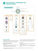
Figure 22.2. Life cycle of Trypanosoma cruzi. T. cruzi possesses four basic
life cycle stages. In the insect, noninfectious epimastigotes replicate (R) in the
midgut and differentiate into infectious but nonreplicating (NR) metacyclics
as they migrate to hindgut. The fecal material of the insect, which contains
metacyclics is deposited on the skin during a bloodmeal and infection occurs
when this material contaminates the insect bite or a mucous membrane, which
the trypomastigotes can penetrate. Within the human host, metacyclics invade
host cells and differentiate into amastigotes, which replicate, burst out of the
cell and either invade other cells or are taken up by another insect. Within
the insect gut, the trypomastigotes differentiate into replicating epimastigotes,
thus completing the cycle.
Pathogenesis of Chagas Disease
Acute T. cruzi infection results from the contamination of wounds or
mucous membranes with insect feces containing expelled infective parasites.
Locally deposited parasites bind to and invade host tissue and transform into
and replicate as intracellular amastigotes. Infection leads to the formation of
parasite “pseudocysts,” so named because the amastigote nests are intracellular.
This stimulates a localized inflammatory response mediated predominantly by
lymphocytes and macrophages. Lymphatic drainage of the infected area into
regional lymph nodes results in activation and proliferation of cells, resulting
in regional lymphadenopathy. As the process continues, the amas transform
into trypomastigotes, escape host cells and disseminate throughout the body.
Infection and lysis of liver cells results in transient increases in serum liver
enzyme levels. In chronic infection, tissue parasites are difficult to detect but
significant interstitial fibrosis occurs, damaging the affected tissue. The molecular
American Trypanosomiasis (Chagas Disease)
157
pathogenesis of Chagas disease is not completely understood, but likely results
from (i) parasite-mediated tissue destruction, (ii) inflammation and fibrosis
resulting from immune responses generated to parasites and residual parasite
antigen, (iii) parasite-induced microvascular spasm and ischemic damage and/
or (iv) autoimmune responses triggered by release of self-antigen during parasite
lysis of host cells. Because there are many outcomes of chronic T. cruzi infection (see below), it is likely that each of these mechanisms occurs in isolation
or in combination in a given individual, depending on the specific pathogenic
potential of the strain of parasite (tissue tropism, replication rate, etc.) and the
immunogenetic susceptibility of the infected individual.
Clinical Syndromes of Chagas Disease
Acute infection by T. cruzi is marked by the development of localized swelling
and erythema at the site of the insect bite, which is termed a chagoma. This is a
result of the local replication of parasites and the influx of fluid and inflammatory cells into the infected area. Infection through the conjunctiva can result in
periorbital swelling, termed Romaña’s sign (Fig. 22.3D). As parasites disseminate
patients experience nonspecific symptoms such as fever, malaise and anorexia.
Parasite infestation of peripheral tissues can give rise to hepatosplenomegaly and,
in some cases, meningeal signs. Initial infection of heart tissue can lead to acute
myocarditis and cardiac sudden death due to parasitization of the cardiac conduction system. The signs and symptoms of acute T. cruzi infection can last from days
to weeks but are often unrecognized due to their nonspecific nature. The disease
then proceeds to a quiescent phase lasting months to years and often decades,
prior to the onset of chronic disease. It should be noted that the majority of T.
cruzi-infected individuals do not develop any parasite-related disease and simply
harbor low levels of parasites for life. Less than one-third of infected people develop
chronic Chagas disease. The two hallmarks, usually mutually exclusive, disorders
22
that occur in chronically infected patients are cardiomyopathy and megaorgan
syndromes (Fig. 22.3E and G. respectively).
Cardiac involvement is heralded by the development of fibrosis within the
heart muscle (Fig. 22.3F) and conduction system which leads to arrhythmias
and heart failure, that latter being predominantly right-sided. Loss of ventricular
muscle leads to wall thinning which can be associated with the development of
apical aneurysms and subsequent formation of thrombi, which may have serious thromboembolic consequences (Fig. 22.3E). In the gastrointestinal tract,
chronic infection leads to parasympathetic denervation, resulting in massive
dilatation of the esophagus and/or colon. Esophageal involvement results in
achalasia, associated odynophagia, dysphagia and esophageal dysmotility, often
resulting in aspiration pneumonia. Colonic involvement results in abdominal
pain, constipation, obstruction with perforation and secondary intrabdominal
infection. Immunosuppression of patients with chronic Chagas disease, regardless of the mechanism (HIV infection, usage of immunosuppressive drugs in
organ transplantation) can lead to recrudescence of parasite replication, massive
parasitosis and death. Clinical disease in this setting is often fulminant with more
extensive involvement of the central nervous system.
22
158
Medical Parasitology
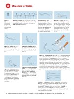
Figure 22.3. Various aspects of Trypanosoma cruzi biology and Chagas disease. A) T. cruzi trypomastigotes stained with Giemsa
(bar = 5 μm). Note the prominent darkly-stained kinetoplast DNA. B) Reduviid bug. C) Nests of amastigotes in heart tissue, often termed
“pseudocysts” since they are intracellular collections of parasites. The inset shows an amastigote with clearly visible nucleus (round
structure) and kinetoplast (bar-like structure). D) Romaña’s sign. E) Apical aneurysm (illuminated by light bulb) can occur after chronic
fibrosis and weakening of the apical wall of left ventricle. F) Histopathology of Chagas heart disease: myofibrillar swelling and degeneration, mononuclear cell infiltration, fibrosis and edema in the absence of parasites are typical. G) Megacolon: a serious sequela of
infection that is poorly understood. Photographs are courtesy of Cheryl Olson (A,C,F), Dr. Chris Beard (B), Dr. Michael Miles (D), Prof.
F. Köberle (E,F,G).
American Trypanosomiasis (Chagas Disease)
159
Diagnosis of T. cruzi Infection
The diagnosis of T. cruzi infection initially requires a high degree of clinical
suspicion. History of potential exposure to T. cruzi is important to document.
Patients with a history of travel to or having had blood transfusion within endemic
areas are at increased risk of T. cruzi infection. The presence of, or recent history
of a chagoma or Romaña’s sign are indicators of recent infection. The mainstay
of diagnosis is detection of trypomastigotes in the blood or the presence of T.
cruzi-specific antibodies in serum to indicate acute or chronic infection, respectively. Direct detection of parasites in blood is easier in immunocompromised
patients in whom the immunologic control of parasites is not as efficient. Heavy
parasite burdens in the tissues of such patients can permit diagnosis via direct
examination of tissue (lymph nodes, or bone marrow) or fluids (cerebrospinal or
pericardial fluid). In addition these specimens can be cultured in vitro in liquid
medium or by growth within uninfected insect vectors (xenodiagnosis). During
chronic infection parasites are frequently not detectable in the blood, and the
presence of T. cruzi IgG, using commercial immunoassays, ELISA, complement
fixation, or hemagglutination based tests, establishes the diagnosis. Direct detection of parasites using PCR based testing has been demonstrated but is not yet
available for routine laboratory diagnosis. Potential blood donors throughout
the Americas are asked questions related to risk factors of T. cruzi infection, but
transfusion-associated disease remains a serious problem. As a result, the blood
in much of South and Central America is screened for T. cruzi-specific antibodies, and many feel that the United States blood supply will be screened beginning
within a few years.
Treatment of T. cruzi Infection
Benznidazole, an imidazole (trade name Rochagan, produced by Roche in
Brazil) and Nifurtimox, a nitrofuran (trade name Lampit, produced by Bayer 22
in Germany), are the two agents approved for treatment of Chagas disease and
are available in the United States through contact with the Centers for Disease
Control in Atlanta, Georgia. These agents have similar efficacy but have many
adverse effects. Benznidazole is given orally for 1-3 months at a dose of 5-7
mg/kg/d in two divided doses. The side effects of this medication include rash and
peripheral neuropathy but can also include bone marrow suppression. In adults,
Nifurtimox is given for 120 days at a dose of 8-10 mg/kg/d in four divided doses.
In children the drug is given for 90 days in four divided doses but the amount
is based on age: 11-16 years (12.5-15 mg/kg/d); and under 11 years (15-20
mg/kg/d). Gastrointestinal maladies (nausea, vomiting, abdominal pain) are the
predominant side effects of this medication but up to 30% of patients can also
experience central nervous system effects such as polyneuritis, confusion or focal
or generalized seizures. Skin rash can also develop in some patients. Individuals
with glucose-6-phosphate dehydrogenase deficiency can experience drug-induced
hemolytic anemia. Treatment is undertaken in cases of acute or congenital infection
natural infection or in cases of accidental laboratory inoculation. Recent systematic
reviews of clinical trials of trypanocidal therapy in patients with chronic T. cruzi
infection suggest that treatment of asymptomatic immunocompetent patients may
result in a reduction of progression to chronic disease (development of megaorgan
Medical Parasitology
160
syndromes, cardiomyopathy and arrhythmia). In contrast, there is no convincing
data that support the use of trypanocidal therapy in patients who have already
manifested end-organ damage as a result of chronic T. cruzi infection. It is clear
that randomized controlled trials are necessary to truly understand the clinical
benefit of trypanocidal therapy in chronic Chagas disease. The management of T.
cruzi induced cardiac failure, achalasia and megacolon are approached in the same
way that these end-organ problems are approached due to other causes.
Prevention of T. cruzi Infection
Limiting exposure to T. cruzi infected insects and blood is the mainstay of
the prevention of Chagas disease. Persons living in or traveling to areas endemic
for T. cruzi should avoid residing in substandard housing frequented by reduviid
bugs. The use of bed nets and insect repellent are also recommended for this purpose. Barrier protection for those working with T. cruzi in the laboratory setting,
such as protective clothing, gloves and eyewear is a must. Since the incidence of
transfusion- and transplantation-associated T. cruzi infection is increasing in the
Americas, serologic screening of donated blood seems advisable. Such is the practice
in endemic countries within South America. As the number of potentially-infected
immigrants to the United States increases, this will likely increase the number
of transfusion-associated T. cruzi infections despite the presence of blood bank
questionnaires.
Suggested Reading
22
1. Engman DM, Leon JS. Pathogenesis of Chagas heart disease: role of autoimmunity.
Acta Trop 2002; 81:123-32.
2. Kirchhoff LV. Trypanosoma Species (American Trypanosomiasis, Chagas
Disease): Biology of Trypanosomes. In: Mandell GL, Douglas RG, Bennett
JE, eds. Principles and Practice of Infectious Diseases, 6th Edition. New York:
Churchill Livingstone, 2005:
3. Mascola L, Kubak B, Radhakrishna S et al. Chagas disease after organ transplantation—Los Angeles, California. MMWR Morb Mortal Wkly Rep 2006;
55:789-800.
4. Villar JC, Marin-Neto JA, Ebrahim S et al. Trypanocidal drugs for chronic
asymptomatic Trypanosoma cruzi infection. Cochrane Database Syst Rev 2002;
CD003463.
5. Tyler KM, Miles MA. American Trypanosomiasis. Norwell: Kluwer Academic
Publishers, 2003.
CHAPTER 23
African Trypanosomiasis
Guy Caljon, Patrick De Baetselier and Stefan Magez
Abstract
African trypanosomiasis is a vector-born disease that severely affects a broad
range of vertebrate hosts, including humans, on the sub-Saharan African continent.
The infection is caused by flagellated unicellular parasites (Trypanosoma sp.) and is
lethal without treatment. Disease manifestations are pleotropic and are dependent
on the host and infection-stage. Currently available diagnostic tests are adapted
for field usage but have a low specificity, while an accurate differential diagnosis
of human pathogenic Trypanosoma subspecies and correct determination of the
infection stage is essential for appropriate treatment. For treatment of human
African trypanosomiasis (HAT), four drugs with significant side-effects are currently available, with only one of them being registered in the last 50 years. This
chapter will introduce the disease, its diagnosis, treatment and prospects for new
therapeutic approaches.
Introduction
African trypanosomes are extracellular protozoan parasites that cause lethal
infections in humans and livestock in large parts of sub-Saharan Africa. The responsible flagellated parasite (Trypanosoma sp.) is approximately twice the size of
erythrocytes (15-30 μm, Fig. 23.1A) and relies on tsetse flies for its transmission
(Fig. 23.1B). These arthropods are obligate bloodsucking insects (genus Glossina),
that get infected through feeding on a parasitized host and accommodate the
trypanosome during their entire lifespan. Engorged trypanosomes colonize the
midgut, proliferate and undergo differentiation while directionally migrating
towards the insect salivary glands. The vertebrate-infective metacyclic form of the
parasite resides in the salivary glands or mouthparts of the fly, using the bloodfeeding behaviour for its transmission to a new host. Upon transmission to the vertebrate host, trypanosomes will transform into actively proliferating (long slender)
forms to allow a systemic colonization of the host. Eventually, trypanosomes in
the bloodstream become quiescent (short stumpy) and pre-adapt to uptake and
subsequent survival in the tsetse fly. During the complex life cycle (Fig. 23.1C)
of the parasite in the insect and vertebrate host, trypanosomes undergo several
metabolic changes for the acquisition of free-energy from different available sources
and modify mechanisms for the uptake of host nutrients, such as iron complexed
with transferrin. In the fly, trypanosomes utilize amino acids (e.g., proline) as
primary energy sources while trypanosomes in the vertebrate hosts metabolize
glucose via glycolysis in a unique organelle, the glycosome. Only two subspecies,
Medical Parasitology, edited by Abhay R. Satoskar, Gary L. Simon, Peter J. Hotez
and Moriya Tsuji. ©2009 Landes Bioscience.