Antioxidant studies and GCMS analysis of the phytochemical compounds of some endangered plant species collected from the western ghats
Bạn đang xem bản rút gọn của tài liệu. Xem và tải ngay bản đầy đủ của tài liệu tại đây (625.97 KB, 17 trang )
Int.J.Curr.Microbiol.App.Sci (2019) 8(6): 3338-3354
International Journal of Current Microbiology and Applied Sciences
ISSN: 2319-7706 Volume 8 Number 06 (2019)
Journal homepage:
Original Research Article
/>
Antioxidant Studies and GCMS Analysis of the Phytochemical Compounds
of Some Endangered Plant Species Collected from the Western Ghats
N. Sumangala1*, M. Jayaramu2 and M.P. Prasad3
1
2
Microbiology, Tumkur University, Karnataka, India
Department of Studies and Research in Environmental Sciences, Tumkur University,
Karnataka, India
3
Sangene Biotech, Bengaluru, Karnataka, India
*Corresponding author
ABSTRACT
Keywords
Antioxidant, DPPH,
Metal ion, Hydroxyl
radical, Superoxide
anion radical
Article Info
Accepted:
26 May 2019
Available Online:
10 June 2019
Plant has an innate ability to produce non-enzymatic antioxidants which have an important
role in the metabolism of Reactive oxygen species (ROS). Several plants serve as the
source of therapeutic agents but the properties depend on the plant nature. Thus, an
evaluation of antioxidant activity is essential to determine the importance of a plant. Three
plants Utleria salicifolia, Plectranthus vettiveroides and Nothapodytes nimmoniana were
selected to carry out the antioxidant study. Methanol assisted leaves extracts were prepared
and subjected to antioxidant assay by means of DPPH radical scavenging assay, Metal ion
Chelating Assay, Superoxide Anion Radical Scavenging Assay and Hydroxyl radical
scavenging assay. All the extracts showed 20-80% inhibition depending on the
concentration of extracts and the type of assay as well. The existence of crucial organic
compounds in the leaf extracts of all the three plants were corroborated by Gas
chromatography analysis. The present results offer supporting evidence for effective use of
selected plant extracts.
Introduction
Free radicals play a crucial role in the
development of tissue damage in pathological
events. Antioxidants are chemical compounds
which have the ability to quench the free
radicals and thereby it prevents the human
body against various diseases. Plants are the
rich sources of antioxidants which contain
secondary metabolites such as phenolic and
flavonoid compounds commonly which act as
antioxidants with redox and metal chelating
properties
(Karimi
and
Jaafar, 2011).
Antioxidants are characterized as free radical
which has an essential role to develop the
damaged tissue in pathological field.
Medicinal plants have been investigated from
long time to evaluate their antioxidant
properties. Natural antioxidants have potential
to interrupt the destruction which is resulted
from oxidative stress. These antioxidants may
be either natural extracts or as an essential
chemical compound of the extract (Zengin et
al., 2011). Though medicinal plants have been
3338
Int.J.Curr.Microbiol.App.Sci (2019) 8(6): 3338-3354
carefully assessed for their toxicity profile,
still the plant derived medicines are safer as
compared to synthetic medicines (Vongtau et
al., 2005; Oluyemi et al., 2007). The ROS and
other oxidant result in disease and disorders
as proved by different evidence. The evidence
has brought the attention of scientists to an
appreciation of antioxidants for prevention
and treatment of diseases, and maintenance of
human health (Halliwell et al., 1981). Human
body has an inherent antioxidative mechanism
and many of the biological functions such as
the anti-mutagenic, anti-carcinogenic, and
anti-aging responses originate from this
property (Gulcin et al., 2012; Gocer et al.,
2011). Antioxidants stabilize or deactivate
free radicals, often before they attack targets
in biological cells (Nunes et al., 2012)
Recently interest in naturally occurring
antioxidants has considerably increased for
use in food, cosmetic and pharmaceutical
products,
because
they
possess
multifacetedness in their multitude and
magnitude of activity and provide enormous
scope in correcting imbalance (Djeridane et
al., 2006; Wannes et al., 2010).
It is well known that free radical reaction is
actively involved in disease pathology
resulting in several chronic and acute disease
in human such as neurodegeneration,
atherosclerosis, immunosuppression, aging
and diabetes (Harman et al., 1998). If the
balance between inherent antioxidant capacity
of the body and ROS is disrupted then
medicinal supplements and dietary are
provided during attacked by disease. Several
researches on vegetables, herbal plants and
fruits specified the presence of antioxidants
including
flavonoids,
phenolics,
proanthocyanidins and tannins. Antioxidant
from medicinal plants offers quite well
protection against disease.
The ingestion of natural antioxidants has been
inversely associated with morbidity and
mortality from degenerative disorders (Gulcin
et al., 2012). Liver diseases remain a serious
health problem. Free radicals result in the
damage of cell by covalent binding as well as
lipid peroxidation. This further causes the
injury to tissue. Antioxidant agents of natural
origin have attracted special interest because
of their free radical scavenging abilities
(Osawa et al., 1990). The use of medicinal
plants with high level of antioxidant
constituents has been proposed as an effective
therapeutic approach for hepatic damages
(Govind et al., 2011).
Reactive oxygen species (ROS) and Reactive
Nitrogen Species (RNS) are the products of
normal cellular metabolism recognized for
playing the either harmful or beneficial effect
in living system. Increase in concentration of
free radicals or decreased endogenous
antioxidant mechanism can lead to oxidative
stress which is responsible for the
development of many degenerative diseases
(Saikat et al., 2014).
GC-MS is an important technique to analyze
the plant extract in order to determine the
presence of essential herb compound which
are often used in pharmaceutical, drug,
cosmetic or food industry, environmental and
forensic applications (Uma et al., 2009). This
technique is the combination of two separate
analytical methods to separate and determine
the chemical components of a given mixture.
Separation is done by Gas Chromatography
whereas the components analysis is carried
out by mass spectroscopy. Chemical studies
have shown that it mainly contains
cardenolides, pregnane glycosides and
volatile components. Maximum volatile
components belong to the class of long chain
unsaturated fatty acids. These are the building
elements of several valuable compounds and
also an essential energy source. Due to these
features, the volatile compounds play vital
role in the biological system (Mu et
3339
Int.J.Curr.Microbiol.App.Sci (2019) 8(6): 3338-3354
al., 2001). In recent years, increasing research
has been carried out on fatty acids and the
results obtained show that they possess
significant sedative and hypnotic effects
(Zhang et al., 1995).
In-vitro Antioxidant Assay
The antioxidant activity of the leaves extracts
was carried out following four protocols.
DPPH radical scavenging assay method
The current study was conducted to prepare
the methanol extract of Utleria salicifolia,
Plectranthus vettiveroides and Nothapodytes
nimmoniana leaves. The extracts were
assessed for their antioxidant activity. The
content of the extracts were determined by
Gas Chromatography - Mass Spectrometry
analysis.
Materials and Methods
Preparation of plant extract
The plant samples were collected from the
following locations, Utleri salicifolia,
Curcuma
zeodatia,
Nothapodytes
nimmoniana from the Western Ghats of
Kerala. Plectranthus vettiveroides from
Tamilnadu. Cayratia pedate from the Western
Ghats bordering Kerala and Tamilnadu and
Karnataka states. Rhaphidophora persuta
from the Western Ghats of Karnataka and
Syzygium
travancoricum
from
fresh
water Myristica swamps of Kerala and Uttar
Kannada district of Karnataka.
Utleria salicifolia, Plectranthus vettiveroides
and Nothapodytes nimmoniana leaves were
selected to study antioxidant activity and GCMS analysis. The collected leaves were
washed thoroughly with tap water followed
by distilled water several times in order to
remove the dust and soil particles.
The leaves were then shade dried and used for
extraction. 100 gm of all the three plant leaves
powder were treated with methanol and
extracted using soxhlet apparatus. The extract
thus obtained was concentrated by
evaporation in rotary vacuum evaporator.
2.8 ml of leaves extract (20-100 µg/ ml) was
mixed with 200 µL of DPPH (100 µM in
methanol) and incubated for 20 min in dark
condition. Absorbance was taken at 517 nm.
A mixture of DPPH and methanol was used
as control. Ascorbic acid was taken as
reference standard. Percentage of DPPH
inhibition was determined according to Prasad
(2015).
(Absorbance of control – Absorbance of test)
× 100
Inhibition (%) = ---------------------------...Eq 1
Absorbance of control
Metal ion chelating assay
This assay was carried out by determining the
chelating potential of Fe ion present in the
extract. 2,2’-bipyridyl competition assay was
conducted by mixing 0.25mL(1mM) FeSO4
solution to the equal volume of concentrated
extract (200-1000 µg/ml). To this mixture
1mL Tris HCl buffer (pH 7.4) and 0.25mL
(0.1%) 2,2’-bipyridyl solution were added
along with 0.4mL hydroxylaminehydro
chloride and 2.5mL ethanol. Final volume of
the solution was adjusted to 5 ml by distilled
water. The resulting solution was incubated at
room temperature for 10 minutes. The
absorbance was taken at 522 nm with EDTA
as reference chelating agent. The Fe2+
chelating activity of the extract was
determined as per the following equation.
(Absorbance of control – Absorbance of test)
× 100
Inhibition (%) = ---------------------------...Eq 2
Absorbance of control
3340
Int.J.Curr.Microbiol.App.Sci (2019) 8(6): 3338-3354
Superoxide anion radical scavenging assay
NBT reduction method was adopted to assess
superoxide anion radical scavenging activity.
0.1 ml concentrated plant extract (200-1000
µg/ml) was mixed with 1mL NBT (in
phosphate buffer pH 7.4) and 1mL of NADH
solution. 100 µL (60 µM) PMS was added to
initiate the reaction and the reaction mixture
was incubated for 15 min at 30°C. The
absorbance was measured at 560 nm with
ascorbic acid as reference standard. The
inhibition percentage was calculated by the
following equation.
of 250°C with split mode of injection and
liner velocity flow control. The pressure
applied for GC is 57.4kpa which gives the
column flow of 1.00ml/min and linear
velocity of 36.5 cm/sec, with a purge flow of
3.0 ml/min and split ratio is 10.0. The ion
source temperature was set at 200°C and the
interface temperature is 300°C, with 2.00 min
of solvent cut time. The Mass Spectra was
taken with intervals of 0.50 sec, with a scan
range of 40-600 m/z with a scan speed of
1250. The total time taken is 34.00 min and
FTD detector is used for detection.
Results and Discussion
(Absorbance of control – Absorbance of test)
× 100
Inhibition (%) = ---------------------------...Eq 3
Absorbance of control
Antioxidant activity
nimmoniana extract
Hydroxyl radical scavenging assay
Hydroxyl radical scavenging activity of the
plant extract was determined using 2-deoxy2- 3+ ribose oxidative degradation in Fe EDTA- 15 Ascorbate-H O system method.
3.5 ml leaves extract was mixed with 28 mM
2-deoxy-2-ribose, 1.04 mM EDTA and 1 mM
ascorbic acid. The resulting solution was
incubated for 1 hr at 37°C. The preventive
effects of extract on deoxyribose damage,
imposed by hydroxyl radicals were
determined spectrophotometrically at 532 nm
against blank for each concentration.
Mannitol was taken as the reference. The
inhibition percentage was calculated as:
(Absorbance of control – Absorbance of test)
× 100
Inhibition (%) = ---------------------------...Eq 4
Absorbance of control
GC-MS analysis of the leaf’s extracts
The GC-MS was run with a column oven
temperature of 60°c and injection temperature
of
Nothapodytes
Table 1 and Figure 1 exhibited the DPPH
radical
scavenging
capabilities
of
Nothapodytes nimmoniana leaf and ascorbic
acid as well. As a standard ascorbic acid
showed higher inhibition percentage as
compared to leaves extract.
Inhibition percentage enhances with an
increase in leaf extract concentration and a
maximum 75% inhibition was observed at
100 µg/ml leaf extract concentration. For
ascorbic acid inhibition became constant from
60 to 100 µg/ml concentration.
Metal ion chelating activity of leaf extract
was compared to EDTA in Table 2 and Figure
2. Inhibition became constant at 35% at leaf
extract concentration of 60 to 100% whereas
inhibition increases with increase in EDTA
content.
Assessment of Superoxide radical scavenging
of leaf extract was depicted in Table 3 and
Figure 3. Maximum 30% inhibition was
observed at 60 µg/ml leaf extract whereas
45% inhibition was obtained at 60 µg/ml
ascorbic acid content.
3341
Int.J.Curr.Microbiol.App.Sci (2019) 8(6): 3338-3354
Hydroxyl radical scavenging assessment of
leaf extract was exhibited in table 4 and
Figure 4. Maximum 25% inhibition was
achieved at 60 µg/ml leaf extract. Further
increase in extract concentration did not affect
the inhibition percentage. 60 µg/ml mannitol
showed 45% inhibition which was the
maximum.
higher inhibition percentage as compared to
leaves extract. Inhibition percentage enhances
with an increase in leaf extract concentration
up to 80 µg/ml and a maximum 45%
inhibition was observed at this concentration.
Antioxidant activity of Utleria salicifolia
extract
Metal ion chelating activity of leaf extract
was compared to EDTA in Table 6 and Figure
6. Inhibition became constant at 50% at leaf
extract concentration of 80 to 100% whereas
inhibition increases with increase in EDTA
content.
The study carried out on the antioxidant
activity of the methanol extract from the
leaves of Utleria salicifolia using DPPH
radical, metal chelating, hydroxyl and super
oxide radical scavenging assays was
described.
Table 5 and Figure 5 exhibits the DPPH
radical scavenging capabilities of Utleria
salicifolia leaf extract and ascorbic acid as
well. As a standard ascorbic acid showed
For ascorbic acid inhibition became constant
from 60 to 100 µg/ml concentration.
Assessment of Superoxide radical scavenging
of leaf extract was depicted in Table 7 and
Figure 7. Maximum 30% inhibition was
observed at 60 µg/ml leaf extract whereas
45% inhibition was obtained at 60 µg/ml
ascorbic acid content.
Table.1 Variation of inhibition percentage with respect to the concentration of leaf extract and
ascorbic acid
Concentration of leaf
extract (µg/ml)
20
40
60
80
100
% of
inhibitions
40
50
60
70
75
Concentration of
ascorbic acid (µg/ml)
20
40
60
80
100
% of
inhibitions
60
80
85
85
85
Table.2 Metal ion chelation activity Assay of methanol extract of Nothapodytes nimmoniana
leaves and standard EDTA
Concentration of leaf
extract (µg/ml)
20
40
60
80
100
% of
inhibitions
20
30
35
35
35
3342
Concentration of EDTA
(µg/ml)
20
40
60
80
100
% of
inhibitions
50
55
60
65
70
Int.J.Curr.Microbiol.App.Sci (2019) 8(6): 3338-3354
Table.3 Superoxide radical scavenging assay of methanol extract of Nothapodytes nimmoniana
leaves and standard Ascorbic acid
Concentration of leaf
extract (µg/ml)
20
40
60
80
100
% of
inhibitions
20
25
30
30
30
Concentration of
Ascorbic acid (µg/ml)
20
40
60
80
100
% of
inhibitions
30
40
45
45
45
Table.4 Hydroxyl radical scavenging assay of methanol extract of Nothapodytes nimmoniana
leaves and standard Mannitol
Concentration of leaf
extract (µg/ml)
20
40
60
80
100
% of
inhibitions
10
20
25
25
25
Concentration of
Mannitol (µg/ml)
20
40
60
80
100
% of
inhibitions
30
40
45
45
45
Table.5 DPPH radical scavenging capabilities of methanol extract of Utleria salicifolia leaves
and standard ascorbic acid
Concentration of leaf
extract (µm/ml)
20
40
60
80
100
% of
inhibitions
20
30
40
45
45
Concentration of
Ascorbic acid (µm/ml)
20
40
60
80
100
% of
inhibitions
60
80
85
85
85
Table.6 Metal ion chelation activity Assay of methanol extract of Utleria salicifolia leaves and
standard EDTA
Concentration of leaf
extract (µm/ml)
20
40
60
80
100
% of
inhibitions
25
30
40
50
50
3343
Concentration of EDTA
(µm/ml)
20
40
60
80
100
% of
inhibitions
50
55
60
65
70
Int.J.Curr.Microbiol.App.Sci (2019) 8(6): 3338-3354
Table.7 Superoxide radical scavenging assay of methanol extract of Utleria salicifolia leaves
and standard Ascorbic acid
Concentration of leaf
extract (µm/ml)
20
40
60
80
100
% of
inhibitions
20
25
30
30
30
Concentration of
Ascorbic acid (µm/ml)
20
40
60
80
100
% of
inhibitions
30
40
45
45
45
Table.8 Hydroxyl radical scavenging assay of methanol extract of Utleria salicifolia leaves and
standard Mannitol
Concentration of leaf
extract (µm/ml)
20
40
60
80
100
% of
inhibitions
25
35
35
35
35
Concentration of
Mannitol (µm/ml)
20
40
60
80
100
% of
inhibitions
30
40
45
45
45
Table.9 DPPH radical scavenging assay of methanol extract of Plectranthus vettiveroides leaves
and standard Ascorbic acid
Concentration of leaf
extract (µm/ml)
20
40
60
80
100
% of
inhibitions
50
60
70
80
85
Concentration of
Ascorbic acid (µm/ml)
20
40
60
80
100
% of
inhibitions
60
80
85
85
85
Table.10 Metal ion chelation activity Assay of methanol extract of Plectranthus vettiveroides
leaves and standard EDTA
Concentration of leaf
extract (µm/ml)
20
40
60
80
100
% of
inhibitions
25
25
25
25
25
3344
Concentration of EDTA
(µm/ml)
20
40
60
80
100
% of
inhibitions
50
55
60
65
70
Int.J.Curr.Microbiol.App.Sci (2019) 8(6): 3338-3354
Table.11 Superoxide radical scavenging assay of methanol extract of Plectranthus vettiveroides
leaves and standard Ascorbic acid
Concentration of leaf
extract (µm/ml)
20
40
60
80
100
% of
inhibitions
30
40
40
40
40
Concentration of
Ascorbic acid (µm/ml)
20
40
60
80
100
% of
inhibitions
30
40
45
45
45
Table.12 Hydroxyl radical scavenging assay of methanol extract of Plectranthus vettiveroides
leaves and standard Mannitol
Concentration of leaf
extract (µm/ml)
20
40
60
80
100
% of
inhibitions
10
20
20
20
20
Concentration of
Mannitol (µm/ml)
20
40
60
80
100
% of
inhibitions
30
40
45
45
45
Fig.1 DPPH radical scavenging capabilities of methanol extract of Nothapodytes nimmoniana
leaves and standard ascorbic acid
3345
Int.J.Curr.Microbiol.App.Sci (2019) 8(6): 3338-3354
Fig.2 Metal ion chelating capabilities of methanol extract of Nothapodytes nimmoniana leaves
and EDTA
Fig.3 Superoxide ion radical scavenging capabilities of methanol extract of Nothapodytes
nimmoniana leaves and Ascorbic acid.
Fig.4 Hydroxyl radical scavenging capabilities of methanol extract of Nothapodytes nimmoniana
leaves and Mannitol
3346
Int.J.Curr.Microbiol.App.Sci (2019) 8(6): 3338-3354
Fig.5 DPPH radical scavenging capabilities of methanol extract of Utleria salicifolia leaves and
standard ascorbic acid
Fig.6 Metal ion chelating capabilities of methanol extract of Utleria salicifolia leaves and EDTA
Fig.7 Superoxide radical scavenging capabilities of methanol extract of Utleria salicifolia leaves
and Ascorbic acid
3347
Int.J.Curr.Microbiol.App.Sci (2019) 8(6): 3338-3354
Fig.8 Hydroxyl radical scavenging capabilities of methanol extract of Utleria salicifolia leaves
and Mannitol
Fig.9 Gas chromatography of Utleria salicifolia leaf extract
3348
Int.J.Curr.Microbiol.App.Sci (2019) 8(6): 3338-3354
Fig.10 DPPH radical scavenging capabilities of methanol extract of Plectranthus vettiveroides
leaves and standard Ascorbic acid
Fig.11 Metal ion chelation capabilities of methanol extract of Plectranthus vettiveroides leaves
and EDTA
3349
Int.J.Curr.Microbiol.App.Sci (2019) 8(6): 3338-3354
Fig.12 Superoxide radical scavenging capabilities of methanol extract of Plectranthus
vettiveroides leaves and standard Ascorbic acid
Fig.13 Hydroxyl radical scavenging capabilities of methanol extract of Plectranthus
vettiveroides leaves and Mannitol
3350
Int.J.Curr.Microbiol.App.Sci (2019) 8(6): 3338-3354
Fig.14 Gas chromatography of Plectranthus vettiveroides leaf extract
3351
Int.J.Curr.Microbiol.App.Sci (2019) 8(6): 3338-3354
Hydroxyl radical scavenging assessment of
leaf extract was exhibited in table 8 and
Figure 8. Maximum 35% inhibition was
achieved at 40 µg/ml leaf extract. Further
increase in extract concentration did not affect
the inhibition percentage. 60 µg/ml mannitol
showed 45% inhibition which was the
maximum.
GC-MS
extract
analysis
of
Utleria
salicifolia
Figure 9 exhibits the chromatogram obtained
from GC-MS analysis of Utleria salicifolia
extract. Presence of essential organic
component was observed while the obtained
peaks were analyzed.
described. Table 9 and Figure 10 exhibited
the DPPH radical scavenging capabilities of
Plectranthus vettiveroides leaf extract and
ascorbic acid as well. As a standard ascorbic
acid showed higher inhibition percentage as
compared to leaves extract. Inhibition
percentage enhances with an increase in leaf
extract concentration to 100 µg/ml and a
maximum 85% inhibition was observed at
this concentration. For ascorbic acid
inhibition became constant at 85% from 60 to
100 µg/ml concentration.
Plectranthus
Metal ion chelating activity of leaf extract
was compared to EDTA in Table 10 and
Figure 11. There is no change of inhibition
with respect to leaf extract concentration
whereas inhibition increases with increase in
EDTA content.
The study carried out on the antioxidant
activity of the methanol extract from the
leaves of Plectranthus vettiveroides using
DPPH radical, metal chelating, hydroxyl and
super oxide radical scavenging assays is
Assessment of Superoxide radical scavenging
of leaf extract is depicted in Table 11 and
Figure 12. Maximum 40% inhibition was
observed at 40 µg/ml leaf extract whereas a
constant 45% inhibition was obtained at 60
µg/ml ascorbic acid content.
Antioxidant activity
vettiveroides extract
of
3352
Int.J.Curr.Microbiol.App.Sci (2019) 8(6): 3338-3354
Hydroxyl radical scavenging assessment of
leaf extract is exhibited in table 12 and Figure
13. Maximum 20% inhibition was achieved at
40 µg/ml leaf extract. Further increase in
extract concentration did not affect the
inhibition percentage. 60 µg/ml mannitol
showed 45% inhibition which was the
maximum.
GC-MS
analysis
vettiveroides extract
of
Plectranthus
Figure 14 exhibits the Chromatogram
obtained from the GC-MS analysis of
Plectranthus vettiveroides leaf extract.
Presence of essential organic component was
observed while the obtained peaks were
analyzed.
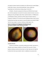
It is concluded in the current study better
activity of all the three plant species was
recorded. This was due to efficient extraction
of phytochemicals. The extracts from three
different plant leaves showed antioxidant
activity which was substantiated by four
different assay methods. Further Gas
Chromatography analysis verified the
presence of important compound in the plant
extracts. Further study can be carried out on
isolating a specific bioactive compound with
commercial value. Being a rich source of
antioxidant, these medicinally important
plants have values as functional ingredients in
food industry.
References
Djeridane, A., Yousfi, M., Nadjemi, B.,
Boutassouna, D., Stocker, P., Vidal, N.
2006. Antioxidant activity of some
Algerian medicinal plant extracts
containing phenolic compounds. Food
Chem. 97, 654-60.
Gocer, H., Gulcin, I., 2011. Caffeic acid
phenethyl ester (CAPE): correlation of
structure and antioxidant properties. Int
J Food Sci Nutr. 62, 821-5.
Gulcin, I., 2012. Antioxidant activity of food
constituents: an overview. Arch
Toxicol. 86, 345-91.
Halliwell, B., Gutteridge, J.M.C., 1981.
Formation of thiobarbituric acid
reactive substances from deoxyribose in
the presence of iron salts: the role of
superoxide and hydroxyl radicals. FEBS
Lett. 128, 347-52.
Harman D: Free radical theory of aging.
Current status. 1998, Amster-dam:
Elsevier, p. 3-7
Ilavarasan, R., Mallika, M., Venkataraman, S.
2005,
Anti-inflammation
and
antioxidant activities of Cassia fistula
Linn. bark extracts. Afr J Trad Compl
Altern Med. 2, 70-85.
Karimi, E., Jaafar, H.Z.E. 2011, HPLC and
GC-MS determination of bioactive
compounds in microwave obtained
extracts of three varieties of Labisia
pumila benth. Molecules. 16, 6791–805.
Mu, Y.M., Yanass, T., et al., 2001. Saturated
FFAs, palmitic acid and stearic acid,
induced apoptosis in human granulose
cells. J Endocrinol. 142, 3590–97.
Nunes, P.X., Silva, S.F., Guedes, R.J.,
Almeida, S. 2012. Biological oxidations
and antioxidant activity of natural
products,
Phytochemicals
as
nutraceuticals - Global Approaches to
Their Role in Nutrition and Health.
Oluyemi, K.A., Okwuonu. U.C., Baxter,
D.G., Oyesola, T.O. 2007, Toxic effects
of methanolic extract of Aspilia
africana leaf on the estrous cycle and
uterine tissues of Wistar rats. Int J
Morphol. 25, 609-14.
Prasad, M.P., 2014. Invitro Phytochemical
Analysis and Anti-oxidant studies of
Hibiscus species. International Journal
of Pure and Applied Biosciences. 2(3),
83-8.
Ruch. R.J., Cheng. S-J, Klaunig, J.E. 1989
Prevention
of
cytotoxicity
and
3353
Int.J.Curr.Microbiol.App.Sci (2019) 8(6): 3338-3354
inhibition
of
intercellular
communication by antioxidant catechins
isolated from Chinese green tea.
Carcinogenesis. 10, 1003-8.
Uma, B., Prabhakar, K., Rajendran, S.,
Sarayu, L.Y. 2009. Studies on GC/MS
spectroscopic
analysis
of
some
bioactive antimicrobial compounds
from Cinnamomum zeylanicum. J Med
Plants. 8(31), 125–31.
Vongtau, H.O., Abbah, J., Chindo, B.A.,
Mosugu, O., Salawu, A.O., Kwanashie,
H.O., Gamaniel, K.S., 2005. Central
inhibitory effects of the methanol
extract of Neorautanenia mitis root in
rats and mice. J Pharm Biol. 43, 113120.
Wannes, W.A., Mhamdi, B., Sriti, J., Jemia,
M.B., Ouchikh, O., Hamdaoui, G.,
Kchouk, M.E., Marzouk, B. 2010,
Antioxidant activities of the essential oil
and methanol extracts from myrtle
(Myrtus communis var. italica L.) leaf,
stem and flower. Food Chem Toxicol.
48, 1362-70.
Zengin, G., Cakmak, Y.S., Guler, G.O.,
Aktumsek, A. 2011. Antioxidant
properties of methanolic extract and
fatty acid composition of Centaurea
urvillei DC. subsp. hayekiana Wagenitz.
Rec Nat Prod. 5, 123-32.
Zhang, Q.X., Wang, Q.L., Han, J.H. 1995.
Effect of fatty oil in Periploca
sepium on neural system in mice. Xi’an
Med Univ. 16, 43–4.
How to cite this article:
Sumangala, N., M. Jayaramu and Prasad, M.P. 2019. Antioxidant Studies and GCMS Analysis
of the Phytochemical Compounds of Some Endangered Plant Species Collected from the
Western Ghats. Int.J.Curr.Microbiol.App.Sci. 8(06): 3338-3354.
doi: />
3354