C-reactive protein for late-onset sepsis diagnosis in very low birth weight infants
Bạn đang xem bản rút gọn của tài liệu. Xem và tải ngay bản đầy đủ của tài liệu tại đây (534.2 KB, 8 trang )
Beltempo et al. BMC Pediatrics (2018) 18:16
DOI 10.1186/s12887-018-1002-5
RESEARCH ARTICLE
Open Access
C-reactive protein for late-onset sepsis
diagnosis in very low birth weight infants
Marc Beltempo1, Isabelle Viel-Thériault2*, Roseline Thibeault2, Anne-Sophie Julien3 and Bruno Piedboeuf2,3
Abstract
Background: Late-onset sepsis in very low birth weight (VLBW) infants is a diagnostic challenge. We aimed to
evaluate the diagnostic utility of the C-Reactive protein (CRP) and the complete blood count (CBC) for late-onset
sepsis in VLBW infants.
Methods: In a 5-year retrospective cohort of 416 VLBW infants born at less than 1500 g, there were 590 separate
late-onset sepsis evaluations. CRP and CBC were drawn at time of initial blood culture (T0), at 16–24 h (T24) and
40–48 h (T48) after. The positive cut-off values for abnormal values were the following: CRP ≥10 mg/L and CBC
with at least one anomaly, including white blood cell count < 5000/mm3, immature neutrophil/total neutrophil
ratio > 0.10, or platelet count < 100,000/uL. Sensitivity and specificity for predicting late-onset sepsis were calculated
for each laboratory test and their combinations. Receiver operating characteristics curves were obtained for each
test and for the absolute change from T0 to T24 in the laboratory value of CRP, white blood cell count and
immature neutrophil/total neutrophil.
Results: At T0, combining the CBC and the CRP had the highest sensitivity of 66% (95% confidence interval [CI],
58–73) compared to both individual tests for predicting late onset sepsis. At T24, CRP’s sensitivity was 84% (95% CI,
78–89) and was statistically higher than the CBC’s 59% (95% CI, 51–67). The combination of CBC at T0 and CRP at
T24 offered the greatest sensitivity of 88% (95% CI, 82–92) and negative predictive value 93% (95% CI, 89–96), with
fewer samples, compared to any other combination of tests. The area under the curve for the change in the white
blood cell count from T0 to T24 was 0.82.
Conclusion: At initial sepsis evaluation (T0), both CBC and CRP should be performed to increase sensitivity. A
highly negative predictive value is reachable with only two tests: a CBC at T0 and a CRP a T24.
Keywords: C-reactive protein, Late-onset sepsis, Neonatology, Very low birth weight
Background
Late-onset sepsis represents significant morbidity and
mortality in the neonatal intensive care unit (NICU) as
it occurs in 16 to 25% of very low birth weight (VLBW)
infants (birth weight < 1500 g) [1–7] It has also been associated with prolonged hospital stay [3, 4, 8] and longterm neurodevelopmental impairment [3, 6, 9]. The
diagnosis of late-onset sepsis in VLBW infants is difficult
due to subtle and non-specific clinical signs [4]. This is
why many studies have proposed the use of laboratory
markers as adjunctive diagnostic tools. C-reactive
protein (CRP) is a well-described acute phase reactant
* Correspondence:
2
Département de pédiatrie, Centre Mère-Enfant Soleil du CHU de Québec,
Université Laval, 2705 Boulevard Laurier, QC, Québec G1V 4G2, Canada
Full list of author information is available at the end of the article
that is synthesized by the liver in response to proinflammatory cytokines 4 to 6 h after an initial trigger,
like infection or tissue injury. It significantly rises 10 to
12 h and peaks 24 to 48 h after the initial insult [10–12].
Many studies have assessed the use of CRP for the
diagnosis of early-onset sepsis in term and late-preterm
infants [13–15]. In these populations, two CRP values of
< 10 mg/L have a negative predictive value of 93 to 97%
[2, 12, 16, 17]. Preterm infants born at ≤32 weeks’
gestational age have a comparable CRP response in
early-onset bacterial infections compared to infants born >
32 weeks’ gestational age [14]. However, less is known
about the CRP response of VLBW infants in late-onset
sepsis. Indeed, coagulase-negative staphylococci (CoNS) are
the most common causative pathogen of late-onset sepsis
© The Author(s). 2018 Open Access This article is distributed under the terms of the Creative Commons Attribution 4.0
International License ( which permits unrestricted use, distribution, and
reproduction in any medium, provided you give appropriate credit to the original author(s) and the source, provide a link to
the Creative Commons license, and indicate if changes were made. The Creative Commons Public Domain Dedication waiver
( applies to the data made available in this article, unless otherwise stated.
Beltempo et al. BMC Pediatrics (2018) 18:16
among VLBW infants in Canadian NICUs [18] and previous studies suggest that CoNS are associated with lower
levels of inflammation compared to other bacteria [19, 20].
Also, little is known about the meaning significance of the
variation of CRP values between two time points.
The complete blood count (CBC) is used by 99% of
the clinicians as part of their initial sepsis evaluation [7].
However, no single marker possesses adequate sensitivity
to rule out late-onset sepsis in VLBW infants [7]. CBC
parameters previously associated with late-onset sepsis
include a total white blood cell count (WBC) < 5000/
mm3, an immature neutrophil/total neutrophil (I/T)
ratio > 0.10 and a platelet count lower than < 100,000/uL
[1]. The use of CBC in combination with CRP for lateonset sepsis evaluation in VLBW infants could potentially be more sensitive than each individual test. It is
also possible that the variation in time (from T0 to T24)
of these tests could be clinically useful even when the
absolute test results are below the cut-off of abnormal
values.
We conducted a retrospective cohort study to evaluate
the use of CRP in the diagnosis of late-onset sepsis in
VLBW infants. Specifically, we aimed (1) to assess sensitivity, specificity, positive and negative predictive values
of CRP compared to the CBC and (2) to identify the
combination of tests that offers the highest sensitivity
for the diagnosis of late-onset sepsis. Additionally, we
assessed the predictive value of the variations from 0 to
24 h after initial evaluation of the CRP, the WBC and
the I/T ratio.
Page 2 of 8
this NICU. CoNS were considered as pathogens if the
infant was symptomatic and treated with antibiotics for
more than two days with clinical improvement. We
excluded known contaminants such as Corynebacterium
and unidentified organisms. Late-onset sepsis evaluations occurring within 14 days from the initial evaluation
in a same patient were excluded to ensure they were
different episodes as opposed to blood culture controls
drawn during treatment. Episodes of sepsis occurring
more than 14 days apart were included as separate
episodes.
Data collection
The hospital clinical database Med-Echo, a national
validated medico-administrative database was used to
collect patient demographics [21]. The local infectious
disease database TDR was used to collect dates of blood
cultures. Individual patient laboratory and clinical data
were collected using electronic medical charts. Time of
initial evaluation (T0) was defined as the of the initial
blood culture. CBC and CRP values were collected at
T0, at 16–24 h (T24) and 40–48 h (T48) after.
Laboratory cut-offs
CRP levels were determined using Vitros CRP slide
method (Vitros 250 Chemistry System, Ortho-Clinical
Diagnostic, Johnson and Johnson). The CRP was considered positive if the laboratory value was ≥10 mg/L. The
CBC was considered abnormal if any of the following
was present: white blood cell count (WBC) < 5000/mm3,
I/T ratio > 0.10 or platelet count < 100,000/uL.
Methods
Study population
Data analysis
This retrospective study was performed at the CHU de
Quebec – Université Laval NICU (Quebec, Canada), between March 2008 and April 2013. The study was approved by the Institutional Research and Ethics Board.
The dataset analyzed for the current study is available
from the corresponding author upon reasonable request.
All neonates with a birth weight < 1500 g and more than
three days old at initial late-onset sepsis evaluation were
included.
Continuous variables with normal distributions are
presented with the mean and standard deviation, while
continuous variables with non-normal distributions are
presented with the median and interquartile range
(IQR). Qualitative variables are presented with frequency
and percentage. Analysis of variance, estimated with
generalized estimated equations (GEE), was used to test
for differences in median CRP results at T0, T24 and
T48 between the different bacterial pathogens. GEE were
used to calculate sensitivity, specificity, positive and
negative predictive values with 95% confidence intervals
(CI) for predicting late-onset sepsis for the CRP and
CBC at T0, T24 and T48 as well as for different combinations of these tests. Logistic regressions were used to
compare sensitivity and specificity between the CRP, the
CBC and different combinations. Multiple comparisons
were corrected using Bonferroni method. Patients with
rapidly resolving clinical symptoms with a normal CBC
and CRP at T0 might not have had repeat testing done
at T24 and T48 and patients with abnormal results
(CBC or CRP) at T0 may not have had repeated tests
Case definition
Late-onset sepsis evaluation was defined as a symptomatic patient having a blood culture drawn. Common
clinical indications for late-onset sepsis evaluations included increased apnea episodes, temperature instability,
feeding intolerance, lethargy and hypotonia. Infants had
proven late-onset sepsis if the blood culture or cerebrospinal fluid culture drawn as part of the initial work-up
was positive for bacterial pathogens. There was no
mandatory requirement for two distinct blood cultures
since it is not the routine practice for VLBW infants in
Beltempo et al. BMC Pediatrics (2018) 18:16
since the early markers were already positive. Both situations could lead to a selection bias by analyzing the
available values at those time points because using only
available data may change the prevalence of the disease
and affect the diagnostic accuracy of the blood tests
[22]. Consequently, last observation carried forward
(LOCF) imputation technique was used for missing
values when data at previous times was available to limit
a potential selection bias.
Receiver-operating characteristic (ROC) curves were
used to assess the diagnostic accuracy of each tesst
through the area under the curve (AUC) estimates.
Specifically, performance of the following markers was
analyzed: the CRP done 24 h after initial workup and the
absolute difference in its values from T0 to T24, the
white blood cell count and the I/T ratio. P values < 0.05
were considered significant. All statistical analyses were
performed with SAS, version 9.3 (SAS Institute Inc.,
Cary, NC), while ROC curves were produced by IBM
SPSS Statistics for Windows, version 22 (IBM Corp.,
Armonk, NY).
Results
Patient characteristics and types of infections
During the 5-year period, 1090 blood cultures were
performed in 416 eligible VLBW infants. A total of 590
distinct late-onset sepsis evaluations met the inclusion
criteria. Of the 500 excluded blood cultures, 481 represented repeated blood cultures done within 14 days of
the initial late-onset sepsis evaluation, 9 were probable
contaminants and 10 had no available associated CRP
data at all three time points. The demographic characteristics of the patients included are detailed in Table 1.
In total, 162 (27%) evaluations were culture proven lateonset sepsis, all had a least one positive blood culture.
CoNS were isolated in 83% of the episodes of infection,
and the remainder were caused by other gram-positive
bacteria (9%), gram-negative bacteria (7%) and fungi
(1%). Among the 162 blood culture-proven late-onset
sepsis episodes, 3 had meningitis (1 fungal and 2 bacterial)
and 6 had a urinary tract infection. There were no cases of
meningitis with a negative blood culture.
CRP increase
After LOCF imputation, there were 575, 583 and 586
available CRP values at T0, T24 and T48 respectively.
CRP peaked at 24 h irrespective of the causative pathogen. At T24, the median CRP values were 38 mg/L for
gram-positive bacterial infections other than CoNS,
40 mg/L for CoNS infections and 90 mg/L for gramnegative bacterial infections (Table 2). At T0, T24 and
T48, all comparisons of CRP values between pathogens
were not statistically significantly different (all P values
> 0.80). There were 116 (48%) false positive CRP tests at
Page 3 of 8
Table 1 Demographic characteristics of the 416 patients
included
Patients characteristics
Value
Gestational age (weeks), mean ± SD
27.9 ± 2.4
Birth weight (g), mean ± SD
1024.8 ± 258.1
Sex
Male, n (%)
231 (56)
Female, n (%)
185 (44)
Twin pregnancy
Yes, n (%)
124 (30)
No, n (%)
292 (70)
C-section delivery
Yes, n (%)
297 (72)
No, n (%)
119 (28)
5 min Apgar < 8
Yes, n (%)
145 (35)
No, n (%)
271 (65)
Death
Yes, n (%)
25 (6)
No, n (%)
391 (94)
≥ 1 positive blood culture
Yes, n (%)
126 (30)
No, n (%)
290 (70)
Age at first sepsis evaluation (days), mean ± SD
15.0 ± 12.8
T24 (CRP > 10 and no late-onset sepsis): 14 were treated
for necrotizing enterocolitis, 8 had tests taken when the
infant was in a postoperative period and 38 had
suspected ventilator-associated pneumonia. At T24,
there were 25 (8%) false negative CRP tests (CRP ≤10
with late-onset sepsis) for which the causative organisms
are listed in Table 3.
Sensitivity, specificity, positive predictive and negative
predictive values of CRP and CBC
The sensitivity, specificity, positive and negative predictive values of the CRP, CBC and their combinations were
calculated at each time point and compared (Table 4).
At initial sepsis work-up (T0), combining the CBC and
CRP offered the highest sensitivity for late-onset sepsis
diagnosis (65%) which was statistically superior to CRP
(49%, p < 0.001) and CBC (49%, p < 0.001) alone. At T24,
the sensitivity of the CRP increased, and was not statistically significantly different than when combined with the
CBC (84% vs 87%, p = 0.36). At T24, the sensitivities of
the individual components of the CBC were 50% for the I/
T ratio, 4% for leukopenia and 12% for thrombocytopenia.
Compared to its performance at T24, the sensitivity of the
CRP at T48 decreased (84% vs 73%, p = 0.08). At T48, the
sensitivity of the combined CBC and CRP was similar to
Beltempo et al. BMC Pediatrics (2018) 18:16
Page 4 of 8
Table 2 Pathogens isolated in blood cultures and their mean serial CRP values
Organisms (N)
CRP at T0 (mg/L), median, [IQR]
CRP at T24 (mg/L), median, [IQR]
CRP at T48 (mg/L), median, [IQR]
Coagulase-negative staphylococci (135)
10 [3–23]
40 [15–58]
19 [9–34]
Non-CoNS gram-positive bacteria (15)
8.0 [2–23]
38 [17–97]
38 [10–97]
Gram-negative bacteria (11)
32 [2–56]
90 [15–140]
93 [13–90]
Fungi (1)
6 [6–6]
6 [6–6]
6 [6–6]
Abbreviations: CoNS Coagulase-negative staphylococci, IQR Interquartile range
Note: At T0, T24 and T48, all comparisons of CRP values between pathogens were not statistically significantly different (all P values > 0.80 obtained by
generalized estimated equations and adjusted with Bonferonni correction for multiples testing)
the individual CRP (76% vs 73%, p = 0.35). The sensitivity
of the CBC at T48 significantly decreased compared to
T24 (21% vs 59%, p < 0.001).
Optimal test combinations
Table 5 presents multiple test combinations at different
times. The maximum sensitivity and negative predictive
values were 88% and 93% respectively, and could be obtained by performing only a CBC at T0 with a CRP at
T24. Also, the sensitivity obtained with three consecutive CRP measurements at T0, T24 and T48 was 86%
which was not superior to the sensitivity of a single CRP
at T24 (p = 0.93).
Absolute variations of CRP, WBC and I/T ration from
T0 to T24
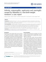
ROC curves and the corresponding AUC of different
tests are presented in Fig. 1. The AUC of a single CRP
measured at T24 was 0.82 and was not statistically
significantly different than the variations in CRP, white
blood cell count and I/T ratio (all P values > 0.75).
Discussion
Diagnostic accuracy of CRP
This study focused on the use of CRP as an adjunctive
diagnostic tool in the evaluation of VLBW infants with
suspected late-onset sepsis. The sensitivity of the CRP at
initial evaluation (T0) was low (49%), which correlates
with previous studies in other neonatal populations
[12, 15]. Nevertheless, at 24 h, CRP had a better sensitivity
of 84% for late-onset sepsis and a high negative predictive
value of 92%. This is similar to previously published results
in in cohorts of infants born at different gestational ages
[12, 15]. Serial measurement of CRP at T0, T24 and T48
was associated with a 93% negative predictive value, which
is lower than what other prospective studies have reported
(98%) [16]. However, those studies included smaller numbers of VLBW infants and had a higher prevalence of
gram-negative bacterial sepsis.
CRP increase and CoNS sepsis
We report a high rate of CoNS sepsis compared to previous studies on CRP [14, 16, 20]. However, these rates
are comparable to the incidence of CoNS infections in
the Canadian Neonatal Network (> 70% of late-onset
sepsis are caused by CoNS) [18]. This is likely attributable to variations in local epidemiology combined with
care practices. Indeed, the rate of fungal infections was
also very low in our cohort and similar to the Canadian
average (incidence < 2% in VLBW infants) [18]. The fact
that we did not mandate two cultures for the diagnosis
of sepsis may have contributed to a higher prevalence of
contaminants. This may have increased the false positive
rate in our cohort, but would have little effect on the
negative predictive value of CRP.
Previous studies concluded that CoNS might induce a
less sustained inflammatory response than gram-negative
bacterial sepsis [13, 14]. However, our findings do not
suggest a statistically significant difference in peak CRP’s
values in infants with CoNS compared to other pathogens,
Table 3 Organisms isolated from positive blood cultures in patients with confirmed infections and CRP < 10 mg/L or
≥10 mg/L at T24
Organisms (N)
CRP < 10 mg/L (N = 25)
CRP ≥ 10 mg/L (N = 137)
Coagulase-negative staphylococci
Coagulase-negative staphylococci (19)
Coagulase-negative staphylococci (116)
Non-CoNS gram-positive bacteria
Staphylococcus aureus (1)
Staphylococcus aureus (6)
Gram-negative bacteria
Enterococcus faecalis (1)
Enterococcus faecalis (2)
Streptococcus agalactiae (1)
Streptococcus agalactiae (4)
Enterobacter cloacae (1)
Enterobacter cloacae (3)
Klebsiella pneumoniae (1)
Klebsiella pneumoniae (4)
Escherchia coli (2)
Fungi
Candida lusitaniae (1)
Beltempo et al. BMC Pediatrics (2018) 18:16
Page 5 of 8
Table 4 Sensitivity, specificity, positive and negative predictive
values of tests at T0, T24 h and T48ha
Test
Sensitivity
Specificity
PPV
NPV
CRP
49% (41–56)
76% (72–80)
43% (37–50) 79% (75–83)
CBC
49% (41–57)
83% (79–86)
52% (44–60) 81% (77–84)
T0
CBC + CRP 65% (57–72)† 66% (61–70)† 42% (36–48) 83% (79–87)
T24 H
CRP
84% (78–89)
70% (66–75)
52% (46–58) 92% (89–95)
CBC
59% (51–67)† 79% (75–83)
53% (46–60) 84% (80–87)
CBC + CRP 87% (80–91)
60% (55–64)† 45% (40–51) 92% (88–95)
T 48H
CRP
73% (66–79)
CBC
21% (15–28)† 92% (88–94)† 50% (39–62) 75% (71–79)
CBC + CRP 76% (69–82)
79% (74–82)
74% (69–78)
57% (50–63) 88% (85–91)
53% (46–59) 89% (85–92)
a
Parenthesis indicate 95% confidence interval
†Sensitivity or specificity significantly different (p < 0.05) compared to the CRP
taken at the same time
however this is not statistically significant given the small
number of gram-negative infections. Likewise, 46% of the
CoNS sepsis included in our cohort were associated with
a CRP value of at least 50 mg/L suggesting that the
inflammatory potential of these organisms is present.
Diagnostic accuracy of the CBC
We found that a CBC obtained at T24 has a low sensitivity (59%) for the diagnosis of late-onset sepsis in VLBW
infants. The I/T ratio was the main contributor to the
diagnostic accuracy of the CBC at T24 (sensitivity 50%).
This is in keeping to what has been reported in more mature newborns. Indeed, Hornik et al. found that the sensitivity of the CBC in infants born < 34 weeks gestational
age was 55% [7]. However, they used a different cut-off for
the I/T ratio (> 0.2). They noted a higher I/T ratio in
gram-negative infections. The low incidence of gramnegative infections in our cohort might explain why a
lower threshold yielded similar results. Moreover, as a
late-onset physiologic neutropenia frequently occurs in
VLBW infants, their I/T ratio might be less accurate [23].
Consequently, using a lower positivity threshold may be
appropriate in order to increase its sensitivity as a screening tool in this specific population. A platelet count <
100,000/uL at T24 had a low sensitivity (12%) for predicting late-onset sepsis. This is similar to previous studies
that found the platelet count has a low discriminative
performance (AUC 0.60) for diagnosing sepsis in preterm
infants [7]. The variation in platelet count based on gestational age and postnatal age may require the use of age
specific cut-offs [24]. Also, platelet parameters like mean
platelet volume and platelet distribution width may
increase diagnostic yield, although these parameters were
not collected in the present study [25].
Optimal test combinations
The combination of the CBC at T0 and CRP at T24 had
the highest negative predictive value (93%) with a
minimal number of tests. Considering every blood
drawn represents a significant volume loss for premature
neonates and carries an inherent infection risk, the use
of a less invasive, but still accurate sepsis work-up strategy is worth considering. Since the majority of clinicians
include a CBC in the initial workup of suspected lateonset sepsis [7], it is unlikely that an approach solely
based on serial CRP measure would be adopted. Also,
two CRP values drawn at T0 and T24 had a nonsuperior negative predictive value than the combined
CBC at T0 and CRP at T24. Further, there were no
additional benefit to repeat any blood tests at T48.
Lastly, if diagnostic accuracy at the time of initial evaluation (T0) is a priority, then the combination of the
CBC and CRP at T0 allows the highest sensitivity and
might assist in clinical decision-making.
Early cessation of antibiotics
An important benefit of using reliable laboratory markers
is to help in the decision to discontinue empirical
Table 5 Sensitivity, specificity, positive and negative predictive values of selected combined testsa
Tests combinations
Sensitivity
Specificity
PPV
NPV
CRP
88% (82–92)
60% (55–65)
46% (41–52)
93% (89–96)
CBC + CRP
CRP
88% (82–92)
60% (55–65)
46% (41–52)
93% (89–96)
CBC + CRP
CBC + CRP
88% (82–92)
56% (51–61)
44% (38–49)
92% (88–95)
T0
T24
CBC
T48
CBC + CRP
65% (57–72)
66% (61–70)
42% (36–48)
83% (79–87)
CBC + CRP
87% (81–92)
59% (54–64)
45% (40–51)
93% (89–95)
CRP
CBC
74% (67–81)
62% (57–67)
43% (37–49)
86% (82–90)
CRP
CRP
84% (78–89)
70% (65–74)
52% (46–58)
92% (88–95)
CRP
CRP
86% (79–90)
69% (64–73)
52% (46–58)
93% (89–95)
a
Parenthesis indicate 95% confidence interval
CRP
Beltempo et al. BMC Pediatrics (2018) 18:16
Page 6 of 8
a
b
c
d
Fig. 1 Receiver operative characteristic (ROC) curves of different tests. a. CRP at T24. AUC = 0.82 (95% CI, 0.78–0.86). b. Absolute difference in the
CRP values obtained at T0 and T24. AUC, 0.84 (95% CI, 0.79–0.88). c. Absolute difference in the white blood cell count at T0 and T24. AUC 0.82
(95% CI, 0.77–0.87). d. Absolute difference in the I/T ratio at T0 and T24. AUC 0.77 (95% CI, 0.70–0.82)
antimicrobial. Recent studies reported a 97% negative predictive value of blood culture at 36 h [26, 27]. The high
negative predictive value of negative blood cultures at 36 h,
combined with the 93% negative predictive value of the
two-step approach described above (CBC at T0 and CRP
at T24) could be used together to support discontinuation
of antimicrobials at that time rather than waiting the traditional 48-h time point. This, in turn, would help mitigate
drug-associated adverse events, decrease the likelihood of
selection of resistant organisms and Candida sp. [26], necrotizing enterocolitis and potential hearing impairment
[28], in addition to reducing healthcare costs [6]. Additionally, discontinuing antibiotics after 36 h reduces blood
draws required for antibiotic dosing when using aminoglycosides and/or vancomycin are used as empiric therapy.
Variations in CRP, white blood cell count and I/T ratio
The analysis of ROC curves associated with the absolute
change from T0 to T24 of the CRP, the WBC count and
the I/T ratio has clinical implications. For example, it
improves the WBC count relevance compared to a single value at T0. These results reinforce the importance
of a marker’s kinetic rather than its absolute value at a
specific timing. However, when trying to minimize the
amount of blood tests, one should consider that the
AUC of the change in white blood cell count from T0 to
T24 was not significantly different than a single CRP
done at T24. Consequently, repeating a CBC to monitor
the white blood cell count variation is not warranted.
Furthermore, as there is no current positivity cut-off for
the absolute increase in white blood cell count, its interpretation is subject to variability.
Strengths and limitations
An important strength of this study is the single cohort
of VLBW infants. The retrospective design and inclusion
of all newborns with a late-onset sepsis evaluation may
have led to the inclusion of patients with other conditions associated with an increased CRP. Indeed, among
infants with false positive CRP results at T24, there were
8 postoperative infants and 14 who were treated for necrotizing enterocolitis, two conditions known to increase
CRP values [2, 10, 29]. Also, infants with a diagnosis of
ventilator-associated pneumonia and elevated CRP were
not considered as having a late-onset sepsis since the
objective of the study was to assess diagnostic accuracy
in predicting culture-proven bloodstream infections or
meningitis. Also, there were no standardized criteria for
Beltempo et al. BMC Pediatrics (2018) 18:16
diagnosis of ventilator-associated pneumonia in VLBW
infants on the NICU during the study period. We did
not collect data on vaccination and intraventricular
hemorrhage which might also have increased false
positive rates [10]. All these conditions, by increasing
the CRP false-positive results may have contributed to
underestimating its specificity. However, the inclusion of
these patients would have had little effect on the negative predictive value. Finally even though missing data
were imputed using LOCF method, their proportion varied between 5% and 33% for the different lab tests and
time points. To ensure that this did not bias the results,
sensitivity analysis without imputation were carried and
showed identical conclusions.
Conclusion
In summary, this study is the first that describes the
combined CRP and CBC for the diagnosis of late-onset
sepsis in a large cohort of VLBW neonates. Our results
emphasize that suspected late-onset sepsis initial workup
should include both CRP and CBC if the decision to
start antibiotics is uncertain. Also, it supports early antibiotics cessation after 36 h of negative cultures if the
combined CBC at T0 and CRP at T24 are negative.
Using a two tests combination strategy could reduce iatrogenic consequences of late-onset sepsis investigations
in VLBW infants. However, further studies are required
to determine if different cut-off values of CRP at different timings during late-onset sepsis evaluation in VLBW
infants could increase sensitivity.
Abbreviations
AUC: Area under the curve; CBC: Complete blood count; CoNS: Coagulasenegative staphylococci; CRP: C-reactive protein; GEE: Generalized estimating
eqs.; I/T: Immature neutrophil/total neutrophil; LOCF: Last observation carried
forward; ROC: Receiver operating characteristics; VLBW: Very low birth weight
Acknowledgements
We would like to thank Vicky Beauchesne, from the department of clinical
performance analyses who helped with data collection and database design.
We also thank the neonatologists of the CHU de Québec neonatal intensive
care unit that supported this project and offered critical review. Our
gratitude is also addressed to the CHU de Québec department of infection
prevention and control that made available the data on blood cultures.
Funding
No funding was obtained for this study.
Availability of data and materials
The data and materials are stored at the Department of Pediatrics,
Neonatology Division, Faculty of Medicine, Laval University. The dataset
supporting the conclusions of this article is available on demand by
contacting the corresponding author.
Author’s contribution
All authors participated in the study design and interpretation of data. The
role of each author is as follows: BP was the principal investigator. He is
guarantor. MB and IVT participated in the research protocol, data collection,
data analysis and interpretation. BP and RT participated in the study design,
data analysis and review of the manuscript. ASJ participated in data analysis
and interpretation. MB and IVT wrote the original draft. All authors read and
approved the final manuscript.
Page 7 of 8
Ethics approval and consent to participate
This study has been approved by the CHU de Québec Research and Ethics
Board (REB). Permission to use the Med-Echo and TDR databases was
obtained from Hospital Director and from CHU de Québec REB. Due to the
retrospective design of the study using medical charts, individual patient
consent was deemed unnecessary by the CHU de Québec REB.
Consent for publication
Not applicable.
Competing interests
The authors declare that they have no competing interests.
Publisher’s Note
Springer Nature remains neutral with regard to jurisdictional claims in
published maps and institutional affiliations.
Author details
1
McGill University Health Centre, Montreal, QC, Canada. 2Département de
pédiatrie, Centre Mère-Enfant Soleil du CHU de Québec, Université Laval,
2705 Boulevard Laurier, QC, Québec G1V 4G2, Canada. 3Centre de recherche
du CHU de Québec, Université Laval, QC, Québec, Canada.
Received: 9 January 2017 Accepted: 22 January 2018
References
1. Hornik CP, Fort P, Clark RH, Watt K, Benjamin DK, Smith PB, et al. Early and
late onset sepsis in very-low-birth-weight infants from a large group of
neonatal intensive care units. Early Hum Dev. 2012;88(Suppl 2):S69–74.
2. Couto RC, Barbosa JA, Pedrosa TM, Biscione FM. C-reactive protein-guided
approach may shorten length of antimicrobial treatment of culture-proven
late-onset sepsis: an intervention study. Braz J Infect Dis. 2007;11(2):240–5.
3. Downey LC, Smith PB, Benjamin DK. Risk factors and prevention of lateonset sepsis in premature infants. Early Hum Dev. 2010;86(Suppl 1):7–12.
4. Fanaroff AA, Korones SB, Wright LL, Verter J, Poland RL, Bauer CR, et al.
Incidence, presenting features, risk factors and significance of late onset
septicemia in very low birth weight infants. The National Institute of Child
Health and Human Development neonatal research network. Pediatr Infect
Dis J. 1998;17(7):593–8.
5. Arnon S, Litmanovitz I, Regev R, Lis M, Shainkin-Kestenbaum R, Dolfin T. The
prognostic virtue of inflammatory markers during late-onset sepsis in
preterm infants. J Perinat Med. 2004;32(2):176–80.
6. Marchant EA, Boyce GK, Sadarangani M, Lavoie PM. Neonatal sepsis due to
coagulase-negative staphylococci. Clin Dev Immunol. 2013;5(8):60–76.
7. Hornik CP, Benjamin DK, Becker KC, Li J, Clark RH, Cohen-Wolkowiez M,
et al. Use of the complete blood cell count in late-onset neonatal sepsis.
Pediatr Infect Dis J. 2012;31(8):803–7.
8. Stoll BJ, Hansen N. Infections in VLBW infants: studies from the NICHD
neonatal research network. Semin Perinatol. 2003;27(4):293–301.
9. Hentges CR, Silveira RC, Procianoy RS, Carvalho CG, Filipouski GR, Fuentefria
RN, et al. Association of late-onset neonatal sepsis with late
neurodevelopment in the first two years of life of preterm infants with very
low birth weight. J Pediatr. 2014;90(1):50–7.
10. Hofer N, Zacharias E, Müller W, Resch B. An update on the use of C-reactive
protein in early-onset neonatal sepsis: current insights and new tasks.
Neonatology. 2012;102(1):25–36.
11. Makhoul IR, Yacoub A, Smolkin T, Sujov P, Kassis I, Sprecher H. Values of Creactive protein, procalcitonin, and staphylococcus-specific PCR in neonatal
late-onset sepsis. Acta Paediatr. 2006;95(10):1218–23.
12. Ng PC, Cheng SH, Chui KM, Fok TF, Wong MY, Wong W, et al. Diagnosis of
late onset neonatal sepsis with cytokines, adhesion molecule, and Creactive protein in preterm very low birthweight infants. Arch Dis Child
Fetal Neonatal Ed. 1997;77(3):F221–7.
13. Dritsakou K, Liosis G, Gioni M, Glynou E, Avdeliodi K, Papagaroufalis K. CRP
levels in extremely low birth weight (ELBW) septic infants. J Matern Fetal
Neonatal Med. 2015;28(2):237–9.
14. Shah SD, Talati AJ, Elabiad MT, Dhanireddy R, Pourcyrous M. Preterm infants
can mount appropriate C-reactive protein responses to early onset sepsis.
Am J Perinatol. 2015;32(13):1281–6.
Beltempo et al. BMC Pediatrics (2018) 18:16
Page 8 of 8
15. Lacaze-Masmonteil T, Rosychuk RJ, Robinson JL. Value of a single C-reactive
protein measurement at 18 h of age. Arch Dis Child Fetal Neonatal Ed.
2014;99(1):F76–9.
16. Benitz WE, Han MY, Madan A, Ramachandra P. Serial serum C-reactive
protein levels in the diagnosis of neonatal infection. Pediatrics.
1998;102(4):E41.
17. Pourcyrous M, Bada HS, Korones SB, Baselski V, Wong SP. Significance of
serial C-reactive protein responses in neonatal infection and other disorders.
Pediatrics. 1993;92(3):431–5.
18. Shah J, Jefferies AL, Yoon EW, Lee SK, Shah PS, Network CN. Risk factors and
outcomes of late-onset bacterial sepsis in preterm neonates born at < 32
Weeks’ gestation. Am J Perinatol. 2015;32(7):675–82.
19. Rewa O, Muscedere J, Reynolds S, Jiang X, Heyland DK. Coagulase-negative
staphylococcus, catheter-related, bloodstream infections and their
association with acute phase markers of inflammation in the intensive care
unit: an observational study. Can J Infect Dis. 2012;23(4):204–8.
20. Chiesa C, Natale F, Pascone R, Osborn JF, Pacifico L, Bonci E, et al. C reactive
protein and procalcitonin: reference intervals for preterm and term
newborns during the early neonatal period. Clin Chim Acta.
2011;412(11–12):1053–9.
21. Firoozi F, Lemiere C, Beauchesne MF, Forget A, Blais L. Development and
validation of database indexes of asthma severity and control. Thorax.
2007;62(7):581–7.
22. Leeflang MM, Rutjes AW, Reitsma JB, Hooft L, Bossuyt PM. Variation of a
test’s sensitivity and specificity with disease prevalence. CMAJ.
2013;185(11):E537–44.
23. Vetter-Laracy S, Balliu PR, Salinas JA, Duran MA. Late-onset neutropenia:
defining limits of neutrophil count in very low birth weight infants.
J Perinatol. 2014;34(1):22–6.
24. Wiedmeier SE, Henry E, Sola-Visner MC, Christensen RD. Platelet reference
ranges for neonates, defined using data from over 47,000 patients in a
multihospital healthcare system. J Perinatol. 2009;29(2):130–6.
25. Andres O, Schulze H, Speer CP. Platelets in neonates: central mediators in
haemostasis, antimicrobial defence and inflammation. Thromb Haemost.
2015;113(1):3–12.
26. Kaiser JR, Cassat JE, Lewno MJ. Should antibiotics be discontinued at 48
hours for negative late-onset sepsis evaluations in the neonatal intensive
care unit? J Perinatol. 2002;22(6):445–7.
27. Kumar Y, Qunibi M, Neal TJ, Yoxall CW. Time to positivity of neonatal blood
cultures. Arch Dis Child Fetal Neonatal Ed. 2001;85(3):F182–6.
28. Coggins SA, Wynn JL, Hill ML, Slaughter JC, Ozdas-Weitkamp A, Jalloh O,
et al. Use of a computerized C-reactive protein based sepsis evaluation in
very low birth weight infants: a five-year experience. PLoS One.
2013;8(11):e78602.
29. Keane M, Fallon R, Riordan A, Shaw B. Markedly raised levels of C-reactive
protein are associated with culture-proven sepsis or necrotising enterocolitis
in extremely preterm neonates. Acta Paediatr. 2015;104(7):e289–93.
Submit your next manuscript to BioMed Central
and we will help you at every step:
• We accept pre-submission inquiries
• Our selector tool helps you to find the most relevant journal
• We provide round the clock customer support
• Convenient online submission
• Thorough peer review
• Inclusion in PubMed and all major indexing services
• Maximum visibility for your research
Submit your manuscript at
www.biomedcentral.com/submit