Gluten-free diet may improve obstructive sleep apnea-related symptoms in children with celiac disease
Bạn đang xem bản rút gọn của tài liệu. Xem và tải ngay bản đầy đủ của tài liệu tại đây (417.19 KB, 5 trang )
Yerushalmy-Feler et al. BMC Pediatrics (2018) 18:35
/>
RESEARCH ARTICLE
Open Access
Gluten-free diet may improve obstructive
sleep apnea-related symptoms in children
with celiac disease
Anat Yerushalmy-Feler1,4†, Riva Tauman2,4†, Ari Derowe3,4, Eran Averbuch3,4, Amir Ben-Tov1,4, Yael Weintraub1,4,
Dror Weiner1,4, Achiya Amir1,4, Hadar Moran-Lev1,4 and Shlomi Cohen1,4*
Abstract
Background: Enlarged tonsils and adenoids are the major etiology of obstructive sleep apnea (OSA) in children.
Lymphatic hyperplasia is common to both OSA and celiac disease. We aimed to investigate the effect of a
gluten-free diet on OSA symptoms in children with celiac disease.
Methods: Children with celiac disease aged 2–18 years were prospectively recruited before the initiation of a
gluten-free diet. Children with negative celiac serology who underwent gastrointestinal endoscopies for other
indications served as controls. All participants completed a validated OSA-related symptoms questionnaire and
the pediatric sleep questionnaire (PSQ) at baseline and 6 months later.
Results: Thirty-four children with celiac disease (mean age 6.6 ± 3.5 years) and 24 controls (mean age 7.3 ± 4.
6 years, P = 0.5) were recruited. There were no significant differences in gender, body mass index or season at
recruitment between the two groups. The rate of positive PSQ scores was higher (more OSA-related symptoms)
in the control group compared to the celiac group, both at recruitment and at the 6-month follow-up (33.3% vs.
11.8%, P = 0.046, and 16.7% vs. 0, P = 0.014, respectively). PSQ scores improved significantly in both groups at the
6-month follow-up (P < 0.001 for both). Improvement was significantly higher in the celiac group compared to
controls (0.1 ± 0.09 vs.0.06 ± 0.06, respectively, P = 0.04).
Conclusions: Children with celiac disease had fewer OSA-related symptoms than controls, but the degree of
improvement following the initiation of a gluten-free diet was significantly higher. These findings suggest that a
gluten-free diet may improve OSA-related symptoms in children with celiac disease.
Keywords: Celiac disease, Gluten-free diet, Obstructive sleep apnea, Pediatrics
Background
Celiac disease is a chronic immune-mediated systemic
disorder caused by a permanent sensitivity to gluten and
related proteins in genetically susceptible individuals. Its
prevalence is approximately 1% of the general population [1, 2]. Mesenteric lymphadenopathy is a potential
part of the clinical course of celiac disease, with resolution after the institution of a gluten-free diet [3, 4].
* Correspondence:
†
Equal contributors
1
Pediatric Gastroenterology Unit, “Dana-Dwek” Children’s Hospital, Tel Aviv
Sourasky Medical Center, Tel Aviv, Israel
4
Sackler Faculty of Medicine, Tel Aviv University, Tel Aviv, Israel
Full list of author information is available at the end of the article
Celiac disease was also associated with mesenteric lymph
node cavitation syndrome [5].
Obstructive sleep apnea (OSA) syndrome is characterized by recurrent episodes of upper airway obstruction
during sleep, associated with intermittent hypoxia,
hypercapnia and sleep fragmentation [6]. The prevalence
of OSA in children is up to 3% in several epidemiological
studies [7]. The incidence peaks in children from 2 to
8 years of age [8]. The major etiology for OSA in children
is adenotonsillar hypertrophy [9, 10]. In addition, children
with OSA may have hypertrophy of the lymphoid tissues
in other regions of the airways, such as the deep cervical
lymph nodes [11, 12]. Recent studies have indicated that a
potential part of the pathophysiology of OSA is localized
© The Author(s). 2018 Open Access This article is distributed under the terms of the Creative Commons Attribution 4.0
International License ( which permits unrestricted use, distribution, and
reproduction in any medium, provided you give appropriate credit to the original author(s) and the source, provide a link to
the Creative Commons license, and indicate if changes were made. The Creative Commons Public Domain Dedication waiver
( applies to the data made available in this article, unless otherwise stated.
Yerushalmy-Feler et al. BMC Pediatrics (2018) 18:35
inflammation in the nasopharyngeal area, including an increase in inflammatory cell proliferation, particularly Tcell lymphocytes [13, 14].
Celiac disease and OSA share common features of
lymphatic hyperplasia and local inflammation. A recent
study by Parisi et al. [15] showed an increased prevalence of sleep-disordered breathing in celiac patients,
with resolution of the symptoms with the introduction
of a gluten-free diet. Considering the common feature of
lymphatic hyperplasia/local inflammation in both disorders, we aimed to investigate the prevalence of OSA in
children with celiac disease and to define the effect of a
gluten-free diet on OSA symptoms in celiac disease.
Methods
All study procedures were approved by the institutional
review board of the Tel Aviv Medical Center (Helsinki
Committee), and parental informed consent was obtained for all participants.
Sample and procedure
The study group included children aged 2 to 18 years
who were diagnosed as having celiac disease and were
recruited between December 2014 and September 2015
at the Pediatric Gastroenterology Unit in “Dana-Dwek”
Children’s Hospital of the Tel Aviv Sourasky Medical
Center. All patients were prospectively recruited after
diagnosis, before the initiation of a gluten-free diet. The
diagnosis of celiac disease was based on the combination
of celiac-related symptoms, a positive serology (anti-tissue transglutaminase levels [TTG] > 10.0 U/mL) and a
characteristic histology according to the European
Society for Pediatric Gastroenterology, Hepatology, and
Nutrition (ESPGHAN) guidelines for the diagnosis of celiac disease [16]. Healthy children who underwent upper
gastrointestinal endoscopy for other indications and who
had a documented negative celiac serology during the
past 3 months and a normal duodenal histology served
as controls. The groups were matched for age, sex and
season of referral. Children with congenital anomalies,
developmental delay or other chronic medical conditions
were excluded, as were children that had received
medical/surgical treatment for OSA.
Measures
We collected demographic data including age and
gender, medical history, height, weight and body mass
index (BMI), clinical symptoms, anti-TTG levels at baseline and 6 months after initiation of a gluten-free diet,
histological findings and Marsh scores. The BMI Z score
was calculated as well [17].
All parents completed the sleep-related breathing disorders scale of the pediatric sleep questionnaire (PSQ) at
the time of recruitment (PSQ1) and 6 months later
Page 2 of 5
(PSQ2), i.e., prior to and 6 months after the initiation of
a gluten-free diet for the celiac group [18]. The pediatric
sleep questionnaire is a well validated symptom inventory that includes 22 items on snoring, apneas, daytime
sleepiness, and inattentive/hyperactive behavior [18].
Responses are “yes” = 1, “no” = 0, or “don’t know” (considered missing). The mean response to non-missing
items is the total score which can vary from 0 to 1.
Higher scores indicate more sleep disordered breathing
(SDB)-related symptoms [18]. A threshold of 0.33, indicating that 33% of symptom-related items are positive, is
considered a positive screen for pediatric SDB [18].
Subscales within the PSQ include a 4-item sleepiness
scale, a 4-item snoring scale, and a 6-item inattention
and hyperactivity scale derived from the Diagnostic and
Statistical Manual of Mental Disorders, Fourth Edition
Criteria for Attention-deficit/hyperactivity Disorder [19].
Since its development, this scale has been used in a variety of research settings [18, 20]. For the purposes of the
current study, we analyzed the total score, as well as the
snoring, sleepiness and the inattentive/hyperactive behavior subscales.
Data and statistical analysis
Analyses were performed with SPSS (version 21.0; SPSS
Inc. Chicago, IL). Between-group comparisons of nonnormally distributed continuous parameters (age, BMI
and PSQ scores) were conducted with the nonparametric Wilcoxon test. Comparisons between preand post-intervention were conducted with paired-tests.
All reported P-values were 2-tailed with statistical
significance set at < 0.05.
Results
Participants
Thirty-four children with celiac disease (mean age 6.6 ±
3.5 years) and 24 controls (mean age 7.3 ± 4.6 years, P =
0.5) were included. Fifteen patients (44.1%) in the celiac
group and 14 patients (58.3%, P = 0.29) in the control
group were males. There was no significant difference in
the BMI z scores between the two groups (Table 1).
Celiac symptoms
The main symptoms in the celiac group were abdominal
pain and growth retardation (9 patients, 26.5% for both
symptoms), anemia (5 patients, 14.7%) and diarrhea (4 patients, 11.7%). Celiac serology was performed as part of
screening (first-degree relatives of celiac patients) in seven
asymptomatic patients (20.5%). The mean anti-TTG antibodies level in the celiac group at diagnosis was 275 ±
34 U/ml (range 17–800 U/ml) and 23 ± 23 U/ml (range
0–76 U/ml) 6 months after the initiation of a gluten-free
diet. Sixteen patients (47%) had normal macroscopic appearance of the duodenum, while 18 patients (53%) had
Yerushalmy-Feler et al. BMC Pediatrics (2018) 18:35
Page 3 of 5
Table 1 Demographic parameters of the celiac and control groups
Age, y
Celiac group
N = 34
Control group
N = 24
P value
6.6 ± 3.5
7.3 ± 4.6
0.5
Male gender
15 (44.1%)
14 (58.3%)
0.29
Body mass index (z-score)
−0.4 ± 1.07
−0.9 ± 2.6
0.3
Recruitment in winter (%)
67.6%
58.3%
0.48
Time interval between questionnaires (months)
5.94 ± 1.25
5.95 ± 0.92
0.97
macroscopic findings consistent with celiac disease, mainly
duodenal scalloping. The duodenal histology of all patients
except one demonstrated villous atrophy, crypt hyperplasia
and an increased number of intraepithelial lymphocytes,
consistent with Marsh 3 celiac disease. One patient had
findings consistent with Marsh 2 celiac disease.
Controls
The control group included 24 patients. Twelve of them
(50%) had abdominal pain with a normal gastric histology,
4 (16.7%) had Helicobacter pylori-positive gastritis, 2
(8.3%) had Helicobacter pylori-negative gastritis, 3 (12.5%)
presented with vomiting (2 with findings consistent with
esophagitis and 1 with normal histology), and 3 (12.5%)
presented with failure to thrive (FTT) with a normal histology. All patients in the control group had normal antiTTG levels as well as a normal duodenal histology.
PSQ
The average time interval between the two PSQ was
5.94 ± 1.25 months in the celiac group and 5.95 ±
0.92 months in the control group (P = 0.97). Most of the
patients were recruited and filled out the first PSQ in
the winter (November to March, 67.6 and 58.3% in the
celiac and control groups, respectively; P = 0.48), and
completed the second PSQ in a non-winter season
(April to October, 79.4 and 75% in the celiac and control
groups, respectively; P = 0.69).
The rate of positive PSQ scores was higher in the control group compared to the celiac group, both at study
onset and at follow-up. Four out of 34 patients (11.8%)
in the celiac group and 8 out of 24 patients (33.3%) in
the control group (P = 0.046) had a positive PSQ score
(≥0.33) at baseline. Six months later, no patient in the
celiac group and 4 out of 24 (16.7%) patients in the control group had a positive PSQ score (P = 0.014).
The total PSQ scores were significantly lower in the
celiac group compared to controls at both baseline and
at the 6-month follow-up. The PSQ subscale scores
(snoring and inattention and hyperactivity scales) were
also significantly higher for the controls compared to the
celiac group both at baseline and at follow-up (Table 2).
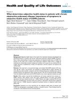
As displayed in Fig. 1, the total PSQ scores improved
significantly in both groups at the 6-month follow-up
compared to baseline (P < 0.001 for both groups). There
was improvement in all three subscales of the PSQ
(snoring, sleepiness and attention/behavioral subscales)
in the celiac group and in two subscales (sleepiness and
attention/behavioral subscales) in the control group. The
degree of improvement (total PSQ1–total PSQ2) was
significantly higher in the celiac group compared to the
controls (0.1 ± 0.09 vs. 0.06 ± 0.06, respectively, P = 0.04,
Fig. 1). As expected, there was a significant correlation
between the total PSQ1 and the total PSQ2 (r = 0.87, P
< 0.0001). No correlation was found between total PSQ
scores (at baseline or at follow-up) and age, BMI Z score
and anti-TTG levels.
Linear regression analysis with PSQ2 as a dependent
variable and group assignment (celiac vs. controls), age,
BMI z score and season as independent variables
revealed that only group assignment was a significant
predictor for PSQ2 score (P < 0.001). Since FTT is also a
manifestation of OSA, we re-analyzed the data after excluding the three patients with FTT and found no differences in the results.
Discussion
This is the first prospective controlled trial that investigated the prevalence of OSA symptoms in children with
celiac disease and the effect of a gluten-free diet on these
symptoms. In this cohort of children, the rate of abnormal PSQ scores, which is a symptom inventory for childhood OSA, was significantly lower among children with
celiac disease compared to age- and gender-matched
controls. The PSQ scores were lower in the celiac group
compared to the controls, while improvements in PSQ
scores were found in both groups 6 months later, and
they were even greater in the celiac group. Our findings
are in contrast to those of Parisi et al. [15] who demonstrated an increased prevalence of sleep-disordered
breathing in celiac patients. Their study, however, used a
different tool for the assessment of sleep and their
sample size (n = 19) was smaller compared to that of the
present study.
The PSQ total scores and the rate of abnormal PSQ
scores were significantly lower in the celiac group compared to controls both at baseline and at follow-up. Both
the snoring subscale and the attention/behavioral
Yerushalmy-Feler et al. BMC Pediatrics (2018) 18:35
Page 4 of 5
Table 2 The pediatric sleep questionnaire (PSQ) scores in the celiac and control groups
Celiac group
P value
Control group
Subscale
PSQ1
PSQ2
P value
PSQ1
PSQ2
P value
PSQ1
(celiac vs controls)
PSQ2
(celiac vs controls)
Snoring
0.07 ± 0.13
0.010.04±
0.01
0.18 ± 0.22
0.16 ± 0.2
0.180
0.022
< 0.001
Sleepiness
0.2 ± 0.23
0.09 ± 0.15
0.001
0.2 ± 0.27
0.11 ± 0.19
0.023
0.75
0.81
Attention/behavioral
0.19 ± 0.29
0.08 ± 0.17
0.001
0.34 ± 0.28
0.29 ± 0.25
0.035
0.014
< 0.001
Total PSQ
0.17 ± 0.14
0.07 ± 0.06
< 0.001
0.26 ± 0.15
0.2 ± 0.11
< 0.001
0.022
< 0.001
subscale scores were lower in the celiac group compared
to controls. These differences cannot be explained by
age, BMI or season of assessment since there were no
differences in these parameters between the two groups.
One possible explanation for these findings is an
increased prevalence of OSA symptoms in the control
group due to factors that were not measured in the
current study, such as prematurity, atopic diseases,
parental smoking or family history of OSA. We speculate, however, that such factors would potentially impact
both groups.
The improvement found in PSQ scores in both groups
at the 6-month follow-up may be explained by the
natural history of OSA, which tends to improve with
age. Indeed, the age range of our cohort is toward the
upper limit of the peak incidence of OSA in children. It
is possible that some children experienced improvement
or even resolution of OSA symptoms during this 6month period. Another explanation is the change in season. Most of our participants were recruited in the winter and so their follow-up assessment was in the spring/
Fig. 1 Average total pediatric sleep questionnaire (PSQ) scores at
baseline and at the 6-month follow-up
summer. As shown in earlier reports, OSA symptoms
may vary with season, and winter is usually associated
with a worsening of OSA symptoms [21–23].
Although PSQ scores were lower in the celiac group
in our study, the degree of improvement in OSA-related
symptoms was significantly higher for those children
compared to controls. This finding suggests that a
gluten-free diet may play a role in this improvement
since there was no difference in the time interval or seasonality between the two groups. This finding is in
agreement with that of Parisi et al. [15] who also showed
resolution of sleep-disordered breathing with a
gluten-free diet. A potential explanation for this finding
is improvement in the lymphatic hyperplasia that is associated with celiac disease and that contributes to OSA
in these patients. The significant improvement in OSA
in celiac patients after the institution of a gluten-free
diet raises the question of whether a gluten-free diet
might have the potential to improve OSA symptoms in
children with celiac and thus serve as an adjuvant therapy for OSA or even replace adenotonsillectomy as the
first line of treatment for OSA for those children.
Our study is based on a well-established tool for identifying OSA symptoms in the pediatric population. It is
the first prospective controlled trial that demonstrated a
possible benefit from a gluten-free diet on OSA in
children with celiac disease. However, the results of the
present study should be interpreted in the context of
several limitations. First, our data are based on a questionnaire and not on objective measures of OSA, such as
polysomnography. Second, we did not assess adenoid
and tonsil size. In addition, the control group is relatively small due to exclusion of patients with chronic
medical conditions, but the results were obtained in this
cohort nevertheless reached a level of significance.
Moreover, three patients in the control group had FTT,
which itself is a potential manifestation of OSA. We decided to include these three patients in the study since
they had similar PSQ scores as those of the control
group. Moreover, as mentioned earlier, the analysis of
the data after excluding these three patients did not
change the results of the study.
In summary, children with celiac disease were found
to have fewer OSA-related symptoms, but the degree of
Yerushalmy-Feler et al. BMC Pediatrics (2018) 18:35
improvement following the initiation of a gluten-free
diet was significantly higher in the celiac group compared to the controls. Our findings suggest that a
gluten-free diet may improve OSA-related symptoms in
children with celiac disease. Further studies are needed
to determine whether a gluten-free diet has the potential
to reduce the need for adenoidectomy and/or tonsillectomy in children with celiac and OSA.
Conclusions
Children with celiac disease had fewer OSA-related
symptoms than controls, but the degree of improvement
following the initiation of a gluten-free diet was significantly higher. These findings suggest that a gluten-free
diet may improve OSA-related symptoms in children
with celiac disease.
Abbreviations
BMI: Body mass index; ESPGHAN: European Society for Pediatric Gastroenterology,
Hepatology, and Nutrition; FTT: Failure to thrive; OSA: Obstructive sleep apnea;
PSQ: Pediatric sleep questionnaire; SDB: Sleep disordered breathing; TTG: Tissue
transglutaminase
Acknowledgements
Not applicable
Funding
No funding to declare.
Availability of data and materials
The datasets used and/or analysed during the current study are available
from the corresponding author on reasonable request.
Authors’ contributions
AYF, RT designed the study, participated in data acquisition and analysis,
drafted the manuscript and approved the final version to be submitted. ABT,
YW, AA,AD, HML, DW, EA participated in the study concept and in data analysis,
revised the manuscript critically, and approved the final version to be
submitted. SC participated in the study concept and in data analysis, revised
the manuscript critically, and approved the final version to be submitted.
Ethics approval and consent to participate
All study procedures were approved by the institutional review board of the
Tel Aviv Medical Center (Helsinki Committee), and written parental informed
consent was obtained for all participants.(0402-14-TLV).
Consent for publication
Not applicable
Competing interests
The authors declare that they have no competing interests.
Publisher’s Note
Springer Nature remains neutral with regard to jurisdictional claims in
published maps and institutional affiliations.
Author details
1
Pediatric Gastroenterology Unit, “Dana-Dwek” Children’s Hospital, Tel Aviv
Sourasky Medical Center, Tel Aviv, Israel. 2Pediatric Sleep Center,
“Dana-Dwek” Children’s Hospital, Tel Aviv Sourasky Medical Center, Tel Aviv,
Israel. 3Pediatric ENT Unit, “Dana-Dwek” Children’s Hospital, Tel Aviv Sourasky
Medical Center, Tel Aviv, Israel. 4Sackler Faculty of Medicine, Tel Aviv
University, Tel Aviv, Israel.
Page 5 of 5
Received: 13 March 2017 Accepted: 30 January 2018
References
1. Fasano A, Berti I, Gerarduzzi T, Not T, Colletti RB, Drago S, Elitsur Y, et al.
Prevalence of celiac disease in at-risk and not at-risk groups in the United
States. Arch Intern Med. 2003;163:286–92.
2. Hoffenberg EJ, MacKenzie T, Barriga KJ, Eisenbarth GS, Bao F, Haas JE, et al.
A prospective study of the incidence of childhood celiac disease. J Pediatr.
2003;143:308–14.
3. Jones B, Bayless TM, Fishman EK, Siegelman SS. Lymphadenopathy in celiac
disease: computed tomographic observations. AJR Am J Roentgenol.
1984;142:1127–32.
4. De Boer WA, Maas M, Tytgat GN. Disappearance of mesenteric
lymphadenopathy with gluten-free diet in celiac sprue. J Clin Gastroenterol.
1993;16:317–9.
5. Hugh JF. Mesenteric lymph node cavitation syndrome. World J
Gastroenterol. 2010;16:2991–3.
6. Balbani AP, Weber SA, Montovani JC. Update in obstructive sleep apnea
syndrome in children. Braz J Otorhinolaryngol. 2005;71:74–80.
7. Anuntaseree W, Rookkapan K, Kuasirikul S, Thongsuksai P. Snoring and
obstructive sleep apnea in Thai school-age children: prevalence and
predisposing factors. Pediatr Pulmonol. 2001;32:322–7.
8. Bower C, Buckmiller L. What’s new in pediatric obstructive sleep apnea. Curr
Opin Otolaryngol Head Neck Surg. 2001;9:352–8.
9. Patino M, Sadhasivam S, Mahmoud M. Obstructive sleep apnoea in children:
perioperative considerations. Br J Anaesth. 2013;111(Suppl 1):i83–95.
10. Tauman R, Gozal D. Obstructive sleep apnea syndrome in children. Expert
Rev Resp Med. 2011;5:425–40.
11. Parikh SR, Sadoughi B, Sin S, Willen S, Nandalike K, Arens R. Deep cervical
lymph node hypertrophy: a new paradigm in the understanding of
pediatric obstructive sleep apnea. Laryngoscope. 2013;123:2043–9.
12. Tan HL, Gozal D, Kheirandish-Gozal L. Obstructive sleep apnea in children: a
critical update. Nat Sci Sleep. 2013;5:109–23.
13. Serpero LD, Kheirandish-Gozal L, Dayyat E, Goldman JL, Kim J, Gozal D. A
mixed cell culture model for assessment of proliferation in tonsillar tissues
from children with obstructive sleep apnea or recurrent tonsillitis.
Laryngoscope. 2009;119:1005–10.
14. Kim J, Bhattacharjee R, Dayyat E, Snow AB, Kheirandish-Gozal L, Goldman JL,
Li RC, et al. Increased cellular proliferation and inflammatory cytokines in
tonsils derived from children with obstructive sleep apnea. Pediatr Res.
2009;66:423–8.
15. Parisi P, Pietropaoli N, Ferretti A, Nenna R, Mastrogiorgio G, Del Pozzo M,
et al. Role of the gluten-free diet on neurological-EEG findings and sleep
disordered breathing in children with celiac disease. Seizure. 2015;25:181–3.
16. Husby S, Koletzko S, Korponay-Szabó IR, Mearin ML, Phillips A, Shamir R,
ESPGHAN Working Group on Coeliac Disease Diagnosis, ESPGHAN
Gastroenterology Committee, European Society for Pediatric
Gastroenterology, Hepatology, and Nutrition, et al. European Society for
Pediatric Gastroenterology, Hepatology, and Nutrition. Guidelines for the
diagnosis of coeliac disease. J Pediatr Gastroenterol Nutr. 2012;54:136–60.
17. Kuczmarski RJ, Ogden CL, Grummer-Strawn LM, Flegal KM, Guo SS, Wei R,
et al. CDC growth charts: United States. Adv Data. 2000;314:1–27.
18. Chervin RD, Hedger K, Dillon JE, Pituch KJ. Pediatric sleep questionnaire
(PSQ): validity and reliability of scales for sleep-disordered breathing,
snoring, sleepiness, and behavioral problems. Sleep Med. 2000;1:21–32.
19. American Psychiatric Association. Diagnostic and statistical manual of
mental disorders. Washington DS: American Psychiatric Association; 1994.
20. O’Brien LM, Lucas NH, Felt BT, Hoban TF, Ruzicka DL, Jordan R, et al.
Aggressive behavior, bullying, snoring, and sleepiness in school children.
Sleep Med. 2011;12:652–8.
21. Nakayama M, Koike S, Kuriyama S, Suzuki M, Nakamura Y, Yamamoto K,
et al. Seasonal variation in a clinical referral pediatric cohort at risk for
obstructive sleep apnea. Int J Pediatr Otorhinolaryngol. 2013;77:266–9.
22. Ingram DG, Matthews CK, Plante DT. Seasonal trends in sleep-disordered
breathing: evidence from internet search engine query data. Sleep Breath.
2015;19:79–84.
23. Greenfeld M, Sivan Y, Tauman R. The effect of seasonality on sleepdisordered breathing severity in children. Sleep Med. 2013;14:991–4.