Differences upon admission and in hospital course of children hospitalized with community-acquired pneumonia with or without radiologically-confirmed pneumonia: A retrospective cohort study
Bạn đang xem bản rút gọn của tài liệu. Xem và tải ngay bản đầy đủ của tài liệu tại đây (964.06 KB, 9 trang )
Simbalista et al. BMC Pediatrics (2015) 15:166
DOI 10.1186/s12887-015-0485-6
RESEARCH ARTICLE
Open Access
Differences upon admission and in hospital
course of children hospitalized with
community-acquired pneumonia with or
without radiologically-confirmed
pneumonia: a retrospective cohort study
Raquel Simbalista1, Dafne C. Andrade2, Igor C. Borges2, Marcelo Araújo3 and Cristiana M. Nascimento-Carvalho1,2,4*
Abstract
Background: The use of chest radiograph (CXR) for the diagnosis of childhood community-acquired pneumonia
(CAP) is controversial. We assessed if children with CAP diagnosed on clinical grounds, with or without
radiologically-confirmed pneumonia on admission, evolved differently.
Methods: Children aged ≥ 2 months, hospitalized with CAP diagnosed on clinical grounds, treated with 200,000 IU/
Kg/day of aqueous penicillin G for ≥ 48 h and with CXR taken upon admission, without pleural effusion, were
included in this retrospective cohort. One researcher, blinded to the radiological diagnosis, collected data on
demographics, clinical history and physical examination on admission, daily hospital course during the first 2 days
of treatment, and outcome, all from medical charts. Radiological confirmation of pneumonia was based on
presence of pulmonary infiltrate detected by a paediatric radiologist who was also blinded to clinical data. Variables
were initially compared by bivariate analysis. Multi-variable logistic regression analysis assessed independent
association between radiologically-confirmed pneumonia and factors which significantly differed during hospital
course in the bivariate analysis. The multi-variable analysis was performed in a model adjusted for age and for the
same factor present upon admission.
Results: 109 (38.5 %) children had radiologically-confirmed pneumonia, 143 (50.5 %) had normal CXR and 31 (11.0 %)
had atelectasis or peribronchial thickening. Children without radiologically-confirmed pneumonia were younger than
those with radiologically-confirmed pneumonia (median [IQR]: 14 [7–28 months versus 21 [12–44] months; P = 0.001).
None died. The subgroup with radiologically-confirmed pneumonia presented fever on D1 (33.7 vs. 19.1; P = 0.015) and
on D2 (31.6 % vs. 16.2 %; P = 0.004) more frequently. The subgroup without radiologically-confirmed pneumonia had
chest indrawing on D1 (22.4 % vs. 11.9 %; P = 0.027) more often detected. By multi-variable analysis, Fever on D2
(OR [95 % CI]: 2.16 [1.15-4.06]) was directly and independently associated with radiologically-confirmed
pneumonia upon admission.
Conclusion: The compared subgroups evolved differently.
* Correspondence:
1
Postgraduate Program in Pathology, Federal University of Bahia School of
Medicine, Salvador, Brazil
2
Postgraduate Program in Health Sciences, Federal University of Bahia School
of Medicine, Salvador, Brazil
Full list of author information is available at the end of the article
© 2015 Simbalista et al. Open Access This article is distributed under the terms of the Creative Commons Attribution 4.0
International License ( which permits unrestricted use, distribution, and
reproduction in any medium, provided you give appropriate credit to the original author(s) and the source, provide a link to
the Creative Commons license, and indicate if changes were made. The Creative Commons Public Domain Dedication waiver
( applies to the data made available in this article, unless otherwise stated.
Simbalista et al. BMC Pediatrics (2015) 15:166
Background
Community acquired pneumonia (CAP) is the leading
cause of mortality in children aged less than 5 years,
accounting for 1.1 million childhood deaths every year –
more than AIDS, measles and malaria all together [1].
Considering CAP control a fundamental step to achieve
the Millennium Development Goal 4 of “reducing by twothirds, between 1990 and 2015, the under-five mortality
rate” [2], the World Health Organization (WHO) proposed in 1990 a standardized case-management protocol
for CAP, based solely on symptoms and signs [3]. In 2005,
a standardized manual for pneumonia recognition on
chest radiograph (CXR) was also produced specifically for
epidemiological studies [4].
However, the use of CXR in the lack of a simple goldstandard exam for pneumonia has been questioned in the
literature as a practice able to improve clinical outcome
[5]. So far, the evidence suggests that an admission CXR
has no effect on the outcome of paediatric outpatients
with CAP [6]. The inability to distinguish between viral
and bacterial aetiology in CAP represents another limitation of CXR analyses [7]. The interpretation of CXR may
also be difficult in young children, when a poor interobserver concordance between attending physicians at the
emergency room is demonstrated [8]. Considering the
aforementioned aspects of CXR, the British Thoracic Society recommended that CXR should not be considered a
routine investigation in children thought to have CAP [9].
Of note, the Pediatric Infectious Diseases Society and
the Infectious Diseases Society of America’s guidelines
state that CXR (postero-anterior and lateral views)
should be obtained in all children hospitalized for management of CAP [10]. It is important to realize that a
significant proportion of paediatric CAP cases diagnosed
on clinical grounds actually have a normal CXR. For
example, in Pakistan, 82 % of the children aged 2–59
months with CAP diagnosed according to the WHO
criteria had a normal CXR [11]. To the best of our
knowledge, the differences in progression of symptoms and signs between children with CAP diagnosed
on clinical grounds with or without radiological confirmation has been assessed only once. That study
included 382 children with non-severe CAP, and demonstrated earlier resolution of the symptoms in children with normal CXR. It was also reported that
persistence of symptoms such as fever and tachypnoea was predictive of radiologically-confirmed pneumonia [12].
The use of aqueous penicillin G is the recommended
antibiotic therapy for all children with CAP who require
hospitalization [10]. The rationale for this approach is to
treat the bacterial CAP cases caused by Streptococcus
pneumoniae, which is the most frequent aetiological
agent of CAP [13]. Moreover, aqueous penicillin G has
Page 2 of 9
treated successfully a massive majority of children hospitalized with CAP [14].
In this context, the aim of this study was to assess if
there were differences in hospital course and in outcome
between groups of children hospitalized with CAP, diagnosed on clinical grounds, treated with aqueous penicillin
G, with or without radiologically-confirmed pneumonia
on admission.
Methods
This retrospective cohort included children aged ≥
2 months hospitalized with CAP and treated intravenously
with 200,000 IU/Kg/day of aqueous penicillin G for at
least 48 h, and with CXR taken on admission, in a 37month period (from October 2002 to October 2005), at
the Federal University of Bahia Hospital, in Salvador,
North-eastern Brazil. The exclusion criteria comprised
underlying debilitating conditions such as heart disease
with hemodynamic repercussion, chronic lung disease
except asthma, severe malnutrition, immunodeficiency,
nosocomial pneumonia from another hospital, transfers to
other hospitals during aqueous penicillin G treatment,
presence of pleural effusion upon admission and radiological diagnoses other than pneumonia or normal CXR
or atelectasis or peribronchial thickening. In accordance
with the recommendation from the Brazilian Society of
Paediatrics, aqueous penicillin G was the standardized
treatment for all children hospitalized with a clinical diagnosis of CAP [15]. Sample size was estimated considering
a smaller expected frequency of 15 % and an expected difference between the compared frequencies of 10 %. The
sample size was thus estimated as 250 cases in the study
group, considering a significance level of 0.05 (95 Confidence Interval [95 %CI]) and power of 80 %.
Based on the hospital admittance log-book, which
contained the list of all hospitalized children and the
respective cause of hospitalization, one researcher (RS)
identified all children hospitalized with CAP during the
study period and collected data from the medical charts
whilst being blinded to the radiological diagnosis. A
paediatric radiologist (MA) blinded to clinical data read
the CXR taken on admission and registered the findings
in a standardized form for the purpose of this study. He
looked for the presence of pulmonary infiltrate, pleural
effusion, atelectasis, hyperinflation, abscess, peribronchial thickening, pneumatocele and pneumothorax, taking into account previously published definitions [4].
The final radiological confirmation of pneumonia was
based on the presence of pulmonary infiltrate [4].
Data on demographics, clinical history, physical
examination on admission, treatment, daily hospital
course during the first 2 days of treatment (cough,
breathlessness, axillary temperature, respiratory rate,
cyanosis, chest indrawing, chest retraction, somnolence,
Simbalista et al. BMC Pediatrics (2015) 15:166
nasal flaring, grunting, seizure), and outcome were collected from the medical charts and recorded on a predefined form. For axillary temperature and respiratory
rate (RR), the highest registered grade was collected.
Fever was defined as axillary temperature ≥ 37.5 °C [16]
Page 3 of 9
and tachypnoea as RR ≥ 50 breaths/min in children
aged 2–11 months, RR ≥ 40 breaths/min in children
from 12 to 59 months of age [17], and RR ≥ 30 in children aged ≥ 60 months [18]. Nutritional evaluation was
performed using the software Anthro, version 1.02
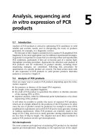
Fig. 1 Flow-chart of the step-by-step selection of children hospitalized with community-acquired pneumonia diagnosed on clinical grounds
Simbalista et al. BMC Pediatrics (2015) 15:166
Page 4 of 9
(CDC [Center for Disease Control and Prevention] and
WHO) and malnutrition and severe malnutrition were
defined as Z-score for weight-for-age index under −2.00
or −3.00, respectively, using the National Centre for
Health Statistics standard [19].
CAP was classified as non-severe, severe or very severe
according to WHO guidelines: patients with chest indrawing were classified as severe CAP and patients with somnolence, seizures, grunting when calm, nasal flaring, cyanosis,
or inability to drink were classified as very severe CAP [17].
If a child had chest indrawing along with any item that
would classify him/her as very severe CAP, the final classification was very severe CAP.
We compared the frequency of demographic and clinical findings detected upon admission and on each day of
hospital course up to the 2nd day between patients with
radiologically-confirmed pneumonia and those with normal CXR or without radiologically-confirmed pneumonia.
Table 1 Baseline and clinical characteristics of children hospitalized with community-acquired pneumonia diagnosed on clinical
grounds
Characteristics
Radiologically-confirmed pneumonia
a
Gender male
Age strataa,
Yes (n = 109)
Normal CXR (n = 143)
P
Noe (n = 174)
P
70 (64.2)
77 (53.8)
0.098
87 (50.0)
0.019
28 (25.7)
63 (44.1)
0.003
73 (42.0)
0.005
b
2-11 months
1-4 years
67 (61.5)
68 (47.6)
0.028
87 (50.0)
0.059
≥ 5 years
14 (12.8)
12 (8.4)
0.250
14 (8.0)
0.188
100 (91.7)
112 (78.3)
0.004
140 (80.5)
0.010
n = 79
n = 80
5 (3–7); 1-30
4(2–6); 1-20
0.093
4(2–7);1-20
0.299
0.720
152 (87.4)
0.483
History of current illness
fevera
c
duration of fever
a
n = 105
cough
92 (84.4)
123 (86.0)
duration of coughc
n = 59
n = 80
7 (4–9); 1-45
4.5 (3–7); 1-31
0.022
5(3–7.5);1-31
0.053
breathlessnessa
67 (61.5)
104 (72.7)
0.058
124 (71.3)
0.087
duration of breathlessnessc
n = 46
n = 77
2 (1–6); 1-30
3 (1–4.5); 1-30
0.894
3(1–5);1-30
0.504
68/85d (80.0)
79/106d (74.5)
0.372
98/131d (74.8)
0.377
d
d
d
n = 93
n = 93
Physical examination findings
tachypnoeaa
a
fever
52/99 (52.5)
62/121 (51.2)
0.849
79/148 (53.4)
0.895
cracklesa
42 (38.5)
86 (60.1)
0.001
100 (57.5)
0.002
wheezinga
32 (29.4)
86 (60.1)
<0.001
99 (56.9)
<0.001
chest retractiona
38 (34.9)
53 (37.1)
0.719
69 (39.7)
0.418
76 (69.7)
76 (53.1)
0.008
97 (55.7)
0.019
severe
22 (20.2)
47 (32.9)
0.025
55 (31.6)
0.036
very severea
11 (10.1)
20 (14.0)
0.351
22 (12.6)
0.515
b
Severity according to WHO
non-severea
a
a
chest indrawing
30 (27.5)
58 (40.6)
0.032
67 (38.5)
0.058
nasal flaringa
7 (6.4)
17 (11.9)
0.143
19 (10.9)
0.202
somnolencea
1 (0.9)
1 (0.7)
1.000
1 (0.6)
1.000
seizurea
1 (0.9)
1 (0.7)
1.000
1 (0.6)
1.000
2 (1.8)
1 (0.7)
0.580
1 (0.6)
0.561
cyanosis
a
CXR indicates chest radiograph
WHO indicates World Health Organization
a
Data are shown as n (%)
b
The frequencies in each age stratum or in the severity groups according to WHO were compared as dichotomic variables
c
Data are shown as median (IQR); minimum-maximum
d
Different denominators are due to missing data
e
Includes normal CXR plus CXR with atelectasis or peribronchial thickening
Simbalista et al. BMC Pediatrics (2015) 15:166
Page 5 of 9
This last group comprised patients with normal CXR or
CXR with atelectasis or peribronchial thickening. A
subgroup comparison was performed when wheezers were
excluded. We also compared the frequency of length of
hospital stay and treatment as well as the final outcome
upon discharge between these groups. Categorical variables were compared by using chi-square or Fisher exact
test as appropriate, and continuous variables were assessed
by using Mann–Whitney U test due to non-parametrical
distribution. Multi-variable logistic regression analysis by
enter method was used to assess independent association
between radiologically-confirmed pneumonia and factors
which significantly differed during hospital course in the
bivariate analysis. The multi-variable analysis was performed in a model adjusted for age and for the same
factor present upon admission. The statistical tests
were two tailed, with a significance level of 0.05. The
software SPSS (version 9.0, IBM, Armonk, New York)
was used for the analysis. The exclusion criteria were
chosen for the purpose of addressing potential confounders. Blinding to the radiological diagnosis during
medical charts review was performed to address potential bias.
The study was conducted according to the principles
expressed in the Declaration of Helsinki and it was
approved by the Ethics Committee at Federal University
of Bahia. Informed consent was deemed unnecessary
due to the retrospective collection of data. Identification
of the patients was kept confidential.
Results
During the study period, 921 cases were detected and
456 patients fulfilled the inclusion criteria. After excluding 132 (29.0 %) cases due to underlying debilitating
illnesses, a further 39 (8.5 %) with pleural effusion
detected on the CXR taken upon admission, and an additional 2 (0.4 %) due to other radiological diagnoses such
as calcification and hilar lymphadenomegaly (Fig. 1), the
final study group comprised 283 (62.1 %) patients. Overall, 157 (55.5 %) patients were males, the median age
was 17 months (IQR [interquartile range]: 9–34 months;
minimum 2 months; maximum 9.2 years) and 101
(35.7 %) patients were aged under 1 year. Upon admission, the most common complaints were cough (86.2 %),
fever (84.8 %), breathlessness (67.5 %), and the most frequent findings were tachypnoea (76.9 %), fever (53.0 %),
crackles (50.2 %), wheezing (46.3 %), chest retraction
(37.8 % ) and chest indrawing (34.3 %). CAP was severe
or very severe among 77 (27.2 %) and 33 (11.7 %) patients, respectively. Malnutrition was detected in 21 (7.4)
cases and severe malnutrition in 1 (0.4 %) case.
The compared subgroups included 109 (38.5 %) children with radiologically-confirmed pneumonia, 143
(50.5 %) children with normal CXR and 31 (11.0 %)
with other radiological diagnoses (atelectasis or peribronchial thickening). In the radiologically-confirmed
pneumonia subgroup, pulmonary infiltrate was classified as alveolar (94.5 %), alveolar-interstitial (3.7 %) or
interstitial (1.8 %). Additional radiological findings were
Table 2 Significant differences during hospital course of children hospitalized with community-acquired pneumonia diagnosed on
clinical grounds
Characteristics
Radiologically-confirmed pneumonia
Yes
Normal CXR
P
Nod
P
a
n = 109
n = 143
Fever
29/86c (33.7)
17/107c (15.9)
0.004
25/131 (19.1)
0.015
0.043
39 (22.4)
0.027
D1
Chest indrawing
b
n = 174
b
13 (11.9)
31 (21.7)
n = 109b
n = 143b
31/98c (31.6)
16/119c (13.4)
0.001
24/148 (16.2)
0.004
Fever
26/62 (41.9)
9/46 (19.6)
0.014
14/59 (23.7)
0.033
Chest indrawing
6/77 (7.8)
9/57 (15.8)
0.147
12/75 (16.0)
0.117
28/69 (40.6)
10/50 (20.0)
0.017
15/68 (22.1)
0.020
D2a
Fever
Without wheezers
D1a
D2a
Fever
Data are shown as n (%)
CXR indicates chest radiograph
a
D1 is the first day after aqueous penicillin G has been initiated (24 h of treatment), D2 is the second day after aqueous penicillin G has been initiated (48 h
of treatment)
b
n = number of evaluated patients in each subgroup on the respective day of hospital course
c
Different denominators due to missing data
d
Includes normal CXR plus CXR with atelectasis or peribronchial thickening
Simbalista et al. BMC Pediatrics (2015) 15:166
Page 6 of 9
atelectasis (2.8 %) and peribronchial thickening (3.7 %).
The baseline characteristics are compared in Table 1.
Children without radiologically-confirmed pneumonia
were younger than those with radiologically-confirmed
pneumonia (median [IQR]: 14 [7–28 months versus 21
[12–44] months; P = 0.001). No difference was found
in the frequency of malnutrition (10 [9.2 %] versus 11
[6.3 %]; P = 0.373).
Overall, the median duration of hospitalization was
7 days (IQR: 5–10; minimum 2; maximum 31), and the
median duration of aqueous penicillin G use was 4 days
(IQR: 3–6; minimum 2; maximum 17). Children with
radiologically-confirmed pneumonia stayed in hospital
for as long as children without radiologically-confirmed
pneumonia (median 7 days [IQR: 4–10] versus median
7 days [IQR: 5–9]; P = 0.903). No difference was found
between the two subgroups regarding duration of penicillin use (radiologically-confirmed pneumonia: median
4 days [IQR: 3–6] versus no radiologically-confirmed
pneumonia: median 4 days [IQR: 3–6]; P = 0.402). Overall, aqueous penicillin G was substituted by other antibiotics
in 29 (10.2 %) cases. Children with radiologically-confirmed
pneumonia had aqueous penicillin G substituted more frequently than those without radiologically-confirmed pneumonia (15.6 % versus 6.9 %; P = 0.019).
No patient died and everyone was discharged after improvement. Table 2 presents the significant differences
found during progression of disease between children
with or without radiologically-confirmed pneumonia or
normal CXR during aqueous penicillin G treatment.
Those with substitution of aqueous penicillin G were
excluded. The comparison of the symptoms and signs
during hospital course which did not demonstrate significant difference is shown in Table 3. Table 4 depicts the
multi-variable analysis of factors whose difference was significant in the bivariate analysis presented in Table 2.
Table 3 Symptoms and signs without significant differences during hospital course of children hospitalized with communityacquired pneumonia diagnosed on clinical grounds
Characteristics
Radiologically-confirmed pneumonia
Yes
Normal CXR
P
a
n = 109
Tachypnoea
54/90c (60.0)
51/107c (47.7)
Cyanosis
0
0
-
0
-
Chest retraction
21 (19.3)
29 (20.3)
0.842
37 (21.3)
0.685
D1
b
n = 143
P
Nod
n = 174
b
b
0.084
65/132c (49.2)
0.115
Somnolence
1 (0.9)
1 (0.7)
1.000
1 (0.6)
1.000
Nasal flaring
4 (3.7)
4 (2.8)
0.730
5 (2.9)
0.737
Grunting
0
1 (0.7)
1.000
1 (0.6)
1.000
Seizure
0
0
-
0
-
Cough
41 (37.6)
44 (30.8)
0.255
55 (31.6)
0.299
Breathlessness
18 (16.5)
33 (23.1)
0.199
42 (24.1)
0.127
n = 109b
n = 143b
D2a
c
n = 174b
c
0.496
67/145c (46.2)
Tachypnoea
45/93 (48.4)
52/119 (43.7)
Cyanosis
1 (0.9)
0
0.433
0
0.385
Chest indrawing
11 (10.1)
14 (9.8)
0.937
19 (10.9)
0.826
Chest retraction
14 (12.8)
16 (11.2)
0.688
20 (11.5)
0.734
Somnolence
2 (1.8)
0
0.186
0
0.148
Nasal flaring
1 (0.9)
1 (0.7)
1.000
1 (0.6)
1.000
Grunting
0
1 (0.7)
1.000
1 (0.6)
1.000
Seizure
0
0
-
0
-
Cough
44 (40.4)
65 (45.5)
0.419
79 (45.4)
0.406
Breathlessness
25 (22.9)
28 (19.6)
0.517
34 (19.5)
0.494
Data are shown as n (%)
CXR indicates chest radiograph
a
D1 is the first day after aqueous penicillin G has been initiated (24 h of treatment), D2 is the second day after aqueous penicillin G has been initiated (48 h
of treatment)
b
n = number of evaluated patients in each subgroup on the respective day of hospital course
c
Different denominators due to missing data
d
Includes normal CXR plus CXR with atelectasis or peribronchial thickening
0.742
Simbalista et al. BMC Pediatrics (2015) 15:166
Page 7 of 9
Table 4 Multi-variable analysis of factors associated with radiologically-confirmed pneumonia during hospital course in bivariate analysis,
adjusted for age and for the same factor upon admission, among children hospitalized with community-acquired pneumonia diagnosed
on clinical grounds
Compared subgroup
Normal CXR
CXR without pneumonia
Without wheezers
Characteristics
OR (95 % CI)
P
OR (95 % CI)
P
OR (95 % CI)
P
Fever on D1a
2.18 (1.07-4.43)
0.031
1.75 (0.92-3.34)
0.091
2.00 (0.89-4.48)
0.094
Age
1.00 (1.00-1.00)
0.062
1.00 (1.00-1.00)
0.023
1.00 (0.99-1.00)
0.089
Report of fever upon admission
4.01 (1.54-10.42)
0.004
3.47 (1.35-8.94)
0.010
1.75 (0.58-5.23)
0.317
Chest indrawing on D1
0.65 (0.31-1.37))
0.259
0.60 (0.29-1.22)
0.160
0.60 (0.20-1.77)
0.354
Age
1.00 (1.00-1.00)
0.033
1.00 (1.00-1.00)
0.024
1.00 (1.00-1.00)
0.072
a
Chest indrawing upon admission
0.67 (0.38-1.19)
0.174
0.74 (0.43-1.28)
0.281
0.67 (0.30-1.46)
0.311
Fever on D2a
2.66 (1.32-5.36)
0.006
2.16 (1.15-4.06)
0.016
2.24 (1.04-4.79)
0.039
Age
1.00 (1.00-1.00)
0.086
1.00 (1.00-1.00)
0.044
1.00 (0.99-1.00)
0.116
Report of fever upon admission
4.15 (1.61-10.67)
0.003
3.65 (1.44-9.23)
0.006
2.01 (0.69-5.83)
0.199
Multi-variable analysis by logistic regression
CXR indicates chest radiograph
CXR without pneumonia includes normal CXR plus CXR with atelectasis or peribronchial thickening
a
D1 is the first day after aqueous penicillin G has been initiated (24 h of treatment), D2 is the second day after aqueous penicillin G has been initiated (48 h
of treatment)
Discussion
This study provides evidence that children hospitalized
with CAP diagnosed on clinical grounds treated with
aqueous penicillin G, present differences during hospital
course when radiologically-confirmed pneumonia cases
are compared to others without radiologically-confirmed
pneumonia or with normal CXR. Notably, patients with
radiologically-confirmed pneumonia were significantly
more feverish on admission and during the first 2 days
of aqueous penicillin G use. This finding remained when
wheezers were excluded from the analysis. It is important to recall that children included in this study were
otherwise healthy and had no significant comorbidity.
Several methodological constraints should be highlighted
in this investigation. As data were collected retrospectively, there was no control on variables measurement
and, as patients were evaluated by different observers,
standardization of evaluations could not be guaranteed.
Also, no aetiological agent was determined. However,
strict criteria for enrolling and grouping the cases were
used, and those with potential confounding variables
were excluded. Moreover, the study was performed in a
teaching hospital where the same standardized procedures for assistance have been used over the period of
the study [15]. Interestingly, all children included in the
analysis had pneumonia diagnosed and were admitted
to hospital by paediatricians.
The presence of fever has been lately associated with
radiologically-confirmed pneumonia. A recent study has
estimated that presence of fever increases the chance of
children hospitalized with lower respiratory tract disease
to have radiologically-confirmed pneumonia by 2.5 times
[20]. Additionally, it has been demonstrated that the inclusion of fever in the WHO criteria for the clinical
diagnosis of CAP substantially increases its specificity,
particularly in children with wheezing [21]. The history
of fever has also been recognized as the symptom with
the greatest sensitivity for the presence of pulmonary infiltrates [22]. Our data provide evidence that persistence
of fever up to the second day of treatment is also more
frequent among hospitalized children with radiologicallyconfirmed pneumonia.
In a previous investigation which compared the progression of symptoms among children with non-severe acute
lower respiratory tract infection with and without a radiological diagnosis of pneumonia, tachypnoea persisted
longer during treatment among those with radiologicallyconfirmed pneumonia [12]. Herein, this finding was not
found, possibly due to sample size. Children without
radiologically-confirmed pneumonia had higher frequency
of wheezing, which is a potential confounding factor for
the diagnosis of CAP among children with tachypnoea
[23, 24]. The high frequency of children with a clinical
diagnosis of CAP and without radiologically-confirmed
pneumonia is in accordance with previous studies. Up to
82 % of children with tachypnoea and wheezing had normal CXR in Pakistan [11]. The prescription of antibiotics
based on only tachypnoea should be restricted to
settings where CXR performance is not feasible. The
lower frequency of fever [23] and the younger age [25]
in the subgroup without radiologically-confirmed pneumonia may also guide the clinical suspicion to lower
respiratory tract diseases other than CAP, for example
bronchiolitis.
Simbalista et al. BMC Pediatrics (2015) 15:166
The evidence that there is no effect of an admission
CXR in the outcome of paediatric outpatients with CAP
was provided in a study in which all those children, irrespective of having CXR taken, received antibiotics. That
means, those who needed antibiotics received antibiotics, as well as those who did not need antibiotics but
instead had a self-limited disease [6]. It has been recently
shown that radiologically-confirmed pneumonia is associated with bacterial infection [26]. Although CXR is
undoubtedly limited in determining the aetiology of
pneumonia [7], it may help identify children with a
lower respiratory tract disease and a probable nonbacterial aetiology, such as bronchiolitis, who can benefit
from not receiving unnecessary antibiotics.
Conclusions
This is the first study to demonstrate the differences in
hospital course between hospitalized children with CAP diagnosed on clinical grounds with or without radiologicallyconfirmed pneumonia. We highlight differences on the
hospital course between the studied subgroups. The performance of CXR may be a tool to select patients who
would not benefit from receiving antibiotics and could be
followed-up instead.
Page 8 of 9
4.
5.
6.
7.
8.
9.
10.
11.
12.
Competing interests
The authors declare that they have no competing interests.
13.
Authors’ contributions
CMN-C designed the study, RS reviewed the medical charts, collected and
entered the data, MA read the chest radiographs, DCA and ICB analyzed the
data. All authors contributed to the interpretation of the results. RS drafted
the manuscript. DCA, ICB and MA contributed to the writing and CMN-C
proofread the manuscript. All authors read and approved the final
manuscript.
Acknowledgments
There was no funding for this investigation. The authors thank the medical
chart unit of the Federal University of Bahia Hospital, in Salvador, Brazil for
their cooperation in getting the medical charts to be reviewed.
14.
15.
16.
17.
Author details
1
Postgraduate Program in Pathology, Federal University of Bahia School of
Medicine, Salvador, Brazil. 2Postgraduate Program in Health Sciences, Federal
University of Bahia School of Medicine, Salvador, Brazil. 3Image Diagnosis,
Image Memorial Unit and Bahia Hospital, Salvador, Brazil. 4Department of
Paediatrics, Federal University of Bahia School of Medicine, Salvador, Brazil.
18.
19.
Received: 16 January 2015 Accepted: 13 October 2015
20.
References
1. Walker CL, Rudan I, Liu L, Nair H, Theodoratou E, Bhutta ZA, et al.
Global burden of childhood pneumonia and diarrhea. Lancet.
2013;381:1405–16.
2. World Health Organization. The United Nations Children’s Fund. Global
Action Plan for Prevention and Control of Pneumonia. 2009. http://
www.whqlibdoc.who.int/hq/2009/WHO_FCH_CAH_NCH_09.04_eng.pdf.
Accessed 20 May 2012.
3. World Health Organization. Programme for the control of acute respiratory
infections. Acute respiratory infections in children: case management in small
hospitals in developing countries. 1990. />1990/WHO_ARI_90.5.pdf. Accessed 26 May 2013.
21.
22.
23.
24.
Cherian T, Mulholland EK, Carlin JB, Ostensen H, Amin R, Campo M, et al.
Standardized interpretation of paediatric chest radiographs for the diagnosis
of pneumonia in epidemiological studies. Bull World Health Organ.
2005;83:353–9.
Swingler GH, Zwarenstein M. Chest radiograph in acute respiratory
infections. Cochrane Database of Syst Rev 2008, Issue 1. Art. No.:
CD001268. doi: 10.1002/14651858.CD001268.pub3. http://
www.onlinelibrary.wiley.com/doi/10.1002/14651858.CD001268.pub3/
abstract. Accessed 14 June 2012.
Swingler GH, Hussey GD, Zwarenstein M. Randomised controlled trial of
clinical outcome after chest radiograph in ambulatory acute lowerrespiratory infection in children. Lancet. 1998;351:404–8.
Korppi M, Don M, Valent F, Canciani M. The value of clinical features in
differentiating between viral, pneumococcal and atypical bacterial
pneumonia in children. Acta Paediatr. 2008;97:943–7.
Johnson J, Kline JA. Intraobserver and interobserver agreement of the
interpretation of pediatric chest radiographs. Emerg Radiol.
2010;17:285–90.
Harris M, Clark J, Coote N, Fletcher P, Harnden A, McKean M, et al.
British Thoracic Society guidelines for the management of
community acquired pneumonia in children: update 2011. Thorax.
2011;66:1–23.
Bradley JS, Byington CL, Shah SS, Alyerson B, Carter ER, Harrison C, et al. The
management of community-acquired pneumonia in infants and children
older than 3 months of age: clinical practice guidelines by the Pediatric
Infectious Diseases Society and the Infectious Diseases Society of America.
Clin Infect Dis. 2011;53:25–76.
Hazir T, Nisar YB, Qazi SA, Khan SF, Raza M, Zameer S, et al. Chest
radiography in children aged 2–59 months diagnosed with non-severe
pneumonia as defined by World Health Organization: descriptive
multicentre study in Pakistan. BMJ. 2006;333:629.
Fontoura MS, Matutino AR, Silva CC, Santana MC, Nobre-Bastos M, Oliveira F,
et al. Differences in evolution of children with non-severe acute lower
respiratory tract infection with and without radiographically diagnosed
pneumonia. Indian Pediatr. 2012;49:363–9.
Gentile A, Bardach A, Ciapponi A, García-Marti S, Aruj P, Glujovsky D, et al.
Epidemiology of community-acquired pneumonia in children of Latin
America and the Caribbean: a systematic review and meta-analysis. Int J
Infect Dis. 2012;16:5–15.
Simbalista R, Araújo M, Nascimento-Carvalho CM. Outcome of children
hospitalized with community-acquired pneumonia treated with aqueous
penicillin G. Clinics. 2011;66:95–100.
Nascimento-Carvalho CM, Souza-Marques HH. Recommendation of the
Brazilian Society of Pediatrics for antibiotic therapy in children and
adolescents with community-acquired pneumonia. Rev Panam Salud
Publica. 2004;15:380–7.
El-Radhi AS, Barry W. Thermometry in paediatric practice. Arch Dis Child.
2006;91:351–6.
World Health Organization. Integrated Management of Childhood Illness
chart booklet (WC 503.2). 2008. />2008/9789241597289_eng.pdf. Accessed 15 January 2009.
Nascimento-Carvalho CM. Physical signs in children with pneumonia. Indian
Pediatr. 2001;38:307–8.
World Health Organization. Training Course on Child Growth Assessment.
2008. />9789241595070_A_eng.pdf. Accessed 13 July 2009.
Key NK, Arajo-Neto CA, Cardoso MR, Nascimento-Carvalho CM.
Characteristics of radiographically diagnosed pneumonia in under-5
children in Salvador, Brazil. Indian Pediatr. 2011;48:873–7.
Cardoso MR, Nascimento-Carvalho CM, Ferrero F, Alves FM, Cousens SN.
Adding fever to WHO criteria for diagnosing pneumonia enhances the
ability to identify pneumonia cases among wheezing children. Arch Dis
Child. 2011;96:58–61.
Lynch T, Platt R, Gouin S, Larson C, Patenaude Y. Can we predict which
children with clinically suspected pneumonia will have the presence of
focal infiltrates on chest radiographs? Pediatrics. 2004;113:e186–9.
Mathews B, Shah S, Cleveland RH, Lee EY, Bachur RG, Neuman MI. Clinical
predictors of pneumonia among children with wheezing. Pediatrics.
2009;124:e29–36.
Castro AV, Nascimento-Carvalho CM, Ney-Oliveria F, Araújo-Neto CA,
Andrade SC, Loureiro LL, et al. Additional markers to refine the World
Simbalista et al. BMC Pediatrics (2015) 15:166
Page 9 of 9
Health Organization algorithm for diagnosis of pneumonia. Indian Pediatr.
2005;42:773–81.
25. Zorc JJ, Hall CB. Bronchiolitis: recent evidence on diagnosis and
management. Pediatrics. 2010;125:342–9.
26. Nascimento-Carvalho CM, Araújo-Neto CA, Ruuskanen O. Association
between bacterial infection and radiologically confirmed pneumonia
among children. Pediatr Infect Dis J. 2015;34:490–3.
Submit your next manuscript to BioMed Central
and take full advantage of:
• Convenient online submission
• Thorough peer review
• No space constraints or color figure charges
• Immediate publication on acceptance
• Inclusion in PubMed, CAS, Scopus and Google Scholar
• Research which is freely available for redistribution
Submit your manuscript at
www.biomedcentral.com/submit