Systemic and local humoral immune response against F-1 and lasota strains of new castle disease virus in chicken
Bạn đang xem bản rút gọn của tài liệu. Xem và tải ngay bản đầy đủ của tài liệu tại đây (184.71 KB, 10 trang )
Int.J.Curr.Microbiol.App.Sci (2019) 8(9): 194-203
International Journal of Current Microbiology and Applied Sciences
ISSN: 2319-7706 Volume 8 Number 09 (2019)
Journal homepage:
Original Research Article
/>
Systemic and Local Humoral Immune Response against F-1 and LaSota
Strains of New Castle Disease Virus in Chicken
Rajesh Singathia1*, Ravindra Sharma2 and Satishkumar Batra2
1
Department of Veterinary Microbiology, College of Veterinary and Animal Sciences,
Navania, Vallabhnagar, Udaipur-313601 (Rajasthan), India
2
Department of Veterinary Microbiology, College of Veterinary Sciences, C.C.S. Haryana
Agricultural University, Hisar, India
*Corresponding author
ABSTRACT
Keywords
Newcastle disease
virus, Chicks,
Humoral immunity,
F-1, LaSota
Article Info
Accepted:
15 July 2019
Available Online:
10 August 2019
F-1 and LaSota strains of NDV were propagated in laboratory using 10-day old
embryonated hen eggs via allantoic cavity route. One group of thirty chicks were infected
with F-1strain and other with LaSota strain with 105 egg infective doses in 100 µl of virus
via oral and ocular route. Sequential sera samples and tracheal samples at day 0, 3,7,14, 21
and 28 post infection were collected and titre of antibodies indicative of humoral immune
response was determined by indirect ELISA. The findings of present study lead to the
conclusion that infection with NDV induces production of humoral immune response in
chickens. Humoral antibodies generated in responses to NDV infection in chickens are
both of local (IgA, IgG) and serum antibodies (IgG and IgM) type. The F-1 strain of NDV
appears to be slightly better immunogenic than that of LaSota strain of NDV.
Introduction
Newcastle disease (ND) is an OIE listed and
highly contagious viral disease affecting over
250 species of birds of all age groups
(Alexander, 1997). ND is one of the lethal,
zoonotic diseases causing colossal economic
losses in poultry industry due to high
morbidity and mortality. The disease is caused
by a single stranded, enveloped, nonsegmented
RNA
virus
i.e.
avian
paramyxovirus
serotype-1
(APMV-1)
classified under genus Avulavirus of family
Paramyxoviridae (Mayo, 2002). The virus
infection occurring through respiratory and/or
gastrointestinal tract results in production of
clinical signs accompanied with high
mortality. Lesions of disease are mainly
produced in respiratory, gastrointestinal and
nervous system. Both inactivated and live
attenuated virus vaccines are being used
commercially for immunization of birds. The
present study was planned to study the
systemic and local humoral immune response
194
Int.J.Curr.Microbiol.App.Sci (2019) 8(9): 194-203
against F-1 and LaSota strains of New castle
disease virus (NDV) in chicken
28 days post inoculation of virus and stored
frozen at -200Ctill further use.
Materials and Methods
Indirect Enzyme linked immunosorbent
assay (ELISA) for antibody assay
Virus
A seed stock of F-1 and LaSota strain of NDV
were procured in lyophilized form and
propagated in 10-day old embryonated eggs
via allantoic cavity route. A rapid slide
haemagglutination (HA) test was performed
on the allantoic fluid to confirm the presence
of virus. The harvested allantoic fluid was
purified by using standard methods. For use as
antigen in ELISA, the purified virus was
inactivated by exposing to ultraviolet light for
40 minute (Reynolds and Maraqa, 2000) and
virus titration was performed by calculating
the Egg infective dose 50 (EID50) of both F-1
and LaSota strain in 10-day old embryonated
chicken eggs by the standard method (Reed
and Muench,1938).
Experimental chicks
Ninety broiler chicks were randomly divided
into three groups of 30 birds each. Of these,
one batch was vaccinated with F-1strain and
second with LaSota strain with 105 egg
infective doses in 100 µl of virus via oral and
ocular route. Third batch were kept as control.
Serum samples collected from immunized and
control birds were subjected to indirect ELISA
for measuring NDV specific serum
immunoglobulins (Igs). The dilution of
antigen, monoclonal antibody and conjugate
was optimized by checker board titration of
these reagents.
Micro ELISA polyvinyl plates (Nunc) were
coated with optimum dilution (1:10) of NDV
antigen in carbonate bicarbonate buffer and
were incubated at 370C for 1 hour (h) and then
kept at 40C overnight.
After three washing with Phosphate buffer
saline (PBS) containing 0.05 percent (v/v)
Tween-20 (PBST), which was the wash buffer
used throughout, the plates were blocked
using 100 μl blocking buffer and incubated at
37°C for 1 h. Thereafter, 50 µl of double fold
dilution of each serum sample (in duplicate) in
blocking buffer was added and incubated at
370C for 1 h followed by washing with PBST.
The optimum dilution of monoclonal antibody
(Kind Gift from Dr. R.C. Jones, University of
Liverpool, U.K.) specific for chicken Igs were
then added and incubated for 1 h.
Collection of samples
Collection of serum
Serum samples were collected from each of
five randomly selected birds from each group
at 0, 3, 7, 14, 21 and 28 days using standard
keys.
Collection of tracheal exudates
Tracheal exudates were obtained from five
chickens from each group at 0, 3, 7, 14, 21 and
The plates were then washed thrice with PBST
and 50 µl of optimally diluted rabbit antimouse Igs Horse raddish peroxidase (HRPO)
conjugate (Sigma Chemical Co.) was added in
all the wells and incubated further for 45
minute at 370C. Plates were washed three
times with PBST. Finally, 50 µl of freshly
prepared substrate solution of orthophenylenediamine (OPD) (Sigma Chemical Co.) was
added in each well and plates were left in dark
for development of color reaction. The
reaction was stopped by adding 50 µl of 1M
195
Int.J.Curr.Microbiol.App.Sci (2019) 8(9): 194-203
H2SO4 / well. The optical density of the wells
was measured in an ELISA reader using 492
nm filter and titer was calculated (Khatri,
2000).
ELISA IgM antibody titres in serum of
broiler chicks immunized with different
strains of NDV using F-1 strain of virus as
coating antigen
Results and Discussion
Humoral immune response against NDV was
determined by assaying titre of different type
of antibodies i.e. IgG, IgM and IgA in serum
and tracheal exudate of infected birds at
different days post immunization (DPI). The
antibody titre was determined by indirect
ELISA using F-1 and LaSota strains of NDV
as coating antigen and anti-chicken
immunoglobulin monoclonals as tracing
antibody.
Antibody titre in serum of broiler chicks
immunized with different strains of NDV
ELISA IgG antibody titres in serum of
broiler chicks immunized with different
strains of NDV using F-1 strain of virus as
coating antigen
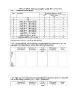
Serum IgG antibody titres as determined by
indirect ELISA on different DPI in different
groups of broiler chicks are shown in table 1.
An increase in the serum IgG antibody titres in
the group of birds infected with F-1 strain of
NDV were observed on 7 DPI and remained
so at 14 DPI which showed an increase on 21st
day and peaked on 28 DPI. Serum IgG
antibody titre in the group of chicks infected
with LaSota strain of NDV were observed on
3 DPI and remained at the same level up to 14
DPI which showed an increase on day 21 and
peaked on 28th DPI. In the serum from control
group of birds, there was no detectable serum
IgG antibody titre. The groups inoculated with
F-1 or LaSota showed a significant higher
antibody titre (P<0.05) than that of control. No
significant variation was observed in serum
IgG titres with in birds immunized with F-1 or
LaSota strain of NDV.
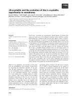
ELISA IgM antibody titres in serum of
different groups of broiler chicks on different
DPI are shown in Table 2. An increase in the
serum IgM antibody titres in the group of
birds infected with F-1 strain of NDV were
observed on 3 DPI and remained so at 7 DPI,
peaked on day 14th followed by a decline by
28 DPI. The serum IgM antibody titres in the
group of chicks immunized with LaSota strain
of NDV were above that of control birds on
3rdDPI and remained almost at the same level
up to day 7 post infection, peaked on 14th day
followed by a decline by 28 DPI. The titre of
serum IgM antibodies in birds immunized
with either F-1 or with LaSota strain of NDV
did not exhibit any statistical significant
(P<0.05) difference at various time intervals
of serum testing but these were significantly
different (P<0.05) as compared to that of
control birds.
Antibody
concentration
in
tracheal
exudates of broiler chicks immunized with
different strains of NDV
ELISA IgA antibody O.D. values in
tracheal exudates of broiler chicks
immunized with different strains of NDV
using F-1 strain of virus as coating antigen
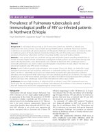
The effect of immunization with different
strains of NDV on induction of IgA antibody
responses in tracheal exudates in different
groups of broiler chicks on different DPI are
shown in Table 3.A rise in IgA antibody
optical density (O.D.) values in tracheal
washing of birds belonging to group
inoculated with F-1 strain was recorded on 7
DPI which peaked on 14th day and remained at
almost the same level upto 28 DPI. The IgA
196
Int.J.Curr.Microbiol.App.Sci (2019) 8(9): 194-203
antibody O.D. in immunized chicks were
significantly higher (P<0.05) compared to that
of control chicks on 7 & 14 DPI.A rise in IgA
antibody O.D. values in birds immunized with
LaSota strain of NDV was observed on 7 DPI
followed by an increase on day 14. It peaked
on 28th DPI. Responses of immunized chicks
were slightly higher as compared to that of
control chicks but statistically these
differences were found to be insignificant
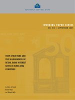
chicks immunized with either F-1 or LaSota
strain of NDV at different DPI are shown in
table 4.A higher than control IgG antibody
O.D. value and hence the antibody
concentration in the group immunized with F1 strain was detected on 14 DPI and it peaked
at 28 DPI. The O.D. values of immunized
chicks were significantly higher (P<0.05)
when compared with that of control chicks on
7, 14 and 28 DPI.
IgG antibody ELISA O.D. in tracheal
exudates of broiler chicks immunized with
different strains of NDV using F-1 strain of
virus as coating antigen
A similar pattern of ascending O.D. values
was noted in tracheal exudates of birds
inoculated with LaSota strain. The O.D.
values of immunized chicks were significantly
higher (P<0.05) as compared with that of
control chicks on 7th and 28th DPI.
IgG antibody O.D. values as obtained in
ELISA test in different groups of broiler
Table.1 ELISA IgG antibody titres in serum of broiler chicks immunized with different strains
of NDV using F-1 strain of virus as coating antigen
Groups
Control
F-1
LaSota
0
<1.30A
0.00
<1.30A
0.00
<1.30A
0.00
Serum antibody titres (log10 )
Days post immunization
3
7
14
± <1.30A ± <1.30B ± <1.30 A ±
0.00
0.00
0.00
A
A
± 1.42
± 1.60
± 1.60A ±
0.07
0.09
0.10
A
AB
± 1.54
± 1.48
± 1.54A ±
0.11
0.07
0.18
21
28
<1.30C ± <1.30B ±
0.00
0.00
A
2.02
± 2.32A ±
0.07
0.12
B
1.66
± 2.20A ±
0.11
0.10
Means with the same letter are not significantly different (P<0.05)
Table.2 ELISA IgM antibody titres in serum of broiler chicks immunized with different strains
of NDV using F-1 strain of virus as coating antigen
Groups
Control
F-1
LaSota
0
<1.30A
0.00
<1.30A
0.00
<1.30A
0.00
Serum antibody titres (log10)
Days post immunization
3
7
14
± <1.30B ± <1.30C ± <1.30B ±
0.00
0.00
0.00
± 1.78A ± 1.72A ± 2.20A ±
0.15
0.07
0.10
AB
B
± 1.60
± 1.53
± 2.26A ±
0.19
0.08
0.11
Means with the same letter are not significantly different (P<0.05)
197
21
28
<1.30B
<1.30A
±0.00
±0.00
1.90A ± 1.68A ±
0.10
0.14
A
1.78
± 1.54A ±
0.12
0.18
Int.J.Curr.Microbiol.App.Sci (2019) 8(9): 194-203
Table.3 ELISA IgA antibody O.D. values in tracheal exudates of broiler chicks immunized with
different strains of NDV using F-1 strain of virus as coating antigen
Groups
3
O. D. values
Days post immunization
7
14
Control
0.03A ±0.00
0.03B ± 0.00
0.04B ± 0.00
0.03A ± 0.00
F-1
0.02A ± 0.01
0.13A ± 0.03
0.26A ± 0.06
0.27A ± 0.12
LaSota
0.02A ± 0.01
0.07AB ± 0.02
0.17AB ± 0.06
0.18A ± 0.04
28
Means with the same letter are not significantly different (P<0.05)
Table.4 ELISA IgG antibody O.D. in tracheal exudates of broiler chicks immunized with
different strains of NDV using F-1 strain of virus as coating antigen
Groups
3
O. D. values
Days post immunization
7
14
Control
0.03A ± 0.00
0.03B ± 0.00
0.04B ± 0.00
0.03B ± 0.00
F-1
0.04A ± 0.00
0.11A ± 0.01
0.23A ± 0.05
0.26A± 0.02
LaSota
0.04A ± 0.01
0.10A ± 0.01
0.11B ± 0.01
0.25A ± 0.03
28
Means with the same letter are not significantly different (P<0.05)
was detectable at 3rd and 7thday post
immunization (DPI), respectively. The
antibody titre increased gradually and peaked
on 28th (DPI). Our finding support the earlier
findings of Zoth et al., (2008) who reported
that IgG induced by low virulent virus was
detectable at day 7 post vaccination. Further
the present study also confirm the observation
of Marquardt et al., (1985)who reported that
the ELISA and HI titres responses began at 7
DPI, rose moderately and peaked at 21st day
in the chicks vaccinated at 2nd week by nostril
and eye route method. Similarly, our findings
are in conformation with the findings of
Ratnaparkhe et al., (1981) who reported that
the HI antibody response increased and
The present study was undertaken to
investigate the kinetics of humoral mediated
immune response against NDV infection in
broiler chicken.
The oral and conjunctival route has been used
to immunize the chicks with either F-1 or
LaSota strain of NDV. Both these routes of
immunization have previously been used
successfully by the other workers (Reynolds
and Maraqa, 2000; Reetha et al., 2001; AlGarib et al., 2003a; Zoth et al., 2008).
In the present study, an increase in IgG
antibody titre in sera of broiler chicks
immunized with LaSota or F-1 strain of NDV
198
Int.J.Curr.Microbiol.App.Sci (2019) 8(9): 194-203
reached a peak at 3rd and 4.5th week after
vaccination with LaSota strain of NDV
inoculated with 107.2 EID50 per chick at 3
week of age by oral and oculonasal route,
respectively. Our finding is also supported by
similar observation of Sharma and Singh
(1986) who reported that the HI antibody titre
started to increase after vaccination and
reached a peak (20.8) on 28th day in the
chicks (6th day of age) infected intranasally
and intraocularly. Similarly, Rahman et al.,
(2004) showed that HI titre, in chickens (7th
day of age) immunized with V4HR-ND
vaccine by eye drops route of inoculation, had
significantly (p<0.01) increased and reached
maximum (5.07±0.50) on 38th day of age.
Similarly, Tanwani and Malik (1978) also
found that chicks vaccinated with different
vaccines by intranasal and intramuscular
routes showed satisfactory HI antibody titres
from 2nd to 6th months of their age and after
that a gradual fall was observed. Otim et al.,
(2005) also reported that HI antibody titres
started to increase and then peaked on 2 week,
then declined after vaccination with LaSota
vaccine in the village chicks (3 week old).
Russell and Ezeifeka (1995) showed that in
chicks (3 day old) immunized with Hitchner
B1 strain of NDV by oculotopical route,
serum IgG antibody response first detectable
on 8th day after vaccination continued to rise
in the titre.
of the IgG, IgA class and HI antibodies was
detectable at day 7 post immunization (PI) in
serum and plateau reached a peak on day 14.
Similarly, Mishra et al., (1985) reported that
chicken vaccinated with different strains of
NDV, a maximum antibody titre of 2560 as
estimated by ELISA was detectable between
14th and 21st days post vaccination. Mast et
al., (2005) have reported that after intranasal
inoculation with LaSota strains of NDV, a
highest IgG antibody titre was observed on
day 15 post infection. Similarly, Dandapat et
al., (2005) observed that in chicks immunized
with conventional RDF vaccine through
occulo nasal route, the peak HI antibody was
log2 6.22 at 2nd week PI which again
increased at 4th week PI after receiving
booster dose. This was followed by a gradual
decline to log2 5.4 at 6th week PI. Similarly,
Kumar et al., (1988) have shown that one
week after vaccination of birds with LaSota
strain of NDV, the HI antibodies reached a
titre of 3.0 logs which peaked on 3rd week
(4.5 logs) and then declined to 1.0 log by 9th
week post exposure. The observations of
Linghua et al., (2007) that chicks vaccinated
with NDV vaccine remained serologically
negative for virus specific antibodies when
tested by ELISA test are in contrast to finding
of the present study where IgG response was
observed in chicks from day 3 onwards.
Ewert et al., (1979) reported that HI antibody
in birds which were immunized at 6th week of
age by intramuscular or local (intratracheally
+ intranasally) route increases after
immunization and reached maximum values
on 10th and 14th days post vaccination,
respectively. Lambrecht et al., (2004)
reported an increase in the HI antibody titre
which peaked on 3rd or 4th week after
vaccination with live NDV vaccine or killed
vaccine respectively.
Our findings are in agreement with that of
Mast et al., (2006) who observed that in
chicks receiving 108 EID50 of F + HN mutant,
the virus specific IgG antibodies were
detectable at day 4 post infection which
gradually increased with age, together with
the increasing HI titres which peaked on 14
day post infection, indicating isotype
switching and active production of NDVspecific IgG. Similarly, Al-Garib et al.,
(2003b) reported that after inoculation with
live NDV virus (LaSota and Roakin) by
oculo-nasal route, an increase in antibody titre
Similarly, Chandrasekar et al., (1988)
observed that in chicks (4 day old) immunized
with RDVF by intranasal route developed a
199
Int.J.Curr.Microbiol.App.Sci (2019) 8(9): 194-203
vaccination and it peaked (3.5 log10) on 8th
DPI. The difference in peak in antibody titre
may be attributed to the use of different strain
of NDV.
gradual increase in HI antibodies level from
the first to 4th week, followed by a fall upto
8th week of immunization. Similar findings
have been reported by Shuaib et al., (2006)
and Satyanarayana and Reddy (1977).
Similarly, Hilgers et al., (1998) found
increase in HI antibody titre in chicks which
were vaccinated with inactivated NDV by
intramuscular route. Carrasco et al., (2008)
found that after infection with Sao Joao do
Meriti strain of NDV, HI titre started to rise
and were maximum at day 21 or 35 DPI. A
similar observation has been reported by
Spradbrow et al., (1987).
Apart from serum IgM and IgG responses,
production of local immune response in the
form of IgA and IgG antibodies was also
investigated in the present study.
In the present study, a detectable increase in
the IgA O.D. values was observed at day 7th
PI which peaked at day 14 PI. A similar
observation has been made by Al-Garib et al.,
(2003b) who reported that after inoculation
with live NDV by oculo-nasal route, IgA
response rose at day 4 post exposure (PE) and
reached a plateau at day 7 PE and then
declined.
Similarly,
Jayawardne
and
Spradbrow (1995) also reported an increase in
IgA antibody titre in tracheal washings,
intestinal washings and lachrymal fluid after
vaccination by intra crop and eye drop
inoculation. In the present study, a low level
of IgA O.D. values observed in tracheal
exudates might be due to dilution of local
antibodies associated with the method of
collection. In contrast to our finding, Perozo
et al., (2007) found that IgA antibody level
remained undetectable upto day 35 DPI after
in ovo vaccination with recombinant avian
adeno-associated vaccine (rAAAV) coding
for NDV haemagglutin in neuramindase. The
delay in mounting of IgA antibody response
was attributed to the failure of recombinant
vaccine virus to stimulate a measurable
mucosal immune response by itself, probably
due to the nature of the antigenic stimulation
induced by the rAAAV which is a replication
defective virus and is dependent upon host
cell machinery to express the HN antigen.
The IgM antibodies are the first to be made by
the B cells in response to any microbial
infection which later on switch to IgG or any
other isotype. The IgM antibody titre in serum
of broiler chicks immunized with F-1 and
LaSota strains of NDV were detectable at
3rdDPI and peaked on day 14 post infection.
These results corroborate the finding of Mast
et al., (2006) who reported a NDV-specific
IgM response which peaked around 14 days
of age in chicks vaccinated at embryonic day
18 with 103 and 104 EID50 of the F + HN
mutant. Similarly, Al-Garib et al., (2003b)
reported that after inoculation with live NDV
virus (LaSota and Roakin) by oculo-nasal
route, an increase in antibody titre of the IgM
class of immunoglobulin was detectable at
day 4th PI in serum and reached a plateau
level at 7th DPI. Mast et al., (2005) have
reported that in one day old chicks after
intranasal inoculation with 106 EID50 of
LaSota strain of NDV, IgM antibody
responses were highest on 10th day post
infection. The variation in peak IgM antibody
responses observed may be due to a different
route of inoculation adopted by the
investigators. Russell and Ezeifeka (1995)
showed that in chicks (3 day old) immunized
with Hitchner B1 strain of NDV by
oculotopical route, serum IgM antibody
response was first detectable on 5th day after
The presence of IgG antibodies was also
detected in tracheal exudates during the
present study. A higher IgG specific O.D.
value detectable at day 7th PI was observed. It
200
Int.J.Curr.Microbiol.App.Sci (2019) 8(9): 194-203
then showed ascending pattern and reached a
peak at day 28 PI. This result conforms to the
findings of Ewert and Eidson (1977), who
also observed a similar pattern of IgG
antibody concentration and postulated that the
Igs other than IgA may have a role in the
protection of tracheal mucosa. Similarly, AlGarib et al., (2003b) reported that after
inoculation with live NDV by oculo-nasal
route; a higher IgG antibody titre was
detected on 7th DPI which reached a plateau at
14th DPI. Zoth et al., (2008) have reported a
similar finding that a significant higher IgG
antibody response was detected on day 21st in
tracheal swabs from the birds immunized with
live NDV vaccine by eye drop route of
inoculation. Ewert et al., (1979) reported that
anti-NDV IgA and anti-NDV IgG levels
increased after immunization and reached
maximum values between 10 and 14 days
post vaccination in birds which were
immunized at 6th week of age by
intramuscularly or intratracheally and
intranasal route.
References
Alexander, D.J. 1997. Newcastle disease and
other avian paramyxoviridae infections.
In: Diseases of Poultry. 10th ed. Calnek,
B.W., Barnes, H.J., Beard, C.W.,
McDougald, L.R., Saif, Y.M. (eds.).
Ames, IA: Iowa State University Press.
pp. 541-569.
Al-Garib, S.O., Gielkens, A.L.J.,Gruys, E.
and Kochi, G. 2003a. Review of
Newcastle disease virus with particular
references to immunity and vaccination.
World Poult. Sci. J.59:185-200.
Al-Garib, S.O., Gielkens, A.L.J.,Gruys, E.,
Hartog, L. and Kochi, G. 2003b.
Immunoglobins class distribution of
systemic and muosal antibody response
to Newcastle disease in chickens. Avian
Dis.47: 32-40.
Carrasco, A.D.O.T., Seki, M.C., Raso,
T.D.F., Paulillo, A.C. and Pinto, A.A.
2008. Experimental infection of
Newcastle disease virus in pigeons
(Columba livia): Humoral antibody
response, contact transmission and viral
genome shedding.Vet. Microbial.129:
89-96.
Chandrasekar,
S.,
Venkatesan,
R.A.,
Padamanaban, V.D. andMasiilamony,
P.R. 1988. Humoral immune response
to Ranikhet disease virus vaccine in
chicks. Indian Vet. J. 65: 653-657.
Dandapat, S., Nagarajan, K., Kataria, J.M.,
Bash, B.B. and Yadav, M.P. 2005.
Humoral and cell mediated immune
response to Ranikhet disease vaccine
delivered through PLG microspheres in
broiler chickens. Indian J. Poult. Sci.
40(1): 47-51.
Ewert, D.L. and Eidson, C.S. 1977. Effect of
bursectomy
and
depletion
of
immunoglobulin A on antibody
production and resistance to respiratory
challenge after local or systemic
vaccination of chickens with Newcastle
The results of the present study indicate that
humoral components of immune system are
stimulated by the vaccine virus. The humoral
components of the immune system respond
by production of both local antibodies (IgA
and IgG) and systemic or serum antibodies
such as IgG and IgM. The F-1 strain of NDV
appears to be more immunogenic than LaSota
strain of NDV. No attempt was made during
the present study to isotype the antibodies
produced. Investigation of the isotype of
antibodies produced in response to NDV virus
will further help in understanding the
mechanism of immune response generated
during NDV infection/vaccination.
Acknowledgment
The Authors thanks the Dean, College of
Veterinary Sciences,
C.C.S.
Haryana
Agricultural University, Hisar for providing
necessary facilities for this study.
201
Int.J.Curr.Microbiol.App.Sci (2019) 8(9): 194-203
disease virus. Infect. Immun. 18: 146150.
Ewert, D.L., Barger, B.O. and Eidson, C.S.
1979. Local antibody synthesis to
Newcastle disease virus by solid-phase
radioimmunoassay
and
immunofluorescene with classicalspecific
antibody
for
chicken
immunoglobulins. Infect. Immun.24(1):
269-275.
Hilgers, L.A.T., Nicolas, I., Lejeune, G.,
Dewil, E. and Boon, B. 1998.Effect of
various adjuvants on secondary immune
response
in
chickens.
Vet.
Immunol.Immunopathol.66:159-171.
Jayawardne, G.W.L. and Spradbrow, P.B.
1995. Mucosal immunity in chickens
vaccinated with V4 stain of Newcastle
disease virus. Vet. Microbiol.46: 69-77.
Khatri, M. 2000. Studies on natural and
vaccine immunity against foot and
mouth disease virus in calves. Ph.D.
Thesis, CCS Haryana Agricultural
university, Hisar.
Kumar, K.U., Swamy, S.K. and Reddy, T.V.
1988.Humoral
and
cell-mediated
immune response in chicks vaccinated
against Newcastle disease. Kerala J.
Vet. Sci.19(2): 116-121.
Lambrecht, B., Gonze, M., Meulemans, G.
and Van de berg, T.P. 2004. Assesment
of the cell-mediated immune response
in chickens by detection of chickens
interferon-γ in response to mitogen and
recall Newcastle disease viral antigen
stimulation. Avian Pathol., 33(3): 343350.
Linghua, Z.,Xingshan, T. and Fengzhen, Z.
2007. Vaccination with Newcastle
disease
vaccine
and
CpG
oligodeoxynucleotides induces specific
immunity and protection against
Newcastle disease virus in SPF chicken.
Vet. Immunol. Immunopathol. 115:
216-222.
Marquardt, W.W., Synder, D.B., Savage,
P.K., Kadavil, S.K. and Yancey, F.S.
1985. Antibody response to Newcastle
disease virus given by two different
routes as measured by ELISA and
Hemagglutination- inhibiton test and
associated tracheal immunity. Avian
Dis.29(1): 71-79.
Mast, J., Nanbru, C., Decaesstecker, M.,
Lambrecht,
B.,
Couvreur,
B.,
Meulemans, G. and Van den berg, T.
2006. Vaccination of chickens embryos
with escape mutants of LaSota
Newcastle disease virus induces a
protective immune response. Vaccine.
24: 1756-1765.
Mast, J., Nanbru, C., Van Den Berg, T. and
Meulemans, G. 2005. Ultrastructual
changes of the tracheal epithelium after
vaccination of day-old chicks with
LaSota strain of the Newcastle disease
virus. AvainPathol.42: 559-565.
Mayo, M.A. 2002. A summary of the
taxonomic changes recently approved
by ICTV.Arch.Virol. 147: 1655-1663.
Mishra, S.C., Rai, A. and Jaiswal, T.N. 1985.
An enzyme linked immunosorbent
assay for estimation of antibodies to
Newcastle disease virus strains.
Acta.Virol.29:151-157.
Otim, M.O., Mukiibi-Muka, G., Christensen,
H. and Bisgaard, M. 2005.Aflatoxicosis,
infectious bursal disease and immune
response
to
Newcastle
disease
vaccination in rural chickens.Avian
Pathol.34(4): 319-323.
Perozo, F., Finol, G. and Mavárez, Y. 2007.
Levels of Immunoglobulin-A in trachea,
gut and Bile samples of chickens
vaccinated against Newcastle Disease.
Rev. Cient. (Maracaibo).17(3): 226230.
Rahman, M.B., Rahman, M.M., Rahman, M.,
Kabir, S.M.L., Nazir, K.H.M.N.H. and
Amin, M.M. 2004. Efficacy of V4HR
Newcastle disease (V4 HR-ND) vaccine
in broiler birds in Bangladesh. Indian. J.
202
Int.J.Curr.Microbiol.App.Sci (2019) 8(9): 194-203
Poult. Sci. 39(5): 365-368.
Ratnaparkhe, P., Tanwani, S.K. and Pathak,
P.N. 1981. Comparative immune
response and antibody response of
CDF-66 strain of New scastle disease
virus with other four lentogenic vaccine
strains. Indian J. Poult. Sci. 16: 235242.
Reed, L.V. and Muench, H. 1938. A simple
method of estimating fifty per cent end
points. Am. J. Hyg. 27:493-497.
Reetha, T.L., Rajeswar, J.J., Dorairajan, N.
and Thangamani, R. 2001. Assessment
of immune response in chicken in
different
commercially
available
Ranikhet disease vaccines by ELISA
test.Indian J. Anim. Hlth. 40(1): 9-12.
Reynolds, D.L. and Maraqa, A.D. 2000.
Protective immunity against Newcastle
disease: the role of cell-mediated
immunity. Avian Dis.44: 145-154.
Russell, P.H. and Ezeifeka, G.O. 1995. The
Hitchner B1 strain of Newcastle disease
virus induces high levels of IgA, IgG
and IgM in newly hatched chicks.
Vaccine. 13: 61- 66.
Satyanarayana, A. and Reddy, A.M.K. 1977.
Pattern of development and duration of
immunity in chicks protected with F
strain Ranikhet disease vaccine. Indian
Vet. J. 54: 509-516.
Sharma, K. and Singh, G. 1986. A note on
serologic and immunologic response to
Ranikhet Disease F strain vaccine in
young broiler chicks. Poult. Adv.19: 3536.
Shuaib, M., Khan, H., Sajid-Ur-Rehman and
Ashfaque, M. 2006. Humoral immune
response to Newcastle disease vaccine
(LaSota strain) in broiler. International
J. Poult. Sci. 5(5): 411-414.
Spradbrow, P.B., Samuel, J.L. and Ibrahim,
A.L. 1987. Serological response of
chickens to oral vaccination with
Newcastle
disease
virus.Vet.
Microbial.16: 255-262.
Tanwani, S.K. and Malik, B.S. 1978. A
comparative study of immune &
antibody response of lentogenic strain
CDF-66 and other vaccine strain of
Ranikhet disease virus. Indian. Vet. J.
55: 267-278.
Zoth, S.C., Gomez, E., Carrillo, E. and
Berinstein, A. 2008. Locally produced
mucosal IgG in chickens immunized
with
conventional
vaccines
for
Newcastle disease virus. Brazilian J.
Med. Biol. Res. 41: 318-323.
How to cite this article:
Rajesh Singathia, Ravindra Sharma and Satishkumar Batra. 2019. Systemic and Local Humoral
Immune Response against F-1 and LaSota Strains of New Castle Disease Virus in Chicken.
Int.J.Curr.Microbiol.App.Sci. 8(09): 194-203. doi: />
203