Common carotid artery intima-media thickness is useful for diagnosis of the acute stage of Kawasaki disease
Bạn đang xem bản rút gọn của tài liệu. Xem và tải ngay bản đầy đủ của tài liệu tại đây (806.36 KB, 6 trang )
Wu et al. BMC Pediatrics 2014, 14:98
/>
RESEARCH ARTICLE
Open Access
Common carotid artery intima-media thickness is
useful for diagnosis of the acute stage of
Kawasaki disease
Ting-Hsin Wu†, Hsuan-Chang Kuo†, You-Lin Tain, Kuan-Miao Lin, Ho-Chang Kuo and Shao-Ju Chien*
Abstract
Background: This study aimed to investigate intima-media thickness (IMT) of the common carotid arteries in
children with acute Kawasaki disease (KD).
Methods: Between 2009 and 2011, patients fulfilling the criteria for KD, including a fever lasting >5 days, were
prospectively enrolled in this study. Laboratory data, echocardiography, and IMT were measured and compared
with matched controls.
Results: A total of 70 common carotid IMTs were measured in 35 children. We studied 21 patients aged 3–60
months old with acute KD and 14 febrile patients aged 3–194 months old with acute infection and similar
characteristics to those of KD patients. Children with KD had a significantly higher IMT compared with the controls
(0.550 ± 0.081 mm vs. 0.483 ± 0.046 mm, P = 0.01).
Conclusions: IMT during the acute stage of KD is increased, suggesting that IMT could be a useful diagnostic tool
in the early diagnosis of KD.
Keywords: Kawasaki disease, Common carotid artery, Intima-media thickness, Acute stage
Background
Kawasaki disease (KD) is an acute systemic vasculitis
that mainly affects medium-sized arteries in multiple
systems and primarily occurs in children under the age
of 5 years. Diagnosis of KD is clinically made using
non-specific diagnostic guidelines, including prolonged
fever, conjunctivitis, diffuse mucosal inflammation,
polymorphous skin rashes, indurative edema of the
hands and feet associated with peeling of the fingertips,
and non-suppurative lymphadenopathy [1,2]. There is currently no definitive laboratory test available for diagnosis
of KD. Diagnosis of KD is further complicated because the
above-listed clinical criteria may be transient. In addition,
the constellation of principle physical findings of KD may
also vary over time or are not obvious either in the early
stages of disease or in cases of incomplete KD. An expedited diagnosis of this disease is crucial to administer
* Correspondence:
†
Equal contributors
Department of Pediatrics, Kaohsiung Chang Gung Memorial Hospital and
College of Medicine, Chang Gung University, 123 Ta-Pei Road, Niaosung,
Kaohsiung, Taiwan
appropriate therapy and to potentially limit the development of coronary artery lesions [1,2]. The most serious
complication in KD is coronary aneurysm due to severe
inflammation and vasculitis of the coronary arteries,
especially when treatment is delayed [3-7]. Treatment
with intravenous immunoglobulin within the first 10 days
after onset of KD is highly effective for the acute phase of
this illness and considerably reduces the prevalence of coronary artery complications [1,3,7-10]. Therefore, early recognition and prompt treatment of KD are crucial.
Measurement of intima-media thickness (IMT) of
common carotid arteries is a widely used and validated
noninvasive imaging technique for the assessment of
early structural changes in the arterial wall. Inflammation
of vessel walls precedes morphological changes and is
believed to be the initial step in many rheumatic diseases,
including Takayasu’s arteritis, [11,12] systemic lupus
erythematosus, [13] rheumatoid arthritis, [14] Behçet’s
disease, [15] and ankylosing spondylitis [16,17]. Only a
few published studies, some of them controversial, have
investigated the development of an increased IMT in the
© 2014 Wu et al.; licensee BioMed Central Ltd. This is an Open Access article distributed under the terms of the Creative
Commons Attribution License ( which permits unrestricted use, distribution, and
reproduction in any medium, provided the original work is properly credited.
Wu et al. BMC Pediatrics 2014, 14:98
/>
common carotid artery in patients diagnosed with KD.
Previous studies have reported that in long-term follow
up, carotid artery IMT is greater in patients with KD, and
patients with coronary artery involvement after KD have
the largest IMT [18,19].
The purpose of this study was to investigate IMT in the
acute stage of KD and to determine the level of vascular
involvement. This study describes the ultrasound findings
of carotid IMT in patients with active KD to evaluate its
utility as a marker of disease activity and to elucidate the
role of carotid IMT in the early diagnosis of KD.
Methods
Between 2009 and 2011, 21 patients, aged 3–60 months
old, treated at the Kaohsiung Chang Gung Children’s
Hospital and fulfilling the criteria for KD (more than four
principle criteria and a fever lasting >5 days) were prospectively enrolled in this study as previously described in detail
[9,20,21]. Fourteen febrile patients, aged 3–194 months old,
with acute infections and similar characteristics to those
of KD patients underwent the same imaging studies
(two-dimensional echocardiography and B-mode ultrasound) for a heart murmur survey, and were used as
the control group. The control group comprised patients
who had pneumococcal pneumonia (n = 5), salmonella enterocolitis (n = 4), or acute pyelonephritis (n = 5). Patients
whose symptoms did not fit the criteria for KD, had an
acute fever for <5 days, or in cases where data were incomplete were excluded. Children with known heart
disease or traditional risk factors of atherosclerosis,
such as hypertension, diabetes mellitus, a family history of premature congestive heart disease, and obesity
(body mass index [BMI] >95th percentile for the agespecific reference group), were also excluded.
In addition to performing echocardiography and ultrasonography of the common carotid arteries, all included
patients underwent clinical evaluation, and their age, sex,
weight, height, (BMI), pulse, and blood pressure were
recorded. Blood pressure was the mean of two consecutive measurements obtained with the patient in a seated
position after resting for >5 min. Laboratory tests routinely performed included a complete blood cell count,
differential white blood cell count, and C-reactive protein
(CRP; reference range, ≤5 mg/L) measurement.
Two-dimensional echocardiography and B-mode
ultrasonography
All patients with KD underwent two-dimensional echocardiography of the coronary artery and high-resolution
B-mode ultrasonography of the bilateral common carotid
arteries to measure IMT prior to intravenous immunoglobulin treatment. All of the children in the control group
underwent the same examinations as KD patients during
acute infections. The left coronary artery was measured
Page 2 of 6
midway between the ostium and the bifurcation of the
circumflex artery and the left anterior descending coronary artery in the parasternal short-axis view. The proximal
right coronary artery was obtained 3 to 5 mm distal to its
origin in the parasternal short-axis view. Coronary artery
dimensions were measured and z-scores were calculated
from the formula derived from Dallaire et al. [22] in 2011
using regression with the square root of body surface area
because of the differences in age and BMI between the
two groups. We chose this method because this equation
yielded pediatric z-scores with an appropriate normal
distribution across the entire range of body surface area
on the basis of a large number of infants, children, and
adolescents. The z-scores beyond the normal limits
(cutoff of Z = ±2) were considered abnormal. The incidence
of abnormal coronary arteries was also analyzed.
A single, experienced pediatric cardiologist who was
blinded to the diagnoses performed all of the carotid
ultrasound assessments. The carotid ultrasonographic
studies were performed under standardized conditions
with the patients in the supine position for at least
10 min in a quiet room prior to examination. For data acquisition, high-resolution ultrasound equipment (SONOS
7500; Phillips Medical Systems, Andover, MA) with an
11-MHz linear array probe was used. All studies were
performed according to a standardized scanning protocol
for the right and left common carotid arteries [21-24].
During the examination, all children were in the supine
position with their heads turned slightly to the side. The
transducer was manipulated so that the near and far walls
of the common carotid arteries were parallel to the transducer footprint and the lumen diameter was maximum in
the longitudinal plane. The entire carotid proximal common carotid artery was observed approximately 1.5 cm
before the bifurcation. We used the distance between the
leading edges of the luminal-intimal interface and the
medial-adventitial interface for the measurement of IMT.
IMT was measured during end diastole as determined by
the R wave on an electrocardiogram. We also scrolled
through the cine loop and measured IMT at the arteries’
largest diameter in two infants in the KD group because
they were not cooperative during monitoring of the
electrocardiogram. All images were stored digitally and
subsequently analyzed offline. We used Qlab Software
(Philips, Germany) to analyze the IMT distance automatically at 64 points within a segment of 10 mm. The
value reported by this software was the arithmetic
mean IMT. The IMT echo was assessed and measured
with calipers within a standardized higher resolution
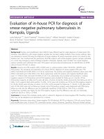
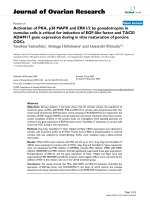
zoom. We chose the image of the best quality with the
clearest edge (Figure 1), which was always obtained with
the interfaces oriented perpendicular to the ultrasound
beam. This image was acquired, temporarily stored in the
cine loop, and consecutively zoomed in Qlab when the
Wu et al. BMC Pediatrics 2014, 14:98
/>
Page 3 of 6
Figure 1 Measurement of carotid IMT in Qlab.
measurements were performed. Manual overreading of
border detection during computed analysis was performed
for all images. Each measurement was accompanied by a
“success rate”, which was the percentage of the intimamedia within the region of interest that was accurately
measured. We only used the measured values that had a
success rate of ≥95%. The use of standard automatic procedures for IMT measurements limits the variability related
to human error and allows comparability between studies.
Our experienced pediatric cardiologist had performed carotid ultrasound for several years. For reproducibility of
IMT measurements, we calculated the intraobserver coefficient of variation, which was 4.1%.
This study protocol was approved by the Institutional
Review Board of Chang Gung Memorial Hospital, Taiwan
and was performed in accordance with the ethical standards laid down in the 1964 Declaration of Helsinki.
Informed consent was obtained from all parents prior
to the children’s inclusion in the study.
with no significant differences in age, sex, body size, or
blood pressure. Because there was no routine oral sedation for the heart murmur survey, clear edges of IMT
were easier to obtain from older children who were
more cooperative for carotid ultrasound. Although the
age in the control group was older than that in the KD
group, this was not significantly different. There were
no significant differences in weight, height, BMI, or
systolic and diastolic blood pressure between the two
groups. There was also no significant difference in CRP
levels or white blood cell counts between the groups.
Platelet counts were significantly higher in KD patients
than in controls. There was no significant difference in
coronary artery diameter between the two groups.
However, the incidence of z-scores beyond the normal
limit (cutoff of Z = ±2) of the left coronary artery and
left anterior descending coronary artery in KD patients
was significantly higher than that in the control group.
Mean carotid IMT
Statistical analysis
All continuous data are expressed as mean ± standard
deviation of the mean and minimum and maximum values.
We used non-parametric statistical procedures because of
the small number of data. Categorical variables were analyzed with Fisher’s exact test and the chi-square test when
appropriate. We also performed multivariate regression
analysis using IMT as the dependent variable, and age,
BMI, and KD as independent variables to assess whether
different factors affected carotid IMT. All analyses were
performed using the Statistical Package for Social Sciences,
version 14.0 for Windows XP (SPSS, Inc., Chicago, USA).
Results
Demographic data and coronary artery diameter
Patient demographics are shown in Tables 1 and 2.
Controls and KD patients had similar characteristics,
Measurement of carotid IMT by Qlab software, accompanied by the IMT within a segment of 10 mm and its
Table 1 Demographic data and blood pressure of patients
with KD and febrile controls
Characteristic
Male sex (%)
Patients with
KD (n = 21)
Controls
(n = 14)
p value*
71.4%
71.4%
1.00
Mean age (months)
21.79 ± 17.91
52.18 ± 56.52
0.13
Body weight (kg)
11.37 ± 3.93
19.17 ± 14.50
0.08
Body height (cm)
82.32 ± 14.91
100.54 ± 29.91
0.21
BMI (kg/m2)
16.45 ± 1.50
18.08 ± 2.64
0.14
Systolic BP (mmHg)
103 ± 16
112 ± 14
0.35
Diastolic BP (mmHg)
63 ± 13
71 ± 11
0.09
Values are mean ± SD or %.
BMI, body mass index; BP, blood pressure; KD, Kawasaki disease.
*Mann–Whitney U test.
Wu et al. BMC Pediatrics 2014, 14:98
/>
Page 4 of 6
Table 2 Inflammation markers, echocardiographic
parameters, and IMT values in patients with KD and
febrile controls
Characteristic
LCA (mm)
z-score >2 or < −2
LAD (mm)
z-score >2 or < −2
LCX (mm)
z-score >2 or < −2
RCA (mm)
z-score >2 or < −2
3
IMT measurements
Patients with
KD (n = 21)
Controls
(n = 14)
p value*
2.79 ± 0.77
2.51 ± 0.56
0.35
52.4% (n = 11)
7.1% (n = 1)
0.01
2.30 ± 0.90
1.90 ± 0.35
0.40
47.6% (n = 10)
0
0.00
1.64 ± 0.63
1.45 ± 0.17
0.57
9.5% (n = 2)
0
0.51
2.57 ± 1.24
2.17 ± 0.54
0.59
17.1% (n = 6)
0
0.06
LVEF (%)
Total WBC (×1000/mm3)
Table 3 Multivariate regression model of IMT
measurements with KD, age, and BMI
65 ± 7
65 ± 6
0.96
16.12 ± 7.01
15.13 ± 6.95
0.69
Platelets (×1000/mm )
487.5 ± 198.0
352.5 ± 161.1
0.01
CRP (mg/L)
110.4 ± 91.5
148.6 ± 89.6
0.17
IMT (mm)
0.550 ± 0.08
0.483 ± 0.05
0.01
Values are mean ± SD or %.
LCA, left coronary artery; LAD, left anterior descending coronary artery; LCX,
left circumflex coronary artery; RCA, right coronary artery; LVEF, left ventricular
ejection fraction; WBC, white blood cells; CRP, C-reactive protein; IMT,
intima-media thickness.
*Mann–Whitney U test.
100% success rate on the right side, are shown in Figure 1.
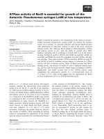
The distribution of IMT is shown in Figure 2. Mean carotid IMT in KD patients (0.550 ± 0.081 mm; range,
0.44–0.69 mm), was significantly higher than that in the
febrile control group (0.483 mm ± 0.046 mm; range,
0.43–0.56 mm; P = 0.01) (Table 2). Even if age and BMI
were not significantly different between KD patients
and controls, the difference between the two groups was
highly relevant. We performed multiple linear regression analysis using IMT measurement as the dependent
variable to exclude the confounding factors of age and
BMI. In multivariate analysis, only KD was consistently
associated with intima-medial thickening (Table 3).
0.8
IMT(mm)
0.7
0.6
0.5
0.4
0.2
0.0
Control
Kawasaki disease
Figure 2 Distribution of IMT measurements. Bars show mean ± SD.
Regression coefficient
Standard error
p value
KD
0.072
0.028
0.017
Age
0.000
0.000
0.465
BMI
0.005
0.006
0.471
Variables
IMT, intima-media thickness; BMI, body mass index; KD, Kawasaki disease.
Discussion
Our study showed that carotid IMT was higher in the
acute stage of KD compared with other acute infections.
Although there was no significant difference in age between
the two groups, higher IMT of KD patients may still be due
to age. Age-associated changes in IMT in young children
have not yet been fully examined. Pauciullo et al. [23]
reported that the mean carotid IMT of healthy children
aged 6 ± 3 years was 0.39 ± 0.03 mm and Ishizu et al.
[24] reported that it was 0.44 ± 0.05 mm among children
aged between 5 to 14 years old. An age-related increase
in carotid IMT with an annual 0.009 mm increase in
healthy children has been reported to reflect the physiological growing process [24]. Theoretically, in our study,
IMT in KD patients should have been much lower than
that in the control group because of their age. However,
we found that IMT was significantly higher in KD patients
who were younger than the control group. Therefore, the
difference between groups is due to the underlying disease
rather than the selection bias of age. Furthermore, we
performed multivariate regression analysis, which showed
that IMT was the only independent factor for KD. All of
the other factors lost their statistical influence on the IMT
thickening. These preliminary results suggest that an increased IMT of the carotid artery in any acutely ill, febrile
child should raise suspicion for KD. Therefore, carotid
ultrasonography is an important diagnostic tool in the
early diagnosis of KD if an increased IMT is shown in
conjunction with additional clinical signs.
Over the last few years, a large number of studies have
emphasized the fundamental role of ultrasonography in
the early diagnosis of vasculitis in adult patients. These
studies demonstrated abnormal ultrasonographic changes
in the acute phase of Takayasu disease, [11,12,24,25].
Mediterranean fever, [26] temporal arteritis, [27] and
Behçet’s disease [15]. These diseases are considered to
have early structural vascular alterations and atherosclerosis because of their ongoing subclinical inflammation. In
these diseases, IMT was used to assess blood vessels and
to help with early detection. Intravascular ultrasound
studies in patients with Takayasu showed thickening and
altered echogenicity of the arterial wall [24]. Most authors
believe that these changes are a result of acute dysfunction
of the endothelium and inflammation of the vascular wall
Wu et al. BMC Pediatrics 2014, 14:98
/>
rather than chronic proliferative and fibrotic changes of
atherosclerosis.
To the best of the authors’ knowledge, no specific IMT
studies of common carotid arteries have been performed to
help diagnose KD. The present study attempted to distinguish KD from other infection diseases on the basis of
IMT. We also attempted to determine the level of vascular
involvement by evaluating the role of IMT in the diagnosis
of KD. In contrast to the combination of cellular and fibrous proliferation, which accumulates during long-term
follow-up after KD, increased IMT in the acute stage is entirely attributed to acute inflammation [28]. Impairment of
endothelial function precedes morphological changes and is
believed to be the initial step in the development of many
other inflammatory rheumatic diseases. Dysfunction of
the endothelium and the presence of macrophages and
activated lymphocytes within the vessel wall lead to
thickening of the intima and media of the vessel wall of
large- and medium-sized muscular arteries [29]. Highresolution B-mode ultrasonography is useful in showing
the same characteristic features of homogenous hyperechogenicity of the thickened arterial wall [11,29].
One noteworthy limitation of this study is that the
ultrasonographic preliminary results were not correlated
with pathological findings. Pathological correlation may
help physicians further understand the causes of increased
wall thickness. This limitation occurred because in KD, biopsy is restricted to opportunities, such as intraoperative
biopsy during vascular reconstruction surgery. Another
limitation of our study is that our KD patients did not
have any coronary artery abnormalities or aneurysms.
During our short-term follow up, we did not find a trend
of persistent increased IMT as in previous studies [17].
These different preliminary results between studies may
be because KD patients enrolled in our study did not have
any considerable coronary artery involvement. This transient IMT phenomenon is similar to their transient coronary
dilation. Among KD children without marked coronary
artery abnormalities (as recruited in our study), intimamedial thickening during the acute phase is probably a transient, subclinical phenomenon without long-term sequelae
on atherosclerotic risk. Considering the small number of
cases included in our study, these results are primarily preliminary and further studies including a larger number of
patients are warranted. IMT can hopefully be used to reliably identify children most at risk for severe disease.
Conclusions
In children with laboratory preliminary results indicative
of vasculitis or suspicion of KD, but who do not fulfill
the criteria of KD, IMT could be an additional diagnostic
tool used to determine the level of vascular involvement.
This will help achieve an early diagnosis of KD and expedite establishment of an appropriate therapy.
Page 5 of 6
Abbreviations
BMI: Body mass index; IMT: Intima-media thickness; KD: Kawasaki disease.
Competing interests
The authors declare that they have no competing interests.
Authors’ contributions
SJC and TSW conceived of and participated in the design of the study, and
drafted the manuscript. YLT and KML participated in the design of the study
and performed the statistical analysis. HsCK and HoCK participated in
coordination of the study and helped to draft the manuscript. All authors
read and approved the final manuscript.
Acknowledgements
This study was supported in part by a grant (NSC 100-2314-B-182-061-MY3)
from the National Science Council of Taiwan and grants (CMRPG8A0481 and
CMRPG8B0151) from the Chang Gung Memorial Hospital, Taiwan. These
institutes had no influence on the collection, analysis, and interpretation of
the data, or on the preparation of the manuscript.
Received: 1 November 2012 Accepted: 13 March 2014
Published: 10 April 2014
References
1. Newburger JW, Takahashi M, Gerber MA, Gewitz MH, Tani LY, Burns JC,
Shulman ST, Bolger AF, Ferrieri P, Baltimore RS, Wilson WR, Baddour LM,
Levison ME, Pallasch TJ, Falace DA, Taubert KA: Diagnosis, treatment, and
long-term management of Kawasaki disease: a statement for health
professionals from the Committee on Rheumatic Fever, Endocarditis and
Kawasaki Disease, Council on Cardiovascular Disease in the Young,
American Heart Association. Circulation 2004, 110:2747–2771.
2. Kuo HC, Chang WC: Genetic polymorphisms in Kawasaki disease. Acta
Pharmacol Sin 2011, 32:1193–1198.
3. Wang CL, Wu YT, Liu CA, Kuo HC, Yang KD: Kawasaki disease: infection,
immunity and genetics. Pediatr Infect Dis J 2005, 24:998–1004.
4. Fukazawa R: Long-term prognosis of Kawasaki disease: increased
cardiovascular risk? Curr Opin Pediatr 2010, 22:587–592.
5. Benseler SM, McCrindle BW, Silverman ED, Tyrrell PN, Wong J, Yeung RS:
Infections and Kawasaki disease: implications for coronary artery
outcome. Pediatrics 2005, 116:e760–e766.
6. Burns JC, Glode MP: Kawasaki syndrome. Lancet 2004, 364:533–544.
7. Senzaki H: Long-term outcome of Kawasaki disease. Circulation 2008,
118:2763–2772.
8. Kuo HC, Yang KD, Chang WC, Ger LP, Hsieh KS: Kawasaki disease: an
update on diagnosis and treatment. Pediatr Neonatol 2012, 53:4–11.
9. Kuo HC, Yang KD, Juo SH, Liang CD, Chen WC, Wang YS, Lee CH, Hsi E, Yu
HR, Woon PY, Lin IC, Huang CF, Hwang DY, Lee CP, Lin LY, Chang WP,
Chang WC: ITPKC single nucleotide polymorphism associated with the
Kawasaki disease in a Taiwanese population. PLoS ONE 2011, 6:e17370.
10. Kuo HC, Liang CD, Yu HR, Wang CL, Lin IC, Liu CA, Chang JC, Lee CP, Chang
WC, Yang KD: CTLA-4, position 49 A/G polymorphism associated with
coronary artery lesions in Kawasaki disease. J Clin Immunol 2011,
31:240–244.
11. Schmidt WA, Nerenheim A, Seipelt E, Poehls C, Gromnica-Ihle E: Diagnosis
of early Takayasu arteritis with sonography. Rheumatology (Oxford) 2002,
41:496–502.
12. Lima DS, Sato EI, Lima VC, Miranda F Jr, Hatta FH: Brachial endothelial
function is impaired in patients with systemic lupus erythematosus.
J Rheumatol 2002, 29:292–297.
13. Van Doornum S, McColl G, Jenkins A, Green DJ, Wicks IP: Screening for
atherosclerosis in patients with rheumatoid arthritis: comparison of two
in vivo tests of vascular function. Arthritis Rheum 2003, 48:72–80.
14. Oflaz H, Mercanoglu F, Karaman O, Kamali S, Erer B, Genchellac H, Pamukcu
B, Umman S, Inanc M, Gul A: Impaired endothelium-dependent
flow-mediated dilation in Behcet’s disease: more prominent endothelial
dysfunction in patients with vascular involvement. Int J Clin Prac 2005,
59:777–781.
15. Poredos P: Intima-media thickness: indicator of cardiovascular risk and
measure of the extent of atherosclerosis. Vasc Med 2004, 9:46–54.
Wu et al. BMC Pediatrics 2014, 14:98
/>
Page 6 of 6
16. Sari I, Okan T, Akar S, Cece H, Altay C, Secil M, Birlik M, Onen F, Akkoc N:
Impaired endothelial function in patients with ankylosing spondylitis.
Rheumatology (Oxford) 2006, 45:283–286.
17. Dalla Pozza R, Bechtold S, Urschel S, Kozlik-Feldmann R, Netz H: Subclinical
atherosclerosis, but normal autonomic function after Kawasaki disease.
J Pediatr 2007, 151:239–243.
18. Cheung YF, Wong SJ, Ho MH: Relationship between carotid intima-media
thickness and arterial stiffness in children after Kawasaki disease.
Arch Dis Childhood 2007, 92:43–47.
19. de Groot E, Hovingh GK, Wiegman A, Duriez P, Smit AJ, Fruchart JC,
Kastelein JJ: Measurement of arterial wall thickness as a surrogate marker
for atherosclerosis. Circulation 2004, 109(23 Suppl 1):III33–III38.
20. Lee YC, Kuo HC, Chang JS, Chang LY, Huang LM, Chen MR, Liang CD, Chi H,
Huang FY, Lee ML, Huang YC, Hwang B, Chiu NC, Hwang KP, Lee PC, Chang
LC, Liu YM, Chen YJ, Chen CH, Chen YT, Tsai FJ, Wu JY: Two new
susceptibility loci for Kawasaki disease identified through genome-wide
association analysis. Nat Genet 2012, 44:522–525.
21. Kuo HC, Yang KD, Liang CD, Bong CN, Yu HR, Wang L, Wang CL: The
relationship of eosinophilia to intravenous immunoglobulin treatment
failure in Kawasaki disease. Pediatr Allergy Immunol 2007, 18:354–359.
22. Dallaire F, Dahdah N: New equations and a critical appraisal of coronary
artery Z scores in healthy children. J Am Soc Echocardiography 2011,
24:60–74.
23. Pauciullo P, Iannuzzi A, Sartorio R, Irace C, Covetti G, Di Costanzo A, Rubba
P: Increased intima-media thickness of the common carotid artery in
hypercholesterolemic children. Arterioscler Thromb 1994, 14:1075–1079.
24. Ishizu T, Ishimitsu T, Yanagi H, Seo Y, Obara K, Moriyama N, Watanabe S,
Yamaguchi I: Effect of age on carotid arterial intima-media thickness in
childhood. Heart Vessels 2004, 19:189–195.
25. Seth S, Goyal NK, Jagia P, Gulati G, Karthikeyan G, Sharma S, Talwar KK:
Carotid intima-medial thickness as a marker of disease activity in
Takayasu’s arteritis. Int J Cardiol 2006, 108:385–390.
26. Park SH, Chung JW, Lee JW, Han MH, Park JH: Carotid artery involvement
in Takayasu’s arteritis: evaluation of the activity by ultrasonography.
J Ultrasound Med 2001, 20:371–378.
27. Peru H, Altun B, Dogan M, Kara F, Elmaci AM, Oran B: The evaluation of
carotid intima-media thickness in children with familial Mediterranean
fever. Clin Rheumatol 2008, 27:689–694.
28. Slyper AH: Clinical review 168: what vascular ultrasound testing has
revealed about pediatric atherogenesis, and a potential clinical role for
ultrasound in pediatric risk assessment. J Clin Endocrinol Metab 2004,
89:3089–3095.
29. Karassa FB, Matsagas MI, Schmidt WA, Ioannidis JP: Meta-analysis: test
performance of ultrasonography for giant-cell arteritis. Ann Intern Med
2005, 142:359–369.
doi:10.1186/1471-2431-14-98
Cite this article as: Wu et al.: Common carotid artery intima-media
thickness is useful for diagnosis of the acute stage of Kawasaki disease.
BMC Pediatrics 2014 14:98.
Submit your next manuscript to BioMed Central
and take full advantage of:
• Convenient online submission
• Thorough peer review
• No space constraints or color figure charges
• Immediate publication on acceptance
• Inclusion in PubMed, CAS, Scopus and Google Scholar
• Research which is freely available for redistribution
Submit your manuscript at
www.biomedcentral.com/submit