POSTOPERATIVE CARE OF THE PATIENTS WITH TOF, CHĂM SÓC BỆNH NHÂN TỨ CHỨNG FALLOT SAU PHẪU THUẬT
Bạn đang xem bản rút gọn của tài liệu. Xem và tải ngay bản đầy đủ của tài liệu tại đây (170.33 KB, 21 trang )
POSTOPERATIVE CARE
OF THE PATIENTS WITH
TOF
BACKGROUND:
Minimal monitoring standards for
children a pediatric cardiac ICU include:
1.ECG, arterial line, CVP.
2.Central and peripheral temperature.
3.Pulse oximetry, temporary pacing
wires.
4.Typically a left atrial line.
5.Pulmonary artery catheters
The monitoring requires a 1:1
nurse:patient.
BACKGROUND (CONTINUE):
Post-op care of patients with TOF is
typically uneventful with most
patients being extubated within 24
hours of surgery.
Patients with TOF increase their
interstitial, pleural, and peritoneal
fluids early postoperatively.
Like other cyanotic individuals,
they can be sensitive to the
damaging effects of CPB.
THE DAMAGING EFFECTS OF CPB: ( BACKGROUND )
Vascular access:
♦Monitoring of arterial blood pressure and intermittent
blood gas by arterial cannulae.
Retention and bleeding:
♦ Checking coagulation times and platelet count.
♦ Bleeding into the pericardium results in the
pericardial tamponade. So, an ECHO should be obtained
soon after line removal. Two common causes of cardiac
tamponade:
1.The presence of blood .
2.Compression of the heart by adjacent structures.
♦ Cardiac tamponade: gradual onset of hypotension,
elevated heart rate, left atrial and CVP and reduction in
CO and hence pulse volume with inspiration(pulsus
paradoxus).
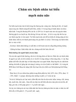
Nutrition: ( CPB continue)
♦The
goals of metabolic and nutritional
support in the paediatric cardiac ICU are first.
♦ Allowance must be made in planning postop feeding, to provide sufficient calories for
ongoing needs and “catch-up” requirement.
.
Age
(years)
Weight
(kg)
Caloric
requirement
(kcal/kg)
<1
>1-6
7-12
12-18
3-10
11-20
21-40
40-70
90-120
75-90
60-75
25-30
I.REPAIR:
1.Residual hemodynamic problems:
* Residual VSD
*RV outflow tract obstruction.
*PV and/or annular stenosis.
*Supravalvar PA stenosis.
*PR
*TR
*RV dysfunction.
*RV outflow tract aneurysm.
*LV dysfunction.
*PHTN.
2.Arrhythmia and conduction disturbance:
*SVT.
*VT.
*Complete heart block.
REPAIR:(continue)
Assessing of the hemodynamic continuously
Measurement of cardiac output.
Identifying of an important right-to-left or left-to-right
shunt by ECHO.
Following arterial desaturation in the early hours after
operation. (Desaturation from right-to-left shunting usually
decreases within 48 hours as RV function improves).
In the absence of shunt, values of PLA and PRA relate the
function of 2 ventricular. (After repair, these are usually
similar).
If PLA is 5 to 10mmHg higher than P RA, a residual left-toright shunt at ventricular or great artery sought promptly
closed by reoperation.
If no shunt, elevated PLA indicates LV hypoplasia or severe
impairment of LV systolic or diastolic function (inotropic
agent and afterload reduction).
REPAIR : (continue)
PRA is rarely 5 to 10 mmHg higher than
PLAindicating important volume or pressure
overload of the RV or RV dysfunction
(Precarious).
PRA /PLA is greater than 0.7, the patient should
reoperate (if a transannular patch was not
used). If a transannular patch is in place,
catecholamine is indicated.
Bleeding (Preoperative polycythemia and
depletion of many clotting factors, extensive
collateral circulation, and damaging effects of
CPB tendency to bleed. (platelet-rich-plasma
and reoperated).
Residual VSD: ( REPAIR : continue.)
Residual VSD may be poorly tolerated:
►The normal LV pre-op without
significant ventricular hypertrophy.
► Present early postoperatively of
congestive HF.
► Cardiac catheterization.
Most residual VSDs are small and important
only in terms of the potential for infective
endocarditis.
If hemodynamic instability occur after
repair, the present of residual VSD should
be promptly and thorough investigated.
REPAIR: (continue)
After the patients leave the ICU,
body weight is followed closely.
(Transient fluid retention is common).
Digoxin is useful in a volume
overload RV for 6 weeks.
Diuretics are used as indicated.
REPAIR: (continue)
Residual right ventricular outflow tract
obstruction ( RRVOTO):
Residual narrowing in the infundibulum, at the
RV pulmonary trunk junction ( with or without a
transannular patch) or more distally.
Stiffening, thickening, and eventually even
calcification of PV cusps cause RV hypertention.
RRVOTO occurred uncommonly, lately.
It includes: valvar stenosis, annular stenosis, and
supravalvar main pulmonary arterial obstruction
The site and severity of them determined by
ECHO
Balloon valvuloplasty or reoperate.
RRVOTO (continue):
Pulmonary artery branch stenosis is
relatively common post-op.
The left pulmonary artery at the site of
prior ductus insertion .
It can be treated by using transcatheter
balloon arterioplasty with or without the
use of stents.
REPAIR: (continue)
Right ventricular dysfunction:
RV systolic hypertention and PR after repair.
RV systolic function and end-diastolic volumn Post-op RV
systolic and diastolic function and a resting systolic pressure up
to 60 to 70 mmHg have little adverse effect.
Higher systolic pressures produce dysfunction.
Low CO may be attributable to RV dysfunction. (Elevated CVP
hepatomegaly, edema, pleural effusion…).
RV dysfunction assessed by ventricular size and EF and severe
PR (3 to 5 days to recover).
The mainstays of therapy are inotropic support, digoxin,
diuretics and ventilatory maneuvers to decrease the P vascular
resistance can reduce RV afterload.
Negative pressure ventilation improve CO well to avoid
secondary organ damage.
TR usually occurs with moderate to severe right ventricular
dilatation secondary to PR and/or right ventricular dysfunction.
When operation is required for P V replacement or correction of
residual outflow obstruction, TV annuloplasty can be a useful
adjunctive procedure.
REPAIR: (continue)
PR commonly accompanies TOF
repair b/c of the frequent need for
transannular patching for adequate
relief of right ventricular outflow tract
obstruction.
PR is usually well tolerated when PA
and RV pressures are low.
REPAIR:(continue)
Right ventricular aneurysms:
Prominent outflow patches were too large to begin with.
The aneurysms may be a false one. ( true aneurysms as
usual)
excessive thinning or devascularization of the RV free wall
or thinning and bulging of pericardium if it has been used
as an infundibular or transannular patch.
Most RV aneurysms develop within 6 months of operation,
and true ones stabilize and rarely progress, whereas false
ones may progress rapidly and rupture.
These patches are akinetic, can contribute to RV
dysfunction.
They should be resected and retailored.
Only 0.9% of patients of TOF underwent reoperation for RV
aneurysms.
REPAIR: (continue)
LV ventricular function:
1.LV systolic and diastolic function are
variable late post-op.
2.Risk factors for poor LV function
include:
►Older age at repair,
► Pre-repair status of LV
► Residual or recurrent defects.
Infective endocarditis: it is rare after
repair.
REPAIR:(continue)
Arrhythmia and conduction disturbance:
Arrhythmia death was 5% to 10% when the patients
underwent repair in adult life.
While it occur 1% of the patients younger than age 5
years at operation.
The RV scar may be arrhythmogenic. Excising the scar
and inserting a patch graft.
Heart block:
Complete heart block is common after repair. It
occurred in 1,3%
Junctional ectopic tachycardia: It occurs
infrequently after repair of TOF. Survival depends
on aggressive treatment in ICU with core cooling
and amiodaron. Thereafter, there is probably little
risk of complete heart block.
II.SYSTEMIC-PULMONARY ARTERIAL SHUNTING:
Careful intraoperative monitoring and control of
PaO2, pH, and buffer base are required.
An intraarterial needle may have been placed
preoperatively, and the baby is returned to the ICU
still intubated.
Using dopamine and epinephrine to establish arterial
blood pressure is 10% to 20% greater than normal to
ensure good flow through the shunt.
Recommending a heparin drip for 24 hours.
A chest radiograph is obtained after procedure and
every 4 hours later.
Hemorrhagic pulmonary edema produces hypoxia
and clinical deterioration. So, many patients died a
few days after shunting operations for TOF.
SYSTEMIC-PULMONARY ARTERIAL
SHUNTING:
Mild renal failure, rarely acute renal failure and
anuria develop after a simple shunting procedure.
A surgically created shunt must function.
If is poorly functioning, prompt reoperation is
indicated. So, auscultation ( excepting large AP
collateral arteries, a continuous murmur is present
pre-operatively) for assessing its patency during
the entire post-op.
If cyanosis has not improved, ECHO or aortography
is indicated.
a hospital mortality approaching zero. Even in the
first month of life, hospital mortality was 0.6%.
SYSTEMIC-PULMONARY ARTERIAL SHUNTING:
The most important risk factor for early death after classic
shunting procedures is PA problems and young age.
Early (less than 30 days) nonfatal shunt closure or narrowing
occurs uncommonly (7%) in patients undergoing classic B-T or
PTEE shunt operations.
Intermediate-term shunt closure or narrowing requiring
reoperation is more common in neonates and young infants than
older patients occuring in 3% to 20%.
Reduced blood flow in the arm on the side of a classic B-T shunt .
Severe blood flow reduction cause gangrene of the hand occur.
Sudden death, without explanation or autopsy is common after
classic shunting procedures (4 months after operation).
Nonfatal brain absess is also common.
Iatrogenic PA problems:Angiographic evidence of PA distortion is
fairy common late post-op.
Beneficial interim results of shunting procedures are increased
Qp, with consequent reduction in cyanosis and polycythemia,
and improved functional capacity.
SYSTEMIC-PULMONARY ARTERIAL
SHUNTING:
NYHA functional class is usually I or II after shunting. SaO2 at
rest is about 80% to 90%, but always decrease with exercise,
at times to as low as 50%.
These benefits are obtained at the expense of increased LV
stroke volumn, a stimulus to gradual development of LV
dysfunction.
Diffuse increase in size of the RPA and LPA.
Severe infundibular or valvar stenoses becomes complete
atresia after a palliative shunting operation.
Important pulmonary vascular disease may develop after a
classic B-T shunt but rarely before 7 years.
The proportion of patients developing hypertention pulmonary
vascular disease increases with increasing shunt duration
(before 5 years).