Experimental and theoretical study of donor-π-acceptor compounds based on malononitrile
Bạn đang xem bản rút gọn của tài liệu. Xem và tải ngay bản đầy đủ của tài liệu tại đây (1.76 MB, 10 trang )
Zayed et al. Chemistry Central Journal (2018) 12:26
/>
Open Access
RESEARCH ARTICLE
Experimental and theoretical study
of donor‑π‑acceptor compounds based
on malononitrile
Mohie E. M. Zayed1, Reda M. El‑Shishtawy1,2*, Shaaban A. Elroby1,3, Khalid O. Al‑Footy1
and Zahra M. Al‑amshany1
Abstract
A set of different donor-π-acceptor compounds having dicyanovinyl as the acceptor and aryl moieties as donors were
synthesized by Knoevenagel condensation. The UV–visible absorption and fluorescence spectra were investigated in
different solvents. The optical band gab energy (Eg) was linearly correlated with the Hammett resonance effect of the
donor to reveal that the higher the value of Hammett resonance effect of a donor, the lower the Eg of the molecule.
The photophysical data revealed that compounds M4–M6 are typical molecular rotors with fluorescence due to
twisted intramolecular charge transfer. Compound M5 revealed the largest Stokes shift (11,089 cm−1) making it a use‑
ful fluorescent sensor for the changes of the microenvironment. The effect of substituents on the optical properties
of donor-π-acceptor compounds having dicyanovinyl as the acceptor are studied using density functional theory and
time-dependent density functional theory (DFT/TD-DFT). The optical transitions are thoroughly examined to reveal
the impact of subtituents on both absorption and fluorescence, mainly through the modification of the structure in
the excited state. The theoretical results have shown that TD-DFT calculations, with a hybrid exchange–correlation
and the long-range corrected density functional PBEPBE with a 6–311++G** basis set, was reasonably capable of
predicting the excitation energies, the absorption and the emission spectra of these molecules.
Keywords: Donor-π-acceptor, Dicyanovinyl, UV–visible and fluorescence spectra, Molecular rotor, DFT, TD-DFT
Introduction
Donor-π-conjugate-electron acceptor (D-π-A) compounds are characterized by having intramolecular
charge transfer (ICT) character. These compounds are of
great interest owing to their high molar absorptivity [1],
amenability of tuning their color by changing the donor,
acceptor, and/or π linker [2, 3] and potential applications
in optoelectronics [4–6], sensors [7, 8], solvent polarity and others [9]. It is known that cyano group is one of
the strongest attracting groups and has been used for the
construction of D-π-A dyes [10–20]. On the other hand,
dimethylamino group is a strong electron donating group
compared with methoxy and/or methyl group.
*Correspondence: ;
1
Chemistry Department, Faculty of Science, King Abdulaziz University,
P. O. Box 80203, Jeddah, Saudi Arabia
Full list of author information is available at the end of the article
In this context, we have designed and prepared as
series of different benzenoid compounds containing different numbers of methoxy groups, methyl group and
dimethylamino group as electron donors compared with
the unsubstantiated benzene ring and using dicyanovinyl
as the electron acceptor group. It was hypothesized that
having acceptor in one side of a conjugated system and
connected with different donors on the other side would
help understanding the ICT character of such compounds and its impact in their photophysical properties.
In recent years, calculations of electronic structures in
the excited states have been a focus of interest because of
the development of computations based on Gaussian and
the time dependent density functional theory (TDDFT)
[21–23]. Also, the solvent effect on the electronic absorption spectra is a useful tool to identify the electronic
transitions of the molecules. This would help in studying the chemical properties of the excited states and to
© The Author(s) 2018. This article is distributed under the terms of the Creative Commons Attribution 4.0 International License
( which permits unrestricted use, distribution, and reproduction in any medium,
provided you give appropriate credit to the original author(s) and the source, provide a link to the Creative Commons license,
and indicate if changes were made. The Creative Commons Public Domain Dedication waiver ( />publicdomain/zero/1.0/) applies to the data made available in this article, unless otherwise stated.
Zayed et al. Chemistry Central Journal (2018) 12:26
0.4
0.2
300
350
400
450
500
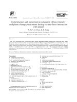
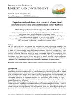
Fig. 1 Normalized absorption (A) and emission (E) spectra of com‑
pound M1 (1 × 10⁻⁵ M) in different solvents
A-Acetonitrile
A-Chloroform
A-Methanol
E-Acetonitrile
E-Chloroform
E-Methanol
1
UV–Visible and fluorescence spectra
0.8
0.6
0.4
0.2
0
270
320
370
420
470
520
570
Wavelength, nm
Fig. 2 Normalized absorption (A) and emission (E) spectra of com‑
pound M2 (1 × 10⁻⁵ M) in different solvents
A Acetonitrile
A Chloroform
A Methanol
E Acetonitrile
E Chloroform
E Methanol
1
Normalized Intensity
Absorption and fluorescence spectra of molecules (M1–
6) recorded in CHCl3, CH3OH and CH3CN and the photophysical properties of these compounds are shown in
Figs. 1, 2, 3, 4, 5, 6 and summarized Table 1, respectively.
The molar absorptivity of these compounds indicates that
their electronic transition is due to π–π*. The effect of
the donor ability of the substituent groups is nicely correlated with the optical data. Substituting hydrogen atom
in compound 1 with different donors shown in Scheme 1
results in a bathochromic shifts in the absorption and in
accordance with the donor ability of the substituents.
As the donor groups are in conjunction with acceptor via π-system, thus it was reasonable to correlate the
0.6
Wavelength, nm
Normalized Intensity
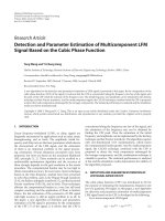
The compounds (M1, M2, M4–6) were obtained by Knoevenagel condensation in a basic medium as shown in
Scheme 1. The structure of these compounds was confirmed by 1H and 13C NMR, mass spectrometry and
FTIR.
0.8
0
250
Results and discussion
Synthesis
A Acetonitrile
A Chloroform
A Methanol
E Acetonitrile
E Chloroform
E Methanol
1
Normalized Intensity
distinguish between the different electronic transitions.
We will use the Continuum Polarizable model (PCM)
[24, 25].
Therefore, computational chemistry is thus necessary to get insight into the molecular structure, although
according to our best knowledge no evidence of similar
study for the dicyanovinyl effect on the ICT character of
the model compounds selected in this study. In this work,
interest resides in correlating the theoretically predicted
electronic parameters with the accurate experimental results so as to provide possible explanations for the
experimentally observed data.
Page 2 of 10
0.8
0.6
0.4
0.2
0
260
310
360
410
460
510
560
Wavelength, nm
Fig. 3 Normalized absorption (A) and emission (E) spectra of com‑
pound M3 (1 × 10⁻⁵ M) in different solvents
Normalized Intensity
1
A Acetonitrile
A Chloroform
A Methanol
E Acetonitrile
E Chloroform
E Methanol
0.8
0.6
0.4
0.2
0
350
400
450
500
550
600
650
Wavelength, nm
Scheme 1 Synthesis of molecular rotors
Fig. 4 Normalized absorption (A) and emission (E) spectra of com‑
pound M4 (1 × 10⁻⁵ M) in different solvents
Normalized Intensity
Zayed et al. Chemistry Central Journal (2018) 12:26
Page 3 of 10
1
A Acetonitrile
A Chloroform
A Methanol
E Acetonitrile
E Chloroform
E Methanol
0.8
0.6
0.4
0.2
0
290
340
390
440
490
540
590
Wavelength, nm
Normalized Intensity
Fig. 5 Normalized absorption (A) and emission (E) spectra of com‑
pound M5 (1 × 10⁻⁵ M) in different solvents
A Acetonitrile
A Chloroform
A Methanol
E Acetonitrile
E Chloroform
E Methanol
1
0.8
0.6
0.4
0.2
0
290
340
390
440
490
540
590
Wavelength, nm
Fig. 6 Normalized absorption (A) and emission (E) spectra of com‑
pound M6 (1 × 10⁻⁵ M) in different solvents
calculated band gap energy of all compounds with Hammett resonance effect [26]. The optical band gap (Eg)
was estimated from the onset wavelength of absorption
using the equation of Eg = 1240/λab, onset. Figure 7 shows
a linear relation between E
g and Hammett resonance
effect of donors. As shown in this figure, the higher the
value of Hammett resonance effect of a donor, the lower
the Eg of the molecule indicating the involvement of an
intramolecular charge transfer (ICT) between donor and
acceptor.
Another interesting feature observed in Table 1 and
Figs. 1, 2, 3, 4, 5, 6 is the enhanced Stokes shift and
bathochromic shift of emission for in different solvents.
Correlating the solvents polarity in terms of their dielectric constants with Stokes shifts and emission wavelengths of M4–6 (Fig. 8) gives a direct linear proportion
indicating that compounds M4–6 are typical molecular
rotors. Molecular rotors are donor-π-acceptor compounds that emit as a result of twisted intramolecular charge transfer (TICT) due to the rotation of donor
and/or acceptor in the ground and excited states around
sigma bond [27]. This TICT is greatly manifested in compound M5 as evidenced by its relatively higher fluorescence intensity (Fig. 9) as well as its largest Stokes shift
(Table 1).
The fluorescent intensity is a function of the free rotation of the molecular rotor and thus a higher fluorescence would be observed dependent on the nature of
TICT and/or the fluorophore microenvironment. Since
the solvents used are non-viscous solvents thus the huge
fluorescence observed in compound M5 compared with
other compounds is reflecting its twisted geometry that
hampers the free rotation. It is worth noting (Table 1)
that compound M5 has the lowest molar absorptivity among all compounds studied indicating a relatively
twisted ground state. The very large Stokes shift observed
in compound M5 is of practical usefulness as such property would reduce the overlap between the UV–vis
absorption and emission spectra of the compound and
consequently minimizing the so-called inner filter effect
and thus rendering compound M5 as an environmentsensitive fluorescent probe [9, 28–30].
Molecular orbital calculations
The optimized geometries obtained by B3LYP/6311++G** level of theory for the ground and excited
states studied molecules are displayed in Figs. 10 and 11,
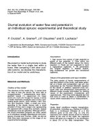
respectively. DFT calculations give planar optimal geometries for ground and excited states. The characterization
of the delocalization of π-electrons along the molecule
Table 1 Photophysical data of compounds M1–6 in different solvents
M
Chloroform
−1
Methanol
−1
−1
Acetonitrile
ε, M
cm−1
× 104
λabs
λem
Stokes shift, cm
ε, M
cm−1
× 104
λabs
λem
M1
2.35
305
377
6262
3.16
306
370
M2
2.92
327
392
5071
2.20
323
370
−1
ε, M−1
cm−1
× 104
λabs
5653
2.35
304 (324)a
341
3569
3933
2.76
321 (348)
399
6090
Stokes shift, cm
λem
Stokes shift, cm−1
M3
3.05
327
390
4940
3.23
321
372
4271
3.06
321 (434)
357
3141
M4
6.02
432
470
1872
5.77
429
481
2520
5.53
430 (403)
485
2637
M5
1.86
329
475
9343
1.86
322
511
11,486
1.92
320 (319)
496
11,089
M6
2.24
369
445
4628
2.16
353
459
6542
2.03
356 (437)
469
6768
a
Data in brackets are theoretical values using PBEPBE/6–311++G** level of theory in acetonitrile solvent
OpƟcal band gap, ev
Zayed et al. Chemistry Central Journal (2018) 12:26
Page 4 of 10
4
p OMe
3
H
p+m (OMe)2
p Me
m (OMe)2
1
p N(Me)2
y = 0.7809x + 3.6434
R² = 0.7251
2
0
0.25
0.5
0.75
1
HammeƩ resonace effect of donors
Fig. 7 Hammett resonance effect of donors versus optical band gap
of compounds M1–6
Fluorescence Intensity, AU
1000
800
600
400
200
0
M1
M2
M3
M4
M5
M6
Fig. 9 Relative fluorescence intensity of compounds M1-6 in acetoni‑
trile (1 × 10⁻⁵ M)
charge density on all over the molecules. Table 2 shows
the bond lengths and differences between single and
double bonds for ground and excited states of the optimal geometries obtained using B3LYP/6-311++G**
level of theory. The difference between C–C and C=C
in M3 and M5 decrease compared to the other compounds in both ground and excited states. This result
indicated that π electron density becomes stronger upon
photoexcitation. The bonds between donor and acceptor groups are C8-C1 and C8=C9. The shorter length
of these bonds favored the charge transfer (CT) within
the studied molecules. Table 2 shows that C8=C9 of
M1, M2, M3, M4, M5 and M6 are 1.363, 1.365, 1.372,
1.367, 1.369 and 1.367 Å respectively, while C8-C1 shows
more single C–C features. The difference between double and single bond lengths are sorted in the order of
M5 > M4 > M6 > M2 > M1, which presents the intensity
of interaction between donor and acceptor groups. For
all the studied molecules, C8-C1 does not change significantly. The difference between double and single bond
lengths are significantly decreased for the excited state
(S1) compared to those in the ground state ( S0), especially
in M3 and M5 molecules. These results indicate that the
connection between acceptor group and donor group for
highly enhanced ICT character, which is important for
the absorption spectra red-shift.
Absorption spectra
Stokes shiŌ, cm 1
12000
y5 = 64.135x + 9061.7
R² = 0.9319
10000
8000
M5
M6
y6 = 69.762x + 4262.1
R² = 1
6000
4000
y 4= 24.15x + 1751.3
R² = 0.9979
2000
0
M4
0
10
20
30
40
Dielectric constant
E,
nm
520
500
M5
M6
y4 = 0.4492x + 467.66
R² = 0.9733
480
460
440
M4
y5 = 0.9108x + 471.52
R² = 0.7578
y6 = 0.6851x + 440.72
R² = 0.9001
0
10
20
30
40
Dielectric constant
Fig. 8 Solvent polarity versus Stokes shift and emission wavelengths
of compounds M4–6
can be estimated by the difference between single and
double bond lengths. The small difference between single and double bond lengths corresponds to delocalized
The vertical excited first three singlet states, transitions
energies, and oscillator strength using TD-DFT (PBEPBE) method started from the optimized structures have
been calculated. The corresponding simulated UV–visible absorption spectra of all molecules in the gas phase
using PBEPBE/6-311++G** level of theory displays in
Fig. 12. Table 3 reveals the calculated absorption λmax
(nm), frontier molecular orbitals contributions and oscillator strength (f ) of the studied compounds (M) collected
in Table 3. As shown in Fig. 13 and Table 3, all compounds exhibit a strong absorption band in the region
around 450 − 200 nm, which can be assigned to an intramolecular charge transfer (ICT) between the various
donating unit and the electron acceptor groups. The λabs
of the studied molecules decreases in the following order
M6 > M3 > M5 > M4 > M2 > M1 which is the same order of
the band gap except with M3. This bathochromic effect
from M1 (304.27 nm) to M3 (397.62) is obviously due to
increased π delocalization. With the increasing of conjugation, the λabs arising from S0 → S1 electronic transition
increase. The first excited states for all studied molecules
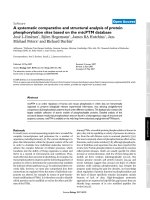
are π → π∗ transitions which differ in the dominant configuration. The natural transition orbitals (NTO) displayed in Fig. 13, which indicate that all transitions are of
π → π∗ and have a pronounced charge-transfer character.
Zayed et al. Chemistry Central Journal (2018) 12:26
Page 5 of 10
Fig. 10 Optimized geometries (bond lengths/Ǻ) of the ground state for the studied compounds using B3LYP/6–311++G** level of theory
HOMO and LUMO show a pronounced electronic density shift from the donor to the acceptor groups.
Experimental section
General
All solvents and reagents were purchased from SigmaAldrich Company and used as received. 2-(4-Methoxybenzylidene)malononitrile (M3) is commercially
available at Life Chemicals, Canada and was used as
received. 1H and 13C NMR spectra were recorded in
CDCl3 solutions on a Bruker Avance 600 MHz spectrometer. Infrared spectra were performed on a PerkinElmer spectrum 100 FTIR spectrometer. Mass spectra
were measured on a GCMS-QP1000 EX spectrometer at
70 eV. UV–visible absorption spectra were recorded with
a Jasco V560 spectrophotometer (Jasco international Co.,
Ltd., Tokyo, Japan). Fluorescence spectra were conducted
on a Perkin-Elmer LS-55 Luminescence Spectrometer
Zayed et al. Chemistry Central Journal (2018) 12:26
Page 6 of 10
Fig. 11 Optimized geometries (bond lengths/Ǻ) of the excited state for studied compounds using B3LYP/6–311++G** level of theory
and uncorrected. Melting points were determined in
open capillary tubes in a Stuart Scientific melting point
apparatus SMP3 and are uncorrected.
Synthesis
General procedure
A mixture of aldehyde derivative (10 mmol), malononitrile (10 mmol), sodium acetate anhydrous (12 mmol)
and ethanol absolute (30 ml) were stirred at room
temperature for 24 h. Then, water was added to the reaction mixture to precipitate the product. The precipitate
was filtered, washed water and then dried. Further purification by silica gel column chromatography afforded the
corresponding product in good yield.
2‑Benzylidenemalononitrile (M1) Solid, m.p:84 °C 1H
NMR (600 MHz, CDCl3): ∂ 7.54 (t, 2H, J = 7.2 Hz, Ar–CH),
7.63 (t, 2H, J = 7.2 Hz, Ar–CH), 7.78 (s, 1H, CH=(CN)2),
Zayed et al. Chemistry Central Journal (2018) 12:26
Page 7 of 10
Table 2 Optimized Selected Bond lengths of the studied
molecules obtained by B3LYP/6–311++G** level
M
Ground state
Excited state
C4–C8
C8–C9
(4-C9)-(8-C9)
C4-C8
C8-C9
M1
1.452
1.363
0.089
1.410
1.444
M2
1.449
1.365
0.084
1.365
1.449
M3
1.436
1.372
0.064
1.458
1.407
M4
1.444
1.367
0.077
1.425
1.429
M5
1.453
1.363
0.090
1.4507
1.417
M6
1.445
1.367
0.078
1.468
1.417
7.90 (d, 2H, J = 7.2 Hz, Ar–CH). 13C NMR (150 MHz,
CDCl3): ∂ 82.88, 112.57, 113.73, 129.67, 130.77, 130.93,
134.69, 159.9; ATR-IR: 3043, 2222, 1589, 1567, 1449; MS
(m/z) for C10H6N2 (M−H)+: Calcd: 153.05, Found: 153.
2‑(4‑Methylbenzylidene)malononitrile
(M2)
Solid,
m.p:135 °C 1H NMR (600 MHz, C
DCl3): ∂ 2.45 (s,
3H, CH3), 7.32 (d, 2H, J = 7.8 Hz, Ar–CH), 7.71 (s, 1H,
CH=(CN)2), 7.8 (d, 2H, J = 7.8 Hz, Ar–CH). 13C NMR
(150 MHz, CDCl3): ∂ 22.05, 81.27, 112.88, 114.04, 128.50,
130.41, 130.95, 146.41, 159.79; ATR-IR: 3035, 2221, 1605,
1584, 1553, 1509; MS (m/z) for C11H8N2 (M−H)+: Calcd:
167.07, Found: 167.
2‑(4‑( D imethyl amino)benzylidene)malononitr ile
(M4) Solid, m.p:180 °C 1H NMR (600 MHz, CDCl3):
∂ 3.14 (s, 6H, N(CH3)2), 6.68 (d, 2H, J = 9 Hz, Ar–CH),
7.46 (s, 1H, CH=(CN)2), 7.81 (d, 2H, J = 9 Hz, Ar–CH).
13
C NMR (150 MHz, C
DCl3): ∂ 40.15, 71.95, 111.60,
114.95, 116.03, 119.31, 133.83, 154.22, 158.16; ATR-IR:
2920, 2207, 1607, 1560, 1515, 1385, 1357; MS (m/z) for
C12H11N3 (M−H)+: Calcd: 196.1, Found: 196.
Table 3 Absorption wavelength (nm), molecular orbital
contribution, energy level of HOMO, LUMO and oscillator
strength calculated by using PBEPBE/6–311 ++G** level
of theory in gas phase
M
M1
Wave length (nm)
f
MO contribution
MO coeff. (%)
304
0.542
HOMO–LUMO
94
297
0.119
HOMO-1-LUMO
91
179
0.414
HOMO-1-LUMO+1
57
M2
331
0.536
HOMO–LUMO
85
335
0.15
HOMO-1-LUMO
84
M3
397
0.705
HOMO–LUMO
98
285
0.216
HOMO-1-LUMO
89
M4
354
0.69
HOMO–LUMO
98
269
0.147
HOMO-2-LUMO
71
M5
393
0.154
HOMO-1-LUMO
63
300
0.519
HOMO-2-LUMO
62
M6
434
0.17
HOMO-1-LUMO
96
413
0.339
HOMO–LUMO
92
294
0.289
HOMO-3-LUMO
79
2‑(3,5‑Dimethoxybenzylidene)malononitrile (M5) Solid,
m.p:89 °C 1HNMR (600 MHz, C
DCl3): ∂ 3.83 (s, 6H,
OCH3), 6.69 (s, 1H, Ar–CH), 7.03 (s, 2H, Ar–CH), 7.68
(s, 1H, CH=(CN)2).13C NMR (150 MHz, CDCl3): ∂ 55.71,
83.12, 107.22, 108.24, 112.66, 113.68, 132.35, 160.14,
161.25; ATR-IR: 2966, 2229, 1603, 1577, 1458, 1426, 1310;
MS (m/z) for C
12H10N2O2 (M−H)+: Calcd: 213.07, Found:
213.
2 ‑ ( 3 , 4 , 5 ‑Tr i m e t h o x y b e n z y l i d e n e) m a l o n o n i t r i l e
(M6) Solid, m.p:145 °C 1H NMR (600 MHz, CDCl3):
∂ 3.90 (s, 6H, OCH3), 3.97 (s, 3H, OCH3), 7.18 (s, 2H,
Ar–CH), 7.65 (s, 1H, CH=(CN)2).13C NMR (150 MHz,
CDCl3): ∂ 56.37, 61.30, 80.60, 108.26, 113.23, 114.02,
M2
M4
M5
M6
M3
M1
1.1
Oscillator strength
1
0.9
0.8
0.7
0.6
0.5
0.4
0.3
0.2
0.1
0
150
200
250
300
350
400
450
500
550
Wavelength, nm
Fig. 12 The UV–visible absorption spectra of the studied compounds calculated using PBEPBE/6–311++G** level of theory in chloroform
600
Zayed et al. Chemistry Central Journal (2018) 12:26
M
Page 8 of 10
HOMO
LUMO
M1
M2
M3
M4
M5
M6
v
Fig. 13 Schematic diagram of NTO’s of four studied dyes calculated at the PBEPBE/6–311++G∗∗ level of theory. The surfaces are generated with
an isovalue at 0.02
Zayed et al. Chemistry Central Journal (2018) 12:26
125.96, 143.97, 153.37, 159.45; ATR-IR: 2942, 2839, 2221,
1568, 1499, 1455, 1247, 1126.92; MS (m/z) for C13H12N2O3
(M−H)+: Caled: 243.1, Found: 243.
Computational methods
All calculations are performed using Gaussian 09 W
[21] program package. In the present work, B3LYP/6–
311++G** level of theory is employed to achieve our aim
from this study. Becke’s three parameter hybrids function
combined with the Lee–Yang–Parr correlation function
(B3LYP) [31–34] predict the best results for molecular
geometry and electronic transition for moderately larger
molecules. B3LYP/6–311++G** frequency analysis calculations were performed to characterize the stationary
points as the minima. HOMO–LUMO energies, absorption wavelengths and oscillator strengths are calculated
using TD-B3LYP [35–37]. These optimized structures
were calculated for the first excitation energy, maximal
absorption wavelength (λmax) and oscillator strengths (f )
for the three states by using TD-B3LYP/6–311++G**
level of theory. Moreover, three density functional,
namely, PBEPBE [38] with same above basis set have
been evaluated in order to find out the suitable functional
that estimates the absorption behavior of the studied
dyes.
Conclusions
In this paper, different donor-π-acceptor compounds
having dicyanovinyl as the acceptor and aryl moieties as
donors were synthesized. Compared with all molecules
investigated, molecule 5 showed the highest Stokes shift
as well as the highest fluorescent intensity indicating a
typical molecular rotor. Also, the energy Eg values were
nicely correlated with the donor ability of the substituent
as presented by Hammett resonance effect. UV–visible
absorption maxima of the compounds were examined
experimentally as well as computationally and the results
obtained have shown that TD-DFT calculations, with a
hybrid exchange–correlation and the long-range corrected density functional PBEPBE with a 6–311++G**
basis set, was reasonably capable of predicting the excitation energies, the absorption and the emission spectra of
these molecules.
Authors’ contributions
RME suggested the research point and did some of the writing up. SAE carried
out the theoretical calculations and the writing up of the theoretical part of
the manuscript. MEMZ, KOA, and ZMA carried out experimental part (prepara‑
tion and characterization). All authors shared equally the revision of the
manuscript. All authors read and approved the final manuscript.
Author details
1
Chemistry Department, Faculty of Science, King Abdulaziz University, P. O.
Box 80203, Jeddah, Saudi Arabia. 2 Dyeing, Printing and Textile Auxiliaries
Department, Textile Research Division, National Research Center, Dokki,
Page 9 of 10
Cairo 12622, Egypt. 3 Chemistry Department, Faculty of Science, Beni-Suef
University, Beni‑Suef 6251, Egypt.
Acknowledgements
This project was funded by the Deanship of Scientific Research (DSR) at
King Abdulaziz University, Jeddah, under Grant Number (337/130/1434). The
authors, therefore, acknowledge with thanks DSR technical and financial
support.
Competing interests
The authors declare no competing interests.
Ethics approval and consent to participate
Not applicable.
Publisher’s Note
Springer Nature remains neutral with regard to jurisdictional claims in pub‑
lished maps and institutional affiliations.
Received: 13 December 2017 Accepted: 23 February 2018
References
1. El-Shishtawy RM, Elroby SA, Asiri AM, Müllen K (2016) Optical absorption
spectra and electronic properties of symmetric and asymmetric squar‑
aine dyes for use in dssc solar cells: DFT and TD-DFT studies. Int J Mol Sci
17:487–495
2. El-Shishtawy RM, Borbone F, Al-Amshany ZM, Tuzi A, Barsella A, Asiri AM,
Roviello A (2013) Thiazole azo dyes with lateral donor branch: synthesis,
structure and second order NLO properties. Dyes Pigments 96:45–51
3. Lu H, Mack J, Yang Y, Shen Z (2014) Structural modification strate‑
gies for the rational design of red/NIR region BODIPYs. Chem Soc Rev
43:4778–4823
4. El-Shishtawy RM (2009) Functional dyes, and some hi-tech applications.
Int J Photoenergy 2009:21
5. Ji C, Yin L, Li K, Wang L, Jiang X, Suna Y, Yanqin L (2015) D–π–A–π–D-type
low band gap diketopyrrolopyrrole based small molecules contain‑
ing an ethynyl-linkage: synthesis and photovoltaic properties. RSC Adv
5:31606–31614
6. El-Shishtawy RM, Al-Zahrani FAM, Afzal SM, Razvi MAN, Al-Mashany ZM,
Bakry AH, Asiri AM (2016) Synthesis, linear and nonlinear optical proper‑
ties of a new dimethine cyanine dye derived from phenothiazine. RSC
Adv 6:91546–91556
7. El-Shishtawy RM, Al-Zahrani FAM, Al-amshany ZM, Asiri AM (2017) Syn‑
thesis of a new fluorescent cyanide chemosensor based on phenothia‑
zine derivative. Sens Actuators B 240:288–296
8. Beer PD, Gale PA (2001) Anion recognition and sensing: the state of the
art and future perspectives. Angew Chem Int Ed 40:486–516
9. Grabowski ZR, Rotkiewicz K, Rettig W (2003) Structural changes accom‑
panying intramolecular electron transfer: focus on twisted intramolecular
charge-transfer states and structures. Chem Rev 103:3899–4032
10. Yanga Y, Li B, Zhang L (2013) Design and synthesis of triphenylaminemalonitrile derivatives as solvatochromic fluorescent dyes. Sens Actuators
B 183:46–51 (and references cited therein)
11. Li X, Kim SH, Son YA (2009) Optical properties of donor-π-(acceptor)
n merocyanine dyes with dicyanovinylindane as acceptor group and
triphenylamine as donor unit. Dyes Pigments 82:293–298
12. Kim SH, Gwon SY, Bae JS, Son YA (2011) The synthesis and spectral
properties of a stimuli-responsive D-π-A charge transfer dye. Spectrochim
Acta Part A 78:234–237
13. Son YA, Gwon SY, Lee SY, Kim SH (2010) Synthesis and property of solva‑
tochromic fluorophore based on D-π-A molecular system: 2-{[3-Cyano4-(N-ethyl-N-(2-hydroxyethyl)amino)styryl]-5,5-dimethylfuran-2(5H)ylidene}malononitrile dye. Spectrochim Acta Part A 75:225–229
14. Li Y, Ren T, Dong W-J (2013) Tuning photophysical properties of triph‑
enylamine and aromatic cyano conjugate-based wavelength-shifting
Zayed et al. Chemistry Central Journal (2018) 12:26
15.
16.
17.
18.
19.
20.
21.
22.
23.
24.
25.
26.
compounds by manipulating intramolecular charge transfer strength. J
Photochem Photobiol A Chem 251:1–9
Bolduc A, Dong Y, Guérinz A, Skene WG (2012) Solvatochromic investiga‑
tion of highly fluorescent 2-aminobithiophene derivatives. Phys Chem
Chem Phys 14:6946–6956
Giordano L, Shvadchak VV, Fauerbach JA, Jares-Erijman EA, Jovin TM
(2012) Highly solvatochromic 7-aryl-3-hydroxychromones. J Phys Chem
Lett 3:1011–1016
Hofmann K, Spange S (2012) Influence of the boron atom on the solva‑
tochromic properties of 4-nitroaniline-functionalized boronate esters. J
Org Chem 77:5049–5055
Kucherak OA, Richert L, Mély Y, Klymchenko AS (2012) Dipolar 3-methox‑
ychromones as bright and highly solvatochromic fluorescent dyes. Phys
Chem Chem Phys 14:2292–2300
Benedetti E, Kocsis LS, Brummond KM (2012) Synthesis and photophysi‑
cal properties of a series of cyclopenta[b]naphthalene solvatochromic
fluorophores. J Am Chem Soc 134:12418–12421
Huang GJ, Ho JH, Prabhakar C, Liu YH, Peng SM, Yang JM (2012) Siteselective hydrogen-bonding-induced fluorescence quenching of
highly solvatofluorochromic GFP-like chromophores. Organic Letters
14:5034–5037
Frisch MJ, Trucks GW, Schlegel HB, Scuseria GE, Robb MA, Cheeseman JR,
Scalmani G, Barone V, Mennucci B, Petersson GA et al (2009) Gaussian 09
Suite of Programs. Gaussian Inc, Wallingford
Burke K, Werschnik J, Gross EKU (2005) Time-dependent density func‑
tional theory: past, present, and future. J Chem Phys 123:062206
Foreman JB, Head-Gordon M, Pople JA (1992) Toward a systematic molec‑
ular orbital theory for excited states. J Phys Chem 96:135
Miertus S, Scrocco E, Tomasi J (1981) Electrostatic interaction of a solute
with a continuum. A direct utilizaion of AB initio molecular potentials for
the prevision of solvent effects. Chem Phys 55:117–129
Miertus S, Tomasi J (1982) Approximate evaluations of the electrostatic
free energy and internal energy changes in solution processes. Chem
Phys 65:239
Hansch C, Leo A, Taft RW (1991) A survey of Hammett substituent con‑
stants and resonance and field parameters. Chem Rev 91(2):165–195
Page 10 of 10
27. Haidekker MA, Theodorakis EA (2010) Environment-sensitive behavior of
fluorescent molecular rotors. J Biol Eng 4:11
28. Liu X, Xu Z, Cole JM (2013) Molecular design of uv–vis absorption and
emission properties in organic fluorophores: toward larger bathochromic
shifts, enhanced molar extinction coefficients, and greater stokes shifts. J
Phys Chem C 117:16584–16595
29. Haidekker MA, Theodorakis EA (2007) Molecular rotors–fluorescent
biosensors for viscosity and flow Org. Biomol Chem 5:1669–1678
30. Demchenko P, Mely Y, Duportail G, Klymchenko AS (2009) Monitoring
biophysical properties of lipid membranes by environment-sensitive
fluorescent probes. Biophys J 96:3461–3470
31. Becke AD (1993) Density-functional thermochemistry. III. The role of exact
exchange. J Chem Phys 98:5648
32. Lee C, Yang W, Parr RG (1988) Development of the Colle-Salvetti
correlation-energy formula into a functional of the electron density. Phys
Rev B 37:785
33. Becke AD (1996) Density-functional thermochemistry. IV. A new dynami‑
cal correlation functional and implications for exact-exchange mixing. J
Chem Phys 104:1040–1046
34. Becke AD (1997) Density-functional thermochemistry. V. System‑
atic optimization of exchange-correlation functionals. J Chem Phys
107:8554–8560
35. Perdew JP, Burke K, Ernzerhof M (1996) Generalized gradient approxima‑
tion made simple. Phys Rev Lett 77:3865–3868
36. Casida ME, Jamorski C, Casida KC, Salahub DR (1998) Molecular excitation
energies to high-lying bound states from time-dependent densityfunctional response theory: characterization and correction of the timedependent local density approximation ionization threshold. J Chem
Phys 108:4439–4449
37. Jacquemin D, Wathelet V, Perpete EA, Adamo C (2009) Extensive TD-DFT
benchmark: singlet-excited states of organic molecules. J Chem Theor
Comput 5:2420–2435
38. Adamo C, Barone V (1999) Toward reliable density functional meth‑
ods without adjustable parameters: the PBE0 model. J Chem Phys
110:6158–6170