Gastrointestinal peptides in children before and after hematopoietic stem cell transplantation
Bạn đang xem bản rút gọn của tài liệu. Xem và tải ngay bản đầy đủ của tài liệu tại đây (993.19 KB, 9 trang )
Skoczeń et al. BMC Cancer
(2020) 20:306
/>
RESEARCH ARTICLE
Open Access
Gastrointestinal peptides in children before
and after hematopoietic stem cell
transplantation
Szymon Skoczeń1, Magdalena Rej1*, Kinga Kwiecińska1, Danuta Pietrys2, Przemysław J. Tomasik3,
Małgorzata Wójcik4, Wojciech Strojny2, Agnieszka Dłużniewska5, Katarzyna Klimasz6, Kamil Fijorek7,
Michał Korostyński8, Marcin Piechota8 and Walentyna Balwierz1
Abstract
Background: Gastrointestinal tract function and it’s integrity are controlled by a number of peptides whose
secretion is influenced by severe inflammation. In stomach the main regulatory peptide is ghrelin. For upper small
intestine cholecystokinin and lower small intestine glucagon-like peptide- 1 are secreted, while fibroblast growth
factor-21 is secreted by several organs, including the liver, pancreas, and adipose tissue [12]. Hematopoietic stem
cell transplantation causes serious mucosal damage, which can reflect on this peptides.
Methods: The aim of the study was to determine fasting plasma concentrations of ghrelin, cholecystokinin,
glucagon- like peptide-1, and fibroblast growth factor-21, and their gene expressions, before and 6 months after
hematopoietic stem cell transplantation.27 children were studied, control group included 26 healthy children.
Results: Acute graft versus host disease was diagnosed in 11 patients (41%, n = 27). Median pre-transplantation
concentrations of gastrointestinal peptides, as well as their gene expressions, were significantly lower in studied
group compared with the control group. Only median of fibroblast growth factor-21 concentration was nearsignificantly higher before stem cell transplantation than in the control group. The post–hematopoietic transplant
results revealed significantly higher concentrations of the studied peptides (except fibroblast growth factor-21) and
respective gene expressions as compare to pre transplant results. Median glucagone like peptide-1 concentrations
were significantly decreased in patients with features of acute graft versus host disease. Moreover, negative
correlation between glucagone like peptide-1 concentrations and acute graft versus host disease severity was
found.
Conclusions: Increased concentrations and gene expressions of gastrointestinal tract regulation peptides can be
caused by stimulation of regeneration in the severe injured organ. Measurement of these parameters may be a
useful method of assessment of severity of gastrointestinal tract complications of hematopoietic stem cell
transplantation.
Keywords: Hematopoietic stem cell transplantation, Peptides regulating gastrointestinal tract functions, Children
* Correspondence:
1
Department of Oncology and Hematology, University Children’s Hospital in
Krakow, Jagiellonian University Medical College, Wielicka St. 265, 30-663
Krakow, Poland
Full list of author information is available at the end of the article
© The Author(s). 2020 Open Access This article is licensed under a Creative Commons Attribution 4.0 International License,
which permits use, sharing, adaptation, distribution and reproduction in any medium or format, as long as you give
appropriate credit to the original author(s) and the source, provide a link to the Creative Commons licence, and indicate if
changes were made. The images or other third party material in this article are included in the article's Creative Commons
licence, unless indicated otherwise in a credit line to the material. If material is not included in the article's Creative Commons
licence and your intended use is not permitted by statutory regulation or exceeds the permitted use, you will need to obtain
permission directly from the copyright holder. To view a copy of this licence, visit />The Creative Commons Public Domain Dedication waiver ( applies to the
data made available in this article, unless otherwise stated in a credit line to the data.
Skoczeń et al. BMC Cancer
(2020) 20:306
Page 2 of 9
Background
Impaired intestinal function is a common complication
of hematopoietic stem cell transplantation (HSCT).
Damage to the gastrointestinal (GI) mucosa in patients
undergoing HSCT is a serious but still poorly understood complication. Toxicity of HSCT conditioning regimens and graft-versus-host disease (GvHD) result in a
5-fold increase of the risk of significant GI complications
compared with other cancer survivors [1, 2]. Chemotherapy and total body irradiation (TBI) can damage GI
mucosa and cause diffuse inflammation of GI tract. This
leads to disruption of integrity of GI mucosa with subsequent transfer of bacterial lipopolysaccharides and other
danger/pathogen-associated
molecular
patterns
(DAMPs/PAMPs) into the circulation [3]. The intestine
is also known as the largest endocrine organ in the body.
It strongly influences other organs, including the brain
via the gut-brain axis [4]. The majority of GI regulatory
peptides are secreted by strictly defined sections of the
intestine [5]. Ghrelin is produced mainly in the stomach
by P/D1 cells, cholecystokinin (CCK) is secreted mainly
by the I cells of the upper small intestine, while glucagone like peptide-1 (GLP-1) is produced by the endocrine L cells in the lower intestine [6–11]. Fibroblast
growth factor-21 (FGF21) is secreted by several organs,
including the liver, pancreas, and adipose tissue [12].
The intensity of GI dysfunction can be assessed using
mucositis grading and parenteral nutrition requirements,
but these tools cannot identify the most severely affected
parts of the GI tract [13]. Endoscopy is rarely performed
in the early post-HSCT phase due to the high risk of severe complications. In addition, the test load with nutrients is unreliable in this phase. Due to the differences in
the anatomic distribution of intestinal endocrine cells,
studies of alterations in GI peptide concentrations might
help to localize the affected sections of the gut and assess the severity of inflammation. Thus, there is a need
to identify simple and noninvasive tests that can assess
the location and severity of gut damage. Additional comparison of marker concentrations before and several
months after HSCT can explain the mechanisms of destruction and restoration of the GI tract [14–16]. The
aim of this study was to determine and analyze the selected GI peptides secreted on different levels of the gut
in patients before and after HSCT.
being over 18, therefore fulfilled inclusion criteria of the
study. The patients were assessed twice—before HSCT
(pre-HSCT group) and approximately 6 months after
HSCT (post-HSCT group). Diseases that were the indication for HSCT are listed in Table 1. Patients with malignancies, except for juvenile myelomonocytic leukemia
(JMML), were referred for HSCT in complete remission.
Characteristics of the transplantation procedures are detailed in Table 2.
In more than half of the patients (16 patients, n = 27) a
conditioning regimen was based on Busulfan/Treosulfan.
Total body irradiation (TBI) was used in 7 of patients, 4
patients received regimen based on Cyclofsphamide. Most
patients (85%) in whom graft-versus-host disease (GvHD)
prophylaxis was used received methotrexate combined
with cyclosporine. Mucositis was diagnosed in 82% cases
(22 patients), grade III and IV mucositis in 26% (7 patients). The key clinical data of the HSCT recipients are
presented in Table 3. Mucositis requiring parenteral nutrition was found in almost half (48%) of the patients. Systemic glucocorticoids were used in 19 children in the
post-HSCT group to treat complications of HSCT. In 11
of patients aGvHD was seen, including intestinal involvement in one. According to the aGvHD grader (agvhd.
com), grade II and III aGvHD was found in 22% cases (6
patients). In two cases multiple locations of aGvHD occurred (II/C - skin+liver, III/C - skin+GI + liver). The patients with aGvHD were treated with additional
immunosuppressive agents, including tacrolimus, mycophenolate mofetil, and etanercept. Six months after
HSCT, four children still received tapered doses of immunosuppressive agents other than glucocorticoids. The
control group consisted of 11 boys and 15 girls aged 4.3 to
16.0 years (median 12.2 years). The control children were
recruited among family donors, siblings of patients treated
with HSCT, and unrelated healthy children. They all had
negative medical history, no signs or symptoms of acute
Methods
Study groups
A group of 27 children aged 1.5–19 years (median 9.6
years) was referred to the Stem Cell Transplantation
Centre of the University Children’s Hospital in Krakow
and was included in this study. One patient of 19 years
old started the treatment being underage and remain for
the treatment and the observation in Children Hospital
Table 1 Indications for HSCT (pre-HSCT group)
Diagnosis
Number (%)
Acute lymphoblastic leukemia (ALL)
11 (40.7)
Acute myeloblastic leukemia (AML)
4 (14.8)
Chronic myelocytic leukemia (CML)
1 (3.6)
Myelodysplastic syndrome (MDS)
1 (3.6)
Juvenile myelomonocytic leukemia (JMML) and AML
1 (3.6)
Neoplastic diseases – total
18 (70)
Severe aplastic anemia (SAA)
4 (14.8)
Chronic granulomatous disease (CGD)
3 (8)
Autoimmune lymphoproliferative syndrome (ALPS)
1 (3.6)
Hyper IgM syndrome (HIgM)
1 (3.6)
Non-neoplastic diseases – total
9 (30)
Skoczeń et al. BMC Cancer
(2020) 20:306
Page 3 of 9
Table 2 Types of HSCT procedures
Table 3 Characteristics of HSCT recipients
Type of HSCT
n (%)
Disease (n)
Number of patients
27
Allogeneic
n = 27 (100%)
MUD - 16 (59)
ALL – 8
Sex
boys-20, girls-7
AML - 4
Age (years)
1.5–19 (mean 9, median 9.6)
CML - 1
Neoplastic diseases, n (n %)
18 (67%)
SAA - 1
Chemotherapy before HSCT,
n (n %)
17 (63%)
Local radiotherapy
5 (CNS-4,Testes-1)
CGD – 2
MSD - 9 (33)
ALL – 3
SAA - 2
JMML and AML - 1
CGD - 1
Neoplastic diseases
Non-neoplastic diseases
Median-1, mean-2; range 0.1–7
Median-1.5, mean-3.8, range 0.1–13
Conditioning regimen based on
busulfan or treosulfan, n (n %)
16(60%)
SAA – 1
Total body irradiation – 12Gy/
6fractions, n (n %)
7 (26%)
ALPS - 1
GvHD prophylaxis, n (n %)
HIgM - 1
MDS - 1
MFD - 2 (8)
Time since diagnosis (years)
ALL acute lymphoblastic leukemia
ALPS autoimmune lymphoproliferative syndrome
AML acute myeloblastic leukemia
CGD chronic granulomatous disease
CML chronic myelocytic leukemia
HIgM hyper IgM syndrome
JMML juvenile myelomonocytic leukemia
MDS myelodysplastic syndrome
SAA severe aplastic anemia
or chronic diseases, and no abnormalities in laboratory
tests (CBC, serum ALT, and creatinine levels).
CSA
4 (15%)
MTX + CSA
23 (85%)
Mucositis, n (%)
22 (82%)
Grade, n
I-7, II-8, III-6, IV-1
Intravenous alimentation due to
mucositis (%)
48
aGvHD, n (n %)
11 (41)
Localisation, %
Gut-9, Skin-91, Liver-27
Grade, n
IA-1, IB-4, IIB-1, IIC-3, IIIC-2
Systemic glucocorticoid
treatment, n (%)
19 (70)
Height and body weight measurements were performed
by an anthropometrist. Body mass index (BMI), BMI
percentile (BMIPerc) and BMI SDS (BMISDS) were calculated using online WHO BMI calculators [17]. The results were compared to regional reference values, and
the reference values were published by the WHO [17–
19]. The BF mass and BF% were measured using bioimpedance and calculated according to the method described by Kushner RF and Schoeller DA [20].
Systemic glucocorticoid
treatment (days)
Median-3.5, mean-3.6; range 0.1–11
Time from discontinuation
of systemic glucocorticoids
to the second assessment
(months)
Median-3.6, mean-4.5; range 0.5–14
Time from discontinuation
of immunosuppressive
treatment to the second
assessment (months)
Median 1.6; range 0–9
Time from HSCT to the
second assessment (months)
Median 6.3 (5.9–19.1)
Study protocol
aGvHD acute graft-versus-host disease, CSA cyclosporin, MTX methotrexate
Anthropometric measurements
Fasting blood samples were collected in the morning.
Patients treated with HSCT were assessed immediately
before conditioning and after a median of 6.3 months
after HSCT. In the control group samples were obtained
once, after enrollment to the study. Blood samples were
collected in EDTA and aprotinin tubes, (Becton-Dickinson; UK), and tubes with no anticoagulants. The tubes
were delivered to the laboratory immediately and centrifuged for 15 min at 3000 rpm using a horizontal rotor.
Serum and plasma samples were stored at -80 °C until
the time of measurement. Subsequently, mononuclear
cells were separated for microarray followed by total
RNA extraction.
Laboratory measurements
Plasma concentrations of the peptides were measured
using EIA kits: ghrelin, CCK, GLP-1 (Phoenix Pharmaceuticals, Inc., USA), and FGF-21 (Millipore Corporation, USA). The sensitivity of the methods are provided
by kit suppliers and are as follows: ghrelin – 0.08 ng/ml
(14% intra- assay and 5% interassay variability),CCK –
0.06 ng/ml (5% intra- assay and 9% interassay variability),
GLP-1 – 0.18 ng/ml (14% intra- assay and 5% interassay
variability), FGF-21 – 0.016 ng/ml (5.8% intra- assay and
9% interassay variability).
Skoczeń et al. BMC Cancer
(2020) 20:306
Page 4 of 9
Microarray analysis
Microarray analysis used a GeneChip Human Gene 1.0
ST Arrays (Affimetrix, Santa Clara, USA) according to
the manufacturer’s protocol. GLP-1 expression data were
not available in the Affimetix database, and thus we
checked the results of GLP-1 receptor gene expression.
Gene loci and Affimetrix codes of the tested peptides
are presented in Table 4.
All the primary microarray data were submitted to
GEO public repository and are accessible using GEO
Series accession number GSE69421 (i.
nlm.nih.gov/geo/query/acc.cgi?acc=GSE69421). In our
study a part of submitted microarray data was used.
adult patients signed a written informed consent before
blood sample collection. Study was conducted in accordance with the Declaration of Helsinki.
Results
When comparing the pre-HSCT and post-HSCT groups
and the control group (Table 5), we noted a significantly
lower BF mass and BF% measured using bioimpedance
(6.46/12.0; 6.65/12.0, p < 0.05). The comparative analysis
of the pre-HSCT group and the post-HSCT group
showed no significant differences in anthropometric
parameters.
Ghrelin
Statistical analysis
Continuous clinical and biochemical variables are presented as the mean values and standard error or as the
median values and quartiles, as appropriate. Categorical
variables are presented as frequencies and percentages.
The Shapiro-Wilk test was used to assess the normal
distribution of the continuous variables. To examine the
differences between two or more independent groups,
ANOVA/Student’s t-test (for variables with normal distribution) or Kruskal-Wallis/Mann-Whitney tests (for
variables with non-normal distribution) were used. To
assess the correlations between two continuous variables, Spearman rank correlation coefficient was calculated. The two-sided p-values < 0.05 were considered
statistically significant. Gene expression data were RMAnormalized and presented as the mean and standard deviation. ANOVA was used to examine the differences in
gene expression between two independent groups. The
Benjamini-Hochberg (B-H)-corrected p-values < 0.05
were considered statistically significant. The statistical
analyses were performed using the R 3.4.3 software.
Ethical issues
The Permanent Ethical Committee for Clinical Studies
of the Jagiellonian University Medical College approved
the study protocol. All parents, adolescent patients, and
The median ghrelin concentrations in the pre-HSCT
group (median 501 pg/ml [first and third quartile 425;
582]) and in post-HSCT group (558 pg/ml [445;701])
were significantly lower compared with the median concentration in the control group (711 pg/ml [596;898])
(p < 0.001 and p = 0.05, respectively). Differences in
ghrelin concentrations between the pre-HSCT and postHSCT groups were statistically significant (p = 0.016)
(Fig. 1). Statistical analysis also revealed a considerable
trend towards significance (p = 0.08) for the decreased
ghrelin concentrations in patients with mucositis. Interestingly, ghrelin levels were increased in patients with
liver aGvHD comparing with those with cutaneous and
intestinal aGvHD (p = 0.02). Analysis of ghrelin gene expression revealed near-significantly (p = 0.07) lower
(6.84+/− 0.41 vs. 6.99+/− 0.25) values in the post-HSCT
group compared with the control group (BenjaminiHochberg corrected p-value (B-H) = 0.09; Table 4).
Cholecystokinin
Median CCK concentration in the pre-HSCT group (1.23
ng/ml; [first and third quartile 0.88;1.70]) was significantly
lower than in the post-HSCT group (2.32 ng/ml [1.42;
6.58]; p < 0.005) and in the control group (3.46 ng/ml
[2.87;5.12]; p < 0.001). CCK concentrations in the postHSCT group and control group showed no significantly
Table 4 Comparison of mean parameters and standard deviation of genes expression of peptides regulating gastrointestinal tract.
P-values after ANOVA and Benjamini-Hochberg correction (B-H) are provided
Gene
Gene locus
Affimetrix code
Expression
Ghrelin
3p26-p25
8,085,293
post-HSCT
6.84 ± 0.41
Control
6.99 ± 0.25
0.07/0.09
p/ B-H
Cholecystocinin
3p22-p21.3
8,086,391
post-HSCT
5.61 ± 0.14
Control
5.89 ± 0.23
0.0014/0.003
GLP-1 receptor
6p21
8,119,338
post-HSCT
6.26 ± 0.08
Control
6.61 ± 0.27
0.0000/0.0001
FGF-21
19q13.1-qter
8,030,105
pre-HSCT
5.46 ± 0.15
Control
5.59 ± 0.16
0.0395/0.4325
post-HSCT
5.36 ± 0.12
Control
5.59 ± 0.16
0.0009/0.0021
Skoczeń et al. BMC Cancer
(2020) 20:306
Page 5 of 9
Table 5 Values of adipose tissue parameters in studied groups and control
Parameter
pre-HSCT
post-HSCT
Control
P value, pre-HSCT
vs post-HSCT
P value, pre-HSCT
vs control
P value, Post-HSCT
vs control
BMIa
18.9 (3.33)
18.3 (3.47)
19.1 (3.00)
0.173
0.794
0.405
BMIPerc
70.4 [44.9;86.4]
51.0 [16.2;90.6]
77.7 [46.7;84.3]
0.170
0.967
0.486
BMISDSa
0.57 (1.21)
0.37 (1.26)
0.61 (0.87)
0.392
0.898
0.455
BF_kg
6.46 (6.42)
6.65 (5.35)
12.0 (8.46)
0.854
0.031
0.029
BF_%a
14.5 (11.0)
15.8 (8.71)
21.1 (8.54)
0.616
0.042
0.062
b
a
a
Mean values (standard deviations), paired Student Test for pre-HSCT vs. post-HSCT, and unpaired Student test for comparison with Control
b
Medians [first and third quartile], Mann- Whitney test p-value
differences (Fig. 1). The analysis of CCK gene expression revealed that mean CCK gene expression was
significantly (p = 0.0014, B-H = 0.03) lower (5.61+/−
0.14 vs. 5.89+/− 0.23) in the post-HSCT group than
in the control group (Table 4).
Glucagon like peptide-1
The lowest median GLP-1 concentrations were seen in
the pre-HSCT group (0.62 ng/ml [first and third quartile
0.47; 0.90]. The values observed in the post-HSCT group
(1.31 ng/ml [0.83;1.82]) and in the control group were
not significantly different (1.26 ng/ml [1.14;1.56]). The
differences between the pre-HSCT group and the postHSCT group, as well as between the pre-HSCT group
and the control group, were significant (p < 0.003, p <
0.001 respectively; Fig. 1). Median concentration of
GLP-1 was significantly decreased in patients with
aGvHD symptoms (p = 0.008, Additional File 1). Moreover, GLP-1 levels negatively correlated with grade of
aGvHD (r = − 0.58). Logistic regression model indicates
that GLP-1 concentration may be a potential biomarker
of aGvHD progression (p = 0.03).
GLP-1 receptor gene expression revealed a significantly lower mean expression (6.26+/− 0.08 vs. 6.61+/−
0.27) in the post-HSCT group compared with the control group (p = 0.000, B-H = 0.0001; Table 4).
Fibroblast growth factor-21
Median FGF-21 concentrations seen in the pre-HSCT
group (146 pg/ml; [first and third quartile 83.9; 303]) were
higher than in the post-HSCT group (64.8 pg/ml [45.9;
135]; p = 0.024) and in the control group (65.3 pg/ml
[51.9;115]; p = 0.068). Analysis of FGF-21 gene expression
revealed that its mean expression was significantly lower
(5.36+/− 0.12 vs. 5.59+/− 0.16, p = 0.0009, B-H =
0.0021) in the post-HSCT group than in the control
group (Table 4). No significant correlations between
conditioning intensity or severity of mucositis grade
and the studied peptide concentrations were found.
No significant differences in the peptide levels were
found between group with chemotherapy with Busulfan
or Cyclophosphamide and TBI (Fig. 2).
Discussion
Conditioning regimens are highly toxic to GI mucosal
cells. The damage to the GI tract as well as other organs
causes adverse effects, like nausea, vomiting, or diarrhea
[21]. The effect of the treatment (chemotherapy and irradiation) of primary disease and effect of HSCT procedure cannot be easily distinguished. After the treatment
of primary disease adverse effects are also observed. In
our study 9 of 27 examined patients were without any
previous treatment (Table 1). Before HSCT procedure
significant difference was noted in CCK concentration in
non- neoplastic disease group compare to neoplastic
(median of 2.02 vs 1.07 ng/ml, p = 0.003). Same comparison 6 months after HSCT has shown significant difference in FGF-21 concentration in non-neoplastic disease
group compare to neoplastic (median of 48.1 vs 114 pg/
ml, p = 0.044, Additional File 2). Clinical symptoms of
GI tract damage are well described, but there are no precise markers of advanced intestinal involvement and/or
recovery. Endoscopic evaluation and intestinal biopsy
are not recommended in patients with acute disease due
the high risk of bleeding from seriously damaged mucosa and perforation. Therefore, there is a need to define
blood biomarker that would correlate with location and
severity of mucositis. Recently, serum citrulline (a nonessential amino acid) was proposed as a biomarker of
small intestinal enterocyte mass and function [22, 23].
Citrulline indicates damage to the small intestine but is
not specific to the intestinal enterocytes, because it is
also produced in hepatocytes [24]. Therefore, we looked
for other possible markers of GI mucositis dedicated to
various levels of the gut. We studied concentrations of
ghrelin produced in the stomach, CCK produced in the
jejunum, GLP-1 produced in the ileum, and FGF-21 produced in the liver, pancreas, and white and brown adipose tissue [25, 26]. Cells of immune system are directly
involved in acute graft versus host disease and they are
infiltrating GI tract abundantly, therefore we hypothesized that this could be a cellular source, taking also into
the account that gastrointestinal tract is one of the largest organs rich in lymphatic and vascular tissue itself.
The expression of GI peptides has been investigated previously, not only in the gastrointestinal tract, but also in
Skoczeń et al. BMC Cancer
(2020) 20:306
Page 6 of 9
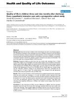
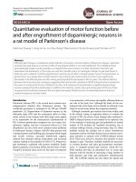
Fig. 1 Boxplots of the distribution of the peptides. From left up: Ghrelin, Cholecystokinin (CCK), Glucagon like peptide-1 (GLP-1), Fibroblast
growth factor-21 (FGF-21). P-values after Kruskall-Wallis or Mann-Whitney test are given above the corresponding boxes
other tissue. For example ghrelin mRNA is naturally
expressed in intestinal tissue but also in lymphocytes,
neutrophils and lymphoid tissue [27]. Cholecytokinin
was considered in some studies as a potential marker for
Ewing Sarcoma in children [28]. GLPR-1 receptor is
expressed on various immune cells and shows antiinflammatory effects - decreasing proliferation of Tcells, increasing number of T regulatory cells [29]. To
our knowledge only FGF-21 was underinvestigated in
this matter. From the fact that expression of this peptides was significant in cells circulating in peripheral
blood we can draw a conclusion that there is
physiological relevance. There is little information in the
literature on examined peptides in disease state, especially in metabolically unstable patients.
The amount of body fat did not influence peptide secretion, as the HSCT subgroups did not differ in terms
of anthropometric parameters. Our study showed that 6
months after conditioning there was a significant increase in the secretion of ghrelin, CCK, and GLP-1.
Plasma concentrations of these peptides were lower in
the pre-HSCT group than the post-HSCT (convalescent)
group and the control group. Kuruca et al. showed that
irradiation during the treatment of intestinal cancers
Skoczeń et al. BMC Cancer
(2020) 20:306
Page 7 of 9
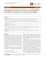
Fig. 2 Median concentrations of peptides (bars) with standard error (lines) before and after HSCT depending on regime used for conditioning
(BUS = Busulfan, CYC = Cyclofosphamide, TBI = total body irradiation). From left up: Ghrelin, Cholecystokinin (CCK), Glucagon like peptide-1 (GLP-1),
Fibroblast growth factor-21 (FGF-21). P-values after Kruskal-Wallis test are given per group analyzed
was associated with a decrease in ghrelin concentrations
[30]. The low concentrations of ghrelin persisted 3
months after irradiation. Statistical analysis of our data
revealed a considerable trend towards significance (p =
0.08) for the decreased ghrelin concentrations in patients
with mucositis. Moreover, we found that 6 months after
irradiation patients had higher levels of ghrelin compared to the values before conditioning. This suggests
recovery of ghrelin secretion. This is favorable because
ghrelin reduces intestinal injury and mortality after irradiation in animal models [31]. Interestingly, ghrelin
levels were increased in patients with liver aGvHD compared with those with cutaneous or intestinal aGvHD
(p = 0.02). This suggests dysregulation of gastric peptide
secretion caused by liver damage.
CCK has anti-inflammatory properties and reduces cell
apoptosis [32, 33]. We found higher concentrations of
CCK after HSCT suggesting regeneration of the upper
small intestine. The median concentration of GLP-1 was
significantly decreased in patients with aGvHD symptoms. Moreover, GLP-1 levels negatively correlated with
severity of aGvHD. In addition, GLP-1 concentrations
Skoczeń et al. BMC Cancer
(2020) 20:306
returned to baseline (the values seen in healthy subjects)
6 months after conditioning. This suggests full recovery
of the ileum. Logistic regression model indicates that
GLP-1 concentration could be a potential biomarker for
progression of aGvHD.
Increased concentrations of FGF-21 before conditioning suggest that hepatic injury may result from prolonged chemotherapy administered before HSCT.
Animal models show that liver damage induces FGF-21
expression [34]. Conditioning adds to an additional liver
injury. FGF-21is recognized as a stress response hepatokine that reduces hepatic damage through increased glucose uptake by adipose tissue. Normalization of FGF-21
concentrations 6 months after HSCT suggests complete
recovery of hepatic function after transplantation. The
FGF-21 gene expression data confirm the findings from
biochemical analysis. Although we found statistically significant differences in peptide concentrations and gene
expression model, the limitation of the current study is
small sample size. On the other hand, examined group is
unique. The presented results seem promising for establishing new diagnostic tools and provide the background
for further investigation.
Conclusions
Conditioning before HSCT and GvHD result in a widespread damage to the GI tract. Our data reveal that the
stomach, jejunum, ileum, and liver are affected by
chemo- and radiotherapy. Ghrelin may be a biomarker
for liver aGvHD, and GLP-1 seems to be a potential
biomarker for the progression of aGvHD. The increases
in the concentrations of the regulatory peptides secreted in all parts of the GI tract suggest intensive regeneration of the mucosa. These alterations seem to be
beneficial. The peptide measurements allow us to
monitor intestinal damage and regeneration. Our study
also showed that dysregulation of peptide secretion in
some segments of the intestine are long-lasting, as 6
months after HSCT increased ghrelin secretion in the
stomach, as well as CCK secretion in the jejunum, did
not return to the values seen in the control group. The
gene expression data are consistent with the biochemical data.
Supplementary information
Supplementary information accompanies this paper at />1186/s12885-020-06790-9.
Additional file 1: Supplementary Table 1. Mean concentrations of
peptides in post-HSCT group in aGvHD, mucositis and regarding localisation of aGvHD. Group n = 27. Freq = Frequency (%). P-values given after
ANOVA test (p < 0.05).
Additional file 2: Supplementary Table 2. Median concentrations
and quaritiles (in brackets) of peptides in treated group, patients with
Page 8 of 9
non- neoplastic and neoplastic disease before and after HSCT. P-values
given after Kruskal-Wallis test.
Abbreviations
ALT: Alanine transaminase; aGvHD: Acute graft-versus- host disease; BF: Body
fat; BMI: Body mass index; BMIPerc: BMI percentile; BMISDS: BMI standardised;
BUS: Busulfan; CBC: Complete blood count; CYC: Cyclofosphamide;
EIA: Enzyme immunoassay; FGF21: Fibroblast growth factor-21;
CCK: Cholecystokinin; GI: Gastrointestional tract; GLP-1: Glucagone like
peptide-1; GvHD: Graft-versus- host disease; HSCT: Hematopoietic stem cell
transplantation; JMML: Juvenile myelomonocytic leukemia; TBI: Total body
irradiation
Acknowledgements
No acknowledgements.
Authors’ contributions
SS and MR- design of the work, data collection, data analysis/interpretation,
drafting article, critical revision of article, DP, KK, AD - data analysis/
interpretation, drafting article, PT, KK- biochemical analysis/interpretation, WS,
MW- data interpretation, critical revision of article, KF, MK, MP - data analysis/
interpretation, WB and SS-supervised the study. All authors read and approved the final manuscript.
Funding
This work was supported by the National Science Centre under grant
number NN 407 198737.
Availability of data and materials
The datasets generated and/or analysed during the current study are
available in the GEO public repository and are accessible using GEO Series
accession number GSE69421 ( />cgi?acc=GSE69421). All remaining datasets used and/or analysed during the
current study are available from the corresponding author on reasonable
request.
Ethics approval and consent to participate
The Permanent Ethical Committee for Clinical Studies of the Jagiellonian
University Medical College approved the study protocol. All parents,
adolescent patients, and adult patients signed a written informed consent
before blood sample collection. Study was conducted in accordance with
the Declaration of Helsinki.
Consent for publication
Not applicable.
Competing interests
Authors declare that they have no competing interests.
Author details
Department of Oncology and Hematology, University Children’s Hospital in
Krakow, Jagiellonian University Medical College, Wielicka St. 265, 30-663
Krakow, Poland. 2Department of Oncology and Hematology, University
Children’s Hospital in Krakow, Wielicka St. 265, 30-663 Krakow, Poland.
3
Department of Clinical Biochemistry, University Children’s Hospital in
Krakow, Jagiellonian University Medical College, Wielicka St. 265, 30-663
Krakow, Poland. 4Department of Pediatric and Adolescent Endocrinology,
University Children’s Hospital in Krakow, Jagiellonian University Medical
College, Wielicka St. 265, 30-663 Krakow, Poland. 5Stem Cell Transplantation
Center, University Children’s Hospital in Krakow, Wielicka St. 265, 30-663
Krakow, Poland. 6Department of Biochemistry, University Children’s Hospital
in Krakow, Wielicka St. 265, 30-663 Krakow, Poland. 7Department of Statistics,
Cracow University of Economics, 27 Rakowicka Str., 31-510 Krakow, Poland.
8
Department of Molecular Neuropharmacology, Institute of Pharmacology of
Polish Academy of Sciences, 12 Smętna St., 31-343 Krakow, Poland.
1
Skoczeń et al. BMC Cancer
(2020) 20:306
Received: 18 October 2019 Accepted: 26 March 2020
References
1. Ghimire S, Weber D, Mavin E, Wang XN, Dickinson AM, Holler E.
Pathophysiology of GvHD and other HSCT-related major complications.
Front Immunol. 2017;8:79.
2. Armenian SH, Sun CL, Kawashima T, Arora M, Leisenring W, Sklar CA, et al.
Long-term health-related outcomes in survivors of childhood cancer treated
with HSCT versus conventional therapy: a report from the bone marrow
transplant survivor study (BMTSS) and childhood Cancer survivor study
(CCSS). Blood. 2011;4:1413–20.
3. Markey KA, MacDonald KP, Hill GR. The biology of graft-versus-host disease:
experimental systems instructing clinical practice. Blood. 2014;17:354–62.
4. Ahlman H, Nilsson O. The gut as the largest endocrine organ in the body.
Ann Oncol. 2001;12(suppl 2):S63–8.
5. Moran TH. Gut peptides in the control of food intake. Int J Obes. 2009;
33(suppl 1):S7–10.
6. Date Y, Kojima M, Hosoda H, Sawaguchi A, Mondal MS, Suganuma T, et al.
Ghrelin, a novel growth hormone-releasing acylatedpeptide, is synthesized
in a distinct endocrine cell type in the gastrointestinal tracts of rats and
humans. Endocrinology. 2000;141:4255–6.
7. Gualillo O, Caminos J, Blanco M, Garcia-Caballero T, Kojima M, Kangawa K,
et al. Ghrelin, a novel placental-derived hormone. Endocrinology. 2001;142:
788–94.
8. Burckhardt B, Delco F, Ensinck JW, Meier R, Bauerfeind P, Aufderhaar J, et al.
Cholecystokinin is a physiological regulator of gastric acid secretion in man.
Eur J Clin Investig. 1994;24:370–6.
9. Liddle RA, Goldfine ID, Rosen MS, Taplitz RA, Williams JA. Cholecystokinin
bioactivity in human plasma. Molecular forms, responses to feeding, and
relationship to gallbalder contraction. J Clin Invest. 1985;75:1144–52.
10. Fehmann HC, Goke R, Goke B. Cell and molecular biology of the incretin
hormones glucagon-like peptide-I and glucose-dependent insulin releasing
polypeptide. Endocr Rev. 1995;15:390–407.
11. George SK, Uttenthal LO, Ghiglione M, Bloom SR. Molecular forms of
glucagon like peptides in man. FEBS Lett. 1985;192:275–8.
12. Iglesias P, Selgas R, Romero S, Dı’ez J. Biological role, clinical significance,
and therapeutic possibilities of the recently discovered metabolic hormone
fibroblastic growth factor 21. Eur J Endocrinol. 2012;167:301–9.
13. WHO Handbook 1979: 15–22.
14. Date Y, Nakazato M, Hashiguchi S, Dezaki K, Mondal MS, Hosoda H, et al.
Ghrelin is present in pancreatic alpha-cells of humans and rats and
stimulates insulin secretion. Diabetes. 2002;5:124–9.
15. Douglas BR, Woutersen RA, Jansen JB, de Jong AJ, Lamers CB. The influence
of different nutriens on plasma cholecystokinin levels in rat. Experientia.
1988;44:21–3.
16. Wren AM, Seal LJ, Cohen MA, Brynes AE, Frost GS, Murphy KG, et al. Ghrelin
enhances appetite and increases food intake in humans. J
ClinEndocrinolMetab. 2001;86:5992–5.
17. WHO. WHO Anthro Survey Analyser and other tools. />childgrowth/software/en/. Accesed June 2019.
18. Gołąb S, Chrzanowska M. Child of Krakow region 2000. Biological
development of children and adolescents in Krakow city. Poziom rozwoju
biologicznego dzieci i młodzieży miasta Krakowa. Studia i Monografie AWF
w Krakowie. 2000:19.
19. Palczewska I, Niedźwiecka Z. Indices of somatic development of Warsaw
children and adolescents. (Wskaźniki rozwoju somatycznego dzieci i
młodzieży warszawskiej). Med. Wieku Rozwoj. 2001;(supl. 1):2.
20. Kushner RF, Schoeller DA. Estimation of total body water by bioelectrical
impedance analysis. Am J ClinNutr. 1986;44:417–24.
21. Rubenstein EB, Peterson DE, Schubert M, Keefe D, McGuire D, Epstein J,
et al. Clinical practice guidelines for the prevention and treatment of cancer
therapy–induced oral and gastrointestinal mucositis. Cancer. 2004;100:2026–
46.
22. Gosselin KB, Feldman HA, Sonis AL, Bechard LJ, Kellog EM, et al. Serum
citrulline as a biomarker of gastrointestinal function during hematopoietic
cell transplantation (HCT) in children. J Pediatr Gastroenterol Nutr. 2014;58:
709–14.
23. Crenn P, Messing B, Cynober L. Citrulline as a biomarker of intestinal failure
due to enterocyte mass reduction. Clin Nutr. 2008;27:328–39.
Page 9 of 9
24. Rabier D, Kamoun P. Metabolism of citrulline in man. Amino Acids. 1995;9:
299–316.
25. Markan KR, Naber MC, Ameka MK, Anderegg MD, Mangelsdorf DJ, Kliewer
SA, et al. Circulating FGF21 is liver derived and enhances glucose uptake
during refeeding and overfeeding. Diabetes. 2014;63(12):4057–63.
26. Huda MS, Wilding JP, Pinkney JH. Gut peptides and the regulation of
appetite. Obes Rev. 2006;7:163–82.
27. Gnanapavan S, Kola B, Bustin SA, Morris DG, McGee P, Fairclough P,
Bhattacharya S, Carpenter R, Grossman AB, Korbonits M. The tissue
distribution of the mRNA of ghrelin and subtypes of its receptor, GHS-R, in
humans. J Clin Endocrinol Metab. 2002;87(16):2988–91.
28. Reubi JC, Koefoed P, Hansen TVO, Stauffer E, Rauch D, Nielsen FC, Rehfeld
JF. Procholecystokinin as marker of human Ewing sarcomas. Clin Cancer
Res. 2004;10(16):5523–30.
29. Moschovaki Filippidou F, Kirsch AH, Thelen M, Kétszeri M, Artinger, et al.
Glucagon-like Peptide-1 receptor Agonism improves nephrotoxic serum
nephritis by inhibiting T-cell proliferation. Am J Pathol. 2020;190(2):400–11.
30. Karaca F, Afsar CU, Gunaldi M, Erkurt E, Ercolak V, Sertdemir Y, et al.
Alterations of ghrelin with weights and correlation among ghrelin, cytokine
and survival in patients receiving chemoradiotherapy for gastrointestinal
cancers. Int J Clin Exp Med. 2015;8:876–82.
31. Wang Z, Yang WL, Jacob A, Aziz M, Wang P. Human ghrelin mitigates
intestinal injury and mortality after whole body irradiation in rats. PLoS One.
2015;10:e0118213.
32. Miyamoto S, Shikata K, Miyasaka K, Okada S, Sasaki M, Kodera R, et al.
Cholecystokinin plays a novel protective role in diabetic kidney through
anti-inflammatory actions on macrophage: anti-inflammatory effect of
cholecystokinin. Diabetes. 2012;61:897–907.
33. Han Y, Su C, Yu D, Zhou S, Song X, Liu S, et al. Cholecystokinin attenuates
radiation-induced lung cancer cell apoptosis by modulating p53 gene
transcription. Am J Transl Res. 2017;15:638–46.
34. Yang C, Lu W, Lin T, You P, Ye M, Huang Y, et al. Activation of liver FGF21 in
hepatocarcinogenesis and during hepatic stress. BMC Gastroenterol. 2013;
17:67.
Publisher’s Note
Springer Nature remains neutral with regard to jurisdictional claims in
published maps and institutional affiliations.