Investigating the effects of additional truncating variants in DNA-repair genes on breast cancer risk in BRCA1-positive women
Bạn đang xem bản rút gọn của tài liệu. Xem và tải ngay bản đầy đủ của tài liệu tại đây (1.14 MB, 12 trang )
Sepahi et al. BMC Cancer
(2019) 19:787
/>
RESEARCH ARTICLE
Open Access
Investigating the effects of additional
truncating variants in DNA-repair genes on
breast cancer risk in BRCA1-positive women
Ilnaz Sepahi1, Ulrike Faust1, Marc Sturm1, Kristin Bosse1, Martin Kehrer1, Tilman Heinrich1,
Kathrin Grundman-Hauser1, Peter Bauer1,2, Stephan Ossowski1,3,4, Hana Susak3,4, Raymonda Varon5, Evelin Schröck6,
Dieter Niederacher7, Bernd Auber8, Christian Sutter9, Norbert Arnold10, Eric Hahnen11, Bernd Dworniczak12,
Shan Wang-Gorke13, Andrea Gehrig14, Bernhard H. F. Weber15, Christoph Engel16, Johannes R. Lemke17,
Andreas Hartkopf18, Huu Phuc Nguyen19, Olaf Riess1 and Christopher Schroeder1*
Abstract
Background: Inherited pathogenic variants in BRCA1 and BRCA2 are the most common causes of hereditary breast
and ovarian cancer (HBOC). The risk of developing breast cancer by age 80 in women carrying a BRCA1 pathogenic
variant is 72%. The lifetime risk varies between families and even within affected individuals of the same family. The
cause of this variability is largely unknown, but it is hypothesized that additional genetic factors contribute to differences in
age at onset (AAO). Here we investigated whether truncating and rare missense variants in genes of different DNA-repair
pathways contribute to this phenomenon.
Methods: We used extreme phenotype sampling to recruit 133 BRCA1-positive patients with either early breast cancer
onset, below 35 (early AAO cohort) or cancer-free by age 60 (controls). Next Generation Sequencing (NGS) was used to
screen for variants in 311 genes involved in different DNA-repair pathways.
Results: Patients with an early AAO (73 women) had developed breast cancer at a median age of 27 years (interquartile
range (IQR); 25.00–27.00 years). A total of 3703 variants were detected in all patients and 43 of those (1.2%) were truncating
variants. The truncating variants were found in 26 women of the early AAO group (35.6%; 95%-CI 24.7 - 47.7%) compared
to 16 women of controls (26.7%; 95%-CI 16.1 to 39.7%). When adjusted for environmental factors and family history, the
odds ratio indicated an increased breast cancer risk for those carrying an additional truncating DNA-repair variant to BRCA1
mutation (OR: 3.1; 95%-CI 0.92 to 11.5; p-value = 0.07), although it did not reach the conventionally acceptable significance
level of 0.05.
Conclusions: To our knowledge this is the first time that the combined effect of truncating variants in DNA-repair genes
on AAO in patients with hereditary breast cancer is investigated. Our results indicate that co-occurring truncating variants
might be associated with an earlier onset of breast cancer in BRCA1-positive patients. Larger cohorts are needed to confirm
these results.
Keywords: Breast cancer, Age at onset, DNA-repair genes, Next-generation-sequencing, Panel sequencing, Extreme
phenotypes, Hereditary breast and ovarian cancer, BRCA1, DNA-repair
* Correspondence:
1
Institute of Medical Genetics and Applied Genomics, University of Tübingen,
Tübingen, Germany
Full list of author information is available at the end of the article
© The Author(s). 2019 Open Access This article is distributed under the terms of the Creative Commons Attribution 4.0
International License ( which permits unrestricted use, distribution, and
reproduction in any medium, provided you give appropriate credit to the original author(s) and the source, provide a link to
the Creative Commons license, and indicate if changes were made. The Creative Commons Public Domain Dedication waiver
( applies to the data made available in this article, unless otherwise stated.
Sepahi et al. BMC Cancer
(2019) 19:787
Background
Breast cancer is the most common cancer among
women with 30% of all new cancer diagnoses [1]. About
one out of eight US women will develop breast cancer
during her lifetime. It is estimated that hereditary genetic factors explain 5–10% of all breast cancer cases [2].
In the mid-1990s, BRCA1 and BRCA2 [3–5] which are
part of the DNA-repair machinery [6] were identified to
play a crucial role in hereditary breast and ovarian cancer (HBOC) [3–5, 7, 8]. Together, pathogenic variants in
these two genes explain about 24% (95%-CI,23.4 to
24.6%) of all HBOC cases [7]. BRCA1 and BRCA2 are
functioning as genome guardians by playing a central
role in the homologous recombination repair (HRR)
pathway. Up to now, more than 300 gene products have
been associated with the DNA-repair machinery and
genome integrity maintenance of which 25 genes [8]
have been linked to HBOC.
In female BRCA1 mutation carriers, the risk of developing breast cancer by the age of 80 is 72% [9]. Moreover, the incidence of breast cancer rises quickly in early
adulthood until age 30 to 40 years in BRCA1 mutation
carriers [9]. Even though pathogenic variants in BRCA1
are associated with the highest penetrance of HBOC, the
cause for the inter-individual and even intra-familial
variation in penetrance is not clear and remains an active field of research. This variation results in difficulties
in risk calculation and genetic counseling. Several environmental factors such as birth cohort [10], age at menarche [11], number of pregnancies [12], therapeutic
abortion [13], oral contraceptives [14], and prophylactic
oophorectomy [15, 16] are suspected to affect the risk of
cancer in BRCA1/2 mutation carriers. Using data from
the Generations Study, Brewer and colleagues showed
that having a first-degree female relative with breast cancer increases the relative risk of breast cancer as compared to those without family history [17]. Moreover, the
variation in penetrance can be due to allelic variation,
which means variation in the variant type (truncating or
missense) and position within the coding region of the
BRCA1 gene [18]. As proposed by Thompson and
Easton in 2001 and 2002 and also Rebbeck et al. [19–
21], the position of the respective causative pathogenic
variant within the coding region of BRCA1/2 can change
breast or ovarian cancer risk. In this context, Rebbeck
and colleagues identified three putative “breast cancer
cluster regions” including BCCR1 which overlaps with
the RING domain of the BRCA1 protein and an “ovarian
cancer cluster region” located in exon 11 [21]. Furthermore, pathogenic variants towards the 3′-end of BRCA1
lead to a lower risk of ovarian cancer compared to breast
cancer [22].
Another cause of differences in penetrance are modifying genes [18]. The Consortium of Investigators of
Page 2 of 12
Modifiers of BRCA1/2 (CIMBA, .
ac.uk/consortia/cimba) screened more than 20,000 mutation carriers and performed Genome Wide Association
Studies (GWAS) to identify genetic modifier loci [23–29]
and described several candidates; each adding a small part
of risk variation in BRCA1 mutation carriers (in total 2.2%
in BRCA1) [23]. The CIMBA consortium suggested using
a combination of different modifier loci to increase the
precision of risk prediction. Unlike GWAS studies that are
based on common variants, this study pursued the goal to
predict BRCA1 penetrance and AAO of breast cancer by
analysing rare variants in genes that are part of the DNA
damage response and genome integrity maintenance pathways as well as genes which are interacting with BRCA1.
Accurate prediction of AAO can become of clinical relevance in order to prevent overtreatment of carriers who
will never develop breast cancer during their lifetime or
may develop it later in life. To address this issue, we aimed
to investigate the differences in AAO of breast cancer
among BRCA1 mutation carriers by studying 311 DNArepair genes which are contributing to genome stability
along with BRCA1 and BRCA2.
Methods
Selection of samples for extreme phenotype sampling
Out of more than 30,000 HBOC index cases registered
in the German Consortium for Hereditary Breast and/or
Ovarian Cancer (GC-HBOC) biobank, 133 BRCA1-positive patients either with a personal history of breast cancer below the age of 35 years (early AAO onset) or
without personal history of breast cancer at the age of
60 years (controls) were selected for this study. Patients
who had undergone prophylactic mastectomy or
prophylactic oophorectomy before the age of 45 years
were excluded from the analysis [30]. Participants had
signed a written informed consent and the study was
approved by the local ethics committee (ethic vote number 053/2017BO2). Relevant information regarding age
at menarche, number of pregnancies, and Oral contraceptive use was collected from the GC-HBOC database.
Sequencing and data analysis
Reviewing published literature, genes were considered
on the basis of a reported breast cancer association. In
addition, all DNA-repair pathway genes were selected
from KEGG GENES database ( />kegg/genes.html, last accessed: 26.11.2013; Additional file 1:
Table S1). A target region of 895.2 kbp consisting of 311
genes was sequenced in total. The coding regions and
exon-intron boundaries ±25 bps were targeted (using
default parameters of Agilent SureDesign, except for
Masking = Most Stringent) and enriched using Agilent
SureSelect custom RNA probes (Agilent, Santa Clara,
CA). Two hundred nanograms of genomic DNA were
Sepahi et al. BMC Cancer
(2019) 19:787
checked for quality and quantity by Qubit dsDNA Assay
(Thermo Fischer Scientific, Waltham, MA, USA) and
fragmented using a Covaris system (Covaris, Inc.,
Woburn, Massachusetts) to generate fragments of 120–
150 base pairs length. Quality and fragment size of
sheared DNA were checked using a TapeStation (Agilent,
Santa Clara, CA). Sequencing libraries were constructed according to the Agilent SureSelectXT protocol. The pre-capture and post-capture libraries were
quantified by a TapeStation. Libraries were sequenced
either on a Miseq (Illumina, San Diego CA), NextSeq500
(Illumina, San Diego CA) or HiSeq2500 (Illumina, San
Diego CA) platform using paired-end reads of 151 bps or
101 bps.
MegSAP, a free-to-use open-source bioinformatics pipeline was used for data analysis (version 0.1–379-gb459ce0,
In brief, adapter and
quality trimming was applied using SeqPurge [31]; sequencing reads were mapped to the human genome version GRCh37 with BWA (v. 0.7.15) [32], and ABRA2 [33]
(v. 2.05) was used for indel realignment; variant calling
was performed by freebayes (v. 1.1.0) [34] and variant annotation was done using snpEff/SnpSift (v. 4.3i) [35].
Quality control was executed on three layers of information including raw reads, mapped reads and variants
(Additional file 2: Table S2). We used Alamut batch (v.
1.5.1, Interactive Biosoftware) for splice site annotation.
Page 3 of 12
Minor Allele Frequency (MAF > 1%) in the 1000
Genomes Project (1KGP), dbSNP, Exome Aggregation
Consortium (ExAC) or ESP6500.
Statistical analysis
Descriptive statistics such as medians, means and
standard deviations for continuous data and proportion and 95%-CI for categorical data was used to
characterize the study population and sequencing results. A multivariable logistic regression was carried
out to control for the potential confounding effect of
family history, age at menarche, parity, and use of
oral contraceptives. Missing data was imputed using
median or mode. The variable additional truncating
DNA-repair variants was coded as yes if the patient
carried a truncating DNA-repair variant and it was
coded as no if the patient was not carrying a truncating DNA-repair variant. The outcome was considered
the incidence of cancer. The regression analysis was
performed in R 3.5.2. Using GraphPad Prism version
6.07 for Windows (GraphPad Software, La Jolla California
USA), we performed Fisher’s exact test to compare the
mutational location in each cohort. All p-values were twotailed and p-values less than 0.05 were considered to be
statistically significant. Maftools was applied to visualize
BRCA1 pathogenic variants with a modified database [38].
Rare variant association study
Variant interpretation
Variants were automatically classified according to an
algorithm based on a modified version of the American College of Medical Genetics and Genomics
(ACMG) guidelines for variant classification [36]. According to this algorithm, splice variants at the position +/− 1 and +/− 2 are classified as likely
pathogenic if the variant disrupts the function of the
gene product unless the population frequency of the
variant is not compatible for a pathogenic variant
(minor allele frequency of 1% was used as a cutoff ).
For intronic variants located outside of the canonical
splice sites including Cartegni splice sites [37] we referred to Alamut Visual (Interactive Biosoftware) incorporated prediction tools such as MaxEntScan,
Splice Site Finder Like, and Human Splicing Finder.
Variants were considered as pathogenic or likely
pathogenic (collectively termed as pathogenic) if they
led to a truncation, initiation loss or canonical splice
site effect or if there was a relevant publication in
favor of pathogenicity and if there was additional evidence in public database like ClinVar. In case there
was no evidence such as functional assessment data
available, missense, synonymous and intronic variants
were classified as variants of unknown significance
(VUS), benign or likely benign according to the
Variants obtained from freebayes in VCF format (see
above) were annotated using the eDiVA platform
( in order to obtain functional annotation (exonic, nonsynonymous, synonymous, splicing
etc.), European population allele frequencies from 1KGP,
Exome Variant Server (EVS) and ExAC databases, as well
as functional impact scores from CADD. Variants not annotated as ‘exonic’ or ‘splicing’, as well as variants within
segmental duplication (SegDup identity > = 0.9) were removed from further analysis. We performed sample quality control by screening for outliers in (a) number of
variants per sample and (b) transition to transversion ratio
per sample. Second, we calculated the first 10 PCA components of all samples using only synonymous SNVs that
were not in linkage disequilibrium and had an allele frequency above 0.005 in EVS. Finally, we compared the rare
variant load per gene between the early AAO cohort and
controls. No outliers were detected in any QC test and
early AAO patients and controls were clustering in a single group in the PCA. Following QC, we removed any
variant with European AF higher than 0.01 in any of the
three databases: EVS, 1KGP, and ExAC. Additionally, we
excluded all variants annotated as synonymous or with a
CADD score below 10 (considered neutral). Using the
remaining rare, likely damaging variants we performed
Burden and SKAT-O association tests implemented in the
Sepahi et al. BMC Cancer
(2019) 19:787
R package SKAT ( />download/) version 1.3.0. The Null model for both tests
was computed using the SKAT_Null_Model function with
output set to dichotomous outcome (out_type = “D”) and
no sample adjustment (Adjustment = FALSE). For the
SKAT-O test we used the SKATBinary function with default parameters except for method that was set to “optimal.adj” (equivalent to SKAT-O method). Minor allele
frequencies (MAF) of variants transformed with Get_Logistic_Weights were used as weights. The burden test was
performed using the same function (SKATBinary) and parameters, except for method that was set to “Burden”.
Results
Participants characteristics
In total, 133 BRCA1 positive women were screened for
truncating variants in 311 DNA-repair genes. The cohort with early AAO consisted of 73 women who developed breast cancer at an age younger than 35 years
(median age at onset, 27 years; interquartile range (IQR)
25–27 years). The controls consisted of 60 participants,
cancer-free by the age of 60 years. Follow-up data
showed that some developed breast cancer at an age
older than 60 years (n = 25; 41.7%) with a median age at
onset of 64 years (IQR, 62–67) or had no history of
breast cancer (n = 35; 58.3% median age, 70 years; IQR,
63–75 years). The demographic characteristics of the
participants are shown in Table 1.
Page 4 of 12
In total, 117 patients from both cohorts carried a
BRCA1 pathogenic single nucleotide variant (SNV), 13
patients carried a large deletion, and three patients carried a large duplication in BRCA1 (Fig. 1). In the early
AAO cohort, 15.1% of all participants carried a frameshift founder mutation [39] in exon 20 of the BRCA1
gene (ENST00000357654: c.5266dupC:p.Gln1756fs). The
European founder missense variant [40] in exon 4
(ENST00000357654: c.181 T > G: p.Cys61Gly) was the
most frequent (10%) pathogenic variant found in the
control cohort (Additional file 3: Table S3). All pathogenic variants in BRCA1 were confirmed by NGS.
With respect to family history, the majority of patients
in the control cohort had at least one first-degree relative with breast and/or ovarian cancer as compared to
the early AAO patients (56.2% versus 98.4%). Women
with larger families who reached older ages are expected
to have more relatives with breast and/or ovarian cancer
on average in comparison to those whose families are
smaller and younger. This can explain the difference between family history of early AAO cohort and control
cohort (Table 1).
Comparison of type and location of BRCA1 pathogenic
variants
To compare allelic variation in type and location of
pathogenic variants across the BRCA1 protein between
the early age at onset and the control cohort, we compared the pathogenic variant accumulation in different
Table 1 Demographic characteristics of the population study
Early age at onset cohort
Control cohort
Total Number
73
60
Breast cancer positive
100%
41.7%
Median age at onset(IQR)
27 (25–27)
64 (62–67) n = 25
BCCR1 (95%-CI)
13.8% (6.1–25.4%)
11.5% (4.4–23.4%)
BCCR2 (95%-CI)
8.6% (2.9–19.0%)
5.8% (1.2–15.9%)
BCCR2’ (95%-CI)
22.4% (12.5–35.3%)
15.4% (6.9–28.1%)
OCCR (95%-CI)
25.9% (15.3–39%)
42.3% (28.7–56.8%)
Frame-Shift-Del
26.0% (16.5–37.6%)
35.0% (23.1–48.4%)
Frame-Shift-Ins
19.2% (10.9–30.1%)
16.7% (8.3–28.5%)
Missense variant
8.2% (3.1–17.0%)
13.3% (5.9–24.6%)
Nonsense variant
26.0% (16.5–37.6%)
21.7% (12.1–34.20%)
Splice-Site variant
5.5% (1.5–13.4%)
5.0% (1.0–13.9%)
CNV
BRCA1 variant location
BRCA1 variant type % (95%-CI)
15.1% (7.8–25.4%)
8.3% (2.8–18.4%)
Family History
Data available for
73 (100%)
60 (100%)
First-degree relative with Breast and/or Ovarian cancer
41 (56.2%)
59 (98.4%)
BCCR Breast cancer cluster region, BCCR1 c.179–505, BCCR2 c.4328–4945, BCCR2’ c.5261–5563, OCCR c.1380–4062, Del Deletion, Ins Insertion, CNV Copy
number variation
Sepahi et al. BMC Cancer
(2019) 19:787
Page 5 of 12
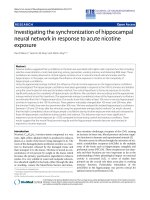
Fig. 1 BRCA1 pathogenic variants. X axis shows the amino acid position and functional domains of the BRCA1 protein. Each lollipop represents a
pathogenic variant and the type of variant is depicted with different colors. The Y axis demonstrates the number of mutation carriers. The
Horizontal bars show the copy number variation. Deletion (red) and duplication (purple) is depicted by different colors. Breast cancer Cluster
Regions (BCCRs) are shown as black bars and Ovarian Cancer Cluster Region (OCCR, Rebbeck and colleagues [21]) are depicted in dark blue.
Splice-site variants are not shown
regions of BRCA1. Whereas no differences were detected for the Breast Cancer Cluster Regions (BCCRs),
which are associated with increased risk of breast cancer
(Additional file 4: Figure S1a), differences were found for
the Ovarian Cancer Cluster Region (OCCR). 22 (45.3%)
patients in the control cohort (Fig. 1, Table 1) carried a
pathogenic variant within the OCCR compared to 15
(25.9%) of patients in the early AAO cohort, though the
statistical significance was not reached (p-value = 0.07).
Patients with large deletions or insertions and splice site
variants were excluded from this analysis since they either span more than one region or their impact on protein function is not certain, respectively. In the early
AAO cohort, 56 patients (76.7%; 95%-CI 65.4 to 85.3%) of
BRCA1 mutation carriers carried a truncating variant
while 6 patients (8.2%; 95%-CI 3.1 to 13.3%) carried a missense pathogenic variant (ENST00000357654: c.181 T > G:
p.Cys61Gly) and 11 patients (15.1%; 95%-CI 7.8 -25.4%)
carried a copy number variation (CNV). In contrast, 47
patients (78.3%; 95%-CI.65.8% to 87.9) carried a truncating
variant in controls, 8 patients (13.3%; 95%-CI 5.9 to
24.6%) carried a missense pathogenic variant (Additional
file 4: Figure S1b) including ENST00000357654: c.181 T >
G: p.Cys61Gly, and c.5096G > A: p.Arg1699Gln and 5 patients (8.3%; 95% CI 2.8 to 18.4%) carried a CNV.
Truncating germline variants in DNA-repair genes
We evaluated 311 genes that maintain genome integrity and/or have been associated with HBOC. The
mean sequencing depth was 456x ± 197.3 SD. Additional
Sepahi et al. BMC Cancer
(2019) 19:787
file 2: Table S2 shows the detailed results and quality parameters of sequencing. A total of 3703 variants was identified and of those 43 (1.2%) truncating variants
(Additional file 5: Table S4) were detected in 36 DNA-repair genes. The affected genes were mainly Single Strand
Break Repair genes (SSBR, 30.6%), Double Strand Break
Repair genes (DSBR, 30.6%), and check-point factor genes
(11.1%). The remaining truncating variants were identified
in genes with other functions such as BRCA1/2 interactors, centrosome formation and signal transduction. In
overall, 42 women had at least one additional DNA-repair
truncating variant. In the early AAO cohort, 26 out of 73
patients (35.6%; 95%-CI 24.7 - 47.7%) carried at least one
additional truncating variant and two cases carried two
additional truncating variants in DNA-repair genes
(Additional file 6: Figure S2a). Among controls, 16 out of
60 participants carried an additional DNA-repair germline
truncating variant (26.7%; 95%-CI 16.1 to 39.7%). In this
cohort, three participants carried two germline DNA-repair truncating variants; at least one of them affected a
DSBR pathway gene (Additional file 6: Figure S2b).
We investigated the effect of additional DNA-repair
truncating variants on the risk of developing breast cancer among BRCA1 mutation carriers, adjusted for age at
menarche, oral contraceptive use, parity and family history. Despite the fact that it did not reach the conventionally accepted p-value of 0.05, the odds ratio is in
favor of increased breast cancer risk for double heterozygote patients (OR: 3.1; 95% CI 0.92 to 11.5, p-value =
0.07). To confirm the validity of our model, the same
analysis was carried out on a subset of subjects who
were matched for family history (early AAO cohort; n =
41 and control cohort; n = 59) adjusted for age at menarche, oral contraceptive use and parity (OR: 3.3; 95%-CI
0.92 to 13.3; p-value = 0.07). Consistent results were obtained for this subset of cohorts.
To test the effect of additional truncating variants
in specific DNA-repair pathways, we compared the
mutational load in DSBR and SSBR genes between
the two cohorts. Among the early AAO cohort, 8/73
women (11.0%; 95%-CI 4.9 -20.5%) carried an additional truncating variant in DSBR compared to 5/60
women (8.3%; 95%-CI 2.8 -18.4%) in the control cohort. Regarding the SSBR genes, we found 8/73
women (11.0% %; 95%-CI 4.9 -20.5%) in the early
AAO cohort carrying additional SSBR truncating variants as compared to 5/60 women (8.3%; 95%-CI 2.
%-20.5) in the control cohort. The mutational load in
DSBR and SSBR did not differ between both cohorts
(Fig. 2). Further comparison has been carried out between SSBR- and DSBR- mutation carriers with noncarriers (Additional file 7: Figure S3; Additional file 8:
Table S5). In none of the cases differences were statistically significant.
Page 6 of 12
Pathological characteristics
Among control cohort, 25 (41.7%) patients developed
breast cancer at a median age of 64. For these patients the tumor characteristics were compared with
the tumor characteristics of the early AAO patients.
The immunohistochemical staining of estrogen and
progesterone receptors did not differ significantly with
respect to the AAO, though the ER and PR negativity
was more frequently found in the early AAO cohort
compared to affected control patients (p-value = 0.28
and 0.76 respectively, Table 2). Tumors of the early
AAO group tended to show a higher histological
grade compared to the tumors of the affected control
patients (Table 2) although the difference failed to
reach the significant level (p-value = 0.24). Expression
of estrogen and progesterone receptors, grading of tumors and histological types of tumors were not significantly different between patients with additional
truncating variants in DNA-repair genes and patients
without additional DNA-repair truncating variants
(Additional file 9: Table S6).
Rare variant association study (RVAS)
To assess the load of rare missense (VUS + pathogenic
variants) variants in DNA-repair genes on the AAO
of breast cancer in BRCA1-positive patients we performed a Burden test and a SNP-set (sequence)
Kernel Association Test (SKAT-O). To this end, a
comprehensive quality control of early AAO cohort
and controls was done (see Methods). No differences
were observed between early AAO cohort and controls in (a) variants per sample, (b) rare variant load
per gene, (c) transition-transversion ratio, and (d) top
10 PCA components. Next, we removed all common
variants (MAF > 1% in EVS, 1KGP, or ExAc) as well
as all synonymous variants from both early AAO and
control cohort. To search for genes conveying an increased risk, we used patients of the early AAO
cohort as cases and patients of the late AAO cohort
as controls (Additional file 10: Table S7). Although
there was no significant gene identified after FDR
correction, several genes showed significant un-corrected p-values in at least one of the two RVAS tests,
requiring more investigation in independent larger cohorts. These candidate genes include MYBBP1A (early
AAO: 13, controls: 3), MRE11 (7:0), TDG (5:0), WRN
(7:1), TP53BP1 (10:3) and REV1 (8:2) as well as one
potential risk reducing factor, PTCH1 (early AAO: 1,
controls: 8).
Patients with both heterozygous pathogenic variants in
BRCA1 and BRCA2
Interestingly, two cases carrying pathogenic variants in
both BRCA genes were found in either cohort. Case 1
Sepahi et al. BMC Cancer
(2019) 19:787
Page 7 of 12
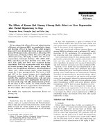
Fig. 2 Distribution of carriers of additional DNA-repair mutation in each cohort regarding the type of pathway. 43 truncating variants were
detected in 36 DNA-repair genes. These truncating variants mainly affected double-strand break repair (DSBR), single-strand break repair (SSBR),
BRCA1/2 interactors, centrosome formation, and check-point factors. No significant difference was found in DSBR, SSBR, BRCA1 / BRCA2 interactors,
checkpoint factors and other pathways mutational load between the two cohorts. Two cases in the early AAO cohort carried an additional
mutation in BRCA1 / BRCA2 interactor genes while no mutation acrrier in these genes was found in control cohort. The width of each block
referes to the porportion of mutated pathway among all mutated pathways and the hight of each block referes to the porportion of mutated
samples in each cohort. Mutated genes in each pathways are shown in boxes
was a patient affected with breast cancer at the age of
26 yrs. She had two first-degree relatives with breast cancer. There was no ovarian cancer and no second-degree
relative with any type of cancer. She carried a BRCA1
pathogenic variant (ENST00000357654: c.1016dupA)
and an additional BRCA2 pathogenic variant
(ENST00000544455.1: c.3585_3686delAAAT). Unfortunately, tumor characteristics were not available for this
patient. Case 2 was diagnosed with breast cancer at the
age of 63.9 years. Her family history was indicative for
HBOC: A first-degree relative with breast cancer and
three first-degree relatives with ovarian cancer. Also, there
was a second-degree relative with breast cancer. She
carried a nonsense variant in BRCA1 (ENST00000357654:
c.1687C > T) and a nonsense variant in BRCA2
(ENST00000544455.1: c.8875G > T). An additional truncating variant was found in EME2, (ENST00000568449:
c.541_544delGCTG) a DSBR gene. The immunohistochemical staining showed a triple negative tumor.
Discussion
Genome-wide case control association studies identified
susceptibility variants and modifiers of penetrance for
BRCA1 mutation carriers [23, 25–29]. Despite the fact
that each modifier explains a small proportion of genetic
variation of breast cancer development in carriers of
Sepahi et al. BMC Cancer
(2019) 19:787
Page 8 of 12
Table 2 Histopathological characteristics of tumors
Early AAO cohort
Number (%)
Affected controls
Number (%)
62 out of 73
22 out of 25
Ductal
53 (85 .5%)
22 (100%)
Medullary
6 (9.7%)
0
Lobular
2 (3.2%)
0
Others
1 (1.6%)
0
66 out of 73
22 out 25
Histological Type
Data available
Histological grade
Data available
Grade III
53 (80.3%)
14 (63.7%)
Grade II
13 (19.7%)
7 (31.8%)
Grade I
P value
0.10
0.24
0
1 (4.5%)
64 out of 73
22 out 25
ER negative
47 (73.4%)
13 (59.1%)
0.28
PR negative
52 (81.3%)
17 (77.3%)
0.76
52 out of 73
19 out of 25
Steroid receptors
Data available
Human Epidermal Receptor
Data available
HER2/neu negative
Triple Negative Breast Cancer
Data Available
TNB
49 (94.2%)
17 (89.5%)
55 out of 73
20 out of 25
32 (58.2%)
11 (55%)
0.60
Data were available for 67 out of 73 patients in the early age at onset cohort and from 25 cases that developed breast cancer in control cohort
ER Estrogen receptor, PR Progesterone receptor, HER2 Human epidermal growth factor receptor 2
BRCA1 pathogenic variants [23], still a large proportion
of risk variation is unknown. The effect of each modifying variant can be combined into poly genic risk scores
(PRSs), which may confer larger relative risks [25, 41].
The approach taken in this study was to enrich for rare
variants via preferentially selecting the carriers who are
most informative cases [42]. For this reason, the extreme
ends of age at onset of hereditary breast cancer were
chosen and we aimed to identify differences in the mutational load in these two highly selected cohorts. We hypothesized that inherited truncating variants in DNArepair genes, which are partner components of BRCA1
in the maintenance of genome integrity, are likely to
interact with BRCA1 by reducing the age at onset of hereditary breast carcinoma.
Previously reported by Thompson and Easton in 2001
and subject of a more recent study by Rebbeck et al.
(2015), it was found that allelic variation in BRCA1
pathogenic variants is one of the reasons of variation in
risk for breast cancer compared to ovarian cancer in
HBOC patients. Rebbeck and colleagues described multiple regions associated with a higher risk for breast cancer compared to ovarian cancer (breast cancer cluster
regions = BCCRs) and, one region with an increased risk
for ovarian cancer compared to breast cancer (OCCR)
[19–21]. The mutational position comparison in our
cohorts showed no difference for BCCRs but a non-significant higher variant load in the OCCR (p-value = 0.07)
among controls. Although the difference was not statistically significant, it is worth considering that pathogenic
variants in OCCR not only lead to increased risk of
ovarian cancer but they also decrease the risk of breast
cancer [21]. Regarding the variant type, there was no difference in truncating or missense variants distribution in
each cohort. While the most common pathogenic missense
variant in both cohort was ENST00000357654: c.181 T >
G: p. Cys61Gly, the missense variant ENST00000357654:
c.5090G > A: p.Arg1699Gln was exclusively found in two
of the patients in the control cohort. This is in line with
previous reports where this variant had reduced cumulative risk of breast cancer by age 70 to 20% [43, 44].
Concerning the sum effect of truncating DNA-repair
variants on the risk of breast cancer among BRCA1 mutation carriers, our results are suggesting an increase in
the breast cancer risk for the BRCA1 mutation carriers
who carry additional truncating DNA-repair variants
(OR: 3.1; 95% CI 0.92 to 11.5; p-value = 0.07). The small
number of old cancer-free BRCA1 mutation carriers was
a limiting factor in this study. The sum effect of pathogenic variants in DNA-repair genes can lead to a different cancer phenotype as shown by Pritchard and
colleagues [45] who reported a higher prevalence of
Sepahi et al. BMC Cancer
(2019) 19:787
germline DNA-repair pathogenic variants in metastatic
prostate cancer patients compared to localized prostate
cancer. More recently, Brohl and colleagues [46] reported a significantly higher frequency of germline
DNA-repair pathogenic variants in patients with Ewing
sarcoma in comparison with general population. By
pathway analysis they uncovered that hereditary breast
cancer genes, and remarkably, genes involved in DSBR
were highly mutated.
Despite the small sample size, we carried out a rare
variant association study (RVAS) using SKAT-O and
Burden tests to shed light on the role of rare variants in
the genetic risk of hereditary breast cancer. The results
of SKAT-O and Burden tests were not statistically significant after multiple testing corrections. The top
ranked gene in the Burden test is MRE11. Mre11 is a
member of MRN (MRE11, RAD50, and NBS1) complex
[47]. This complex is involved in the sensing of DNA
double strand breaks and it initiates the processing of
double strand break repair [48–50]. Studies showed that
hypomorphic mutations in MRE11 and NBS1 lead to
Ataxia telangiectasia disorder and Nijmegen breakage
syndrome, a rare autosomal recessive disorder [51, 52].
Pathogenic variants in the MRN complex were also
linked to cancer predisposition. Recently Gupta and colleagues showed an association between triple negative
breast cancer and MRE11 defects [53]. The top ranked
gene in SKAT-O test and the third top ranked gene in
burden test is MYBBP1 which inhibits colony formation
and tumorigenesis and enhances the anoikis in a p53
dependent manner [54].
We also evaluated the tumor histology and immunohistochemical characteristics of the tumors and whether
they were influenced by AAO among BRCA1 mutation
carriers. Although the clinicopathological features of
BRCA1 associated breast tumors are studied widely and
previous studies showed that BRCA1 positive tumors
demonstrated higher tumor grade, lower estrogen receptor expression, and lower progesterone receptor expression [55–57], the status of ER and PR expression among
young and older BRCA1 associated breast cancer patients is less well studied. Vaziri and colleagues [58] observed that the ER and PR negativity was more common
in BRCA1-positive patients with an age at onset younger
than 50 years compared to above 50 years of age. In
2005, Eerola and colleagues [59] showed similar results
by studying BRCA1/2 positive families in comparison
with BRCA1/2 negative families. They observed a significant difference in ER negativity for BRCA1 positive, premenopausal patients (age of diagnosis below 50 years).
These patients also suffered from higher-grade tumors
compared to postmenopausal patients. Our results also
demonstrate that carrying a truncating variant in DNArepair genes in addition to a BRCA1 pathogenic variant
Page 9 of 12
does not change tumor characteristics since the differences in histology and histochemical features of tumors
did not differ in those with additional truncating variants
in DNA-repair genes compared to those without.
As part of the study we also identified double heterozygotes for pathogenic BRCA1 and BRCA2 variants.
While the frequency of pathogenic variants in BRCA1
and BRCA2 is high in the Ashkenazi Jewish population
[60, 61], it was found that 0.3% of all Ashkenazi Jewish
breast cancer patients were double heterozygotes for
BRCA1/2 pathogenic variants [62]. In contrast, double
heterozygosity for the two major breast cancer genes is
expected to be less common phenomenon in other populations. Several studies reported double heterozygous
females including a report by Heidemann and colleagues (2012), showing that double heterozygotes
were not younger at the time of first diagnosis compared to other patients. Interestingly, they reported a
more severe phenotype in double heterozygote females in comparison with their single heterozygote
relatives [63]. In the present study, we identified two
cases with double heterozygosity in BRCA1/2. One of
them was found in early AAO cohort whereas another double heterozygote BRCA1/2 female had a late
breast cancer manifestation. These results advocate
panel testing, since panel testing allows detection of
variants in different genes simultaneously. The presence of additional truncating variants is also of high
relevance for the families and segregation analysis
should be offered in families with known pathogenic
variants to identify patients with high risk for cancer
predisposing syndromes.
Conclusions
In the last few years, several attempts were made to
elucidate the variable penetrance of BRCA1 pathogenic variants. GWA analyses identified several loci,
which can modify the penetrance of BRCA1/2 pathogenic variants and the age at onset of hereditary
breast and ovarian cancer to some extent. To our
knowledge, this is the first time that germline truncating variants in DNA-repair pathways were studied
for their effect on age of breast cancer onset among
BRCA1 carriers. The odds ratio observed in this study
indicates a potential effect of co-occurring DNA-repair truncating variants and pathogenic variants in
BRCA1 on the earlier onset of breast cancer. Limitations of this study are the small sample size due to
low numbers of asymptomatic BRCA1 mutation carriers and the large number of missense variants in
DNA-repair genes which are of uncertain significance.
Further studies and larger cohorts are needed to confirm the results obtained in this study.
Sepahi et al. BMC Cancer
(2019) 19:787
Additional files
Additional file 1 : Table S1 List of 311 DNA repair and cancer
predisposition syndrome genes as well as the pathways. DSBR: Double
Strand Break Repair, SSBR: Single Strand Break Repair, HR: Homologous
Recombination, NER: Nucleotide Excision Repair, BER, Base Excision Repair,
FA: Fancony Anemia, NHEJ: Non-Homologous End Joining. (XLSX 20 kb)
Additional file 2 : Table S2 The quality parameters of Next Generation
Sequencing. (DOCX 14 kb)
Page 10 of 12
data. KB, MK, TH, KGH, RV, ES, DN, BA, CSu, NA, EH, BD, SWG, AG, BHFW, JL,
AH, HHPN performed genetic counseling and/or testing and interpreting of
respective results. All the authors contributed in critical revision of the
manuscript. All authors read and approved the final manuscript.
Funding
The study was supported by a Fortüne Project grant of the Medical Faculty
of the University of Tübingen (Nr.2253-0-0). The funding body was not
involved in the design of study, collection, analysis and interpretation of data
and writing the manuscript.
Additional file 3 : Table S3 BRCA1 pathogenic variants. (DOCX 22 kb)
Additional file 4 : Figure S1 Comparison of type and location of BRCA1
pathogenic variants in two cohorts: a) Accumulation of pathogenic
variants in BCCR (Breast Cancer Cluster Region) and OCCR (Ovarian
Cancer Cluster Region) are compared in both cohorts. b) Comparison of
type of pathogenic variants in two cohorts; Del: deletion; Ins: insertion;
CNV: Copy Number Variation. (TIFF 13270 kb)
Additional file 5 : Table S4 List of putative truncating variants in DNA
-repair genes. 43 truncating variants were detected in 36 DNA-repair
genes. (XLSX 12 kb)
Additional file 6 : Figure S2 Additional truncating variants carriers vs
non-carriers . The lollipop plot shows the position of BRCA1 pathogenic
variants in two cohorts: (a) early AAO and (b) Control cohort; with and
without additional truncating variant in DNA-repair genes. X axis shows
the functional domain of BRCA1 protein and amino acid position and Y
axis demonstrates the number of carriers. Each lollipop represents the
location of a BRCA1 pathogenic variant of those with (red) and without
(blue) additional truncating variants . Horizontal bars depict the copy
number variations of those with (red) and without (blue) additional
truncating variant. Splice-site variants are not shown. (TIFF 13653 kb)
Additional file 7 : Figure S3 Comparison of AAO between DSBR/SSBR
gene mutation carriers and non-carriers. (TIFF 18234 kb)
Additional file 8 : Table S5 Comparison of AAO between carriers of
DSBR and SSBR truncating variants in both cohorts. DSBR: Double Strand
Break Repair; SSBR: Single Strand Break Repair. (DOCX 14 kb)
Additional file 9 : Table S6 Comparison of histopathological
characteristics of DNA-repair mutation carriers with non-carriers. There
was no significant difference in tumors of patients carrying additional
truncating variant in DNA-repair genes compare to non-carriers in each
cohort. ER: Estrogen receptor; PR: Progesterone receptor; HER2: Human
Epidermal growth factor receptor 2. (DOCX 16 kb)
Additional file 10 : Table S7 The top 8 genes that stood out in the
Burden test. q value after FDR correction. (DOCX 13 kb)
Abbreviations
1KGP: 1000 Genomes Project; AAO: Age at (cancer) onset; BCCR: Breast
cancer cluster region; BRCA1: Breast Cancer 1 gene; CNV: Copy number
variation; CPS: Cancer predisposing syndrome; DSBR: Double Strand Break
Repair; ER: Estrogen; HBOC: Hereditary breast and ovarian cancer;
HER2: Human epidermal growth factor receptor 2; Indel: Insertion/Deletion;
OCCR: Ovarian cancer cluster region; PR: Progesterone; RHR: The Ratio of
Hazard Ratio; SNV: Single Nucleotide Variation; SSBR: Single Strand Break
Repair; VUS: Variant of Unknown Significance
Acknowledgements
We acknowledge support by Deutsche Forschungsgemeinschaft and Open
Access Publishing Fund of University of Tübingen. We would like to thank all
the patients who kindly participated in this study, and the German
consortium of Hereditary Breast and Ovarian Cancer (GC-HBOC) for providing
us with the DNA samples.
Authors’ contributions
IS and CSc analyzed the data and drafted the manuscript. HS and SO
performed the RVAS. CSc, OR and PB designed the study. MS and CSc
performed the bioinformatics analysis of the data. IS, UF and MH contributed
in variant interpretation. UF supervised the variant interpretation and data
analysis. OR and HHPN contributed in expert editing of the manuscript. CE
and EH provided the DNA samples and collected the clinical and genetic
Availability of data and materials
The dataset produced or analyzed in this study is not publicly available due
to privacy reasons but it will be available from the corresponding author
upon reasonable request.
Ethics approval and consent to participate
This study was approved by the ethics committee of the Medical faculty of
the Eberhard-Karls University and the University Hospital of Tübingen (project
number 053/2017BO2). Members of the committee were: Prof. Dr. med
Henner Giedke, Prof. Dr. med Jürgen Honegger, Prof. Dr. med. Holger Lerche,
Prof. Dr. med. Dieter Luft, Prof. Dr. med. Klaus Mörike, Prof. Dr. med. Christian
F. Poets, Prof. Dr. iur. Dr. h.c. Georg Sandberger, Prof. Dr. Dr. Siegmar Reinert,
Prof. Dr. med. Dr. phil. Urban Wiesing. All participants signed a written
informed consent before study enrollment.
Consent for publication
Not applicable.
Competing interests
The authors declare that they have no competing interests.
Author details
Institute of Medical Genetics and Applied Genomics, University of Tübingen,
Tübingen, Germany. 2CENTOGENE AG, Rostock, Germany. 3Centre for
Genomic Regulation (CRG), The Barcelona Institute of Science and
Technology, Barcelona, Spain. 4Universitat Pompeu Fabra (UPF), Barcelona,
Spain. 5Institute of Medical and Human Genetics, Charité Universitätsmedizin
Berlin, Berlin, Germany. 6Institute for Clinical Genetics, Dresden, Germany.
7
Department of Obstetrics and Gynaecology, Düsseldorf University Hospital,
Düsseldorf, Germany. 8Department of Human Genetics, Hannover Medical
School, Hannover, Germany. 9Institute of Human Genetics, University Hospital
Heidelberg, Heidelberg, Germany. 10Department of Gynaecology and
Obstetrics and Institute of Clinical Molecular Biology, University Hospital of
Schleswig-Holstein, Christian-Albrechts-University of Kiel, Kiel, Germany.
11
Centre for Hereditary Breast and Ovarian Cancer, University of Cologne and
University Hospital Cologne, Cologne, Germany. 12Institute of Human
Genetics, University Hospital Münster, Münster, Germany. 13Department of
Gynaecology and Obstetrics, University Hospital Ulm, Ulm, Germany. 14Centre
of Familial Breast and Ovarian Cancer, Department of Medical Genetics,
Institute of Human Genetics, University Würzburg, Würzburg, Germany.
15
Institute of Human Genetics, University of Regensburg, Regensburg,
Germany. 16Institute for Medical Informatics, Statistics and Epidemiology,
University of Leipzig, Leipzig, Germany. 17Institute of Human Genetics,
University of Leipzig Hospitals and Clinics, Leipzig, Germany. 18Department of
Obstetrics and Gynecology, University of Tuebingen, Tuebingen, Germany.
19
Department of Human Genetics, Ruhr-University Bochum, Bochum,
Germany.
1
Received: 16 May 2018 Accepted: 16 July 2019
References
1. Siegel RL, Miller KD, Jemal A. Cancer statistics. CA Cancer J Clin. 2016;66:7–30.
2. Campeau PM, Foulkes WD, Tischkowitz MD. Hereditary breast cancer:
new genetic developments, new therapeutic avenues. Hum Genet.
2008;124:31–42.
3. Miki Y, Swensen J, Shattuck-Eidens D, Futreal P, Harshman K, Tavtigian S, et
al. A strong candidate for the breast and ovarian cancer susceptibility gene
BRCA1. Science (80-. ). 1994;266:66–71 American Association for the
Advancement of Science.
Sepahi et al. BMC Cancer
4.
5.
6.
7.
8.
9.
10.
11.
12.
13.
14.
15.
16.
17.
18.
19.
20.
21.
22.
23.
24.
25.
26.
27.
28.
(2019) 19:787
Wooster R, Neuhausen S, Mangion J, Quirk Y, Ford D, Collins N, et al.
Localization of a breast cancer susceptibility gene, BRCA2, to chromosome
13q12-13. Science (80-. ). 1994;265:2088–90.
Wooster R, Bignell G, Lancaster J, Swift S, Seal S, Mangion J, et al.
Identification of the breast cancer susceptibility gene BRCA2. Nature.
1995;378:789–92.
Roy R, Chun J, Powell SN. BRCA1 and BRCA2: different roles in a common
pathway of genome protection. Nat Rev Cancer. 2012;12:68–78.
Kast K, Rhiem K, Wappenschmidt B, Hahnen E, Hauke J, Bluemcke B, et al.
Prevalence of BRCA1/2 germline mutations in 21 401 families with breast
and ovarian cancer. J Med Genet. 2016;53:465 LP–471.
Nielsen FC, van Overeem HT, Sørensen CS. Hereditary breast and ovarian
cancer: new genes in confined pathways. Nat Rev Cancer. 2016;16:599–612.
Kuchenbaecker KB, Hopper JL, Barnes DR, Phillips K-A, Mooij TM, Roos-Blom
M-J, et al. Risks of breast, ovarian, and contralateral breast cancer for BRCA1
and BRCA2 mutation carriers. JAMA. 2017;317:2402.
Antoniou A, Pharoah PDP, Narod S, Risch HA, Eyfjord JE, Hopper JL, et al.
Average risks of breast and ovarian cancer associated with BRCA1 or BRCA2
mutations detected in case series unselected for family history: a combined
analysis of 22 studies. Am J Hum Genet. 2003;72:1117–30.
Kotsopoulos J, Lubinski J, Lynch HT, Neuhausen SL, Ghadirian P, Isaacs C, et
al. Age at menarche and the risk of breast cancer in BRCA1 and BRCA2
mutation carriers. Cancer Causes Control. 2005;16:667–74.
Cullinane CA, Lubinski J, Neuhausen SL, Ghadirian P, Lynch HT, Isaacs C, et
al. Effect of pregnancy as a risk factor for breast cancer in BRCA1/BRCA2
mutation carriers. Int J Cancer. 2005;117:988–91 Wiley Subscription Services,
Inc., A Wiley Company.
Friedman E, Kotsopoulos J, Lubinski J, Lynch HT, Ghadirian P, Neuhausen SL,
et al. Spontaneous and therapeutic abortions and the risk of breast cancer
among BRCAmutation carriers. Breast Cancer Res. 2006;8:R15.
Narod SA. Modifiers of risk of hereditary breast cancer. Oncogene. 2006;25:5832–6.
Kauff ND, Satagopan JM, Robson ME, Scheuer L, Hensley M, Hudis CA, et al.
Risk-reducing salpingo-oophorectomy in women with a BRCA1 or BRCA2
mutation. N Engl J Med. 2002;346:1609–15.
Rebbeck TR, Lynch HT, Neuhausen SL, Narod SA, Van’t Veer L, Garber JE, et
al. Prophylactic oophorectomy in carriers of BRCA1 or BRCA2 mutations. N
Engl J Med. 2002;346:1616–22.
Brewer HR, Jones ME, Schoemaker MJ, Ashworth A, Swerdlow AJ. Family
history and risk of breast cancer: an analysis accounting for family structure.
Breast Cancer Res Treat. 2017;165:193–200 Springer.
Narod SA. Modifiers of risk of hereditary breast and ovarian cancer. Nat Rev
Cancer. 2002;2:113–23.
Thompson D, Easton D. Variation in cancer risks, by mutation position, in
BRCA2 mutation carriers. Am J Hum Genet. 2001;68:410–9.
Thompson D, Easton D. Variation in BRCA1 cancer risks by mutation
position. Cancer Epidemiol Biomark Prev. 2002;11:329–36.
Rebbeck TR, Mitra N, Wan F, Sinilnikova OM, Healey S, McGuffog L, et al.
Association of type and location of BRCA1 and BRCA2 mutations with risk
of breast and ovarian cancer. JAMA. 2015;313:1347–61 NIH Public Access.
Gayther SA, Warren W, Mazoyer S, Russell PA, Harrington PA, Chiano M,
et al. Germline mutations of the BRCA1 gene in breast and ovarian
cancer families provide evidence for a genotype-phenotype correlation.
Nat Genet. 1995;11:428–33.
Milne RL, Antoniou AC. Genetic modifiers of cancer risk for BRCA1 and
BRCA2 mutation carriers. Ann Oncol. 2011;22(Suppl 1):i11–7.
Gaudet MM, Kuchenbaecker KB, Vijai J, Klein RJ, Kirchhoff T, McGuffog L, et
al. Identification of a BRCA2-specific modifier locus at 6p24 related to breast
cancer risk. PLoS Genet. 2013;9:e1003173.
Couch FJ, Wang X, McGuffog L, Lee A, Olswold C, Kuchenbaecker KB, et al.
Genome-wide association study in BRCA1 mutation carriers identifies novel
loci associated with breast and ovarian cancer risk. PLoS Genet. 2013;9:
e1003212 Hunter KW, editor. Public Library of Science.
Bojesen SE, Pooley KA, Johnatty SE, Beesley J, Michailidou K, Tyrer JP, et
al. Multiple independent variants at the TERT locus are associated with
telomere length and risks of breast and ovarian cancer. Nat Genet.
2013;45:371–84 384e1-2.
Antoniou AC, Sinilnikova OM, McGuffog L, Healey S, Nevanlinna H, Heikkinen T,
et al. Common variants in LSP1, 2q35 and 8q24 and breast cancer risk for
BRCA1 and BRCA2 mutation carriers. Hum Mol Genet. 2009;18:4442–56.
Antoniou AC, Spurdle AB, Sinilnikova OM, Healey S, Pooley KA,
Schmutzler RK, et al. Common breast cancer-predisposition alleles are
Page 11 of 12
29.
30.
31.
32.
33.
34.
35.
36.
37.
38.
39.
40.
41.
42.
43.
44.
45.
46.
47.
48.
49.
associated with breast cancer risk in BRCA1 and BRCA2 mutation
carriers. Am J Hum Genet. 2008;82:937–48.
Antoniou AC, Wang X, Fredericksen ZS, McGuffog L, Tarrell R, Sinilnikova
OM, et al. A locus on 19p13 modifies risk of breast cancer in BRCA1
mutation carriers and is associated with hormone receptor-negative breast
cancer in the general population. Nat Genet. 2010;42:885–92.
Heemskerk-Gerritsen BA, Seynaeve C, van Asperen CJ, Ausems MG,
Collee JM, van Doorn HC, et al. Breast cancer risk after salpingooophorectomy in healthy BRCA1/2 mutation carriers: revisiting the
evidence for risk reduction. J Natl Cancer Inst. 2015:107. Oxford
University Press. />Sturm M, Schroeder C, Bauer P. SeqPurge: highly-sensitive adapter trimming
for paired-end NGS data. BMC Bioinformatics. 2016;17:208 BioMed Central.
Li H, Durbin R. Fast and accurate short-read alignment with burrowswheeler transform. Bioinformatics. 2009;25:1754–60 Oxford University Press.
Mose LE, Wilkerson MD, Hayes DN, Perou CM, Parker JS. ABRA: improved
coding indel detection via assembly-based realignment. Bioinformatics.
2014;30:2813–5 Oxford University Press.
Garrison E, Marth G. Haplotype-based variant detection from short-read
sequencing; 2012.
Cingolani P, Platts A, Wang LL, Coon M, Nguyen T, Wang L, et al. A program
for annotating and predicting the effects of single nucleotide
polymorphisms, SnpEff: SNPs in the genome of Drosophila melanogaster
strain w1118; iso-2; iso-3. Fly (Austin). 2012;6:80–92 Taylor & Francis.
Richards S, Aziz N, Bale S, Bick D, Das S, Gastier-Foster J, et al. Standards and
guidelines for the interpretation of sequence variants: a joint consensus
recommendation of the American College of Medical Genetics and
Genomics and the Association for Molecular Pathology. Genet Med. 2015;
17:405–23 American College of Medical Genetics and Genomics.
Cartegni L, Chew SL, Krainer AR. Listening to silence and
understanding nonsense: exonic mutations that affect splicing. Nat
Rev Genet. 2002;3:285–98.
Mayakonda A, Koeffler HP. Maftools: efficient analysis, visualization and
summarization of MAF files from large-scale cohort based cancer studies.
bioRxiv. 2016.
Roa B, Boyd AA, Volcik K, Richards CS. Ashkenazi Jewish population
frequencies for common mutations in BRCA1 and BRCA2. Nat Genet.
1996;14:185–7.
Janavičius R. Founder BRCA1/2 mutations in the Europe: implications for
hereditary breast-ovarian cancer prevention and control. EPMA J. 2010;1:
397–412 Dordrecht: Springer Netherlands.
Kuchenbaecker KB, McGuffog L, Barrowdale D, Lee A, Soucy P, Dennis J, et
al. Evaluation of polygenic risk scores for breast and ovarian cancer risk
prediction in BRCA1 and BRCA2 mutation carriers. J Natl Cancer Inst. 2017;
109. Oxford University Press. />Lee S, Abecasis GR, Boehnke M, Lin X. Rare-variant association analysis:
study designs and statistical tests. Am J Hum Genet. 2014;95:5–23 Elsevier.
Spurdle AB, Whiley PJ, Thompson B, Feng B, Healey S, Brown MA, et al.
BRCA1 R1699Q variant displaying ambiguous functional abrogation confers
intermediate breast and ovarian cancer risk. J Med Genet. 2012;49:525–32
BMJ Publishing Group Ltd.
Moghadasi S, Meeks HD, Vreeswijk MP, Janssen LA, Borg Å, Ehrencrona H, et
al. The BRCA1 c. 5096G>A p.Arg1699Gln (R1699Q) intermediate risk variant:
breast and ovarian cancer risk estimation and recommendations for clinical
management from the ENIGMA consortium. J Med Genet. 2017;1:15–20
BMJ Publishing Group Ltd.
Pritchard CC, Mateo J, Walsh MF, De Sarkar N, Abida W, Beltran H, et al.
Inherited DNA-repair gene mutations in men with metastatic prostate
cancer. N Engl J Med. 2016;375:443–53.
Brohl AS, Patidar R, Turner CE, Wen X, Song YK, Wei JS, et al. Frequent
inactivating germline mutations in DNA repair genes in patients with Ewing
sarcoma germline mutations in Ewing sarcoma. Genet Med. 2017;19:955–8.
Trujillo KM, Yuan SSF, Lee EYHP, Sung P. Nuclease activities in a complex of
human recombination and DNA repair factors Rad50, Mre11, and p95. J Biol
Chem. 1998;273:21447–50.
D’Amours D, Jackson SP. The MRE11 complex: at the crossroads of DNA
repair and checkpoint signalling. Nat Rev Mol Cell Biol. 2002;3:317–27
Nature Publishing Group.
Stracker TH, Morales M, Couto SS, Hussein H, Petrini JHJ. The carboxy
terminus of NBS1 is required for induction of apoptosis by the MRE11
complex. Nature. 2007;447:218–21.
Sepahi et al. BMC Cancer
(2019) 19:787
50. Falck J, Coates J, Jackson SP. Conserved modes of recruitment of ATM, ATR
and DNA-PKcs to sites of DNA damage. Nature. 2005;434:605–11.
51. Stewart GS, Maser RS, Stankovic T, Bressan DA, Kaplan MI, Jaspers NG, et al.
The DNA double-strand break repair gene hMRE11 is mutated in individuals
with an ataxia-telangiectasia-like disorder. Cell. 1999;99:577–87.
52. Varon R, Vissinga C, Platzer M, Cerosaletti KM, Chrzanowska KH, Saar K, et al.
Nibrin, a novel DNA double-strand break repair protein, is mutated in
Nijmegen breakage syndrome. Cell. 1998;93:467–76 Cell Press.
53. Gupta GP, Ho AY, Feng W, Fan C, Akram M, Brogi E, et al. Mre11
dysfunction is associated with triple-negative breast cancer and confers
sensitivity to DNA damaging therapy. Int J Radiat Oncol Biol Phys. 2017;
96:S43 Elsevier.
54. Akaogi K, Ono W, Hayashi Y, Kishimoto H, Yanagisawa J. MYBBP1A
suppresses breast cancer tumorigenesis by enhancing the p53 dependent
anoikis. BMC Cancer. 2013;13:65 BioMed Central.
55. Foulkes WD, Chappuis PO, Wong N, Brunet J-S, Vesprini D, Rozen F, et al.
Primary node negative breast cancer in BRCAI mutation carriers has a poor
outcome. Ann Oncol. 2000;11:307–13 Oxford University Press.
56. Noguchi S, Kasugai T, Miki Y, Fukutomi T, Emi M, Nomizu T.
Clinicopathologic analysis of BRCA1- or BRCA2-associated hereditary
breast carcinoma in Japanese women. Cancer. 1999;85:2200–5 John
Wiley & Sons, Inc.
57. Loman N, Johannsson O, Bendahl P-O, Borg Å, Fernö M, Olsson H.
Steroid receptors in hereditary breast carcinomas associated with BRCA1
or BRCA2 mutations or unknown susceptibility genes. Cancer. 1998;83:
310–9 John Wiley & Sons, Inc.
58. Vaziri SAJ, Krumroy LM, Elson P, Budd GT, Darlington G, Myles J, et al. Breast
tumor immunophenotype of BRCA1-mutation carriers is influenced by age
at diagnosis. Clin Cancer Res. 2001;7:1937 LP–1945.
59. Eerola H, Heikkilä P, Tamminen A, Aittomäki K, Blomqvist C, Nevanlinna H.
Relationship of patients’ age to histopathological features of breast tumours
in BRCA1 and BRCA2and mutation-negative breast cancer families. Breast
Cancer Res. 2005;7:R465.
60. Hartge P, Struewing JP, Wacholder S, Brody LC, Tucker MA. The prevalence
of common BRCA1 and BRCA2 mutations among Ashkenazi Jews. Am J
Hum Genet. 1999;64:963–70.
61. Rennert G, Bisland-Naggan S, Barnett-Griness O, Bar-Joseph N, Zhang S,
Rennert HS, et al. Clinical outcomes of breast cancer in carriers of
BRCA1 and BRCA2 mutations. N Engl J Med. 2007;357:115–23
Massachusetts Medical Society.
62. Lavie O, Narod S, Lejbkowicz F, Dishon S, Goldberg Y, Gemer O, et al.
Double heterozygosity in the BRCA1 and BRCA2 genes in the Jewish
population. Ann Oncol. 2011;22:964–6 Oxford University Press.
63. Heidemann S, Fischer C, Engel C, Fischer B, Harder L, Schlegelberger B, et al.
Double heterozygosity for mutations in BRCA1 and BRCA2 in German breast
cancer patients: implications on test strategies and clinical management.
Breast Cancer Res Treat. 2012;134:1229–39.
Publisher’s Note
Springer Nature remains neutral with regard to jurisdictional claims in
published maps and institutional affiliations.
Page 12 of 12