Prognostic value of biomarkers EpCAM and αB-crystallin associated with lymphatic metastasis in breast cancer by iTRAQ analysis
Bạn đang xem bản rút gọn của tài liệu. Xem và tải ngay bản đầy đủ của tài liệu tại đây (2.52 MB, 11 trang )
Zeng et al. BMC Cancer
(2019) 19:831
/>
RESEARCH ARTICLE
Open Access
Prognostic value of biomarkers EpCAM and
αB-crystallin associated with lymphatic
metastasis in breast cancer by iTRAQ
analysis
Liang Zeng1, Xiyun Deng2* , Jingmin Zhong3, Li Yuan1, Xiaojun Tao4, Sai Zhang5, Yong Zeng6, Guangchun He2,
Pingping Tan7 and Yongguang Tao8*
Abstract
Background: Metastasis is responsible for the majority of deaths in a variety of cancer types, including breast
cancer. Although several factors or biomarkers have been identified to predict the outcome of patients with breast
cancer, few studies have been conducted to identify metastasis-associated biomarkers.
Methods: Quantitative iTRAQ proteomics analysis was used to detect differentially expressed proteins between
lymph node metastases and their paired primary tumor tissues from 23 patients with metastatic breast cancer.
Immunohistochemistry was performed to validate the expression of two upregulated (EpCAM, FADD) and two
downregulated (NDRG1, αB-crystallin) proteins in 190 paraffin-embedded tissue samples. These four proteins were
further analyzed for their correlation with clinicopathological features in 190 breast cancer patients.
Results: We identified 637 differentially regulated proteins (397 upregulated and 240 downregulated) in lymph
node metastases compared with their paired primary tumor tissues. Data are available via ProteomeXchange with
identifier PXD013931. Furthermore, bioinformatics analysis using GEO profiling confirmed the difference in the
expression of EpCAM between metastases and primary tumors tissues. Two upregulated (EpCAM, FADD) and two
downregulated (NDRG1, αB-crystallin) proteins were associated with the progression of breast cancer. Obviously,
EpCAM plays a role in the metastasis of breast cancer cells to the lymph node. We further identified αB-crystallin as
an independent biomarker to predict lymph node metastasis and the outcome of breast cancer patients.
Conclusion: We have identified that EpCAM plays a role in the metastasis of breast cancer cells to the lymph node.
αB-crystallin, a stress-related protein that has recently been shown to be important for cell invasion and survival,
was identified as a potential prognostic biomarker to predict the outcome of breast cancer patients.
Keywords: Breast cancer, Metastasis, EpCAM, FADD, NDRG1, αB-crystallin, Biomarker, iTRAQ proteomic analysis
* Correspondence: ;
2
Key Laboratory of Translational Cancer Stem Cell Research, Hunan Normal
University, Changsha, Hunan, China
8
Key Laboratory of Carcinogenesis and Cancer Invasion, Ministry of
Education, Key Laboratory of Carcinogenesis, Ministry of Health, Cancer
Research Institute, Xiangya Hospital, Central South University, Changsha,
Hunan, China
Full list of author information is available at the end of the article
© The Author(s). 2019 Open Access This article is distributed under the terms of the Creative Commons Attribution 4.0
International License ( which permits unrestricted use, distribution, and
reproduction in any medium, provided you give appropriate credit to the original author(s) and the source, provide a link to
the Creative Commons license, and indicate if changes were made. The Creative Commons Public Domain Dedication waiver
( applies to the data made available in this article, unless otherwise stated.
Zeng et al. BMC Cancer
(2019) 19:831
Background
Breast cancer is the most frequently diagnosed cancer and
the leading cause of cancer death among females worldwide [1]. While the incidence rates are generally higher in
more developed areas, such as North America and
Australia, the incidence of breast cancer in developing
countries has been increasing in recent years. In China,
breast cancer has become the most common cancer in females and the leading cause of cancer-related death in
younger women, especially in highly urbanized regions,
which is possibly due to changes in lifestyle and reproductive behavior [2, 3]. With breast cancer, it is not the
primary tumors but the metastasis that is responsible for
the death of over 90% of breast cancer patients [4, 5].
Some breast cancer patients who initially present with distant metastases and resection are diagnosed with latestage disease that is nearly incurable. It is possible that the
seeds of metastasis are sown at a very early stage in the
primary tumor development in the breast [5–8]. Other patients, who have no detectable metastases at the time of
diagnosis, ultimately develop metastatic lesions, often
months or years after the initial diagnosis [9, 10]. Therefore, the identification of metastasis-related factors warrants further investigation.
Enormous efforts have been made in identifying
metastasis-related factors that can be used as prognostic
markers to predict the transition from primary to systemic diseases [11–15]. Established prognostic factors
that have been confirmed to be involved in breast cancer
metastasis include tumor size, axillary lymph node status, and histological grade/subtype. New potential prognostic biomarkers of breast cancer metastasis are
continuously being uncovered, which include uPA/PAI1,
ER, PR, HER2/ErbB2, circulating tumor cells, the presence of epithelial cells in the bone marrow [12, 16], Ecadherin [17] and, more recently, nucleobindin-2 [18].
Unfortunately, each of these prognostic markers has limited prognostic value in only certain subgroups of patients with breast cancer. Moreover, metastasis to the
lymph node, primarily the axillary nodes, is the earliest
sign of the metastatic spread of breast cancer [19] and
this process occurs at a higher rate than any single distant organ metastasis [20]. In addition to the wellknown CXCL12/CXCR4 axis in directing the migration
of breast cancer cells through the lymphatics [21, 22],
very few studies have been conducted to identify biomarkers associated with the lymph metastasis of breast
cancer.
Profiling the tumor tissue proteomics provides important information of biomarker discovery. This potentially
useful strategy, however, is limited by the sensitivity of
the currently available methods [16]. Isobaric tags for
relative and absolute quantitation (iTRAQ) has been
widely employed in quantitative proteomic studies in
Page 2 of 11
complex biological systems [23, 24] and has been successful in the characterization of protein bioindicators of diverse effects [25]. Recently, the combination of iTRAQ
isobaric labeling, multidimensional liquid chromatography
and ultrahigh resolution mass spectrometry has been used
to identify tumor biomarkers in cancer, including breast
cancer [26–30]. In this study, primary breast tumor tissues
and paired lymph node metastases from breast cancer patients were analyzed in parallel by the quantitative iTRAQ
proteomic method. Four differentially regulated proteins
were validated by immunohistochemistry. Through further clinicopathological correlation and bioinformatic
studies, we identified αB-crystallin as a potential prognostic biomarker to predict the occurrence of lymph metastasis and the clinical outcome of breast cancer patients.
Methods
Human subjects
This study was approved by the Research Ethics Committee of Central South University, China, and informed
consent was obtained from all of the patients. All patients were diagnosed by two senior pathologists as invasive breast cancer (invasive ductal carcinoma or invasive
lobular carcinoma) without radiotherapy or chemotherapy before surgery.
Mass collection methods for breast cancer
Select the cases with large lesions (> 1.5 cm × 1.5 cm × 1
cm) which were diagnosed as breast cancer by frozen
section. Tissue samples were cut the tumors (> 0.5 cm ×
0.5 cm × 0.5 cm) and preserved them in liquid nitrogen.
We then decided whether to join the group according to
routine diagnosis and lymph node metastasis.
Methods for collecting lymph node metastases
The lymph nodes with the largest diameter (> 1 cm)
were selected, the adipose tissue around the lymph
nodes was removed, the lymph nodes were cut along the
largest diameter, and the color of the section was observed by naked eyes. The selected lymph nodes were divided into two parts, half of which were stored in liquid
nitrogen, and the other half were stained with H&E and
observed under a microscope to determine whether the
lymph nodes really existed. In breast cancer metastasis,
the criterion for admission was that metastatic cancer
accounted for more than 90% of lymph nodes. The collected breast cancer tissues and matched metastatic
lymph nodes were preserved in liquid nitrogen.
iTRAQ proteomics
Twenty-three paired fresh primary tumors and metastatic axillary LNs were collected from Hunan Cancer
Hospital between November 2013 and March 2014. Each
collected tissue sample was divided into two parts; one
Zeng et al. BMC Cancer
(2019) 19:831
part was used for routine pathological examination, and
the other part was stored in liquid nitrogen for the proteomic study. To minimize the influence of residual lymphoid tissues on protein identification, only the axillary LNs
with > 95% neoplastic cells according to H&E examination
were used for the proteomic study. Relative quantitative
proteomics was performed using the Fitgene iTRAQ Proteomics Platform () according to
the standard procedure [28, 30]. Briefly, the prepared lysates (200 μg) were treated with 4 μL of reducing reagent
for 1 h at 60 °C and then blocked with 2 μL of cysteine
blocking reagent for 10 min at room temperature. After
centrifugation, the supernatant was collected and incubated with trypsin and TEAB overnight at 37 °C. The samples were then mixed with the iTRAQ reagents and
subjected to two-dimensional LC-MS/MS analysis and a
database search. An expression change greater than 1.5fold was considered a difference between the primary
tumor tissues and the paired metastatic LN tissues.
The raw data acquired from LC-MS/MS was processed
with AB Sciex ProteinPilot 4.0 (AB Sciex, Concord, Ontario, Canada), and protein identification and quantification were achieved by searching the UniProt database
(Release 2014.5.14). Proteomics profiling and database
searching based on the TripleTOF® 5600+ System (AB
Sciex) and ProteinPilot 4.0 (AB Sciex) were performed
following the manufacturer’s recommendations. The parameters were set as follows: Unused ≥1.3; Credibility
≥95%; C.V. ≤ 0.5; AVG. ≥ 1.5 or ≤ 0.67; T.TEST < 0.05;
Peptides (95%) ≥ 4. To ensure the reliability and stability
of the reported data, we performed the following steps
for data quality control. First, before database searching,
we selected “Run False Discovery Rate Analysis” in the
software AB Sciex ProteinPilot for FDR control. Second,
we removed the results identified by the reverse database. Third, we removed those proteins with extremely
high or low ratios. Finally, we removed those proteins
with abnormal quantification between technical repetition and biological repetition.
The coefficients of variation (CV) of biological repetition
were analyzed for data from different groups of samples.
By observing the experimental data, when the coefficient
of variation is within (+ 50%), 60% of the identified proteins can be covered. Most of the data exceeding the coefficient of variation are caused by individual differences of
organisms. In subsequent analysis, this part of data will be
excluded from the scope of analysis. The mass spectrometry proteomics data have been deposited to the ProteomeXchange Consortium via the PRIDE [31] partner
repository with the dataset identifier PXD013931.
Immunohistochemical analysis
A total of 106 paired paraffin-embedded tissue samples
with lymph node metastasis were obtained from female
Page 3 of 11
patients with breast disease who were operated on in
Hunan Cancer Hospital between May 1996 and May
2008. None of the patients underwent preoperative
chemotherapy or radiotherapy. The tissue samples were
fixed with 10% formaldehyde in PBS, embedded in paraffin and cut into consecutive 4-μm sections. Breast cancer was staged according to the Nottingham modified
program of Bloom-Richardson scoring system.
For immunohistochemistry, a two-step polymer-based
detection method (EnVison™) was used according to our
recently published protocol [18]. The primary antibodies
(all diluted 1:200) were rabbit monoclonal antibodies obtained from Abcam (Cambridge, MA, USA) (EpCAM
[ab124825], FADD [ab108601], αB-crystallin [ab76467])
or CST (Danvers, MA, USA) (NDRG1 [#9485]). The
staining was examined by two senior pathologists, and
the total immunostaining score (TIS) was calculated as
described.
Clinicopathological correlation study
A total of 190 breast cancer patients admitted to Hunan
Cancer Hospital between May 1996 and March 2005
were followed up for over 10 years, and the clinicopathological parameters, including age at diagnosis, tumor
size, axillary node status, clinical stage, histological type/
grade, ER/PR/HER2 status, and menstruation history,
were recorded. These parameters were correlated with
the expression levels of the four metastasis-associated
proteins.
GEO analysis
The difference in the expression levels of αB-crystallin
between normal breast tissues and breast cancers was
analyzed online in the Gene Expression Omnibus (GEO)
profile ( using the
search terms of “invasive breast cancer” and “CRYAB”.
Statistical analysis
The statistical analysis was performed using SPSS 2.0
Software. A Wilcoxon signed-rank test was used to compare the expression of the metastasis-associated proteins
between the paired primary tumors and the metastatic
lesions of breast cancer on immunohistochemistry. A
chi-square (χ2) test was used to evaluate the metastasisassociated proteins with the clinicopathological parameters. Survival analysis was performed using the KaplanMeier method. The Student’s t test was used to compare
the mRNA expression of FADD and αB-crystallin between normal breast and breast cancer tissues from the
GEO profile. A p value of less than 0.05 was considered
statistically significant.
Zeng et al. BMC Cancer
(2019) 19:831
Results
Identification of lymph metastasis-associated proteins in
breast cancer patients
To identify the proteins associated with lymph metastasis of breast cancer, we first analyzed 23 paired primary
tumors and axillary lymph node metastases from patients with metastatic breast cancer using iTRAQ-based
proteomic analysis. The quantitative data are presented
in Additional file 3: Table S1. A total of 637 differentially
regulated proteins (397 upregulated and 240 downregulated) between the primary sites and the lymph node
metastases of breast cancer were identified based on a
95% confidence interval and a difference ratio of ≥1.5 for
up-regulated protein, and ratio ≤ 0.67 for downregulated. The top 30 upregulated and downregulated
proteins are presented in Additional file 4: Table S2 and
Table S3, respectively.
To gain insights into the biological and molecular
characteristics of these proteins, gene ontology (GO)
analysis was performed on the differentially regulated
proteins. An analysis of the biological process annotations of the 397 proteins that were upregulated in metastatic sites is shown in Additional file 1: Figure S1A.
These proteins were predominantly involved in cellular nitrogen compound metabolism and biosynthesis, followed
by signal transduction, small molecule metabolism, and
stress responses. The GO enrichment analysis of cellular
components indicated that these upregulated proteins
were primarily distributed in the nucleus and the cytoplasm (Additional file 1: Figure S1B). In terms of molecular functions, the majority of these upregulated proteins
were involved in binding activities, such as RNA binding
and ion binding (Additional file 1: Figure S1C). The 240
proteins that were downregulated in lymph node metastases were primarily associated with signal transduction,
anatomical structure development, stress response, and
cell differentiation (Additional file 1: Figure S1D). For
cellular distribution, the downregulated proteins were
predominantly localized in the extracellular region,
the organelles, and the cytoplasm (Additional file 1:
Figure S1E). The most significant molecular function
of these downregulated proteins was ion binding
(Additional file 1: Figure S1F).
Validation of differentially regulated proteins
We filtered out four proteins (two upregulated proteins
and two downregulated proteins) for further validation.
These proteins were chosen based on the following criteria: 1) they had a fold-change of greater than 1.5 (for
the upregulated proteins) or less than 0.67 (for the
downregulated proteins); 2) they had a peptide number
of greater than 3 in the iTRAQ identification; and 3)
they are known to be related to cancer cell invasion/metastasis based on previous studies. These four proteins
Page 4 of 11
were EpCAM (epithelial cell adhesion molecule) [32],
FADD (Fas-associated death domain protein) [33],
NDRG1 (N-myc downstream-regulated gene 1) [34] and
αB-crystallin (Alpha-crystallin B chain) [35], and their
ratios of metastatic vs. primary tumor sites were 1.85,
1.51, 0.33, and 0.34, respectively. The mass annotated
product ion spectra of these four proteins were obtained
(data not shown). The biological processes, cellular locations, and molecular functions of these four individual
proteins (Additional file 4: Table S4) were analyzed
using the UniProt knowledgebase (prot.
org/), which was in agreement with the abovementioned
GO analysis results.
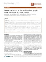
Next, we used immunohistochemistry to verify the expression of the four breast cancer lymph metastasisassociated proteins in 106 cases of paraffin-embedded
paired primary tumors and lymph metastasis tissues obtained from metastatic breast cancer patients. The representative staining images are presented in Fig. 1, and the
quantitatively analyzed results, which are presented as
total immunostaining score (TIS), are summarized in
Table 1. As shown in Fig. 1, most of the EpCAM was localized on the plasma membrane, which is in agreement
with its known cellular localization. FADD was primarily
localized in the cytoplasm and the nucleus. NDRG1 was
located in the plasma membrane and the cytoplasm. The
αB-crystallin protein was primarily expressed on the
plasma membrane and in the cytoplasm. Consistent with
the iTRAQ data, NDRG1 and αB-crystallin were downregulated at the metastatic sites compared with the primary tumors in terms of TIS (Table 1) (P = 0.0003
[NDRG1] or P = 0.046 [αB-crystallin]). However, the expression levels of EpCAM and FADD were also lower at
the metastatic sites compared with the primary tumors
(P = 0.0005).
Correlation of metastasis-associated proteins with the
clinicopathological features of breast cancer patients
To clarify the clinical relevance of the proteins identified
from iTRAQ proteomics that were associated with
lymph metastasis, we analyzed the relationship between
these four proteins and the clinicopathological parameters of 190 cases of breast cancer patients. We showed
that EpCAM was not correlated with any of the clinicopathological parameters examined (Table 2). However,
FADD expression was positively correlated with a younger age at diagnosis (P = 0.049) and lymph node metastasis (P = 0.003). NDRG1 expression was correlated with
worse histological grade (P = 0.041) but not with lymph
node metastasis (P = 0.655). αB-crystallin expression was
inversely correlated with lymph node metastasis (P <
0.001), clinical stage (P = 0.001), histological grade (P =
0.037), ER (P < 0.001), and PR status (P = 0.007).
Zeng et al. BMC Cancer
(2019) 19:831
Page 5 of 11
Fig. 1 Immunohistochemical analysis of the expression of four breast cancer metastasis-associated proteins. The expression levels of EpCAM,
FADD, NDRG1, and αB-crystallin were evaluated by the immunohistochemical staining of paraffin-embedded paired primary and metastatic tissue
sections that were obtained from patients with metastatic breast cancer
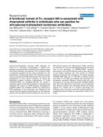
Association of metastasis-associated proteins with overall
survival of breast cancer patients
In addition, we followed up 190 breast cancer patients for
over 10 years and conducted a survival analysis for the
positivity of expression (EpCAM, FADD, and αBcrystallin) or the level of expression (NDRG1) in the primary tumor sites. The results revealed that the patients
who had positive expression of EpCAM or FADD survived
for a shorter time compared with those with negative expression (Fig. 2a-b). Those who had positive expression of
αB-crystallin survived longer than those with negative expression (Fig. 2d). However, the expression level of
NDRG1 had no prognostic value for breast cancer patients
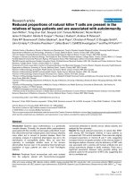
(Fig. 2c). Moreover, the prognostic value of EpCAM only
applied to patients with lymph node metastasis (Fig. 3a-d).
Univariable analysis linked with tumor diameter, TNM
stage and histology stage and type, but multivariable analysis assigned significance only to histology type (lobular
carcinoma vs. duct carcinoma) (Table 3).
Downregulation of αB-crystallin mRNA expression in
breast cancer
Finally, to examine whether αB-crystallin (gene name:
CRYAB) was also involved in human breast cancer
development, using the public database, we reviewed the
mRNA expression of CRYAB in normal breast and invasive breast cancer tissues in Gene Expression Omnibus
(GEO) (Expression Profile GDS3324). The results are
presented in Additional file 2: Figure S2. The expression
of CRYAB was significantly lower in breast cancer tissues
compared with normal breast tissues (P = 0.001). We
further found that the level of expression of αBcrystallin was indeed lower in breast cancer tissues compared with benign breast lesions, with metastatic breast
cancer having the lowest expression (Table 4). These
findings support the tumor-suppressive role of αBcrystallin in the development of breast cancer.
Discussion
Metastasis is one of the most important factors that
causes the death of patients with breast cancer. Detection of breast cancer metastasis at the earliest possible
stage is critical for the successful management of breast
cancer progression. Therefore, it is very important to
search for effective biomarkers for breast cancer metastasis and prognosis. In proteomic comparative studies of
breast cancer metastasis, with tumor tissue as the research object, the commonly used method is based on
Table 1 Summary of the expression of the four metastasis-associated proteins in the paired primary and metastatic tissues of breast
cancer
Zeng et al. BMC Cancer
(2019) 19:831
Page 6 of 11
Table 2 The association between the four metastasis-associated proteins and the clinicopathological features of 190 breast cancer
patients
the comparison of lymph node metastasis or other organ
metastases, gene expression or protein expression of primary breast cancer with metastasis and without metastasis. In this study, we used the iTRAQ proteomic
technique to analyze the differentially regulated proteins
between the primary tumor sites and their corresponding lymph node metastases in metastatic breast cancer
patients, and this comparison method can more accurately compare the differences in protein expression of
breast cancer cells with varying metastatic capacity. Four
proteins (EpCAM, FADD, NDRG1, and αB-crystallin)
were chosen for validation by immunohistochemistry.
Specially, αB-crystallin could potentially be addressed as
a potential prognostic biomarker to predict the lymph
node metastasis and clinical outcomes of breast cancer
patients.
αB-crystallin, also called HspB5, is a member of the αcrystallin family small heat shock proteins and is an important component of the vertebrate lens [36]. In nonlens
tissues, αB-crystallin is an integral part of the cellular proteostasis system, which is associated with a broad
spectrum of human diseases, including cancer [37]. αBcrystallin plays an important role in stress responses, such
as heat shock and radiation poisoning. As a molecular
chaperone, αB-crystallin is expressed in human cells at a
higher level under pathological conditions. The expression
of αB-crystallin in human renal carcinogenesis, triplenegative (basal-like) breast cancer, hepatocellular carcinoma, and squamous cell carcinoma of the head and neck
is related to poor prognosis [36, 37], suggesting an oncogenic role for αB-crystallin in promoting tumorigenesis. In
breast cancer, αB-crystallin has been shown to be an
oncoprotein that predicts poor prognosis [38–41] and resistance to neoadjuvant chemotherapy, especially for
triple-negative breast cancer [40, 42]. However, the role of
αB-crystallin as a tumor suppressor has also been reported
[43]. These contradictory findings indicate that the role of
αB-crystallin in carcinogenesis is complicated. The present
study demonstrated that αB-crystallin was downregulated
in the lymph metastases compared with the primary
breast tumors. This finding is inconsistent with the previous finding that αB-crystallin expression promotes the
brain metastasis of breast cancer [38, 44]. Recently, the
majority of lymphatic and distant metastases were shown
to originate differently in human colorectal cancer [45].
This phenomenon is also true for breast cancer metastasis,
in which approximately 1/3 of breast cancer patients without lymph metastasis develop distant metastasis [46].
These observations suggest that the two routes of cancer
spreading may occur independently and may use different
Zeng et al. BMC Cancer
(2019) 19:831
Page 7 of 11
Fig. 2 The association between four metastasis-associated proteins and the overall survival of breast cancer patients. Kaplan-Meier plots of the
association between the expression of EpCAM (a), FADD (b), NDRG1 (c), and αB-crystallin (d) and the overall survival probability of breast
cancer patients
sets of molecular routers to drive the metastatic spread of
cancer cells through either the lymphatics or the blood
vessels. Reconciling our data with the previous reports, it
is possible that αB-crystallin plays a role of router to
switch between lymphatic and hematogenous spreading.
That is, the role of αB-crystallin in breast cancer progression needs to be reevaluated. It is speculated that αBcrystallin may function as a tumor promoter in
hematogenous metastasis – to the brain, for example, but
αB-crystallin may function as a tumor suppressor in
lymph node metastasis. However, this speculation should
be validated experimentally through in vitro and in vivo
studies. Clearly, our findings further support a tumorsuppressor role for αB-crystallin in breast cancer
development.
Many studies have shown that there is close link between FADD and many cancers, such as nonsmall cell
lung cancer [47], gastric cancer [48] and hepatocellular
carcinoma (HCC) [49]. In the first two of these cancers,
the expression of FADD was correlated with lymph node
metastasis and the poor prognosis of patients, and the
loss of FADD expression plays an important role in
HCC carcinogenesis. FADD expression is associated
with T stage and perineural invasion [50]. An increase in
FADD expression was shown to be associated with a
higher incidence of lymph node metastasis at presentation and with a shorter DMFI when lymph node metastases are present [33]. These studies only involved the
comparison between cancer and the surrounding normal
tissues, whereas we focused on the differences in FADD
expression between primary tumors and metastases.
Using proteomic results, we determined that the expression of FADD was upregulated in metastasis. Furthermore, the IHC results revealed that there were
significant differences in FADD expression between the
primary tumors and metastases, but the rate of FADDpositive tumors decreased, which is inconsistent with
the proteomic results. The possible reason for this inconsistency is that proteomics analyzes the relative
quantity of protein expression, whereas immunohistochemistry analyzes the positive rate of protein expression, and thus results from these two methods are not
Zeng et al. BMC Cancer
(2019) 19:831
Page 8 of 11
Fig. 3 The association between four metastasis-associated proteins and the overall survival in breast cancer patients with metastasis. Kaplan-Meier
plots of the association between the expression of EpCAM (a), FADD (b), NDRG1 (c), and αB-crystallin (d) and the overall survival probability in
breast cancer patients with metastasis
always consistent. In addition, we also investigated potential correlations between FADD expression and the
clinical pathological characteristics of 190 patients with
breast cancer. We performed a 120-months survival analysis and found that FADD expression was associated
with lymph node metastasis. Furthermore, higher expression levels of FADD were identified in patients with
breast cancer, which were also correlated with a shorter
survival time. These finding suggest that there is a close
relationship between FADD expression and the lymph
node metastasis and poor prognosis of breast cancer.
Moreover, the regulatory mechanism of FADD in breast
cancer metastasis warrants further investigation.
NDRG1 has been reported to function as a metastasis
suppressor gene, and it is downregulated in gastric cancer [34], prostate [51, 52], pancreatic cancer [53] and
breast cancers [45]. However, compared with normal tissue, NDRG1 expression was shown to be upregulated in
homologous hepatocellular carcinoma [54] and oral
squamous cell carcinoma [55]. In this study, all of the
proteomics and IHC results revealed that NDRG1 expression was downregulated in metastases compared to
the primary tumors. The expression of NDRG1 in various
tissues may be affected by many factors, such as metal
ions, oxygen, proto-oncogenes, tumor suppressor genes,
hormones or vitamins. For example, NDRG1 expression
in prostate cancer cells was shown to be affected by androgens, whereas NDRG1 expression in breast cancer cells
is mainly associated with estradiol. Thus, the expression of
NDRG1 is variable. In the clinical pathology and survival
analysis, significant differences in NDRG1 expression were
not detected in this study.
EpCAM is a transmembrane glycoprotein and appears
to play a role in tumorigenesis and metastasis of carcinomas [56]. EpCAM is frequently upregulated in carcinomas but is not expressed in cancers of non-epithelial
origin. At present, the FDA approves the automated cell
detection method for EpCAM as biomarker, and this
method has been used to detect circulating tumor cells
in patients with breast [57], prostate [32, 58] and
esophageal cancer [59]. The expression of EpCAM was
shown to be high in laryngeal carcinoma but low in bone
marrow as a metastatic niche for disseminated cancer
cells [60]. These findings are consistent with our IHC
Zeng et al. BMC Cancer
(2019) 19:831
Page 9 of 11
Table 3 Univariate and Multivariate Analysis by a Cox Proportional Hazards Regression Model in Cohort
Variable
OS
Univariate
Multivariate
HR (95% CI)
P Value
HR (95% CI)
Age, years (> 45 vs. ≤ 45)
0.898 (0.581–1.390)
0.630
NA
ER (positive vs. negative)
1.114 (0.711–1.745)
0.637
NA
PR (positive vs. negative)
1.213 (0.775–1.899)
0.398
NA
CrebB-2 (positive vs. negative)
1.128 (0.705–1.806)
0.615
NA
Menstrual history (presence vs. absence)
1.381 (0.851–2.241)
0.191
NA
0.649
NA
Modified radical mastectomy vs. radical correction
1.150 (0.727–1.820)
0.550
NA
Other operation vs. radical correction
Operation
0.642 (0.155–2.663)
0.542
NA
FADD (positive vs. negative)
1.580 (0.995–2.509)
0.053
NA
NDRG1 (low vs. high)
1.302 (0.762–2.226)
0.335
NA
CRYAB (positive vs. negative)
1.561 (0.902–2.701)
0.112
NA
> 5 vs. > 2 and ≤ 5
1.923 (1.019–3.636)
0.043
> 5 vs. ≤2
2.230 (1.093–4.549)
Tumor diameter, cm
0.072
TNM stage
P Value
NS
0.027
< 0.0001
NS
III vs. I
4.329 (1.824–10.273)
0.001
III vs. II
2.101 (1.333–3.311)
0.001
Histology stage (poorly differentiation vs. high-middle differentiation)
2.286 (1.100–4.751)
0.027
Histology type (lobular carcinoma vs. duct carcinoma)
1.720 (1.025–2.886)
0.040
1.846 (1.093–3.118)
0.022
Lymph node metastasis (presence vs. absence)
2.810 (1.694–4.662)
< 0.0001
2.801 (1.688–4.649)
< 0.0001
EpCAM (positive vs. negative)
2.306 (1.218–4.367)
0.010
2.585 (1.351–4.944)
0.004
NS
Data in bold are P values < 0.05
Conclusions
In summary, we discovered differentially regulated proteins
between the primary breast tumors and their lymph node
metastatic sites using the iTRAQ proteomics analysis.
Through further immunohistochemical study, clinicopathological correlation analysis, and GEO profiling, we identified
αB-crystallin as an independent biomarker to predict the
outcome of breast cancer patients in the lymph node. Obviously, αB-crystallin plays a role in the metastasis of breast
cancer cells to the lymph node, but its exact role in each
step of breast cancer metastasis and the underlying signaling mechanism remain to be fully clarified. EpCAM, FADD
and NDRG1 expression were shown to be associated with
the progression of breast cancer, but the questions of how
certain oncogenes may initiate dissemination before triggering aggressive proliferation and how tumor-suppressor
pathways suppress metastasis in breast cancer warrant further investigation.
Table 4 Summary of the expression of CRYAB in different
stages of breast tissues
Additional files
results. However, EpCAM expression was increased in
the metastatic group compared to the nonmetastatic
group according to both iTRAQ and the proteomics
analysis. Furthermore, the survival analysis showed that
the survival rate was lower in the EpCAM-positive
group. Therefore, the expression of EpCAM should be
further clarified in breast cancer metastasis. Taken together, these data suggest that EpCAM plays a critical
role in the metastatic process of breast cancer.
Tissue
Benign
P
TIS
0
1–4
5–8
9–12
1
24
16
6
Non-metastatic
46
28
6
3
Metastatic
189
24
1
0
0.0003
Additional file 1: Figure S1. GO analysis of the differentially regulated
proteins in lymph node metastases vs. primary breast tumor tissues. The
upregulated (A-C) and downregulated (D-F) proteins identified by the
iTRAQ proteomics were analyzed by the GO Consortium and categorized
according to their biological processes, cellular locations, and molecular
functions. (TIF 5559 kb)
Zeng et al. BMC Cancer
(2019) 19:831
Additional file 2: Figure S2. GEO analysis of CRYAB mRNA expression in
normal breast and breast cancer tissues. (A) The mRNA expression of
CRYAB in normal breast tissues (n =5) and breast cancer tissues (n = 28)
was analyzed from the Affymetrix Human Genome Microarray at the GEO
website ( for αBcrystallin). (B) Quantification of the mRNA expression of CRYAB in normal
breast tissues and breast cancer tissues. (TIF 4929 kb)
Additional file 3: Table S1. Identification of differentially expressed
proteins between primary breast cancer tissues and metastatic lymph
node tissues by the iTRAQ technique. (XLS 1215 kb)
Additional file 4: Table S2. Partial up-regulated proteins in metastatic
lymph node compared with primary tumor in breast cancer. Table S3.
Partial down-regulated proteins in metastatic lymph node compared with
primary tumor in breast cancer. Table S4. UniProt analysis of the biological
processes, cellular locations, and molecular functions of the four metastasisassociated proteins. (DOCX 29 kb)
Abbreviations
EpCAM: Epithelial cell adhesion molecule; FADD: Fas-associated death
domain; GEO: Gene expression omnibus; GO: Gene ontology;
HCC: Hepatocellular carcinoma; iTRAQ: Isobaric tags for relative and absolute
quantitation; NDRG1: N-myc downstream-regulated gene 1; TIS: Total
immunostaining score
Acknowledgements
We thank the Proteomic technique platform from Lei Xue by FitGene
Biotechnology Co., Ltd. (Guangzhou, P. R. China, ) for
the iTRAQ proteomics analysis and the PRIDE partner repository. The clerical
assistance from Kassey Deng, Lu Lu, Chao Chen and Huimei Yi is highly
appreciated.
Authors’ contributions
LZ, XD, JZ, YZ, GH, PT and YT conceived of the study. LZ, XD, LY, XT, JZ, YZ,
GH, SZ, PT and YT analyzed and interpreted the data. LZ, XD, LY, XT, JZ, YZ,
GH, and YT performed the histological examination of the tumor tissue. LZ,
JZ, YZ, LY, XT, GH, SZ, PT and YT participated in the study design and
coordination, helped to interpret the data, and helped to draft the
manuscript. LZ, SZ, PT and YT helped to interpret the data and to draft the
manuscript. LZ, XD, JZ, and YT drafted the manuscript, and LZ, XD, and YT
were major contributors in writing the manuscript. All authors read and
approved the final manuscript.
Funding
This work was supported by the Hunan Province Science and Technology
Project (2014FJ6090 to LZ) and the National Natural Science Foundation of
China (81472496 to XD) in the design of this study and collection, analysis,
and interpretation of data.
Availability of data and materials
The mass spectrometry proteomics data have been deposited to the
ProteomeXchange Consortium via the PRIDE [1] partner repository with the
dataset identifier PXD013931.
Ethics approval and consent to participate
All participants signed informed consent forms, and the Research Ethics
Committee of Central South University, China approved this study, reference
number is EC20101220005.
Consent for publication
Not applicable.
Competing interests
The authors declare that they have no competing interests.
Author details
1
Department of Pathology, Guangzhou Women and Children’s Medical
Center, Guangzhou Medical University, Guangzhou, Guangdong, China. 2Key
Laboratory of Translational Cancer Stem Cell Research, Hunan Normal
University, Changsha, Hunan, China. 3Department of Pathology, Union
Hospital, Tongji Medical College, HuaZhong University of Science and
Page 10 of 11
Technology, WuHan, China. 4Department of Pharmacy, Hunan Normal
University School of Medicine, Changsha, Hunan, China. 5Department of
Oncology, Institute of Medical Sciences, Xiangya Hospital, Central South
University, Changsha, Hunan, China. 6College of Life Science, Hunan Normal
University, Changsha, Hunan, China. 7Department of Pathology, Hunan
Cancer Hospital & The Affiliated Cancer Hospital of Xiangya School of
Medicine, Central South University, Changsha, Hunan, China. 8Key Laboratory
of Carcinogenesis and Cancer Invasion, Ministry of Education, Key Laboratory
of Carcinogenesis, Ministry of Health, Cancer Research Institute, Xiangya
Hospital, Central South University, Changsha, Hunan, China.
Received: 23 March 2018 Accepted: 5 August 2019
References
1. Torre LA, Bray F, Siegel RL, Ferlay J, Lortet-Tieulent J, Jemal A. Global cancer
statistics, 2012. CA Cancer J Clin. 2015;65(2):87–108.
2. Chen W, Zheng R, Baade PD, Zhang S, Zeng H, Bray F, Jemal A, Yu XQ, He J.
Cancer statistics in China, 2015. CA Cancer J Clin. 2016;66(2):115–32.
3. Chen W, Zheng R, Zuo T, Zeng H, Zhang S, He J. National cancer incidence
and mortality in China, 2012. Chin J Cancer Res. 2016;28(1):1–11.
4. Jin X, Mu P. Targeting breast Cancer metastasis. Breast Cancer (Auckl). 2015;
9(Suppl 1):23–34.
5. Schwartz RS, Erban JK. Timing of metastasis in breast Cancer. N Engl J Med.
2017;376(25):2486–8.
6. DeMichele A, Yee D, Esserman L. Mechanisms of resistance to neoadjuvant
chemotherapy in breast Cancer. N Engl J Med. 2017;377(23):2287–9.
7. Harper KL, Sosa MS, Entenberg D, Hosseini H, Cheung JF, Nobre R, AvivarValderas A, Nagi C, Girnius N, Davis RJ, et al. Mechanism of early
dissemination and metastasis in Her2(+) mammary cancer. Nature. 2016;
540(7634):588.
8. Hosseini H, Obradovic MM, Hoffmann M, Harper KL, Sosa MS, Werner-Klein
M, Nanduri LK, Werno C, Ehrl C, Maneck M, et al. Early dissemination seeds
metastasis in breast cancer. Nature. 2016;540(7634):552.
9. O'Shaughnessy J. Extending survival with chemotherapy in metastatic
breast cancer. Oncologist. 2005;10(Suppl 3):20–9.
10. Spolverato G, Vitale A, Bagante F, Connolly R, Pawlik TM. Liver resection for
breast Cancer liver metastases: a cost-utility analysis. Ann Surg. 2017;265(4):
792–9.
11. Ohsfeldt RL, Ward MM, Schneider JE, Jaana M, Miller TR, Lei Y, Wakefield DS.
Implementation of hospital computerized physician order entry systems in
a rural state: feasibility and financial impact. J Am Med Inform Assoc. 2005;
12(1):20–7.
12. van de Vijver MJ, He YD, van't Veer LJ, Dai H, Hart AA, Voskuil DW, Schreiber
GJ, Peterse JL, Roberts C, Marton MJ, et al. A gene-expression signature as a
predictor of survival in breast cancer. N Engl J Med. 2002;347(25):1999–2009.
13. Chandran UR, Ma C, Dhir R, Bisceglia M, Lyons-Weiler M, Liang W,
Michalopoulos G, Becich M, Monzon FA. Gene expression profiles of
prostate cancer reveal involvement of multiple molecular pathways in the
metastatic process. BMC Cancer. 2007;7:64.
14. Appierto V, Di Cosimo S, Reduzzi C, Pala V, Cappelletti V, Daidone MG. How
to study and overcome tumor heterogeneity with circulating biomarkers:
the breast cancer case. Semin Cancer Biol. 2017;44:106–16.
15. Ramaswamy S, Ross KN, Lander ES, Golub TR. A molecular signature of
metastasis in primary solid tumors. Nat Genet. 2003;33(1):49–54.
16. Lorusso G, Ruegg C. New insights into the mechanisms of organ-specific
breast cancer metastasis. Semin Cancer Biol. 2012;22(3):226–33.
17. Fry SA, Sinclair J, Timms JF, Leathem AJ, Dwek MV. A targeted
glycoproteomic approach identifies cadherin-5 as a novel biomarker of
metastatic breast cancer. Cancer Lett. 2013;328(2):335–44.
18. Zeng L, Zhong J, He G, Li J, Zhou W, Liu W, Zhang Y, Huang S, Liu Z, Deng
X: Identification of Nucleobindin-2 as a Potential Biomarker for Breast
Cancer Metastasis Using iTRAQ-based Quantitative Proteomic Analysis. J
Cancer 2017: />19. Castle J, Shaker H, Morris K, Tugwood JD, Kirwan CC. The significance of
circulating tumour cells in breast cancer: a review. Breast. 2014;23(5):552–60.
20. Lu X, Kang Y. Organotropism of breast cancer metastasis. J Mammary Gland
Biol Neoplasia. 2007;12(2–3):153–62.
21. Patsialou A, Wang Y, Lin J, Whitney K, Goswami S, Kenny PA, Condeelis JS.
Selective gene-expression profiling of migratory tumor cells in vivo predicts
clinical outcome in breast cancer patients. Breast Cancer Res. 2012;14(5):R139.
Zeng et al. BMC Cancer
(2019) 19:831
22. van’t Veer LJ, Dai H, van de Vijver MJ, He YD, Hart AA, Mao M, Peterse HL,
van der Kooy K, Marton MJ, Witteveen AT, et al. Gene expression profiling
predicts clinical outcome of breast cancer. Nature. 2002;415(6871):530–6.
23. Zieske LR. A perspective on the use of iTRAQ reagent technology for
protein complex and profiling studies. J Exp Bot. 2006;57(7):1501–8.
24. Evans C, Noirel J, Ow SY, Salim M, Pereira-Medrano AG, Couto N, Pandhal J,
Smith D, Pham TK, Karunakaran E, et al. An insight into iTRAQ: where do we
stand now? Anal Bioanal Chem. 2012;404(4):1011–27.
25. Putz SM, Boehm AM, Stiewe T, Sickmann A. iTRAQ analysis of a cell culture
model for malignant transformation, including comparison with 2D-PAGE
and SILAC. J Proteome Res. 2012;11(4):2140–53.
26. Zeidan B, Manousopoulou A, Garay-Baquero DJ, White CH, Larkin SET, Potter
KN, Roumeliotis TI, Papachristou EK, Copson E, Cutress RI, et al. Increased
circulating resistin levels in early-onset breast cancer patients of normal
body mass index correlate with lymph node negative involvement and
longer disease free survival: a multi-center POSH cohort serum proteomics
study. Breast Cancer Res. 2018;20(1):19.
27. Bouchal P, Dvorakova M, Roumeliotis T, Bortlicek Z, Ihnatova I, Prochazkova
I, Ho JT, Maryas J, Imrichova H, Budinska E, et al. Combined proteomics and
transcriptomics identifies carboxypeptidase B1 and nuclear factor kappaB
(NF-kappaB) associated proteins as putative biomarkers of metastasis in low
grade breast Cancer. Mol Cell Proteomics. 2015;14(7):1814–30.
28. Bouchal P, Roumeliotis T, Hrstka R, Nenutil R, Vojtesek B, Garbis SD.
Biomarker discovery in low-grade breast cancer using isobaric stable
isotope tags and two-dimensional liquid chromatography-tandem mass
spectrometry (iTRAQ-2DLC-MS/MS) based quantitative proteomic analysis. J
Proteome Res. 2009;8(1):362–73.
29. Jesneck JL, Mukherjee S, Yurkovetsky Z, Clyde M, Marks JR, Lokshin AE, Lo JY.
Do serum biomarkers really measure breast cancer? BMC Cancer. 2009;9:164.
30. Ruppen I, Grau L, Orenes-Pinero E, Ashman K, Gil M, Algaba F, Bellmunt J,
Sanchez-Carbayo M. Differential protein expression profiling by iTRAQ-twodimensional LC-MS/MS of human bladder cancer EJ138 cells transfected
with the metastasis suppressor KiSS-1 gene. Mol Cell Proteomics. 2010;9(10):
2276–91.
31. Perez-Riverol Y, Csordas A, Bai J, Bernal-Llinares M, Hewapathirana S, Kundu
DJ, Inuganti A, Griss J, Mayer G, Eisenacher M, et al. The PRIDE database and
related tools and resources in 2019: improving support for quantification
data. Nucleic Acids Res. 2019;47(D1):D442–50.
32. Ni J, Cozzi PJ, Duan W, Shigdar S, Graham PH, John KH, Li Y. Role of the
EpCAM (CD326) in prostate cancer metastasis and progression. Cancer
Metastasis Rev. 2012;31(3–4):779–91.
33. Pattje WJ, Melchers LJ, Slagter-Menkema L, Mastik MF, Schrijvers ML, Gibcus JH,
Kluin PM, Hoegen-Chouvalova O, van der Laan BF, Roodenburg JL, et al. FADD
expression is associated with regional and distant metastasis in squamous cell
carcinoma of the head and neck. Histopathology. 2013;63(2):263–70.
34. Chang X, Xu X, Ma J, Xue X, Li Z, Deng P, Zhang S, Zhi Y, Chen J, Dai D.
NDRG1 expression is related to the progression and prognosis of gastric
cancer patients through modulating proliferation, invasion and cell cycle of
gastric cancer cells. Mol Biol Rep. 2014;41(9):6215–23.
35. Liu S, Yan B, Lai W, Chen L, Xiao D, Xi S, Jiang Y, Dong X, An J, Chen X, et al.
As a novel p53 direct target, bidirectional gene HspB2/alphaB-crystallin
regulates the ROS level and Warburg effect. Biochim Biophys Acta. 2014;
1839(7):592–603.
36. Cvekl A, McGreal R, Liu W. Lens development and Crystallin gene
expression. Prog Mol Biol Transl Sci. 2015;134:129–67.
37. Haslbeck M, Peschek J, Buchner J, Weinkauf S. Structure and function of
alpha-crystallins: Traversing from in vitro to in vivo. Biochim Biophys Acta.
2016;1860(1 Pt B):149–66.
38. Malin D, Strekalova E, Petrovic V, Deal AM, Al Ahmad A, Adamo B, Miller CR,
Ugolkov A, Livasy C, Fritchie K, et al. alphaB-crystallin: a novel regulator of
breast cancer metastasis to the brain. Clin Cancer Res. 2014;20(1):56–67.
39. Malin D, Strekalova E, Petrovic V, Rajanala H, Sharma B, Ugolkov A, Gradishar
WJ, Cryns VL. ERK-regulated alphaB-crystallin induction by matrix
detachment inhibits anoikis and promotes lung metastasis in vivo.
Oncogene. 2015;34(45):5626–34.
40. Petrovic V, Malin D, Cryns VL. alphaB-crystallin promotes oncogenic
transformation and inhibits caspase activation in cells primed for apoptosis
by Rb inactivation. Breast Cancer Res Treat. 2013;138(2):415–25.
41. Wang F, Chen X, Li C, Sun Q, Chen Y, Wang Y, Peng H, Liu Z, Chen R, Liu K,
et al. Pivotal role of augmented alphaB-crystallin in tumor development
induced by deficient TSC1/2 complex. Oncogene. 2014;33(34):4352–8.
Page 11 of 11
42. Chen Z, Ruan Q, Han S, Xi L, Jiang W, Jiang H, Ostrov DA, Cai J. Discovery of
structure-based small molecular inhibitor of alphaB-crystallin against basallike/triple-negative breast cancer development in vitro and in vivo. Breast
Cancer Res Treat. 2014;145(1):45–59.
43. Huang Z, Cheng Y, Chiu PM, Cheung FM, Nicholls JM, Kwong DL, Lee AW,
Zabarovsky ER, Stanbridge EJ, Lung HL, et al. Tumor suppressor alpha B-crystallin
(CRYAB) associates with the cadherin/catenin adherens junction and impairs NPC
progression-associated properties. Oncogene. 2012;31(32):3709–20.
44. Voduc KD, Nielsen TO, Perou CM, Harrell JC, Fan C, Kennecke H, Minn AJ,
Cryns VL, Cheang MCU. alphaB-crystallin expression in breast Cancer is
associated with brain metastasis. NPJ Breast Cancer. 2015;1:15014.
45. Bandyopadhyay S, Pai SK, Hirota S, Hosobe S, Takano Y, Saito K, Piquemal D,
Commes T, Watabe M, Gross SC, et al. Role of the putative tumor metastasis
suppressor gene Drg-1 in breast cancer progression. Oncogene. 2004;23(33):
5675–81.
46. Weigelt B, Peterse JL, van’t Veer LJ. Breast cancer metastasis: markers and
models. Nat Rev Cancer. 2005;5(8):591–602.
47. Cimino Y, Costes A, Damotte D, Validire P, Mistou S, Cagnard N, Alifano M,
Regnard JF, Chiocchia G, Sautes-Fridman C, et al. FADD protein release
mirrors the development and aggressiveness of human non-small cell lung
cancer. Br J Cancer. 2012;106(12):1989–96.
48. Yoo NJ, Lee SH, Jeong EG, Lee JW, Soung YH, Nam SW, Kim SH, Lee JY, Lee
SH. Expression of nuclear and cytoplasmic phosphorylated FADD in gastric
cancers. Pathol Res Pract. 2007;203(2):73–8.
49. Tu W, Luo M, Wang Z, Yan W, Xia Y, Deng H, He J, Han P, Tian D.
Upregulation of SATB1 promotes tumor growth and metastasis in liver
cancer. Liver Int. 2012;32(7):1064–78.
50. Choi EJ, Yun JA, Jabeen S, Jeon EK, Won HS, Ko YH, Kim SY. Prognostic
significance of TMEM16A, PPFIA1, and FADD expression in invasive ductal
carcinoma of the breast. World J Surg Oncol. 2014;12:137.
51. Bandyopadhyay S, Pai SK, Gross SC, Hirota S, Hosobe S, Miura K, Saito K,
Commes T, Hayashi S, Watabe M, et al. The Drg-1 gene suppresses tumor
metastasis in prostate cancer. Cancer Res. 2003;63(8):1731–6.
52. Bandyopadhyay S, Wang Y, Zhan R, Pai SK, Watabe M, Iiizumi M, Furuta E,
Mohinta S, Liu W, Hirota S, et al. The tumor metastasis suppressor gene
Drg-1 down-regulates the expression of activating transcription factor 3 in
prostate cancer. Cancer Res. 2006;66(24):11983–90.
53. Maruyama Y, Ono M, Kawahara A, Yokoyama T, Basaki Y, Kage M, Aoyagi S,
Kinoshita H, Kuwano M. Tumor growth suppression in pancreatic cancer by
a putative metastasis suppressor gene Cap43/NDRG1/Drg-1 through
modulation of angiogenesis. Cancer Res. 2006;66(12):6233–42.
54. Cheng J, Xie HY, Xu X, Wu J, Wei X, Su R, Zhang W, Lv Z, Zheng S, Zhou L.
NDRG1 as a biomarker for metastasis, recurrence and of poor prognosis in
hepatocellular carcinoma. Cancer Lett. 2011;310(1):35–45.
55. Chang JT, Wang HM, Chang KW, Chen WH, Wen MC, Hsu YM, Yung BY, Chen
IH, Liao CT, Hsieh LL, et al. Identification of differentially expressed genes in
oral squamous cell carcinoma (OSCC): overexpression of NPM, CDK1 and
NDRG1 and underexpression of CHES1. Int J Cancer. 2005;114(6):942–9.
56. Grover PK, Cummins AG, Price TJ, Roberts-Thomson IC, Hardingham JE.
Circulating tumour cells: the evolving concept and the inadequacy of their
enrichment by EpCAM-based methodology for basic and clinical cancer
research. Ann Oncol. 2014;25(8):1506–16.
57. Gastl G, Spizzo G, Obrist P, Dunser M, Mikuz G. Ep-CAM overexpression in
breast cancer as a predictor of survival. Lancet. 2000;356(9246):1981–2.
58. Ni J, Cozzi P, Hao J, Beretov J, Chang L, Duan W, Shigdar S, Delprado W, Graham
P, Bucci J, et al. Epithelial cell adhesion molecule (EpCAM) is associated with
prostate cancer metastasis and chemo/radioresistance via the PI3K/Akt/mTOR
signaling pathway. Int J Biochem Cell Biol. 2013;45(12):2736–48.
59. Driemel C, Kremling H, Schumacher S, Will D, Wolters J, Lindenlauf N, Mack
B, Baldus SA, Hoya V, Pietsch JM, et al. Context-dependent adaption of
EpCAM expression in early systemic esophageal cancer. Oncogene. 2014;
33(41):4904–15.
60. Romeu C, Farre X, Cardesa A, Nadal A. Expression of ep-CAM, but not of
E48, associates with nodal involvement in advanced squamous cell
carcinomas of the larynx. Histopathology. 2013;62(6):954–61.
Publisher’s Note
Springer Nature remains neutral with regard to jurisdictional claims in
published maps and institutional affiliations.