A primary undifferentiated pleomorphic sarcoma of the lumbosacral region harboring a LMNA-NTRK1 gene fusion with durable clinical response to crizotinib: A case report
Bạn đang xem bản rút gọn của tài liệu. Xem và tải ngay bản đầy đủ của tài liệu tại đây (1.65 MB, 8 trang )
Zhou et al. BMC Cancer (2018) 18:842
/>
CASE REPORT
Open Access
A primary undifferentiated pleomorphic
sarcoma of the lumbosacral region
harboring a LMNA-NTRK1 gene fusion with
durable clinical response to crizotinib: a
case report
Ning Zhou1,2, Reinhold Schäfer2, Tao Li3, Meiyu Fang4 and Luying Liu1*
Abstract
Background: High-grade spindle cell sarcomas are a subtype of rare, undifferentiated pleomorphic sarcomas (UPSs)
for which diagnosis is difficult and no specific treatment strategies have been established. The limited published
data on UPSs suggest an aggressive clinical course, high rates of local recurrence and distant metastasis, and poor
prognosis.
Case presentation: Here we present the unusual case of a 45-year-old male patient with a lumbosacral UPS extending
into the sacrum. An initial diagnosis of a low-grade malignant spindle cell tumor was based on a tumor core biopsy.
After complete extensive resection, the diagnosis of an UPS of the lumbosacral region was confirmed by excluding
other types of cancers. Despite treatment with neoadjuvant radiotherapy, extensive resection, and adjuvant
chemotherapy, the patient presented with multiple pulmonary metastases 3 months after surgery. The patient
then began treatment with crizotinib at an oral dose of 450 mg per day, based on the detection of a LMNANTRK1 fusion gene in the tumor by next-generation sequencing. Over 18 months of follow-up through July
2018, the patient maintained a near-complete clinical response to crizotinib.
Conclusions: The LMNA-NTRK1 fusion was likely the molecular driver of tumorigenesis and metastasis in this
patient, and the observed effectiveness of crizotinib treatment provides clinical validation of this molecular
target. Molecular and cytogenetic evaluations are critical to accurate prognosis and treatment planning in cases
of UPS, especially when treatment options are limited or otherwise exhausted. Molecularly targeted therapy of
these rare but aggressive lesions represents a novel treatment option that may lead to fewer toxic side effects
and better clinical outcomes.
Keywords: Undifferentiated pleomorphic sarcoma, Spindle cells, Lumbosacral, LMNA-NTRK1 gene fusion,
Crizotinib therapy
* Correspondence:
1
Department of Abdominal Radiotherapy, Zhejiang Cancer Hospital,
Hangzhou, Zhejiang 310022, People’s Republic of China
Full list of author information is available at the end of the article
© The Author(s). 2018 Open Access This article is distributed under the terms of the Creative Commons Attribution 4.0
International License ( which permits unrestricted use, distribution, and
reproduction in any medium, provided you give appropriate credit to the original author(s) and the source, provide a link to
the Creative Commons license, and indicate if changes were made. The Creative Commons Public Domain Dedication waiver
( applies to the data made available in this article, unless otherwise stated.
Zhou et al. BMC Cancer (2018) 18:842
Background
Undifferentiated pleomorphic sarcoma (UPS), which is
also referred to as malignant fibrous histiocytoma (MFH)
according to the 2002 World Health Organization classification, is a rare and aggressive type of mesenchymal malignancy with no definitive cell of origin or specific
recurrent genetic hallmarks. Extensive immunohistochemical characterization is required to differentiate UPS
from other tumors. While UPS can occur throughout the
body, these tumors are commonly found in the extremities and in the retroperitoneum [1, 2], and superficial lesions (subcutaneous) are rare. High-grade spindle cell
sarcomas are one subtype of UPSs that is particularly challenging to accurately diagnose and effectively treat. The
current 5-year overall survival rate for patients with UPSs
is only 65–70%, highlighting the need for more effective
treatment options [3].
At present, UPSs should be treated according to
current guidelines for soft tissue sarcoma (STS), because
no standard treatment strategy specific for UPSs has
been established. Extensive excision and radiotherapy remain the cornerstones of treatment for non-metastatic
tumors. With the majority of these tumors being high
grade at diagnosis, localized treatments commonly result
in poor local control and poor survival. Perioperative
chemotherapy was recently reported to be beneficial in
terms of overall survival [4], and doxorubicin as a single
agent or in combination with ifosfamide is the first
choice of chemotherapy in cases of UPS metastasis. A
more complete understanding of the molecular characteristics and cytogenetics of these tumors will aid in the
differentiation of sarcoma subtypes and development of
specifically targeted therapies. Here we report a rare case
of UPS in the lumbrosacral region and review the diagnostic procedures applied in this case as well as the
treatment decisions and outcomes.
Case presentation
A 45-year-old male patient presented with a complaint
of progressive pain and soreness in the lumbosacral region persisting for more than 3 months. The pain radiated to the left thigh and perineum but did not affect
walking. Magnetic resonance imaging (MRI) and computed tomography (CT) scans with and without intravenous contrast showed a tumor mass adjacent to the
left side of the fifth lumbar spinous process. The tumor
was located in the lower left part of the erector spinae
and extended onto the fifth lumbar vertebra, the first sacral
vertebra, and the iliac wing. Positron emission tomography
with CT (PET/CT) showed a hypermetabolic lesion in the
erector spinae adjacent to the left side of the fifth lumbar
spinous process. No sites of regional or distant metastases
were found. A core biopsy of the tumor mass revealed
spindle-shaped cells with infiltrating inflammatory cells.
Page 2 of 8
Together the morphological and immunohistochemical
features indicated a low-grade inflammatory myofibroblastic tumor. The expression profile based on immunostaining was as follows: overall positive for vimentin, CD34,
ALK (SP8), and p53; focally positive for smooth muscle
actin (SMA); sporadically positive for S-100; partially positive for CD68; and negative for cytokeratin (CK) (AE1/
AE3), desmin, and CD117. The Ki-67 nuclear labeling
index was 10%.
The patient reported no other symptoms. Physical examinations revealed no neuro-pathological signs or
symptoms. He denied smoking, alcohol, or illicit drug
usage. He also denied recent radiation or toxin exposure.
He had no history of unintentional weight loss, fever, or
chills. He had no family history of malignant or other
chronic diseases, with the exception of a sister who had
breast cancer.
The treatment plan of the case was discussed by our
multi-disciplinary team including experts from orthopedics, neurosurgery, chemotherapy, radiotherapy, pathology, and radiology. Considering that the boundary of
the tumor was unclear and involved the sacrum, a
complete resection would be difficult. Therefore, we administered neoadjuvant radiotherapy to the affected area
at a dose of DT 5000 cGy in 25 fractions to the planning
target volume (PTV). After shrinkage of the tumor
volume, the patient underwent complete extensive resection at 1 month after radiotherapy. Postoperative pathology confirmed that resection of a lesion measuring
7.5 cm × 4 cm × 3.5 cm achieved negative histological
margins and indicated a classification of the specimen as
a mesenchymal-derived malignant tumor involving the
sacrum. Histologic examination of the resected tumor
revealed undifferentiated pleomorphic spindle cells surrounding an area of geographic necrosis with frequent
atypical mitosis. Microscopically, the morphology conformed to that of a high-grade spindle cell sarcoma consistent with UPS. The result from MDM2 amplification
using fluorescence in situ hybridization was negative,
and thus, lipogenesis on histology could be excluded
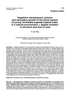
(Additional file 1). The expression profile of the UPS tissue is described in Table 1, and representative images of
staining tumor tissue are presented in Fig. 1.
Table 1 Expression profile of UPS tumor based on
immunohistochemical staining of surgically resected tumor tissue
Positive
INI-1 (+), vimentin (+), S-100 (focally+), p53 (partially+), Bcl-2
(partially+), CD99 (+), calponin (sporadically+), Ki-67 (+, 15%),
transducin-like enhancer of Split 1 (TLE1+), melan-A (focally
weak+).
Negative AE1/AE3 (−), desmin (−), CD31 (−), caldesmon (−), CK (−),
EMA (−), ALK (−), SMA (−), CD117\c-kit (−), CD34 (−), MyoD1 (−),
myogenin (−), CK/LMW (−), CK5/6 (−), 34βE12 (−), CAM5.2 (−),
HMB45 (−), SOX10 (−), MITF (−).
Zhou et al. BMC Cancer (2018) 18:842
Page 3 of 8
Fig. 1 Histopathological staining of surgically resected tumor tissue. Pathology revealed high-grade spindle cell sarcoma consistent with UPS.
a Hematoxylin and eosin (H&E); magnification, 100×. b H&E, 400×. c H&E, 400×. d Ki-67, 200×. Brown nuclear staining for this proliferation marker
is seen in many tumor cells
A postsurgical MRI scan obtained 1 month after surgery showed postoperative changes and no obvious mass
in the surgical area. The patient underwent adjuvant
chemotherapy with liposomal doxorubicin and ifosfamide but had to discontinue chemotherapy after 2 cycles
due to intolerance of grade 3 fatigue and grade 2 nausea.
At 3 months after surgery, three new lesions were discovered in the bilateral pulmonary region on a routine
follow-up CT scan (Fig. 2a). Further radiographic imaging
with PET/CT showed hypermetabolic metastases involving the erector spinae of the left posterior sacral, fifth lumbar spine, sacrum, left ilium, and twelfth thoracic vertebra,
Fig. 2 Chest CT images. (a) Follow-up chest CT images taken 3 months after surgery on January 10, 2017 demonstrated three new lesions
(arrows) in the bilateral pulmonary region, before treatment with cizotinib. b Follow-up chest CT images taken on February 20, 2017 at 4 weeks
after the initiation of oral crizotinib administration indicated improvement
Zhou et al. BMC Cancer (2018) 18:842
accompanied by multiple lung lesions and a suspected
metastasis adjacent to the spleen (Fig. 3a). At this stage,
the patient refused further chemotherapy.
With the standard therapeutic options exhausted, primary tumor tissue was subjected to DNA sequencing via
next-generation sequencing (NGS) with an ILLUMINA
Nextseq 500 (3DMedicines, Inc.). The MasterView 381
cancer-gene panel covered 4557 exons of 365 cancer-related genes and 47 introns of 25 genes frequently rearranged in 381 cancer-related genes (Additional file 2).
The genomic DNA was extracted with the QIAamp
DNA formalin-fixed paraffin-embedded tissue kit (Qiagen) following the manufacturer’s protocol and quantified with the Qubit™ dsDNA HS Assay kit (Invitrogen).
Bioinformatics analyses involved analyzing the clipped
reads, which can be extracted by the tag information of
bam files, which mapped the individual reads to the reference human genome (hg19) with bwa aligner v0.7.12.
Candidate reads that were discordant or aligned in the
same direction were filtered. Read pairs with reads
mapped to separate chromosomes or separated by a distance of over 2 kb on the same chromosome were kept
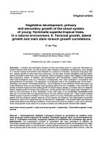
for fusion detection at the probe level. Output rearrangements contained translocation, inversion, long deletion, etc. [5]. Through this profiling, a LMNA-NTRK1
gene fusion encoding exons 1–2 of lamin A/C and exons
11–17 of the NTRK1 gene was identified (Fig. 4), and
the other unlisted genes were all wild-type. The sequencing results for the LMNA-NTRK1 gene fusion are presented in Additional files 3 and 4.
After extensive discussion and consultation with the
patient and his family, we initiated crizotinib therapy per
os at 450 mg per day on January 23, 2017. One month
later, chest CT scanning showed that all lesions in the bilateral lungs had almost disappeared, and the patient
had achieved a near-complete clinical response (CCR,
Fig. 2b). PET/CT imaging was repeated after 4 months
of treatment and continued to show the same response
to crizotinib therapy. PET/CT revealed that local FDG
metabolism was slightly increased at the lesions of the
fifth lumbar spine, sacrum, left ilium and left paraspinal
muscle. However, with crizotinib treatment, the FDG
metabolism was significantly reduced in comparison
with that seen in the first PET-CT examination. The bilateral pulmonary nodules had disappeared, and the
twelfth vertebra, which had shown osteolytic bone destruction, now showed signs of healing, with an increased density and a lower FDG metabolism. The
volume of the left front nodule of the spleen was significantly reduced after treatment (Fig. 3b). A timeline of
the treatment course is presented in Fig. 5. As of July
2018, clinical assessments in this patient showed an ongoing near-CCR of 18 months. In general, the side effects of oral administration of crizotinib at 450 mg per
Page 4 of 8
day were tolerable for the patient. During the course of
treatment, the patient experienced grade 3 myelosuppression and grade 2 weakness, but myelosuppression
could be alleviated with granulocyte colony-stimulating
factor (G-CSF)-based supportive treatment.
Discussion and conclusions
Approximately 5–15% of STS lesions cannot be differentiated by current molecular technologies or immunohistochemical criteria and are therefore classified as UPSs
in an exclusion-based diagnosis [6]. The morphology of
the primary tumor in the present case showed an ordered storiform pattern on hematoxylin and eosin
(H&E) staining and progressively dedifferentiated to a
highly pleomorphic tumor without definite true histiocytic differentiation. In addition, the tumor cells were
mainly spindly with elongated, tapering nuclei. Considering also the findings on immunohistochemical staining
after surgery, we finally confirmed a diagnosis of
high-grade spindle cell UPS. The main pathology-based
differential diagnosis among different potential histological entities was based on morphology as well as the
expression profile of a panel of immunocytochemical
markers. Before rendering the diagnosis of UPSs, the differential diagnoses that must be excluded include dedifferentiated liposarcoma, pleomorphic liposarcoma, pleomorphic
leiomyosarcoma, pleomorphic rhabdomyosarcoma, high
grade and epithelioid variant of myxofibrosarcoma, poorly
differentiated carcinoma, and melanoma [7]. The diagnosis
of primary UPS is made easier by extensive tumor sampling, evaluation of the overall morphologic pattern, careful
searching for the best-differentiated area, and determination of the specific immunophenotype to evaluate a particular lineage of differentiation. In the present case, the
initial diagnostic classification was difficult.
Current knowledge on UPSs suggests an aggressive
clinical course, high incidence of recurrence and metastasis compared with other histologic STS subtypes [8].
Treatment with surgery only leads to poor rates of local
control and even survival. To date, the clinical benefit of
adjuvant chemotherapy and radiation remains unclear.
More recently, genetic studies have contributed to an increased understanding of sarcomas and provided possible therapeutic advancements by identifying genetic
markers of patients most likely to respond. In the
present case, we identified a LMNA-NTRK1 fusion gene
comprising exons 11–17 of the NKRT1 gene and exons
1–2 of LMNA gene in the patient’s tumor. The NTRK1
gene encodes tropomyosin receptor kinase A (TrkA),
which is a membrane-bound receptor that, upon neurotrophin binding, undergoes autophosphorylation and activates members of the mitogen activated protein kinase
(MAPK) pathway [9, 10]. The LMNA gene (localized at
chromosome 1q22) encodes a key component of the
Zhou et al. BMC Cancer (2018) 18:842
Page 5 of 8
Fig. 3 PET-CT images showing visible regression of the multiple metastases after 16 weeks of crizotinib monocherapy. a Follow-up PET-CT image
taken on January 10, 2017 at 3 months after surgery showed hypermetabolic metastases in multiple regions, before the start of cizotinib treatment.
b Follow-up PET-CT images taken on May 19, 2017 at 4 months after initiation of crizotinib showed near-CCR
Zhou et al. BMC Cancer (2018) 18:842
Page 6 of 8
Fig. 4 Schematic presentation of the LMNA–NTRK1 gene fusion. The fusion consisted of LMNA exons 1–2 followed by NTRK1 exons 11–17
nuclear lamina that is involved in nuclear assembly and
chromatin organization. TrkA does not appear to be an
oncogene, but gene fusions involving NTRK1 have been
shown to be oncogenic, resulting in constitutive TrkA
activation [11]. Activation of this receptor initiates several key downstream signal transduction cascades, including the MAPK, phosphatidylinositol 3-kinase (PI3K),
and phospholipase C-γ (PLC-γ) pathways [12] as well as
promotes phosphorylation of the AKT, ERK, and
PLC-γ1 fusion proteins in vitro. Strong activation of the
MAPK, PLC-γ1 and PI3K pathways can be inhibited by
the NTRK1 inhibitor AZ-23 [13].
At present, no direct kinase inhibitors with NTRK1 fusions have been approved by the U.S. Food and Drug Administration. Doebele et al. [14] reported the case of a
41-year-old woman with an undifferentiated soft tissue
sarcoma and lung metastasis harboring a LMNA-NTRK1
gene fusion who consented to treatment with the Trk inhibitor LOXO-101. Her tumors underwent rapid and
Fig. 5 Timeline of the patient’s clinical course
substantial regression, with improvements in pulmonary
dyspnea, oxygen saturation and reductions in plasma
tumor markers. In another case of congenital infantile
fibrosarcoma harboring a LMNA-NTRK1 gene fusion, a
complete response to crizotinib therapy over 12 weeks
was reported [15]. Crizotinib is a multi-active kinase inhibitor that blocks TrkA autophosphorylation and cell
growth in cells expressing NTRK1 fusion proteins [11].
Notably, targeted crizotinib therapy is superior to standard
chemotherapy in lung cancer patients with ALK fusions
[16]. Based on the report of a minor response to crizotinib
in a case of non-small cell lung cancer harboring a
NTRK1 fusion as well as preclinical data [11], we started
oral administration of crizotinib (450 mg QD) in the UPS
patient described in this report. Over the follow-up period,
the patient did not experience intolerable adverse effects
from treatment and continued crizotinib monotherapy
with no evidence of disease for more than 18 months as
of July 2018. To our knowledge, this is the first case of
Zhou et al. BMC Cancer (2018) 18:842
UPS with a LMNA-NTRK1 gene fusion showing a durable
response to crizotinib.
After screening a total of 1272 soft tissue sarcomas,
Doebele et al. [14] identified five cases with a NTRK1
gene fusion, including three pediatric cases aged < 5 years
and two adults. Thus, the detection rate for NTRK1 fusions in STS was less than 1% in their study. Haller et al
[17] also reported four cases of sarcomas harboring
NTRK1 gene fusions. The patients were two children
aged 11 months and 2 years and two adults aged 51 and
80 years. The histomorphology in these cases was also
described as characteristic spindle cell features, corresponing well to observations in the present case. These
findings highlight the importance of further large research series with genetic testing of any sarcomatous
neoplasm with similar histomorphology features for
NTRK1 gene fusion and the application of such testing
in the routine clinical diagnostic setting. The tumor regression and clinical response observed in the present
case establishes that this LMNA-NTRK1 fusion may be
a molecular driver of carcinogenesis in this patient and
provides clinical validation of a molecular target in oncology. The oncogene driver may be the dominant factor
in determining the response to targeted therapy, rather
than the histologic subtype. We will continue following
the clinical course of the patient to monitor the duration
of the response, investigate how crizotinib has impacted
the tumor, and track the potential development of treatment resistance.
In summary, this case provides robust evidence for the
importance of molecular evaluation in cases of these rare
but aggressive lesions and stresses the need for the development of drugs for better molecularly targeted STS treatment, especially when standard-of-care options have been
exhausted or treatment options are unavailable.
Additional files
Additional file 1: FISH result of MDM2 amplification. (PDF 299 kb)
Additional file 2: The MasterView 381 cancer-gene panel. (PDF 77 kb)
Additional file 3: LMNA BLAST. (PDF 42 kb)
Additional file 4: NTRK1 BLAST. (PDF 47 kb)
Abbreviations
CCR: Complete clinical response; CK: Cytokeratin; CT: Computed
tomography; G-CSF: Granulocyte colony-stimulating factor;
H&E: Hematoxylin and eosin; MAPK: Mitogen-activated protein kinase;
MFH: Malignant fibrous histiocytoma; MRI: Magnetic resonance imaging;
NGS: Next-generation sequencing; PET/CT: Positron emission tomography
−computed tomography; PI3K: Phosphatidylinositol 3-kinase; PLCγ: Phospholipase C-γ; PTV: Planning target volume; SMA: Smooth muscle
actin; STS: Soft tissue sarcoma; TrkA: Tropomyosin receptor kinase A;
UPS: Undifferentiated pleomorphic sarcoma
Acknowledgments
The first author is an MD candidate in Charité Universitätsmedizin Berlin and
is sponsored by Zhejiang Cancer Hospital.
Page 7 of 8
Availability of data and materials
The datasets used and/or analyzed during the current study are available
from the corresponding authors on reasonable request.
Authors’ contributions
NZ treated the patient and participated in study conception, acquisition of
data and drafting the article. RS participated in drafting and revising the
article. TL performed the surgery. MYF provided treatment advice. LYL is
responsible for the patient’s entire management, treatment, participation in
conception, critical review and supervision. All the authors read and approved
the final paper.
Ethics approval and consent to participate
Informed consent as documented by signature was obtained from this
patient.
Consent for publication
Written informed consent was obtained from the patient for publication of
the Case Report and any accompanying images.
Competing interests
The authors declare that they have no competing interests.
Publisher’s note
Springer Nature remains neutral with regard to jurisdictional claims in
published maps and institutional affiliations.
Author details
1
Department of Abdominal Radiotherapy, Zhejiang Cancer Hospital,
Hangzhou, Zhejiang 310022, People’s Republic of China. 2Comprehensive
Cancer Center, Charité Universitätsmedizin Berlin, Charitéplatz 1, D-10117
Berlin, Germany. 3Department of Bone and Soft-tissue Surgery, Zhejiang
Cancer Hospital, Hangzhou, Zhejiang 310022, People’s Republic of China.
4
Department of Integration of Traditional Chinese and Western Medicine,
Zhejiang Cancer Hospital, Hangzhou, Zhejiang 310022, People’s Republic of
China.
Received: 21 March 2018 Accepted: 14 August 2018
References
1. Inoue R, Aoki M, Matsumoto Y, Haraoka S, Kazekawa K, Nabeshima K.
Prolactin-producing pituitary adenoma with atypical spindle cell
morphology: a case report. World J Surg Oncol. 2015;13:229.
2. Mahore A, Ramdasi R, Dange N, Epari S. Malignant fibrous histiocytoma of
the skull base: a neurosurgical nuance. Asian J Neurosurg. 2015;10(2):135–8.
3. Goldblum JR. An approach to pleomorphic sarcomas: can we subclassify,
and does it matter? Mod Pathol. 2014;27(Suppl 1):S39–46.
4. Gronchi A, Ferrari S, Quagliuolo V, Broto JM, Pousa AL, Grignani G, Basso U,
Blay J-Y, Tendero O, Beveridge RD, et al. Histotype-tailored neoadjuvant
chemotherapy versus standard chemotherapy in patients with high-risk
soft-tissue sarcomas (ISG-STS 1001): an international, open-label,
randomised, controlled, phase 3, multicentre trial. Lancet Oncol. 2017;18(6):
812–22.
5. Su D, Zhang D, Chen K, Lu J, Wu J, Cao X, Ying L, Jin Q, Ye Y, Xie Z, et al.
High performance of targeted next generation sequencing on variance
detection in clinical tumor specimens in comparison with current
conventional methods. J Exp Clin Cancer Res. 2017;36(1):121.
6. Brennan MF, Antonescu CR, Moraco N, Singer S. Lessons learned from the
study of 10,000 patients with soft tissue sarcoma. Ann Surg. 2014;260(3):
416–22.
7. Liegl-Atzwanger B, Hofmann G, Leithner A, Beham A. Undifferentiated highgrade pleomorphic sarcoma (UHPS): diagnostic criteria, differential
diagnosis, and treatment. An attempt to erasure the misnomer “MFH”. Eur
Surg. 2009;41(4):143–9.
8. Savina M, Le Cesne A, Blay JY, Ray-Coquard I, Mir O, Toulmonde M, Cousin
S, Terrier P, Ranchere-Vince D, Meeus P, et al. Patterns of care and outcomes
of patients with METAstatic soft tissue SARComa in a real-life setting: the
METASARC observational study. BMC Med. 2017;15(1):78.
9. Nakagawara A. Trk receptor tyrosine kinases: a bridge between cancer and
neural development. Cancer Lett. 2001;169(2):107–14.
Zhou et al. BMC Cancer (2018) 18:842
10. Sossin WS. Tracing the evolution and function of the Trk superfamily of
receptor tyrosine kinases. Brain Behav Evol. 2006;68(3):145–56.
11. Vaishnavi A, Capelletti M, Le AT, Kako S, Butaney M, Ercan D, Mahale S,
Davies KD, Aisner DL, Pilling AB, et al. Oncogenic and drug-sensitive NTRK1
rearrangements in lung cancer. Nat Med. 2013;19(11):1469–72.
12. Alberti L, Carniti C, Miranda C, Roccato E, Pierotti MA. RET and NTRK1 protooncogenes in human diseases. J Cell Physiol. 2003;195(2):168–86.
13. Wiesner T, He J, Yelensky R, Esteve-Puig R, Botton T, Yeh I, Lipson D, Otto G,
Brennan K, Murali R, et al. Kinase fusions are frequent in Spitz tumours and
spitzoid melanomas. Nat Commun. 2014;5:3116.
14. Doebele RC, Davis LE, Vaishnavi A, Le AT, Estrada-Bernal A, Keysar S, Jimeno
A, Varella-Garcia M, Aisner DL, Li Y, et al. An oncogenic NTRK fusion in a
patient with soft-tissue sarcoma with response to the tropomyosin-related
kinase inhibitor LOXO-101. Cancer Discov. 2015;5(10):1049–57.
15. Wong V, Pavlick D, Brennan T, Yelensky R, Crawford J, Ross JS, Miller VA,
Malicki D, Stephens PJ, Ali SM, et al. Evaluation of a congenital infantile
Fibrosarcoma by comprehensive genomic profiling reveals an LMNA-NTRK1
gene fusion responsive to Crizotinib. J Natl Cancer Inst. 2016;108(1):307–10.
16. Shaw AT, Kim DW, Nakagawa K, Seto T, Crino L, Ahn MJ, De Pas T, Besse B,
Solomon BJ, Blackhall F, et al. Crizotinib versus chemotherapy in advanced
ALK-positive lung cancer. N Engl J Med. 2013;368(25):2385–94.
17. Haller F, Knopf J, Ackermann A, Bieg M, Kleinheinz K, Schlesner M, Moskalev
EA, Will R, Satir AA, Abdelmagid IE, et al. Paediatric and adult soft tissue
sarcomas with NTRK1 gene fusions: a subset of spindle cell sarcomas
unified by a prominent myopericytic/haemangiopericytic pattern. J Pathol.
2016;238(5):700–10.
Page 8 of 8