Chloroquine reduces hypercoagulability in pancreatic cancer through inhibition of neutrophil extracellular traps
Bạn đang xem bản rút gọn của tài liệu. Xem và tải ngay bản đầy đủ của tài liệu tại đây (2.58 MB, 12 trang )
Boone et al. BMC Cancer (2018) 18:678
/>
RESEARCH ARTICLE
Open Access
Chloroquine reduces hypercoagulability in
pancreatic cancer through inhibition of
neutrophil extracellular traps
Brian A. Boone1,6* , Pranav Murthy1, Jennifer Miller-Ocuin1, W. Reed Doerfler1, Jarrod T. Ellis1, Xiaoyan Liang1,
Mark A. Ross2, Callen T. Wallace2, Jason L. Sperry1, Michael T. Lotze1,3,4,5, Matthew D. Neal1 and Herbert J. Zeh III1
Abstract
Background: The hypercoagulable state associated with pancreatic adenocarcinoma (PDA) results in increased risk
of venous thromboembolism, leading to substantial morbidity and mortality. Recently, neutrophil extracellular traps
(NETs), whereby activated neutrophils release their intracellular contents containing DNA, histones, tissue factor,
high mobility group box 1 (HMGB1) and other components have been implicated in PDA and in cancer-associated
thrombosis.
Methods: Utilizing an orthotopic murine PDA model in C57/Bl6 mice and patient correlative samples, we studied the
role of NETs in PDA hypercoagulability and targeted this pathway through treatment with the NET inhibitor chloroquine.
PAD4 and RAGE knockout mice, deficient in NET formation, were used to study the role of NETs in platelet aggregation,
release of tissue factor and hypercoagulability. Platelet aggregation was assessed using collagen-activated impedance
aggregometry. Levels of circulating tissue factor, the initiator of extrinsic coagulation, were measured using ELISA.
Thromboelastograms (TEGs) were performed to assess hypercoagulability and changes associated with treatment.
Correlative data and samples from a randomized clinical trial of preoperative gemcitabine/nab-paclitaxel with and
without hydroxychloroquine were studied and the impact of treatment on venous thromboembolism (VTE) rate was
evaluated.
Results: The addition of NETs to whole blood stimulated platelet activation and aggregation. DNA and the receptor for
advanced glycation end products (RAGE) were necessary for induction of NET associated platelet aggregation. PAD4
knockout tumor-burdened mice, unable to form NETs, had decreased aggregation and decreased circulating tissue factor.
The NET inhibitor chloroquine reduces platelet aggregation, reduces circulating tissue factor and decreases
hypercoagulability on TEG. Review of correlative data from patients treated on a randomized protocol of
preoperative chemotherapy with and without hydroxychloroquine demonstrated a reduction in peri-operative
VTE rate from 30 to 9.1% with hydroxychloroquine that neared statistical significance (p = 0.053) despite the
trial not being designed to study VTE.
Conclusion: NETs promote hypercoagulability in murine PDA through stimulation of platelets and release of
tissue factor. Chloroquine inhibits NETs and diminishes hypercoagulability. These findings support clinical study
of chloroquine to lower rates of venous thromboembolism in patients with cancer.
(Continued on next page)
* Correspondence:
1
Department of Surgery, University of Pittsburgh, Pittsburgh, PA, USA
6
UPMC Cancer Pavilion, University of Pittsburgh, Suite 417, 5150 Centre Ave,
Pittsburgh, PA 15232, USA
Full list of author information is available at the end of the article
© The Author(s). 2018 Open Access This article is distributed under the terms of the Creative Commons Attribution 4.0
International License ( which permits unrestricted use, distribution, and
reproduction in any medium, provided you give appropriate credit to the original author(s) and the source, provide a link to
the Creative Commons license, and indicate if changes were made. The Creative Commons Public Domain Dedication waiver
( applies to the data made available in this article, unless otherwise stated.
Boone et al. BMC Cancer (2018) 18:678
Page 2 of 12
(Continued from previous page)
Trial registration: This study reports correlative data from two clinical trials that registered with clinicaltrials.
gov, NCT01128296 (May 21, 2010) and NCT01978184 (November 7, 2013).
Keywords: Chloroquine, Autophagy, Neutrophil extracellular traps (NETs), Hypercoagulability, Venous
thromboembolism
Background
Pancreatic cancer is associated with a hypercoagulable
state resulting in a high risk of venous thromboembolism (VTE), which affects up to 40% of patients during
their course of disease [1–3]. Development of VTE in
patients with pancreatic cancer is associated with a poor
prognosis [4, 5]. Despite various approaches for thromboprophylaxis, both VTE and subsequent treatments for
it are significant sources of morbidity and mortality.
Novel pathways and therapeutic approaches to prevent
VTE events are needed [6].
A recently described phenomenon that occurs in activated neutrophils, neutrophil extracellular trap formation
or NETs, has been described as a potential contributor to
hypercoagulability. NETs have been linked to thrombosis
in autoimmune conditions and sterile inflammation [7, 8]
and more recently implicated in cancer associated thrombosis [9–11]. Neutrophil extracellular traps (NETs) occur
when activated neutrophils release their intracellular contents, including DNA, histones, granules and proteins,
into the surrounding tissue or circulation [12]. We have
previously demonstrated that pancreatic cancer primes
neutrophils to become more prone to NET formation and
identified NETs within pancreatic tumors [13].
Autophagy, a cancer cell survival mechanism whereby
damaged organelles, proteins and other intracellular
components are recycled, appears to be critical for NET
formation in pancreatic cancer [13]. Furthermore, the
autophagy inhibitor chloroquine inhibits NET formation
[13, 14]. We sought to further elucidate the mechanism of
NET mediated hypercoagulability in pancreatic cancer
and evaluate the role for NET inhibition with chloroquine
in reversing this hypercoagulability. NETs and downstream signaling pathways represent a novel target for
further research on cancer associated thrombosis [15].
Methods
Murine studies and treatments
All experimental procedures were reviewed and
approved by the Institutional Animal Care and Use
Committee of the University of Pittsburgh (Protocol #
14084123) and performed in accordance with the guidelines established by the University of Pittsburgh Division
of Laboratory Animal Services and the American Veterinary Medical Association and in accordance with the
Guide for the Care and Use of Laboratory Animals.
Euthanasia was performed using CO2 inhalation or
under the surgical plane of anesthesia via cardiac puncture resulting in exsanguination followed by cervical dislocation. Mice were housed in ventilated caging units in
the Hillman Cancer Center Specific Pathogen Free (SPF)
facility with standard housing and husbandry and free
access to food and water.
C57/Bl6 wild-type mice (10–12-week female weighing
20–30 g) were purchased from Taconic (Hudson, NY,
USA). Mice genetically deficient in protein arginine deiminase 4 (PAD4 KO), an enzyme required for NET formation were a generous gift from the late Kerri Mowen
(Scripps Institute). The generation of these mice from a
C57/Bl6 background has been previously described [16].
Knockout mice deficient in the receptor for advanced glycation end products (RAGE−/−, SVEV129 x C57/BL6), a
critical inducer of autophagy and NET formation in pancreatic cancer, were also studied and made available by the
late Angelika Bierhaus (Heidelberg). For the orthotopic
pancreatic cancer model, wild type, RAGE KO and PAD4
KO mice were randomly allocated and injected with 1 ×
106 Panc02 cells (National Cancer Institute repository,
2008) into the tail of the pancreas through a limited laparotomy. Anesthesia was induced using isoflurane (2–5%
inhalation), ketamine (90 mg/kg IP) and xylazine (10 mg/
kg IP). Buprenex (0.1 mg/kg IP BID for 3 days) was administered for postoperative pain control. Animals were
sacrificed 4 weeks following injection at which time they
had palpable left upper quadrant abdominal tumors. Prior
to injection, cells were cultured in RPMI 1640 media
(Hyclone, Logan, UT, USA) with 10% fetal bovine serum,
and PenStrep antibiotic (Gemini, West Sacramento, CA,
USA) in a humidified incubator with 5% CO2. Mice were
treated with oral chloroquine administered in the drinking
water (0.5 mg/mL, MP Biomedicals, Solon, OH, USA).
Mice were treated with DNase I (Sigma Aldrich, St Louis,
MO, USA) for 5 consecutive daily intraperitoneal injections (5 mg/kg) prior to sacrifice. The n for each experiment reports the number of individual animals.
Ex vivo neutrophil extracellular trap formation
Neutrophils were harvested from healthy volunteer
blood or murine bone marrow using density gradient
centrifugation [17]. Cells were initially plated in Hank’s
Balanced Salt Solution (HBSS, Gibco, Grand Island, NY,
USA), then to form NETs, HBSS was removed and cells
Boone et al. BMC Cancer (2018) 18:678
were stimulated with 500 nM phorbol 12-myristate
13-acetate (PMA, Sigma, St. Louis, MO, USA) in RPMI.
Supernatant was collected after 4 h and the formation of
NETs was confirmed by measuring supernatant DNA
using Quant-iT Picogreen (Invitrogen, Grand Island, NY,
USA, MP07581) and by fluorescence microscopy to
visualize NET formation using DNA staining with
Hoechst (Additional file 1: Figure S1).
Platelet activation and aggregation
Platelet activation was assessed by analyzing expression
of P-selectin (CD62P) by flow cytometry using an
APC-conjugated anti-CD62P monoclonal antibody
(2 μg/ml, mouse IgG1κ; eBioscience, San Diego, CA) or
isotype control antibody (eBioscience) in platelet rich
plasma (PRP), obtained by platelet isolation centrifugation. A BD Accuri C6 Plus (BD Biosciences, San Jose,
CA) flow cytometer and FlowJo software (Tree Star,
Ashland, OR) were used to measure %CD62P positive
platelets. Platelets were gated based on their characteristic scatter properties. Whole blood platelet aggregation
was measured using impedance aggregometry (ChronoLog aggregometer, Model 700, Havertown, PA, USA).
Platelets were activated with collagen (2 μg/ml; ChronoLog) and aggregation was measured for 6 min at 37 °C
with a stir speed of 1200 rpm and gain of 0.01. Data analysis was then performed using the aggrolink-8 software
(ChronoLog). Data is reported as the area under the
curve (AUC), which incorporates both the slope and
amplitude of the aggregation curve. Murine whole blood
was tested after submandibular bleed or cardiac puncture into 3.4% sodium citrated with 10 units/mL heparin. Human (500 μL) and murine (300 μL) whole blood
was treated with 50 to 100 μL of NET supernatant for
10 min. RPMI media with 500 nM PMA was added to
whole blood for a control. 1 mg/mL treatment of DNase
I (Sigma Aldrich, St. Louis, MO, USA) was added to
NET supernatant for 10 min prior to treatment of whole
blood. 100 μg/mL chloroquine (MP Biomedicals) was
added to whole blood for 10 min prior to aggregation.
Page 3 of 12
hydroxychloroquine was initiated 48 h before the first dose
of chemotherapy and continued until the day before
surgery. These studies were not powered to evaluate the
exploratory endpoints including in the current manuscript.
Patient blood was drawn pre- and post-chemotherapy treatment. Plasma was collected from blood drawn into 3.2%
sodium citrate tubes. Serum was collected after blood was
allowed to clot and then spun at 1000 g for 10 min. Serum
and plasma samples were stored at − 80 °C.
Resected pancreatic specimens from patients with
pancreatic adenocarcinoma were stained and imaged using
the following protocol. Following standard IHC deparaffinization protocol, sections were subject to antigen retrieval
using 10 mM Citric acid buffer. Post antigen retrieval,
sections were washed three times with phosphate buffered
saline (PBS), followed by 3× washes with solution of 0.5%
BSA in PBS. Sections were blocked with 5% donkey serum
in BSA solution for 45 min. The slides were incubated for
1 h at room temperature (RT) with primary antibodies for
rabbit anti neutrophil elastase (ab68672, Abcam) at 1:200,
sheep anti fibrinogen (ab61352, Abcam) 1:1000, and mouse
anti tissue factor (ab17375, Abcam) 1:200, in 0.5% BSA
solution. Slides were washed three times with BSA solution
and incubated for 1 h at RT with Alexa 488 donkey anti
mouse secondary antibody (A21202, Invitrogen) diluted
1:500, combined with donkey anti rabbit CY3 (711–
165-152, Jackson Immuno) 1:1000, and donkey anti sheep
Cy5 (713–175-147, Jackson) in BSA solution. Nuclei were
stained with Hoechst dye (bisbenzamide 1 mg/100 ml
water) for 30 s. After three rinses with PBS, sections were
cover slipped with Gelvatol mounting media. Large area
scan images were captured with a Nikon A1confocal
microscope (NIS Elements 4.4, Tokyo, Japan).
For clinical outcomes, venous thromboembolism was
defined as any venous thrombosis including deep vein
thrombosis, pulmonary embolism, mesenteric thrombosis and catheter associated thrombosis. Venous
thromboembolism was reported from the initiation of
treatment through the 90 day postoperative period.
Tissue factor analysis
Clinical correlative samples and trial protocols
Clinical data and samples from two recently completed,
Institutional Review Board (IRB) approved clinical trial protocols of patients with resectable and borderline resectable
biopsy proven pancreatic cancer treated with preoperative
hydroxychloroquine were evaluated. The first trial was a
dose escalation Phase I/II trial of preoperative gemcitabine
with hydroxychloroquine for patients with high risk pancreatic adenocarcinoma (UPCI 09–122, IRB Protocol
#10010028) [18]. A more recent trial randomized patients
to two cycles of preoperative gemcitabine/nab-paclitaxel
with or without 1200 mg/day oral hydroxychloroquine
(UPCI 13–074, IRB Protocol #13080444). In both trials,
Serum was collected after blood was allowed to clot for
30 min and then spun at 1000 g for 10 min. A 10 fold
dilution was performed and mouse tissue factor levels
were measured using the F3 / CD142 / Tissue factor
ELISA per the manufacturer’s instruction (LS Bio,
LS-F14709, Seattle, WA, USA). The human F3/CD142/
Tissue factor ELISA kit was used to measure tissue
factor in patient blood samples (LS Bio, LS-F433).
Thromboelastography (TEG)
TEG was performed on 340 μl murine whole blood
drawn via submandibular bleed mixed with 1:9 dilution
of 3.4% sodium citrate and 10 units/mL heparin using a
Boone et al. BMC Cancer (2018) 18:678
Haemoscope 5000 analyzer (Haemonetics, Braintree,
MA, USA) as previously described [19]. Samples were
placed into TEG cups 2 IU of Heparinase I and 20 μL of
0.2 mol/l CaCl2 was added. Curve analysis was
performed using Haemonetics TEG software (version
4.2.3) and the R, K, angle, and MA were measured. The
primary outcome for hypercoagulability was the coagulation index, a value that incorporates all measurements
from the TEG curve [20].
Statistical analysis
Data are expressed as mean ± standard deviation. Results
are reported from at least two independent experiments
performed with at least duplicate samples. Analysis was
performed by using Student’s two tailed t-test or 1-way
ANOVA with Tukey’s post-hoc test using Graph Pad
Prism software (GraphPad, San Diego CA, USA). Pre and
post-treatment results were compared using paired t-test.
P-values < 0.05 were considered statistically significant.
Results
NETs promote platelet aggregation through a DNA/RAGE
dependent mechanism
The interaction between NETs and platelets has been implicated in the pathogenesis of deep vein thrombosis [21].
To determine the role of NETs in platelet aggregation in
our cancer model, we first examined platelet activation
and aggregation in mice injected with orthotopic tumor
and sham injected controls. Mice from tumor bearing
animals demonstrated significantly greater platelet aggregation in response to collagen stimulation (Fig. 1a) and
had heightened platelet activation as measured by
%CD62P positive platelets (Additional file 1: Figure S1A).
To determine if NETs played a role in this enhanced platelet function, we treated whole blood from C57/Bl6 wild
type mice and healthy human volunteers with NET supernatant for 10 min and assessed platelet activation and
aggregation. Treatment with NET supernatant induced
platelet aggregation in both human (Fig. 1b) and murine
(Fig. 1c) blood in a dose dependent fashion and increased
platelet activation (Additional file 2: Figure S2B). Furthermore, staining of resected human pancreatic tumors
demonstrated focal areas of neutrophil and fibrinogen
conjugates (Additional file 3: Figure S3), suggesting
potential interaction between neutrophils and platelets
in thrombosis within the pancreatic tumor
microenvironment.
To substantiate the role of NETs in upregulated platelet function, we injected orthotopic tumor into the pancreas of PAD4 KO and syngeneic wild type controls.
PAD4 KO mice are unable to form NETs as a result of
genetic deficiency in protein arginine deiminase 4, an
enzyme critical for NET formation that citrullinates histones to allow for DNA unwinding and expulsion from
Page 4 of 12
the cell [22]. PAD4 KO tumor bearing mice demonstrated decreased platelet activation (Additional file 2:
Figure S2A) and aggregation compared with WT tumor
bearing controls (Fig. 1d). Together these findings support that enhanced platelet function in tumor bearing
mice is associated with NETs.
During the formation of NETs, DNA is the principle
factor released, however many other intracellular components including tissue factor, myeloperoxidase, and
histones are also released. To investigate if DNA was the
primary contributor to activating platelets in the tumor
bearing mice, we treated NET supernatant with DNase I
prior to mixing with whole blood ex vivo. Treatment of
NET supernatant with DNase diminished platelet aggregation (Fig. 2a). Next, we treated tumor bearing mice with
DNase I and observed a significant reduction in platelet
aggregation (Fig. 2b). Because the receptor for advanced
glycation end products (RAGE) is a known receptor for
DNA [23] and induces autophagy and NET formation in
pancreatic cancer [13], we sought to evaluate the role of
RAGE in NET mediated platelet aggregation. Platelet
aggregometry was performed on RAGE knockout (RAGE
KO) animals, which have global genetic depletion of
RAGE. RAGE KO tumor bearing mice had decreased
platelet aggregation compared to WT tumor bearing mice
(Fig. 2c). Furthermore, treatment of whole blood from
RAGE KO mice with NET supernatant led to diminished
platelet aggregation compared with WT mice (Fig. 2d).
These findings implicate a role for DNA and RAGE in
NET induced platelet aggregation.
NETs increase circulating tissue factor
Tissue factor, a transmembrane receptor in subendothelial cells, is a key initiator of the extrinsic coagulation
cascade and is a contributor to hypercoagulability in
pancreatic cancer [24]. Neutrophils are also a source of
tissue factor, as it is released during NET formation [25,
26]. Since NETs are known to release tissue factor, we
evaluated levels of circulating tissue factor in our murine
models of pancreatic cancer. Tumor bearing mice had
elevated levels of serum tissue factor compared with
sham controls (Fig. 3a & b). Inhibiting NET formation
by genetic depletion of PAD4 resulted in a decrease in
serum tissue factor (Fig. 3a). Furthermore, RAGE KO
mice, which have diminished NET formation, also had
lower levels of serum tissue factor (Fig. 3b).
Chloroquine decreases NET mediated platelet
aggregation and release of tissue factor
Because chloroquine (CQ) inhibits formation of neutrophil extracellular traps [13], we sought to determine if
chloroquine treatment would reverse the NET mediated
platelet activation and aggregation, and release of tissue
factor in tumor bearing animals. Both in vitro treatment
Boone et al. BMC Cancer (2018) 18:678
Page 5 of 12
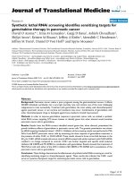
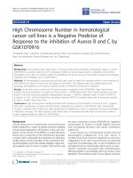
Fig. 1 NETs promote hypercoagulability through platelet aggregation. Tumor bearing mice have elevated platelet aggregation compared with sham
controls (a, AUC 40.2 ± 5.5 vs. 25.8 ± 1.5, n = 5). Treatment of human (b) and murine (c) blood with NET supernatant led to a dose dependent increase
in platelet aggregation compared with treatment with media control. Tumor bearing PAD4 KO mice had decreased platelet aggregation compared to
WT (AUC 8.4 ± 2.4 vs. 3.7 ± 1.7, n = 7) with no difference in sham controls (d). *p < 0.05
of whole blood (Fig. 4a) and in vivo treatment of mice
(Fig. 4b) with chloroquine resulted in decreased platelet
aggregation and activation (Additional file 2: Figure
S2C). To elucidate the potential mechanism of decreased
platelet aggregation after CQ treatment, we treated
PAD4KO mice with CQ and found that it had minimal
effect in these mice, suggesting that CQ mediates decreased platelet aggregation through inhibition of NETs
(Fig. 4c). Chloroquine treatment led to a significant
reduction in serum tissue factor levels in tumor bearing
mice with no significant change in sham mice (Fig. 4d).
We next examined the impact of hydroxychloroquine
(HCQ) on circulating tissue factor in patients with
pancreatic cancer using serum from our recently completed randomized clinical trial of preoperative gemcitabine/nab-paclitaxel with or without HCQ. There was
no difference in pretreatment patient demographics
between the two randomized groups (Additional file 4:
Table S1). HCQ led to a statistically greater reduction
in tissue factor in those patients who had elevated tissue factor prior to treatment, defined by preoperative
level greater than the median (40 ng/mL), (− 240 ± 120
Boone et al. BMC Cancer (2018) 18:678
Page 6 of 12
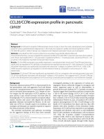
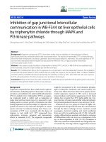
Fig. 2 NET upregulation of platelet aggregation is mediated by neutrophil DNA and platelet RAGE. Removing DNA from NET supernatant using
DNase I treatment prior to exposure to whole blood reversed the treatment effects of NET supernatant on platelet aggregation in human blood
(a, 25.9 ± 2.2 vs. 11.35 ± 0.31, n = 4, p < 0.05). In vivo treatment with DNase I resulted in decreased aggregation in tumor bearing mice (b, AUC
22.1 ± 2.3 vs. 38.4 ± 2.1, n = 4, p < 0.05). Tumor bearing RAGE KO mice have decreased platelet aggregation compared to WT mice (c, AUC 30.6 ±
1.5 vs. 40.2 ± 5.5, n = 4, p < 0.05). Blood from RAGE knockout mice had decreased aggregation after treatment with 100 μL of NET supernatant
compared with WT (d, AUC 25.5 ± 2.6 vs. 43.3 ± 3.9, n = 4, p < 0.05). *p < 0.05
vs. -8.74 ± 26.1 pg/mL, p < 0.05, Fig. 4e). There was no
difference in change in tissue factor with HCQ treatment in those patients with normal pre-treatment levels
(mean change with treatment − 55 ± 63 vs. + 3.1 ±
14 pg/mL, p = 0.38, n = 19 gem/nab-paclitaxel alone, n
= 18 gem/nab-paclitaxel + HCQ).
Chloroquine inhibition of NETs reverses
hypercoagulability
To study the effects of chloroquine inhibition of NETs
and subsequent decrease in platelet aggregation and circulating tissue factor on the hypercoagulable state seen
in pancreatic cancer, we performed thromboelastograms
Boone et al. BMC Cancer (2018) 18:678
Page 7 of 12
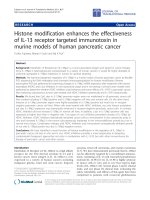
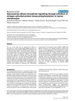
Fig. 3 NETs promote hypercoagulability in PDA by releasing circulating tissue factor. Tissue factor ELISA was performed on serum from
orthotopic mice, demonstrating that tumor burdened mice had elevated levels of circulating tissue factor compared to sham (a, 255 ± 49 vs. 159
± 26 pg/mL, p < 0.05). Genetic deletion of PAD4, thereby inhibiting NET formation, resulted in a substantial decrease in circulating tissue factor
levels in tumor bearing mice (269 ± 26 vs. 202 ± 30 pg/mL, p < 0.05). Blue = WT, Red = PAD4 KO, Circle = Sham, Triangle = Tumor. RAGE
knockout tumor bearing mice, who we have previously shown have decreased NET formation, also had lower levels of tissue factor compared to
WT controls (b, 331 ± 39 vs. 390 ± 34 pg/mL, p < 0.05). *p < 0.05. Blue = WT, Red = RAGE KO, Circle = Sham, Triangle = Tumor
(TEG) in mice with pancreatic adenocarcinoma to assess
hypercoagulability as measured by the coagulation index,
which takes into account all of the TEG parameters
(Additional file 5: Table S2). Tumor mice had an elevated coagulation index compared with sham controls,
suggestive of hypercoagulability (Fig. 5a). Treatment
with CQ resulted in a decrease in the coagulation index
in cancer burdened animals (Fig. 5b).
We next assessed the rate of venous thromboembolism
(VTE) in patients treated with pre-operative hydroxychloroquine as part of two separate clinical trial protocols.
In patients treated as part of a phase I/II dose escalation
trial of preoperative hydroxychloroquine with gemcitabine, the 90 day VTE rate was 3% (n = 1 of 33) [18]. Of
note, the lone patient who developed VTE was treated as
part of the dose escalation at 800 mg per day rather than
at the maximum dose of 1200 mg. In a more recent
randomized trial of preoperative gemcitabine and
nab-paclitaxel with or without hydroxychloroquine, the
VTE rate of patients treated with hydroxychloroquine was
9.1% compared to 30% in patients treated with
gemcitabine/nab-paclitaxel alone (p = 0.053, Fig. 5c).
Mean plasma DNA decreased with treatment in the HCQ
group, consistent with potential NET inhibition (601 ±
129 vs. 539 ± 114 ng/mL, p < 0.05), but not in the gemcitabine/nab-paclitaxel alone group (588 ± 144 vs. 543 ±
166 ng/mL, p = 0.09). Among all patients, those with VTE
had a mean increase of 6 ng/mL with treatment compared
with decrease of 70 ng/mL in those that did not have VTE
(p < 0.05). There was a trend towards change in plasma
DNA with treatment being associated with development
of VTE in patients treated with gemcitabine/nab-paclitaxel alone. Gemcitabine/nab-paclitaxel treated patients
who had a VTE had a mean increase of 20 ng/mL following treatment compared with a mean decrease of 76 ng/
mL in patients who did not develop VTE (p = 0.08). There
was no correlation between plasma DNA and VTE in
HCQ treated patients.
Discussion
It has long been recognized that patients with pancreatic
cancer are prone to venous thrombosis and it continues to
be a major source of morbidity and mortality [27]. After
initially being described in sepsis, neutrophil extracellular
traps (NETs) were discovered in malignancy and promote
tumor growth [28], development of metastases [29, 30]
and serve as a potential contributor to cancer associated
thrombosis [10]. The current work explores upregulation
of platelet function and release of tissue factor as two
mechanisms through which NETs contribute to hypercoagulability and thrombosis in pancreatic cancer. Furthermore, because autophagy is critical for NETs in pancreatic
cancer, we investigated the use of the autophagy/NET
inhibitor chloroquine to reverse NET mediated hypercoagulability in murine models and human patients.
Neutrophil-platelet interactions are increasingly recognized as an important collaboration in promoting malignancy and thrombosis [31]. Activated platelets are capable
of inducing NETs [32] and NETs in turn promote platelet
aggregation as observed in sepsis and deep vein thrombosis [33, 34]. Cancer induced platelet activation
Boone et al. BMC Cancer (2018) 18:678
Page 8 of 12
Fig. 4 CQ inhibition of NETs reverses platelet aggregation and decreases tissue factor. In vitro treatment of whole blood with CQ led to a significant
reduction in platelet aggregation in blood harvested from tumor bearing mice (a, AUC 50 ± 2.4 vs. 68.1 ± 8.8, n = 4, p < 0.05). Treatment of mice with
CQ led to a decrease in aggregation in tumor bearing animals with no change in sham (b, AUC 52.6 ± 5.3 vs. 68.1 ± 8.8, n = 4, p < 0.05). Importantly,
CQ had minimal effects in PAD4KO mice, suggesting that it decreases platelet aggregation through inhibition of NETs (c). CQ treatment led to a
decrease in circulating tissue factor in tumor bearing mice (d, 186.9 ± 5.6 vs. 228.2 ± 21 pg/mL, p < 0.05). Hydroxychloroquine treatment resulted in
significant reduction in tissue factor levels in patients with elevated preoperative serum tissue factor compared to control, with a mean response to
treatment of − 240 ± 120 versus − 8.74 ± 26 pg/mL (p < 0.05, n = 10 gem/nab-paclitaxel, n = 7 HCQ). Waterfall plot demonstrating individual treatment
response to gemcitabine/nab-paclitaxel with and without hydroxychloroquine in patients with elevated preoperative levels (e)
contributes to tumor growth, development of metastases
and thrombosis [35, 36]. The current study identifies
NETs as a key contributor to platelet aggregation in pancreatic cancer. During NET formation, PAD4 mediated
histone citrullination leads to unwinding and release of
DNA from neutrophils [37]. Since DNA is known to increase platelet aggregation in sepsis and deep vein thrombosis, we suspected that DNA released during NETosis
would also mediate platelet aggregation in pancreatic cancer [33, 34, 38]. Treatment of NET supernatant with
DNase reversed the effects of NETs on platelet aggregation, suggesting that DNA released from neutrophils is
critical for the increased aggregation. Similarly, Razak et
al. also showed that pancreatic cancer NETs promoted
platelet adhesion and that these effects could be reversed
with DNase [11]. We confirmed these observations and
expanded on this mechanism to include the receptor for
advanced glycation end products (RAGE), a known receptor for extracellular DNA, as a critical component of NET
mediated platelet aggregation in pancreatic cancer. The
addition of NET supernatant to RAGE knockout blood
did not result in increased platelet aggregation. Additionally, RAGE knockout mice had no differences in platelet
aggregation at baseline, but had decreased platelet aggregation in tumor burdened mice compared with wild type.
While these findings point to extracellular DNA and
RAGE promoting NET mediated platelet aggregation,
there are many components released from NETs that may
also have an impact on hypercoagulability and were not
evaluated in the current analysis.
Boone et al. BMC Cancer (2018) 18:678
Page 9 of 12
Fig. 5 Chloroquine reverses hypercoagulability in pancreatic cancer. Representative TEG curves demonstrating orthotopically injected mice are
hypercoagulable compared with sham controls (a). Treatment with CQ reverses the hypercoagulability on TEG as measured by coagulation index
(b). The 90 day VTE rate for patients treated with 2 cycles of preoperative gemcitabine/abraxane + HCQ was 9.1% (n = 3 of 33) compared to 30%
(n = 9 of 30) in patients treated with gemcitabine/abraxane alone (c, p = 0.053)
Tissue factor, a transmembrane receptor typically
found in subendothelial cells that binds to factor VII to
initiate the extrinsic pathway when the endothelium is
damaged is also released from neutrophils during NET
formation [25, 26]. Tissue factor thought to be derived
from tumor associated microparticles has been linked to
pancreatic cancer thrombosis [39–42] and levels of tissue factor predict venous thromboembolism in cancer
patients [43]. We identified NETs as a potential source
of circulating tissue factor in pancreatic cancer, as
genetic deletion of PAD4, an enzyme critical for NET
formation, resulted in significant reduction in circulating
tissue factor in tumor bearing mice. Importantly, PAD4
also citrullinates and inhibits antithrombin [44, 45], suggesting another possible mechanism of hypercoagulability in pancreatic cancer. This does potentially confound
our results in PAD4 knockout mice and must be taken
into account when considering our findings.
Because autophagy is critical to the process of NET formation, we studied the novel use of the autophagy inhibitor
chloroquine to target NET mediated hypercoagulability.
Chloroquine has been used for many years to treat patients
with malaria, lupus, and rheumatoid arthritis, but more
recently, hydroxychloroquine has been evaluated as a treatment for pancreatic cancer, with encouraging preliminary
results [18]. Chloroquine has previously been studied for
prevention of perioperative VTE in orthopedic surgery patients, however these studies had mixed results and the
precise mechanism was not completely understood [46,
47]. Subsequent studies have established that HCQ has direct effects on platelet activation and aggregation [48, 49].
However, our group and others have demonstrated that
chloroquine prevents NET formation [13, 14]; therefore
some of the antiplatelet effects of HCQ may be secondary
to reduction in NET mediated DNA release which increases platelet aggregation. In the current study, inhibition
of NETs with chloroquine resulted in decreased platelet
aggregation and lower levels of circulating tissue factor. In
patients who had elevated levels of pre-treatment tissue
factor, HCQ treatment led to a significant reduction, suggesting that the greatest effect of HCQ is seen in patients
who may have upregulation of NETs at baseline. Based on
this data, inhibition of NET formation may also explain the
previously recognized reduction in VTE rate. Importantly,
Boone et al. BMC Cancer (2018) 18:678
treatment with CQ in PAD4 KO mice, incapable of forming NETs, had minimal effect, suggesting that CQ
decreases platelet aggregation through inhibition of NETs.
However, because CQ also has direct antiplatelet effects, it
is difficult to completely attribute all its effects to inhibition
of NETosis.
Traditional coagulation tests such as prothrombin
time (PT), partial thromboplastin time (PTT), and international normalized ratio (INR) are frequently normal in
hypercoagulability and provide limited information regarding the mechanisms driving a prothrombotic state.
To study the role of chloroquine inhibition of NETs and
hypercoagulability using a more informative and clinically translatable approach, we utilized thromboelastograms to evaluate whether treatment with chloroquine
decreases hypercoagulability in orthotopic murine pancreatic cancer. TEG has been most thoroughly studied
in patients during massive bleeding from trauma as a
rapidly available test to direct transfusion of blood products, however, it is becoming more frequently utilized to
identify hypercoagulability [20]. Hypercoagulable
changes are detectable on rotational thromboelastometry, similar to TEG, in patients with abdominal malignancy [50]. We demonstrate that tumor burdened mice
are hypercoagulable on TEG and treatment with chloroquine reverses this hypercoagulopathy. Importantly, control sham mice appear to have a subtle increase in
coagulation index with CQ treatment. It is possible that
CQ may only serve a beneficial role in reducing hypercoagulability in the cancer burdened state, where NETs
are upregulated. This could explain why prior randomized trials of CQ to decrease VTE in non-malignant
orthopedic patients were inconclusive [46, 47].
Given its well-established use, favorable safety profile
and anti-tumor effects, CQ is a suitable treatment to decrease VTE rate in patients with pancreatic cancer. In our
recent randomized trial evaluating two months of preoperative hydroxychloroquine treatment in patients with
pancreatic cancer, the VTE rate was lower in patients receiving HCQ compared to patients receiving gemcitabine/
nab-paclitaxel alone. Although designed and powered to
study the effects of HCQ on pathologic treatment response and decrease in Ca 19–9, the reduction in VTE
rate neared statistical significance. Additionally, the 90 day
postoperative reduction in VTE occurred despite HCQ
stopping at time of surgery. We identified a trend towards
an increase in plasma DNA with treatment and development of VTE, which has been previously recognized as a
marker for risk of VTE [51]. DNA is released from neutrophils into the circulation during NET formation, therefore
this data suggests that NETs may play a role in VTE in patients with pancreatic cancer. However, given that DNA is
a nonspecific marker for NETs and that circulating DNA
in cancer patients is likely derived from multiple sources
Page 10 of 12
[52] we are unable to conclude that DNA released from
NETs is driving VTE in these patients. Nonetheless, these
findings support a clinical trial designed specifically to
study reduction in VTE by treatment of cancer patients
with perioperative HCQ.
Conclusion
We demonstrate in murine models of pancreatic cancer
that NETs promote hypercoagulability by increasing
platelet aggregation through DNA release and RAGE as
well as by release of tissue factor. Treatment with the
autophagy inhibitor chloroquine results in a reversal of
hypercoagulability in pancreatic cancer by diminishing
NET mediated platelet aggregation and release of circulating tissue factor and improving coagulation index on
TEG. We have for the first time also provided evidence
that these pathways play a role in human pancreatic cancer. All together our findings support additional clinical
trials with hydroxychloroquine to examine the ability of
NET inhibition to lower the venous thromboembolism
rate in patients with pancreatic and other cancer types.
Additional files
Additional file 1: Figure S1. Formation of ex vivo NETs. Microscopy of
isolated neutrophils stimulated with platelet activating factor (PAF) and
stained with Hoechst to visualize extracellular DNA, demonstrating ex
vivo neutrophil extracellular trap (NET) formation. (DOCX 221 kb)
Additional file 2: Figure S2. Neutrophil Extracellular Traps (NETs)
promote platelet activation in murine pancreatic adenocarcinoma.
Platelet activation was assessed by measuring % CD62P positive cells by
flow cytometry. Tumor burdened mice had heightened platelet
activation compared to sham controls (A). PAD4 KO mice, unable to form
NETs had diminished platelet activation. Addition of NET supernatant to
murine whole blood increased platelet activation in a dose dependent
fashion (B). Chloroquine treatment reversed the tumor associated
increase in platelet activation (C). (DOCX 109 kb)
Additional file 3: Figure S3. Neutrophil and fibrinogen conjugates in
the pancreatic tumor microenvironment. Pancreatic tumor specimens
from resected patients with pancreatic adenocarcinoma were stained for
neutrophil elastase (red) and fibrinogen (white). Representative images
from three individual patients are shown, demonstrating focal areas of
elastase and fibrinogen in the tumor, suggesting interactions between
neutrophils and thrombosis in the tumor microenvironment. (DOCX 489
kb)
Additional file 4: Table S1. Select results of randomized trial of
potentially resectable pancreatic cancer patients treated with
preoperative gemcitabine/nab-paclitaxel with and without
hydroxychloroquine (HCQ). There were no significant differences in
pretreatment patient demographics or characteristics. Correlative markers
of NET formation including circulating levels of DNA and tissue factor
were also assessed as discussed in the manuscript. Pre-tx = Pre-treatment,
CCI=Charlson Comorbidity Index, EUS = Endoscopic ultrasound. (DOCX 15
kb)
Additional file 5: Table S2. CQ reverses hypercoagulability in tumor
burdened mice. Thromboelastogram (TEG) values for orthotopic tumor
and sham mice with and without chloroquine (CQ) treatment,
demonstrating that tumor mice have hypercoagulable elevations in K,
angle, maximum amplitude (MA) and coagulation index (CI) compared
with sham controls and that CQ reverses hypercoagulability as assessed
by the CI. *p < 0.05 vs. Sham, **p < 0.05 vs. Tumor. (DOCX 14 kb)
Boone et al. BMC Cancer (2018) 18:678
Abbreviations
AUC: Area under the curve; CQ: Chloroquine; HBSS: Hank’s balanced salt
solution; HCQ: Hydroxychloroquine; HMGB1: High mobility group box 1;
IRB: Institutional Review Board; NET: Neutrophil extracellular trap; PAD
4: Protein arginine deiminase 4; PDA: Pancreatic ductal adenocarcinoma;
PMA: Phorbol 12-myristate 13-acetate; RAGE: Receptor for advanced
glycation end products; TEG: Thromboelastogram; VTE: Venous
thromboembolism
Acknowledgements
We appreciate the efforts of Stacy Stull, Peter Adams and MACRO
(Multidisciplinary Acute Care Research Organization) research, University of
Pittsburgh, in running TEG samples.
Funding
This work was supported in part by R01CA181450 from the National Cancer
Institute (HJZ and MTL) and by 1R35GM119526–01 (MDN). The content is
solely the responsibility of the authors and does not necessarily represent
the official views of the National Cancer Institute or the U.S. National
Institutes of Health. Funding was also graciously provided by philanthropic
donors, including the Emma Clyde Hodge Memorial Fund.
Page 11 of 12
3.
4.
5.
6.
7.
8.
9.
Availability of data and materials
The datasets used and/or analyzed during the current study are available
from the corresponding author on reasonable request.
10.
Authors’ contributions
BAB, PM, HJZ, MDN, and MTL contributed to experimental concept and
design, interpreted the results, wrote the manuscript and critically reviewed
the manuscript. JMO, XL, MAR, CTW, JLS, WRD, and JTE analyzed and
interpreted the data and provided critical review of the manuscript. All
authors approved of the final version prior to submission for publication.
11.
Ethics approval and consent to participate
All experimental animal procedures were reviewed and approved by the
Institutional Animal Care and Use Committee of the University of Pittsburgh
(Protocol # 14084123).
Correlative patient samples and data were included from two clinical trial
protocols that were approved by the Institutional Review Board for the
University of Pittsburgh (Protocol #10010028 and #13080444). All patients
signed informed consent prior to participation in these clinical protocols.
12.
13.
14.
15.
Consent for publication
Not applicable.
16.
Competing interests
The authors declare that they have no competing interests.
17.
18.
Publisher’s Note
Springer Nature remains neutral with regard to jurisdictional claims in
published maps and institutional affiliations.
Author details
1
Department of Surgery, University of Pittsburgh, Pittsburgh, PA, USA.
2
Center for Biologic Imaging, University of Pittsburgh, Pittsburgh, PA, USA.
3
Departments of Thoracic Surgery, University of Pittsburgh, Pittsburgh, PA,
USA. 4Immunology, University of Pittsburgh, Pittsburgh, PA, USA.
5
Bioengineering, University of Pittsburgh, Pittsburgh, PA, USA. 6UPMC Cancer
Pavilion, University of Pittsburgh, Suite 417, 5150 Centre Ave, Pittsburgh, PA
15232, USA.
19.
20.
21.
22.
Received: 6 April 2018 Accepted: 12 June 2018
References
1. Petterson TM, Marks RS, Ashrani AA, Bailey KR, Heit JA. Risk of site-specific
cancer in incident venous thromboembolism: a population-based study.
Thromb Res. 2015;135(3):472–8.
2. Wun T, White RH. Venous thromboembolism (VTE) in patients with cancer:
epidemiology and risk factors. Cancer Investig. 2009;27(Suppl 1):63–74.
23.
24.
Kruger S, Haas M, Burkl C, Goehring P, Kleespies A, Roeder F, et al.
Incidence, outcome and risk stratification tools for venous
thromboembolism in advanced pancreatic cancer - a retrospective cohort
study. Thromb Res. 2017;157:9–15.
Chew HK, Wun T, Harvey D, Zhou H, White RH. Incidence of venous
thromboembolism and its effect on survival among patients with common
cancers. Arch Intern Med. 2006;166(4):458–64.
Mandala M, Reni M, Cascinu S, Barni S, Floriani I, Cereda S, et al. Venous
thromboembolism predicts poor prognosis in irresectable pancreatic cancer
patients. Annals of oncology: official journal of the European society for.
Med Oncol. 2007;18(10):1660–5.
Krepline AN, Christians KK, George B, Ritch PS, Erickson BA, Tolat P, et al.
Venous thromboembolism prophylaxis during neoadjuvant therapy for
resectable and borderline resectable pancreatic cancer-is it indicated? J
Surg Oncol. 2016;114(5):581–6.
Meng H, Yalavarthi S, Kanthi Y, Mazza LF, Elfline MA, Luke CE, et al. In vivo
role of neutrophil extracellular traps in antiphospholipid antibody-mediated
venous thrombosis. Arthritis Rheum. 2017;69(3):655–67.
Doring Y, Soehnlein O, Weber C. Neutrophil extracellular traps in
atherosclerosis and Atherothrombosis. Circ Res. 2017;120(4):736–43.
Demers M, Wagner DD. NETosis: a new factor in tumor progression and
cancer-associated thrombosis. Semin Thromb Hemost. 2014;40(3):277–83.
Demers M, Krause DS, Schatzberg D, Martinod K, Voorhees JR, Fuchs TA, et
al. Cancers predispose neutrophils to release extracellular DNA traps that
contribute to cancer-associated thrombosis. Proc Natl Acad Sci U S A. 2012;
109(32):13076–81.
Abdol Razak N, Elaskalani O, Metharom P. Pancreatic Cancer-induced
neutrophil extracellular traps: a potential contributor to Cancer-associated
thrombosis. Int J Mol Sci. 2017;18(3):487.
Brinkmann V, Reichard U, Goosmann C, Fauler B, Uhlemann Y, Weiss DS, et
al. Neutrophil extracellular traps kill bacteria. Science. 2004;303(5663):1532–5.
Boone BA, Orlichenko L, Schapiro NE, Loughran P, Gianfrate GC, Ellis JT, et
al. The receptor for advanced glycation end products (RAGE) enhances
autophagy and neutrophil extracellular traps in pancreatic cancer. Cancer
Gene Ther. 2015;22(6):326–34.
Smith CK, Vivekanandan-Giri A, Tang C, Knight JS, Mathew A, Padilla RL, et
al. Neutrophil extracellular trap-derived enzymes oxidize high-density
lipoprotein: an additional proatherogenic mechanism in systemic lupus
erythematosus. Arthritis Rheum. 2014;66(9):2532–44.
Demers M, Wagner DD. Neutrophil extracellular traps: a new link to cancerassociated thrombosis and potential implications for tumor progression.
Oncoimmunology. 2013;2(2):e22946.
Hemmers S, Teijaro JR, Arandjelovic S, Mowen KA. PAD4-mediated
neutrophil extracellular trap formation is not required for immunity against
influenza infection. PLoS One. 2011;6(7):e22043.
Swamydas M, Luo Y, Dorf ME, Lionakis MS. Isolation of mouse neutrophils.
Curr Protoc Immunol. 2015;110(3):20. 21–23 20 15
Boone BA, Bahary N, Zureikat AH, Moser AJ, Normolle DP, Wu WC, et al.
Safety and biologic response of pre-operative autophagy inhibition in
combination with gemcitabine in patients with pancreatic adenocarcinoma.
Ann Surg Oncol. 2015;22(13):4402–10.
Ding N, Chen G, Hoffman R, Loughran PA, Sodhi CP, Hackam DJ, et al. Tolllike receptor 4 regulates platelet function and contributes to coagulation
abnormality and organ injury in hemorrhagic shock and resuscitation. Circ
Cardiovasc Genet. 2014;7(5):615–24.
Zohav E, Almog B, Cohen A, Levin I, Deutsch V, Many A, et al. A new
perspective on the risk of Hypercoagulopathy in ovarian Hyperstimulation
syndrome using Thromboelastography. Reprod Sci. 2017;24(12):1600-6.
Fuchs TA, Brill A, Wagner DD. Neutrophil extracellular trap (NET) impact
on deep vein thrombosis. Arterioscler Thromb Vasc Biol. 2012;32(8):
1777–83.
Leshner M, Wang S, Lewis C, Zheng H, Chen XA, Santy L, et al. PAD4
mediated histone hypercitrullination induces heterochromatin
decondensation and chromatin unfolding to form neutrophil extracellular
trap-like structures. Front Immunol. 2012;3:307.
Sirois CM, Jin T, Miller AL, Bertheloot D, Nakamura H, Horvath GL, et al.
RAGE is a nucleic acid receptor that promotes inflammatory responses to
DNA. J Exp Med. 2013;210(11):2447–63.
Khorana AA, Ahrendt SA, Ryan CK, Francis CW, Hruban RH, Hu YC, et al.
Tissue factor expression, angiogenesis, and thrombosis in pancreatic cancer.
Clin Cancer Res. 2007;13(10):2870–5.
Boone et al. BMC Cancer (2018) 18:678
25. Kambas K, Mitroulis I, Apostolidou E, Girod A, Chrysanthopoulou A,
Pneumatikos I, et al. Autophagy mediates the delivery of thrombogenic
tissue factor to neutrophil extracellular traps in human sepsis. PLoS One.
2012;7(9):e45427.
26. Kambas K, Chrysanthopoulou A, Vassilopoulos D, Apostolidou E, Skendros P,
Girod A, et al. Tissue factor expression in neutrophil extracellular traps and
neutrophil derived microparticles in antineutrophil cytoplasmic antibody
associated vasculitis may promote thromboinflammation and the
thrombophilic state associated with the disease. Ann Rheum Dis. 2014;
73(10):1854–63.
27. Ansari D, Ansari D, Andersson R, Andren-Sandberg A. Pancreatic cancer and
thromboembolic disease, 150 years after Trousseau. Hepatobiliary Surg Nutr.
2015;4(5):325–35.
28. Demers M, Wong SL, Martinod K, Gallant M, Cabral JE, Wang Y, et al.
Priming of neutrophils toward NETosis promotes tumor growth.
Oncoimmunology. 2016;5(5):e1134073.
29. Cools-Lartigue J, Spicer J, McDonald B, Gowing S, Chow S, Giannias B, et al.
Neutrophil extracellular traps sequester circulating tumor cells and promote
metastasis. J Clin Invest. 2013;123(8):3446–58.
30. Tohme S, Yazdani HO, Al-Khafaji AB, Chidi AP, Loughran P, Mowen K, et
al. Neutrophil extracellular traps promote the development and
progression of liver metastases after surgical stress. Cancer Res. 2016;
76(6):1367–80.
31. Olsson AK, Cedervall J. NETosis in Cancer - platelet-neutrophil crosstalk
promotes tumor-associated pathology. Front Immunol. 2016;7:373.
32. Maugeri N, Campana L, Gavina M, Covino C, De Metrio M, Panciroli C, et al.
Activated platelets present high mobility group box 1 to neutrophils,
inducing autophagy and promoting the extrusion of neutrophil extracellular
traps. J Thromb Haemost. 2014;12(12):2074–88.
33. McDonald B, Davis RP, Kim SJ, Tse M, Esmon CT, Kolaczkowska E, et al. Platelets
and neutrophil extracellular traps collaborate to promote intravascular
coagulation during sepsis in mice. Blood. 2017;129(10):1357–67.
34. Fuchs TA, Brill A, Duerschmied D, Schatzberg D, Monestier M, Myers DD Jr,
et al. Extracellular DNA traps promote thrombosis. Proc Natl Acad Sci U S A.
2010;107(36):15880–5.
35. Heinmoller E, Schropp T, Kisker O, Simon B, Seitz R, Weinel RJ. Tumor cellinduced platelet aggregation in vitro by human pancreatic cancer cell lines.
Scand J Gastroenterol. 1995;30(10):1008–16.
36. Yan M, Jurasz P. The role of platelets in the tumor microenvironment: from
solid tumors to leukemia. Biochim Biophys Acta. 2016;1863(3):392–400.
37. Martinod K, Demers M, Fuchs TA, Wong SL, Brill A, Gallant M, et al. Neutrophil
histone modification by peptidylarginine deiminase 4 is critical for deep vein
thrombosis in mice. Proc Natl Acad Sci U S A. 2013;110(21):8674–9.
38. Gould TJ, Vu TT, Swystun LL, Dwivedi DJ, Mai SH, Weitz JI, et al. Neutrophil
extracellular traps promote thrombin generation through plateletdependent and platelet-independent mechanisms. Arterioscler Thromb Vasc
Biol. 2014;34(9):1977–84.
39. Woei AJFJ, Tesselaar ME, Garcia Rodriguez P, Romijn FP, Bertina RM, Osanto
S. Tissue factor-bearing microparticles and CA19.9: two players in pancreatic
cancer-associated thrombosis? Br J Cancer. 2016;115(3):332–8.
40. Geddings JE, Mackman N. Tumor-derived tissue factor-positive
microparticles and venous thrombosis in cancer patients. Blood. 2013;
122(11):1873–80.
41. Kambas K, Mitroulis I, Ritis K. The emerging role of neutrophils in
thrombosis-the journey of TF through NETs. Front Immunol. 2012;3:385.
42. Thomas GM, Brill A, Mezouar S, Crescence L, Gallant M, Dubois C, et al.
Tissue factor expressed by circulating cancer cell-derived microparticles
drastically increases the incidence of deep vein thrombosis in mice. J
Thromb Haemost. 2015;13(7):1310–9.
43. Khorana AA, Kamphuisen PW, Meyer G, Bauersachs R, Janas MS, Jarner MF,
et al. Tissue factor as a predictor of recurrent venous thromboembolism in
malignancy: biomarker analyses of the CATCH trial. J Clin Oncol. 2016;35(10):
1078-85.
44. Chang X, Yamada R, Sawada T, Suzuki A, Kochi Y, Yamamoto K. The
inhibition of antithrombin by peptidylarginine deiminase 4 may contribute
to pathogenesis of rheumatoid arthritis. Rheumatology. 2005;44(3):293–8.
45. Ordonez A, Martinez-Martinez I, Corrales FJ, Miqueo C, Minano A, Vicente V,
et al. Effect of citrullination on the function and conformation of
antithrombin. FEBS J. 2009;276(22):6763–72.
46. Pilcher DB. Hydroxychloroquine sulfate in prevention of thromboembolic
phenomena in surgical patients. Am Surg. 1975;41(12):761–6.
Page 12 of 12
47. Carter AE, Eban R. Prevention of postoperative deep venous thrombosis in
legs by orally administered hydroxychloroquine sulphate. Br Med J. 1974;
3(5923):94–5.
48. Espinola RG, Pierangeli SS, Gharavi AE, Harris EN. Hydroxychloroquine
reverses platelet activation induced by human IgG antiphospholipid
antibodies. Thromb Haemost. 2002;87(3):518–22.
49. Nosal R, Jancinova V, Danihelova E. Chloroquine: a multipotent inhibitor of
human platelets in vitro. Thromb Res. 2000;98(5):411–21.
50. Thorson CM, Van Haren RM, Ryan ML, Curia E, Sleeman D, Levi JU, et al. Preexisting hypercoagulability in patients undergoing potentially curative
cancer resection. Surgery. 2014;155(1):134–44.
51. Diaz JA, Fuchs TA, Jackson TO, Kremer Hovinga JA, Lammle B, Henke PK, et
al. Plasma DNA is elevated in patients with deep vein thrombosis. J Vasc
Surg Venous Lymphat Disord. 2013;1(4):341-8.
52. Jahr S, Hentze H, Englisch S, Hardt D, Fackelmayer FO, Hesch RD, et al. DNA
fragments in the blood plasma of cancer patients: quantitations and
evidence for their origin from apoptotic and necrotic cells. Cancer Res.
2001;61(4):1659–65.