Integrator complex subunit 6 (INTS6) inhibits hepatocellular carcinoma growth by Wnt pathway and serve as a prognostic marker
Bạn đang xem bản rút gọn của tài liệu. Xem và tải ngay bản đầy đủ của tài liệu tại đây (2.59 MB, 12 trang )
Lui et al. BMC Cancer (2017) 17:644
DOI 10.1186/s12885-017-3628-3
RESEARCH ARTICLE
Open Access
Integrator complex subunit 6 (INTS6)
inhibits hepatocellular carcinoma growth
by Wnt pathway and serve as a prognostic
marker
Ka Yin Lui2, Hui Zhao3, Chunhui Qiu3, Chuo Li4, Zhigang Zhang6, Haoran Peng5, Rongdang Fu3, Hu-an Chen3*
and Min-qiang Lu1*
Abstract
Background: Integrator complex subunit 6 (INTS6) was found to play a tumour suppressing role in certain types of
solid tumours. In this study, we wanted to determine the expression level of INTS6 in hepatocellular carcinoma
(HCC) and evaluate its clinical characteristics and mechanisms in HCC patients (Lui and Lu, European Journal of
Cancer, 51:S94, 2015).
Methods: First, we used a microarray analysis to explore the mRNA expression levels in HCC and paired normal
liver tissues; second, we used qRT-PCR to measure the INTS6 mRNA levels in a cohort of 50 HCC tissues and
adjacent normal liver tissues; third, we used Western blot analyses to detect the INTS6 protein levels in 20 paired
HCC and normal liver tissues; fourth, we used immunohistochemistry to determine the INTS6 expression levels in 70
archived paraffin-embedded HCC samples. Finally, we investigated the suppressive function of INTS6 in the Wnt
pathway.
Results: Herein, according to the microarray data analysis, the expression levels of INTS6 were dramatically downregulated in HCC tissues vs. those in normal liver tissues (p<0.05). qRT-PCR and Western blot analyses showed that
the INTS6 mRNA and protein expression was significantly down-regulated in tumour tissues compared to the
adjacent normal liver tissues (p<0.05). Immunohistochemical assays revealed that decreased INTS6 expression was
present in 62.9% (44/70) of HCC patients. Correlation analyses showed that INTS6 expression was significantly
correlated with serum alpha-fetoprotein levels (AFP, p =0.004), pathology grade (p =0.005), and tumour recurrence
(p =0.04). Kaplan-Meier analysis revealed that patients with low INTS6 expression levels had shorter overall and
disease-free survival rates than patients with high INTS6 expression levels (p =0.001 and p =0.001). Multivariate
regression analysis indicated that INTS6 was an independent predictor of overall survival and disease-free survival
rates. Mechanistically, INTS6 increased WIF-1 expression and then inhibited the Wnt/β-catenin signalling pathway.
Conclusion: The results of our study show that down-regulated INTS6 expression is associated with a poorer
prognosis in HCC patients. This newly identified INTS6/WIF-1 axis indicates the molecular mechanism of HCC and
may represent a therapeutic target in HCC patients.
Keywords: INTS6, Hepatocellular carcinoma, Prognosis, Wnt/β-catenin
* Correspondence: ;
3
Department of Hepatic Surgery, the Third Affiliated Hospital of Sun Yat-sen
University, Guangzhou 510630, China
1
Department of Hepatobiliary Surgery, Guangzhou First People’s Hospital,
Guangzhou 510180, China
Full list of author information is available at the end of the article
© The Author(s). 2017 Open Access This article is distributed under the terms of the Creative Commons Attribution 4.0
International License ( which permits unrestricted use, distribution, and
reproduction in any medium, provided you give appropriate credit to the original author(s) and the source, provide a link to
the Creative Commons license, and indicate if changes were made. The Creative Commons Public Domain Dedication waiver
( applies to the data made available in this article, unless otherwise stated.
Lui et al. BMC Cancer (2017) 17:644
Background
Hepatocellular carcinoma (HCC) is one of the most
common cancers in the world and has characteristics
of high mobility, high recurrence rates, and poor
prognosis [1]. Approximately 110,000 people die of
HCC each year in China [2]. This mortality rate accounts for 45% of the total deaths from HCC in the
world. Potentially curative therapies for HCC include
surgical resection and liver transplantation [3]. In recent years, tremendous progress has been made towards understanding the causes of HCC, such as
hepatitis B virus or hepatitis C virus infection, alcohol
consumption, and water contamination [4, 5]. The
discovery of some significant causes of HCC makes
this disease somewhat preventable. Hence, in part,
early treatment reduces its mortality. However, the 5year survival rate of HCC is still very low [6].
Multi-step processes including genetic and epigenetic alterations are thought to play a cumulative role
in the progression of HCC [7]. Most of the abnormally expressed genes play a key role in the process
of the malignant transformation of liver cells, such as
the regulation of the cell cycle, cell growth, apoptosis,
cell migration and diffusion [8]. The Wnt/β-catenin
signalling pathway, generally activated by genetic and
epigenetic alterations, has been linked to several types
of tumours, including HCC [9]. Common epigenetic
changes include DNA hypermethylation in the promoter region of WIF-1 [10]. The screening and identification of molecular targets involved in hepatic cell
malignant transformation are very important, and
these may become potential clinical therapeutic targets in HCC patients [11].
Integrator complex subunit 6 (INTS6), which was previously known as the gene encoding deleted in cancer
cells 1 (DICE1) (OMIM 604331), was identified to
localize with the microsatellite marker D13S284 in
13q14.3, a region frequently affected by allelic deletion
in many solid tumours, such as prostate carcinoma, cervical carcinoma and oesophageal squamous cell carcinoma [12–14]. Some studies have identified the promoter
of the tumour suppressor gene INTS6, which is downregulated in prostate cancer, and have revealed that the
INTS6 promoter is hypermethylated in prostate cancer
cell lines [15].
Although INTS6 is known to play a key role in
many solid tumours, including in HCC [16], the relationship between INTS6 expression and the clinicopathological characteristics of HCC and its molecular
mechanisms are poorly unknown. The current study
detected the expression of INTS6 in HCC using
quantitative reverse transcriptase polymerase chain reaction (qRT-PCR), Western blotting, and immunohistochemistry analyses. After these experiments, we
Page 2 of 12
wanted to determine the relationship between INTS6
expression levels and the clinicopathological features
of HCC and one of its pathways.
Methods
Patients and HCC tissue samples.
All of the clinical samples (including HCC tissues and
adjacent normal liver tissues) were obtained from the
Third Affiliated Hospital, Sun Yat-sen University
(Guangzhou, China). All of the patients gave informed
consent. This project was approved by the Clinical
Research Ethics Committee of the Third Affiliated
Hospital, Sun Yat-sen University.
Hepatocellular carcinoma tissues and their matched
adjacent normal tissues (not less than 2 cm away
from the tumour) were obtained from 3 patients and
were used for the discovery of specific mRNA
changes from the microarrays. A total of 50 HCC
tumour tissues and matched adjacent normal liver tissues were obtained from patients, and 70 FFPE samples with pathologist-diagnosed HCC were obtained
from the Third Affiliated Hospital of Sun Yat-sen
University between the years 2008 and 2012. The liver
and tumour tissues were immediately frozen in liquid
nitrogen after surgery and stored at −80 °C until the
extraction of total RNA.
We used the TNM classification of the 6th edition
American Joint Committee on Cancer (AJCC) to classify
the tumour stage. The patients included both men and
women with ages ranging from 29 to 71 (mean age:
48.2 years). None of the patients who participated in this
study received any pre-operative treatments, including
TACE or radiofrequency ablation.
Cell lines and culture condition
The HCC cell lines MHCC97L (catalogue number
CC0109), Huh7 (catalogue number TCHu182),
Hep3B (catalogue number TCHu106) and HepG2
(catalogue number TCHu 72) and a normal human
hepatocyte (HH) (catalogue number GNHu 6) cell
line were obtained from the Cell Bank of the
Chinese Academy of Sciences, and all the cell lines
were grown in DMEM (Gibco, Invitrogen, USA) supplemented with 10% foetal bovine serum (FBS)
(Gibco, Invitrogen, USA), penicillin (100 units/ml)
and streptomycin (100 units/ml) in 5% CO2 at 37 °C
in a humidified incubator.
HCC mRNA microarray analysis
The Arraystar Human lncRNA Array v2.0 was used to
profile both lncRNAs and messenger RNAs (mRNAs) in
the human genome of 3 pairs of human HCC and the
matched normal tissues. Sample labelling and array
hybridization were performed according to the Agilent
Lui et al. BMC Cancer (2017) 17:644
One-Color Microarray-Based Gene Expression Analysis
protocol (Agilent Technologies) with minor modifications [15] (the data have been deposited in the GEO:
GSE64633).
Plasmid DNA construction and transfection
The MSCV-based bicistronic retroviral vector MIEG3
was used to express INTS6 as described in our previous
study [16]. All plasmid DNAs were verified by DNA sequencing. For plasmid transfection, HCC cell lines were
grown in 6-well plates; the next day, cells were transfected with Lipofectamine 2000 (Invitrogen) according
to the manufacturer’s recommendations. siRNA oligonucleotides targeting INTS6 or the negative control were
transfected into the HCC cell lines using Lipofectamine
RNAiMAX (Invitrogen, USA), according to the manufacturer’s protocol. Target gene expression levels were
measured 72 h post-transfection. RNA and protein were
acquired 72 h after transfection.
Western blot analysis
Total frozen HCC and adjacent normal liver tissue proteins and HCC cell proteins were extracted using RIPA
lysis buffer (KeyGen, China) according to the manufacturer’s’ instructions. For Western blotting, primary polyclonal antibodies against INTS6 (rabbit anti-INTS6,
Abcam, 1:2000 dilution), WIF-1 (rabbit anti-WIF-1,
Abcam, 1:2000 dilution), and β-catenin (rabbit anti-Beta
Catenin, Abcam, 1:5000 dilution) were used. An actin
antibody served as the internal control. The online software ImageJ was used to quantify the density of the
bands.
Quantitative real-time PCR analysis
All the RNA samples were reverse transcribed to
synthesize cDNA using the PrimeScript® RT reagent kit
with a gDNA Eraser (Takara, China).
QRT-PCR was employed to determine the relative expression levels of the target genes using a LightCycler
480 SYBR Green I Master (Roche).
The qRT-PCR reactions were performed in triplicate
using the LC480 Real-Time PCR Detection System
(Roche).
The primers for qRT-PCR were as follows: INTS6 forward 5′-AGCTGCCAGTTCTTGGAATG-3′ and reverse
5′-AGGCCAGACAGCTCTGATGT-3′; GAPDH, forward
5′-GTCCACCACCCTGTTGCTGTA-3′ and reverse 5′CTTCAACAGCGACACCCACTC-3′. Additionally, we detected the expression levels of the downstream target genes
ZEB1 and MMP13 from the Wnt/β-catenin pathway. The
primers for qRT-PCR were as follows: ZEB1 forward 5′
-TGCACTGAGTGTGGAAAAGC-3′ and reverse 5′TGGTGATGCTGAAAGAGACG-3′; MMP13 forward
Page 3 of 12
5′ - ACTGAGAGGCTCCGAGAAATG-3′ and reverse
5′- GAACCCCGCATCTTGGCTT-3′.
The cycle at which the reaction crossed an arbitrarily
placed threshold (Ct) was determined for each gene. The
mRNA expression level of each gene was calculated
using the △Ct method: △Ct = CtmRNA – CtGAPDH.
Immunohistochemistry
A total of 70 HCC tissue samples were collected by the
Department of Pathology, and all the samples were fixed
in formalin and embedded in paraffin.
The HE-stained sections of each specimen from a single random block were measured by a senior
pathologist.
Both percent positivity and staining intensity of
tumour cells were viewed according to a double blinded
method. Immunohistochemistry was performed based
on a previously described method [17]. The percent
positivity was scored as “0” (<5%, negative), “1” (5–25%,
sporadic), “2” (25–50%, focal), or “3” (>50%, diffuse).
The staining intensity was scored as “0” (no staining),
“1” (weakly stained), “2” (moderately stained), or “3”
(strongly stained). The final INTS6 expression score was
calculated using the value of the percent positivity cell
score × the staining intensity score [18], which ranged
from 0 to 9. The INTS6 expression level was defined as
follows: “−” (score 0–1), “+” (score 2–4), “++” (score 5–
6), and “+++” (score > 6). According to previous research, a low expression level of INTS6 was defined as a
total score ≤ 4, and a high expression level of INTS6
was defined as a total score > 4.
Statistical analysis
The statistical analyses were performed with SPSS 14.0
software. A p-value of <0.05 was considered significant.
The non-parametric statistical test was used to analyse
the differences in the protein and mRNA expression
levels of INTS6 in HCC tissue and cell lines. The χ2 test
was used to analyse the relationship between the expression levels of INTS6 and the clinicopathological characteristics of HCC patients. Survival curves were
calculated using the Kaplan-Meier method and were
compared using the log-rank test. Finally, univariate and
multivariate Cox regression analyses were used to evaluate the survival rates of HCC patients.
Results
Expression of INTS6 mRNA in HCC patients determined
by microarray analysis
To discover the dysregulated genes in HCC tissue, we
collected 3 HCC tissues and their matched adjacent normal tissues and used microarray analysis. According to
the microarray data analysis (2.0-fold up- or downregulated and p<0.05) (the data have been deposited in
Lui et al. BMC Cancer (2017) 17:644
the GEO: GSE64633), the expression levels of INTS6
were dramatically down-regulated in HCC vs. matched
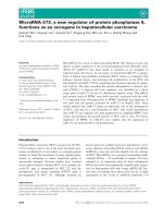
normal liver tissues (p = 0.015) (Fig. 1a, b). Detailed information and the analysis file for the differential mRNA
expression are summarized in the supplementary material (Additional files 1).
Page 4 of 12
normal human hepatocytes (HH) (p<0.05, Fig. 3). Then,
the INTS6 protein levels in the same HCC samples used
for qRT-PCR were examined by using Western blotting.
From our results, both the INTS6 protein and mRNA
expression levels were reduced in the HCC tissues compared to the corresponding adjacent normal tissues
(Fig. 4a, b).
INTS6 mRNA and protein expression was down-regulated
in HCC compared to the corresponding adjacent liver
tissues
INTS6 expression was correlated with the
clinicopathological features of HCC
To further explore the array data, we collected a larger
cohort of human HCC and paired normal liver tissues
and detected the expression levels of INTS6. We found
that the INTS6 expression was down-regulated in 68.0%
(34/50) of the HCC tissues, compared to the expression
in the normal liver tissues.
From the analysis of the 50 paired HCC samples, we
identified a remarkable difference in the expression
levels of INTS6 between the HCC and adjacent liver tissues (p = 0.0066, Fig. 2). What is more, the expression
of INTS6 was down-regulated in HCC cell lines (Huh7,
MHCC97L, HepG2 and Hep3B) when compared to
Immunohistochemistry was performed on 70 archived
paraffin-embedded HCC samples. The results revealed
that INTS6 expression localized primarily to the nuclei
of tumour cells (Fig. 5). Low INTS6 expression was
present in 44 (62.9%) of 70 HCC cases. The correlation
between INTS6 expression and the clinicopathological
features of HCC was analysed using the chi-square test
(Table 1). The expression level of INTS6 was significantly associated with the serum alpha-fetoprotein level
(AFP, p = 0.004), pathology grade (p = 0.005), and
tumour recurrence (p = 0.040). Moreover, from our
qRT-PCR analysis of HCC tissues and adjacent normal
Fig. 1 a. INTS6 microarray data from HCC tissues vs. normal adjacent tissues (3 pairs, p < 0.01). b. Heat map from the microarray of HCC tissues
vs. normal adjacent tissues
Lui et al. BMC Cancer (2017) 17:644
Page 5 of 12
a
Fig. 2 Relative expression level of INTS6 mRNA in 50 paired HCC
tissues and adjacent normal tissues by qRT-PCR (p = 0.0066)
*
3
*
2
3
4
0
Fig. 4 a. Expression of INTS6 protein in HCC and adjacent normal
tissue samples using Western blotting (T = tumour, N = normal
tissue). *p<0.05, **p<0.01. b. Expression of INTS6 mRNA in the
corresponding HCC and adjacent normal tissue samples
(T = tumour, N = normal tissue). *p<0.05, **p<0.01
*
*
*
2
*
3
4
*
1
1
INTS6 (Cell RNA)
*
1
B
H
ep
3
2
ep
G
H
M
H
H
C
C
97
L
uh
7
H
0
H
Relative INTS6 mRNA Expression
Survival curves were plotted using the Kaplan-Meier
method and were compared using the log-rank test for
70 archived paraffin-embedded HCC patient samples.
Patients with low INTS6 expression had shorter overall
Tumor
Normal Tissue
4
2
Low expression of INTS6 was correlated with poor
prognosis in HCC patients
b
Re l a ti ve I NTS 6 Ex pre ssi on m RNA
liver tissues (Table 2), the expression level of INTS6 was
remarkably associated with the serum alpha-fetoprotein
levels (p = 0.004) and pathology grade (p = 0.006). Therefore, HCC patients with low INTS6 expression had a
higher tendency to have high AFP levels, a poor pathology
grade, and tumour recurrence. However, neither immunohistochemistry nor qRT-PCR analyses of INTS6 expression had statistically significant associations with age,
gender, HBsAg positivity, tumour size, or cirrhosis.
Fig. 3 qRT-PCR analysis of INTS6 expression levels in different liver
cell lines. GAPDH was used as an endogenous control for
normalizing experimental data. *p<0.05
and disease-free survival time than patients with high
INTS6 expression (p = 0.001 and p = 0.001, Fig. 6a-b).
Stratified survival analysis explored the prognostic value
of INTS6 according to poor pathology grade and tumour
recurrence. Patients with low INTS6 expression had
shorter survival time with poor pathology grades II–III
and tumour recurrence (p<0.001 and p = 0.007, Fig. 6c,
e). In addition, they also had shorter disease-free survival
time with poor pathology grades II–III and tumour
Lui et al. BMC Cancer (2017) 17:644
Page 6 of 12
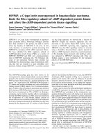
Fig. 5 Immunohistochemical staining of INTS6 in HCC. INTS6 protein expression localized mainly to the nuclei in tumour cells. Different INTS6
staining intensities [negative: 0, weak: 1, moderate: 2, strong: 3] are indicated in the micrographs
recurrence (p<0.001 and p = 0.001, Fig. 6d, f ) than patients with high INTS6 expression.
INTS6 was an independent predictor of overall and
disease-free survival rates in HCC patients
Univariate and multivariate Cox analyses explored the impacts of the expression of INTS6 and other clinicopathological parameters on the overall and disease-free survival
rates in 70 archived paraffin-embedded HCC patient samples. Univariate Cox regression analyses revealed that the
expression of INTS6 (p = 0.001), pathology grade
(p = 0.005), tumour size (p = 0.035), vascular invasion
(p = 0.001) and recurrence (p = 0.002) correlated with
overall survival (Table 3). In addition, multivariate Cox regression analysis also indicated that the expression of
INTS6 (p = 0.040), recurrence (p = 0.041) and vascular invasion (p = 0.024) were independent predictors of overall
survival in HCC patients (Table 2). Furthermore, the expression of INTS6 (p = 0.001), tumour size (p = 0.004),
pathology grade (p = 0.009) and vascular invasion
(p = 0.001) were correlated with disease-free survival
(Table 3). In addition, multivariate Cox regression analysis
also indicated that the expression of INTS6 (p = 0.025)
and vascular invasion (p = 0.001) were independent predictors of disease-free survival in HCC patients (Table 4).
INTS6 inhibited the Wnt/β-catenin signalling pathway by
decreasing the expression levels of WIF-1
qRT-PCR analysis showed that WIF-1 mRNA levels in the
HCC cell lines were significantly increased by INTS6 overexpression compared with those in the control cells (Fig. 7).
Consistent with the qRT-PCR results, the protein expression levels of WIF-1 in Huh7 and MHCC97L
cells were dramatically increased after INTS6 overexpression (Fig. 8). What is more, the protein levels of
WIF-1 were slightly changed by the knockdown of INTS6
by siRNA. Furthermore, the inverse correlation between
INTS6 and WIF-1 expression was also detected in 15
HCC clinical tissues by using qRT-PCR (Fig. 9). Then, the
concentrations of β-catenin in HCC in Huh7 and
MHCC97L cells were measured, and decreased β-catenin
expression was observed after INTS6 overexpression by
Western blot analysis (Fig. 10). Additionally, we found
that the downstream target genes just like ZEB1 and
MMP13, which play an important role in tumour regulation, were decreased in the INTS6 overexpressing cells
(Fig. 11). Based on our results, it was suggested that
INTS6 increased WIF-1 expression and then inhibited the
Wnt/β-catenin signalling pathway.
Discussion
Currently, many epigenetic changes are found in HCC
and play a crucial role in aetiology. In addition, mRNA
microarray analyses revealed that the gene expression
changes in HCC were variable, with some types of HCC
being defined based on its mRNA levels [19, 20]. From
our mRNA microarray results, the expression level of
INTS6 has been shown to be dramatically downregulated in HCC vs. normal liver tissues. Therefore, we
report that INTS6 is a potential and significant potent
tumour suppressor in HCC. Several studies have suggested that INTS6 plays an important role as a tumour
Lui et al. BMC Cancer (2017) 17:644
Page 7 of 12
Table 1 Correlation between expression level of INTS6 and
clinicopathlogical features of HCC in Immunohistochemistry
clinicopathological features
All cases
INTS6 expression
low
high
44
26
Total
P-value
70
Age
Table 2 Correlation between expression level of INTS6 and
clinicopathlogical features of HCC in qRT-PCR
clinicopathological features
All cases
0.808
INTS6 expression
low
high
34
16
Total
50
Age
0.154
≥ 50
17
13
30
≥ 50
14
4
18
<50
27
13
40
<50
28
12
40
Male
40
23
63
Male
28
15
43
Female
4
3
7
Female
6
1
7
Gender
0.104
HBsAg
0.096
Gender
0.204
HBsAg
0.495
Positive
37
22
59
Positive
31
14
45
Negative
7
4
11
Negative
3
2
5
≤ 20
9
16
25
≤ 20
10
11
21
>20
35
10
45
>20
24
5
45
Serum AFP(ng/ml)
0.004*
Tumor size(cm)
0.387
Serum AFP(ng/ml)
0.004*
Tumor size(cm)
0.314
≥5
18
6
24
≥5
19
3
22
<5
26
20
46
<5
15
13
28
4
15
19
I(well)
2
5
7
Pathology grade
I(well)
0.005*
Pathology grade
0.006*
II(moderate)
37
11
48
II(moderate)
28
11
39
III(poor)
3
0
3
III(poor)
4
0
4
Yes
38
17
55
Yes
25
9
34
No
6
9
15
No
9
7
16
Cirrhosis
0.197
Recurrence
0.040*
Cirrhosis
0.233
Vascular invasion
0.169
Yes
35
10
45
Yes
13
2
15
No
9
16
25
No
21
14
35
Yes
24
3
27
No
20
23
43
Vascular invasion
0.378
*p<0.05
suppressor in some human cancers. In mechanistic studies, INTS6 tends to induce the Gap 1 (G1) arrest, thus
explaining its tumour suppressor role in prostate cancer [18]. Moreover, another study of INTS6 in prostate cancer shows that lower expression of INTS6 can
cause hypermethylation of the promoter region CpG
[9]. Moreover, INTS6 functions are involved in cell
cycle regulation and the cell-cell communication
pathway, similar to its regulatory role in the Wnt signalling pathway [18, 21]. However, the relationship
between INTS6 expression and the clinicopathological
characteristics of HCC and its mechanism are still
largely unknown.
P-value
*p<0.05
Here, we are the first to report that the downregulation of INTS6 strongly correlates with high AFP
levels, poor pathology grades, and tumour recurrence.
Some studies show that serum AFP levels have considerable predictive value for HCC malignancy and prognosis
[22]. HCC patients with AFP levels ≤20 ng/ml may
benefit the most from hepatectomy as a primary treatment, but patients with AFP levels >20 ng/ml need comprehensive therapy, surgical resection, and close followup examinations. Cirrhosis is commonly considered one
of the most important factors associated with HCC
prognosis [23]. However, in our study, we found that
INTS6 expression is not associated with cirrhosis.
Therefore, we propose that the expression level of
INTS6 was largely due to the tumour itself rather than
cirrhosis and HBsAg-positivity.
Lui et al. BMC Cancer (2017) 17:644
Page 8 of 12
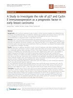
Fig. 6 Kaplan-Meier analysis of overall and disease-free survival time in HCC patients (log-rank test, p = 0.001 and p = 0.001) (a-b). Kaplan-Meier
analysis of overall survival and disease-free survival rates in subclassified HCC patients Stratified survival analysis of overall and disease-free survival
rates according to poor pathology grades II–III (c–d) and tumour recurrence (e–f)
It has been reported that some clinicopathology features, including tumour size, nodule number, macro/μ
invasion and pre-operative AFP, are considered as survival indices that affect the prognosis of HCC patients.
Recently, a correlation between obesity, lifestyle factors
(smoking, drinking status and exercise) and HCC prognosis was emphasized. However, there is not enough evidence to prove the relationships between clinical
features and HCC prognosis. Therefore, if we want to
gain a thorough understanding of how those factors
affect the prognosis and provide the basis for personalized therapy for our patients, molecular studies may be
the key to open the door. In our study, we found that
the correlation between a higher level of AFP
(AFP ≥ 20 ng), lower pathology grade (poor) and tumour
recurrence and a lower expression level of INTS6 may
be associated with poor prognosis in HCC patients.
Moreover, survival curves indicate that patients with low
Lui et al. BMC Cancer (2017) 17:644
Page 9 of 12
Table 3 Univariate analysis and multivariate analysis overall survival time
Variables
Univariate analysis
P value
Exp(B)
Multivariate analysis
95.0% CI
Lower
Upper
Gender
0.911
0.934
0.282
3.094
Age
0.240
0.980
0.948
1.013
Pathology grade
0.005*
0.331
0.152
0.720
HBsAg
0.626
1.300
0.452
3.736
AFP
0.091
2.023
0.894
4.577
Tumor size
0.035*
2.196
1.055
4.568
Cirrhosis
0.079
2.919
0.883
9.654
P
value
Exp(B)
95.0% CI
Lower
Upper
Recurrence
0.002*
23.148
3.136
170.849
0.041*
9.110
1.054
75.895
Vascular invasion
<0.001*
7.685
3.340
17.682
0.024*
2.847
1.151
7.046
INTS6
<0.001*
0.125
0.038
0.416
0.040*
0.221
0.047
1.304
*p<0.05
INTS6 expression have shorter overall and disease-free
survival rates. Our results strengthen the hypothesis that
low INTS6 expression is associated with a poor prognosis in HCC patients.
Cox regression analysis also showed that INTS6 and
vascular invasion are the independent predictors of overall survival and disease-free survival. The reasons why
AFP, tumour size, pathology grade are not independent
predictors for HCC may be due to the effects of multiple
collinearity and the small sample size. It suggested that
INTS6 may be used as a new prognostic marker to identify HCC patients at a high risk of poor prognosis. The
current data show that INTS6 could be used as a potential and independent predictor of prognosis in HCC.
Mechanistically, the INTS6/WIF-1 regulatory model
investigated in this study provides a new perspective
on WIF regulation. In fact, many studies have
reported that WIF-1 down-regulation is involved in
tumours, including in HCC [24–26], and could trigger
the Wnt/β-catenin signal pathway [27]. WIF-1 is one
of the endogenous antagonists that inhibits the Wnt
pathway by directly binding to Wnt proteins in the
extracellular space [28]. Down-regulation of the expression level of WIF-1 due to its hypermethylated
promoter has been reported in bladder cancer, melanoma, lung cancer, and HCC [29]. Moreover, in Hu’s
study [30], it was shown that Wnt antagonists WIF1Fc and SFRP1-Fc inhibit Wnt signalling and exert
antitumour activity by inducing apoptosis in tumour
cells, which indicates that Wnt antagonists would be
a promising molecular treatment for HCC. All the
above studies indicate that WIF-1 plays an important
role in the Wnt/β-catenin signalling pathway, especially in HCC. In our previous study [16], we learned
Table 4 Univariate analysis and multivariate analysis disease-free survival time
Variables
Univariate analysis
Multivariate analysis
P value
Exp(B)
95.0% CI
P value
Lower
Upper
Gender
0.193
1.778
0.748
4.227
Age
0.443
0.990
0.964
1.016
Pathology grade
0.009*
0.409
0.209
0.803
HBsAg
0.460
1.383
0.585
3.269
AFP
0.081
1.741
0.934
3.246
Exp(B)
95.0% CI
Lower
Upper
Tumor size
0.004*
2.398
1.317
4.367
Cirrhosis
0.077
2.076
0.925
4.660
Vascular invasion
<0.001*
6.724
3.542
12.764
<0.001*
5.013
2.415
10.408
INTS6
<0.001*
0.266
0.131
0.542
0.025*
0.425
0.201
0.896
*p<0.05
Lui et al. BMC Cancer (2017) 17:644
2.0
Overexpression
*
*
1.5
1.0
0.5
C
97
uh
C
H
M
Fig. 7 Relative expression levels of WIF-1 mRNA in the HCC cell lines
Huh7 and MHCC97L by qRT-PCR. *p<0.05
*
0.8
*
5
0
Fig. 9 Relative expression levels of WIF-1 and INTS6 mRNA in 15
HCC tumour tissues by qRT-PCR. The relative mRNA level was
normalized to GAPDH
In conclusion, this is the first report to demonstrate the clinical significance of INTS6 expression
and its mechanism in HCC. This research is limited
by its sample size and the inclusion of only patients
with resectable tumours. Larger prospective studies
examining INTS6 are necessary to further validate
the usefulness of this biomarker. INTS6 overexpression may prevent invasive progression and metastatic relapse, which would improve the prognosis
and quality of life for HCC patients after hepatic
resection.
Vector
siRNA
Overexpression
0.6
0.4
0.2
C
97
uh
7
L
0.0
M
H
C
H
Relative W IF-1 Expression Protein
that INTS6 inhibits HCC cell growth and migration
and promotes apoptosis. We also found that INTS6
functions as a competitive endogenous RNA (ceRNA)
to up-regulate its tumour suppressor pseudogene
INTS6P1 to inhibit HCC. In this study, it was hypothesized that INTS6 overexpression inhibited the
WNT signal pathway by increasing WIF-1 expression.
As expected, there was a significantly decrease in the
expression level of β-catenin in the HCC cell lines,
and the downstream target genes ZEB1 and MMP13
were decreased after INTS6 overexpression.
1.0
10
Samples (n=15)
L
7
0.0
INTS6
WIF-1
15
1
2
3
4
5
6
7
8
9
10
11
12
13
14
15
Control
H
Relative W IF-1 mRNA Expression
WIF-1 (Cell RNA)
Relative mRNA Expression Level
Page 10 of 12
Fig. 8 Expression of WIF-1 protein in the HCC cell lines Hun7 and
MHCC97L using Western blotting (EV = Vector, si = siRNA,
Over = Overexpression). *p<0.05
Fig. 10 Expression of β-catenin protein in the HCC cell lines Hun7
and MHCC97L using Western blotting (EV = Vector, si = siRNA,
Over = Overexpression). *p<0.05
Lui et al. BMC Cancer (2017) 17:644
Page 11 of 12
Vector
1.5
Consent for publication
Not applicable.
Overexpression-INTS6
Competing interests
The authors declare that they have no competing interests.
1.0
*
*
0.5
Springer Nature remains neutral with regard to jurisdictional claims in
published maps and institutional affiliations.
M
M
1
P1
3
0.0
ZE
B
Publisher’s Note
Fig. 11 Expression of downstream target genes MMP13 and ZEB1
measured by qRT-PCR. *p<0.05
Conclusions
The results of our study show that down-regulated
INTS6 expression was associated with a poor prognosis
in HCC patients. This newly identified INTS6/WIF-1
axis indicates the molecular mechanism of HCC and
may represent a therapeutic target in HCC patients.
Additional file
Additional file 1: Microarray data files of the differential mRNA
expression in HCC. (XLS 2230 kb)
Abbreviations
AJCC: American Joint Committee on Cancer; DICE1: Deleted in cancer cells 1;
FFPE: Formalin-fixed paraffin-embedded; HCC: Hepatocellular carcinoma;
IHC: Immunohistochemistry; INTS6: Integrator complex subunit 6;
MMP13: Matrix Metallopeptidase 13; qRT-PCR: Quantitative reverse
transcriptase polymerase chain reaction; ZEB1: Zinc finger E-box-binding
homeobox 1
Acknowledgments
We would like to thank Baiyun Tang, MD (Department of Critical Care
Medicine, the First Affiliated Hospital of Sun Yat-sen University) for helping
our research group to improve English language writing.
Funding
This study was supported by the National Natural Science Foundation of
China (Grant No. 81170450). The funder had no role in study design, data
collection and analysis, decision to publish, or preparation of the manuscript.
Availability of data and materials
The dataset supporting the conclusions of this article is available upon
request. Please contact Prof. Minqiang Lu ().
Authors’ contributions
KL, HZ performed the experiments. HP, CQ, CL, ZZ, RF analyzed the data. KL
wrote the manuscript. ML, HC conceived and designed the experiments. All
authors read and approved the final manuscript.
Ethics approval and consent to participate
All clinical samples (tissues, blood, serum) collected and analyzed in this
study were approved by the patients and all patients signed with informed
consent. The experiments were carried out under a protocol approved by
the Ethics Committee of the Third Affiliated Hospital of Sun Yat-sen
University.
Author details
1
Department of Hepatobiliary Surgery, Guangzhou First People’s Hospital,
Guangzhou 510180, China. 2Department of Critical Care Medicine, the First
Affiliated Hospital of Sun Yat-sen University, Guangzhou 510080, China.
3
Department of Hepatic Surgery, the Third Affiliated Hospital of Sun Yat-sen
University, Guangzhou 510630, China. 4Obstetric Laboratory, the Third
Affiliated Hospital of Sun Yat-sen University, Guangzhou 510630, China.
5
Transitional Year, Gwinnentt Medical Center, Lawrenceville, GA, USA.
6
Department of Pathology, the Third Affiliated Hospital of Sun Yat-sen
University, Guangzhou 510630, China.
Received: 14 February 2016 Accepted: 28 August 2017
References
1. EASL-EORTC clinical practice guidelines. management of hepatocellular
carcinoma. J Hepatol. 2012;56(4): 908–943.
2. Chen WQ, Zheng RS, Zhang SW. Liver cancer incidence and mortality in
China, 2009. Chin J Cancer. 2013;32(4):162–9.
3. Abdel-Rahman O. Systemic therapy for hepatocellular carcinoma (HCC):
from bench to bedside. J Egypt Natl Canc Inst. 2013;25:165–71.
4. Turati F, Edefonti V, Talamini R, Ferraroni M, Malvezzi M, Bravi F, et al. Family
history of liver cancer and hepatocellular carcinoma. Hepatology. 2012;55:
1416–25.
5. Arzumanyan A, Reis HM, Feitelson MA. Pathogenic mechanisms in HBV- and
HCV-associated hepatocellular carcinoma. Nat Rev Cancer. 2013;13(2):123–
35.
6. Nault JC, De Reynies A, Villanueva A, Calderaro J, Rebouissou S, Couchy G,
et al. A hepatocellular carcinoma 5-gene score associated with survival of
patients after liver resection. Gastroenterology. 2013;145:176–87.
7. ZG X, JJ D, Zhang X, Cheng ZH, Ma ZZ, Xiao HS, Yu L, Wang ZQ, Li YY, Huo
KK, Han ZGA. Novel liver-specific zona pellucida domain containing protein
that is expressed rarely in hepatocellular carcinoma. Hepatology. 2013;38:
735–44.
8. Burgess R, Jenkins R, Zhang Z. Epigenetic changes in gliomas. Cancer Biol
Ther. 2008;7(9):1326–34.
9. Röpke A, Buhtz P, Böhm M, Seger J, Wieland I, Allhoff EP, Wieacker PF.
Promoter CpG hypermethylation and down-regulation of DICE1 expression
in prostate cancer. Oncogene. 2005;24:6667–75.
10. Ying Y, Tao Q. Epigenetic disruption of the WNT ⁄ beta-catenin signaling
pathway in human cancers. Epigenetics. 2009;4:307–12.
11. Satow R, Shitashige M, Kanai Y, et al. Combined functional genome survey
of therapeutic targets for hepatocellular carcinoma. Clin Cancer Res. 2010;
16:2518–28.
12. Wieland I, Arden KC, Michels D, Klein-Hitpass L, Böhm M, Viars CS, Weidle
UH. Isolation of DICE1: a gene frequently affected by LOH and downregulated in lung carcinomas. Oncogene. 1999;18:4530–7.
13. Wieland I, Röpke A, Stumm M, Sell C, Weidle UH, Wieacker PF. Molecular
characterization of the DICE1 (DDX26) tumor suppressor gene in lung
carcinoma cells. Oncol Res. 2001;12:491–500.
14. Li WJ, Hu N, Su H, Wang C, Goldstein AM, Wang Y, Emmert-Buck MR, Roth
MJ, Guo WJ, Taylor PR. Allelic loss on chromosome 13q14 and mutation in
deleted in cancer 1 gene in esophageal squamous cell carcinoma.
Oncogene. 2003;22:314–8.
15. Hernández M, Papadopoulos N, Almeida TA. Absence of mutations in
DICE1/DDX26 gene in human cancer cell lines with frequent 13q14
deletions. Cancer Genet Cytogenet. 2005;163:91–2.
16. Peng H, Ishida M, Li L, Saito A, et al. Pseudogene INTS6P1 regulates its
cognate gene INTS6 through competitive binding of miR-17-5p in
hepatocellular carcinoma. Oncotarget. 2015;6(8):5666–77.
Lui et al. BMC Cancer (2017) 17:644
Page 12 of 12
17. Li PD, Zhang WJ, Zhang MY, Yuan LJ, Cha YL, Ying XF, et al. Overexpression
of RPS6Kb1 predicts worse prognosis in primary HCC patients. Med Oncol.
2012;29:3070–6.
18. Filleur S, Hirsch J, Wille A, Schon M, Sell C, Shearer MH, Nelius T, Wieland I.
INTS6/DICE1 inhibits growth of human androgen-independent prostate
cancer cells by altering the cell cycle profile and Wnt signaling. Cancer Cell
Int 2009; 9:28.
19. Thorgeirsson SS, Lee JS, Grisham JW. Functional genomics of hepatocellular
carcinoma. Hepatology. 2006;43:S145–50.
20. Szabo G, Bala S. MicroRNAs in liver disease. Nat Rev Gastroenterol Hepatol.
2013;10:542–52.
21. Wieland I, Sell C, Weidle UH, Wieacker P. Ectopic expression of DICE1
suppresses tumor cell growth. Oncol Rep. 2004;12(2):207–11.
22. Ma WJ, Wang HY, Teng LS. Correlation analysis of preoperative serum alphafetoprotein (AFP) level and prognosis of hepatocellular carcinoma (HCC)
after hepatectomy. World J Surg Oncol. 2013;11:212.
23. Sasaki K, Matsuda M, Ohkura Y, et al. Factors associated with early cancerrelated death after curative hepatectomy for solitary smdlhepato-cellular
carcinoma without macroscopic vascular invasion. J Hepatobiliary PancreatSci. 2014;21(2):142–7.
24. Yun Deng, Bin Yu, Qin Cheng, et al. Epigenetic silencing of WIF-1 in
hepatocellular carcinomas. J Cancer Res Clin Oncol, 2010, 136:1161–1167.
25. Cai J, Guan H, Fang L, et al. MicroRNA-374a activates Wnt ⁄ beta-catenin
signaling to promote breast cancer metastasis. J Clin Invest. 2013;123:566–
79.
26. Alvarez C, Tapia T, Cornejo V, et al. Silencing of tumor suppressor genes
RASSF1A, SLIT2, and WIF-1 by promoter hypermethylation in hereditary
breast cancer. Mol Carcinog. 2012;52:475–87.
27. Rubin EM, Guo Y, Tu K, Xie J, Zi X, Hoang BH. Wnt inhibitory factor 1
decreases tumorigenesis and metastasis in osteosarcoma. Mol Cancer Ther.
2010;9:731–41.
28. Hsieh JC, Kodjabachian L, Rebbert ML, et al. A new secreted protein that
binds to Wnt proteins and inhibits their activities. Nature. 1999;398:431–6.
29. Deng Y, Yu B, Cheng Q, et al. J Cancer Res Clin Oncol. 2010;136:1161–7.
30. Jie Hu, Aiwen Dong, Veronica Fernandez-Ruiz, Juanjuan Shan, Milosz Kawa,
Eduardo Martínez-Ansó, Jesus Prieto, Cheng Qian, Blockade of Wnt
Signaling Inhibits Angiogenesis and Tumor Growth in Hepatocellular
Carcinoma. Cancer Res, Sep, 2009: (69) (17):6951–6959;
Submit your next manuscript to BioMed Central
and we will help you at every step:
• We accept pre-submission inquiries
• Our selector tool helps you to find the most relevant journal
• We provide round the clock customer support
• Convenient online submission
• Thorough peer review
• Inclusion in PubMed and all major indexing services
• Maximum visibility for your research
Submit your manuscript at
www.biomedcentral.com/submit