Assessment of Ki67 and uPA/PAI-1 expression in intermediate-risk early stage breast cancers
Bạn đang xem bản rút gọn của tài liệu. Xem và tải ngay bản đầy đủ của tài liệu tại đây (625.44 KB, 10 trang )
Deluche et al. BMC Cancer (2017) 17:662
DOI 10.1186/s12885-017-3648-z
RESEARCH ARTICLE
Open Access
Assessment of Ki67 and uPA/PAI-1
expression in intermediate-risk early
stage breast cancers
Elise Deluche1*, Laurence Venat-Bouvet1, Sophie Leobon1, Veronique Fermeaux2, Joelle Mollard3, Nadira Saidi4,
Isabelle Jammet5, Yves Aubard3 and Nicole Tubiana-Mathieu1
Abstract
Background: The objective of this study was to compare the efficacy of biomarkers in assessing the risk of breast
cancer recurrence in patients with node-negative or micrometastatic grade II breast cancer. Specifically, we compared
risk assessments based on the St. Gallen clinicopathological criteria, Ki67 expression and urokinase plasminogen
activator (uPA)/plasminogen activator inhibitor-1 (PAI-1) expression.
Methods: This retrospective study included 347 patients with breast cancer followed at Limoges University
Hospital. The optimal cut-off for high Ki67 expression (Ki67hi) was established as 20%. The threshold for uPA
and PAI-1 positivity was 3 ng/mg and 14 ng/mg, respectively.
Results: Ki67 expression was lower in uPA/PAI-1-negative than in uPA/PAI-1-positive tumours (227 tumours;
P = 0.04). The addition of Ki67 status to the St. Gallen criteria resulted in a 28% increase in the rate of identification of
high-risk tumours with a potential indication for chemotherapy (P < 0.001). When considering uPA/PAI-1 levels
together with the St Gallen criteria (including Ki67 expression), the number of cases identified as having a
high recurrence risk with a potential indication for adjuvant chemotherapy increased by 20% (P < 0.001). Adjuvant
chemotherapy was 9% less likely to be recommended by a multidisciplinary board when using the current criteria
compared with using a combination of the St. Gallen criteria and Ki67 and uPA/PAI-1 status (P = 0.03).
Conclusions: Taken together, our data show discordance among markers in identifying the risk of recurrence, even
though each marker may prove to be independently valid.
Keywords: uPA/PAI-1, Ki67, Subtypes, Grade II, Breast cancer
Background
The indication for adjuvant therapy for breast cancer
has led to a search for efficient prognostic and predictive
biomarkers for patients at greatest risk of local and/or
distant recurrence and with a potential indication for
chemotherapy (i.e. patients requiring adjuvant therapy).
The main objective is to distinguish patients with a low
risk of recurrence, for whom little evidence supports the
need for chemotherapy, from those with high-risk disease, for whom chemotherapy is clearly justified. The
2007 [1] and 2013 [2] St. Gallen criteria used to define
* Correspondence:
1
Department of Medical Oncology, University Hospital, 2 avenue Martin
Luther King, F-87042 Limoges, France
Full list of author information is available at the end of the article
high-risk breast cancer are patient age < 35 years,
tumour size >2 cm, tumour grade III, presence of extensive peritumoural vascular invasion, oestrogen receptor
(ER) and/or progesterone receptor (PR) negativity,
human epidermal growth factor receptor 2 (HER2) overexpression or HER2 amplification, high Ki67 expression
(in grade II tumours) and >3 positive lymph nodes. The
presence of any one of these factors is considered sufficient for defining a high risk of recurrence with an indication for adjuvant chemotherapy.
Although grade I and III tumours are biologically and
clinically distinct, it is difficult to predict the outcomes
of node-negative or micrometastatic (N0) grade II
tumours because of their intermediate risk of recurrence
[3]. Furthermore, the ultimate benefit of adjuvant
© The Author(s). 2017 Open Access This article is distributed under the terms of the Creative Commons Attribution 4.0
International License ( which permits unrestricted use, distribution, and
reproduction in any medium, provided you give appropriate credit to the original author(s) and the source, provide a link to
the Creative Commons license, and indicate if changes were made. The Creative Commons Public Domain Dedication waiver
( applies to the data made available in this article, unless otherwise stated.
Deluche et al. BMC Cancer (2017) 17:662
chemotherapy for these patients is uncertain. Promising
biomarkers used to stratify patients into different risk
groups include Ki67 and urokinase plasminogen activator
(uPA)/plasminogen activator inhibitor-1 (PAI-1) [4–7].
Reproducible data at a I-B level of evidence (LoE) suggest
that Ki67 is a prognostic marker in early stage breast cancer [8], as well as a positive predictive factor for adjuvant
chemotherapy [9], especially in patients with luminal B
tumours [10]. In addition to the traditional parameters,
guidelines recommend using proliferation markers, such
as Ki67, to define patient subgroups [2].
The prognostic and predictive abilities of the tumourassociated proteolytic factor uPA, and its inhibitor PAI1, in patients with N0 disease have been demonstrated
at the highest LoE (LoE I-A) [11]. In the Chemo N0 trial,
uPA/PAI-1 was identified as a clinically significant risk
discriminator in the clinically relevant grade II breast
cancer subgroup [12]. Furthermore, in N0 breast cancer,
especially grade II tumours, uPA and PAI-1 are predictive markers for the response to cyclophosphamide,
methotrexate and 5-fluorouracil (CMF) chemotherapy
(LoE I-A) [13]. Based on the high LoE, using uPA/PAI-1
status as an indicator for adjuvant chemotherapy for ER/
PR-positive, HER2-negative (node-negative) breast cancer has been recommended by international guidelines
[11]. uPA/PAI-1 expression distinguishes high-risk patients expected to receive a major benefit from chemotherapy from low-risk patients with a low probability of
benefitting from chemotherapy.
The objective of this study was to assess the predictive
value of the St. Gallen clinicopathological criteria, Ki67
status and uPA/PAI-1 status in patients with N0 grade II
breast cancer.
Methods
This retrospective study was performed from December
2007 to October 2015 at Limoges University Hospital,
France.
Patients diagnosed with breast cancer, and with complete
data available regarding their surgically resected tumours,
including tumour Ki67, ER, PR, HER2 and uPA/PAI-1 status, were eligible for this study. Exclusion criteria were
missing data for any of the abovementioned tumour
markers, macroscopic lymph node involvement, previous
breast cancer, initial metastatic breast cancer or prior neoadjuvant chemotherapy. Patients with microscopic lymph
node involvement or isolated cells were included, because
these factors do not influence the decision to perform adjuvant chemotherapy [14]. Clinical data were collected in accordance with French bioethics laws regarding patient
information and consent. Patient consent to the use of their
data and biological material was sought prior to the commencement of medical care.
Page 2 of 10
Clinicopathological subtypes, as well as the risk of
tumour recurrence, were defined according to the St.
Gallen criteria [1, 2]. All Ki67 staining was performed by
the same pathological laboratory using the MIB1 monoclonal antibody (1:80 dilution; Dako, Glostrup, Denmark);
the largest tumour area, including the most proliferative
zone, was assessed. The Ki67 score was calculated as the
percentage of immunostained cells. The optimal cut-off
for a high versus low Ki67 score was defined as 20% (i.e. ≥
20% staining was defined as Ki67hi), according to the 2015
recommendations [15].
Quantitative evaluation of uPA and PAI-1 concentrations was performed at the Biological Oncology Laboratory (Marseille, France) using the commercially available
FEMTELLE® enzyme-linked immunosorbent assay. Positive uPA expression was defined as >3 ng/mg protein,
and positive PAI-1 expression as >14 ng/mg protein.
uPA/PAI-1 positivity, defined as an elevation of at least
one of these markers [16], has been used to identify
high-risk tumours [11]. The uPA/PAI-1 markers can be
used independently of the St. Gallen criteria [17].
Our multidisciplinary breast cancer team set up the
treatment program for the patients. Regional recommendations were based on the St. Gallen criteria and uPA/
PAI-1 status. According to a high LoE (I-A), uPA
and/or PAI-1 are the preferred markers used to indicate adjuvant chemotherapy for N0 grade II, ER/PRpositive tumours.
Statistical analyses
Nominal variables were compared among groups using
the chi-square test or Fisher’s exact test, as appropriate.
Means were compared using the nonparametric MannWhitney U-test for continuous variables, and the
Kruskal-Wallis test was used for comparisons of ordinal
variables among more than two groups. A P value <0.05
was considered to indicate statistical significance.
Statistical analyses were performed using STATVIEW®
software (SAS Institute Inc., Cary, NC, USA).
Results
Clinical and histological characteristics
We screened 2300 patients with breast cancer treated
from December 2007 to October 2015. Application of
our study inclusion criteria resulted in a final cohort of
347 patients, selected primarily because they had available uPA/PAI-1 data. All tumours were evaluated
(Tables 1–3), and the results from grade II tumours were
used specifically, because the use of uPA/PAI-1 expression as a recurrence marker has been validated in these
tumours only.
Table 1 summarises the patient and tumour characteristics according to Ki67 and uPA/PAI-1 status. Ki67
expression was considered low in 250 (72%) tumours
25
(25)
P-value
135
(39)
212
(61)
Negative
Positive
61
(33–86)
60
(38–87)
0.07
45
(21)
40
(30)
61
(36–85)
60
(24)
85
(25)
0.7
97
(28)
High
61.5
(33–87)
62
(33–87)
Premenopausal
Menopausal status
uPA/PAI-1 status
250
(72)
Low
Ki67 status
Age, Years
Median (range)
P-value
347
Total
n (%)
167
(89)
95
(70)
72
(75)
190
(76)
262
(75)
Postmenopausal
0.08
152
(71)
79
(59)
0.02
53
(55)
178
(71)
231
(66)
T1
55
(26)
53
(39)
43
(44)
65
(26)
108
(31)
T2
4
(2)
2
(1)
1
(1)
5
(2)
6
(2)
T3
Tumour classification
Table 1 Patient and tumour characteristics according to Ki67 expression or uPA/PAI-1 status (n = 347)
1
(1)
1
(1)
0
(0)
2
(1)
2
(1)
T4
0.09
161
(76)
99
(74)
<0.0001
64
(66)
196
(78)
260
(75)
ER+/PR +
32
(15)
30
(22)
13
(13)
49
(20)
62
(18)
ER+/PR-
Hormonal status
ER−/PR+
0
(0)
0
(0)
0
(0)
0
(0)
0
(0)
19
(9)
6
(4)
20
(21)
5
(2)
25
(7)
ER−/PR-
Deluche et al. BMC Cancer (2017) 17:662
Page 3 of 10
202
(95)
0.5
10
(5)
131
(97)
P-value
4
(3)
0.1
P-value
6
(6)
91
(94)
uPA/PAI-1 status
8
(3)
242
(97)
Ki67 status
14
(4)
333
(96)
Total
<0.001
113
(53)
77
(57)
<0.0001
1
(1)
189
(76)
190
(55)
71
(33)
49
(36)
72
(74)
48
(19)
120
(34)
Luminal B HER2negative
Luminal A
Negative
Positive
Clinicopathological subtypes
HER2 status
9
(4)
3
(2)
4
(4)
8
(3)
12
(3)
Luminal B HER2positive
1
(1)
1
(1)
2
(2)
0
(0)
2
(1)
HER2positive
0.1
18
(9)
5
(4)
<0.0001
18
(19)
5
(2)
23
(7)
Triplenegative
159
(75)
86
(64)
85
(88)
160
(64)
245
(71)
Invasive ductal
carcinoma
Histological type
Table 1 Patient and tumour characteristics according to Ki67 expression or uPA/PAI-1 status (n = 347) (Continued)
4
(19)
8
(6)
2
(2)
10
(4)
12
(3)
Invasive ductal +
lobular carcinoma
36
(17)
32
(24)
9
(9)
59
(24)
68
(20)
Invasive lobular
carcinoma
13
(7)
9
(7)
1
(1)
21
(9)
22
(6)
Other
57
(59)
170
(68)
227
(66)
II
0.1
38
(18)
36
(27)
142
(67)
85
(63)
<0.0001
3
(3)
71
(28)
74
(21)
I
32
(15)
14
(10)
37
(38)
9
(4)
46
(13)
III
Histological grade
Deluche et al. BMC Cancer (2017) 17:662
Page 4 of 10
Deluche et al. BMC Cancer (2017) 17:662
Page 5 of 10
(median Ki67 level = 10%; range: 1–15%) and high in 97
(28%) tumours (median Ki67 level = 30%; range: 20–
80%). uPA/PAI-1 expression was negative in 135 (39%)
tumours and positive in 212 (61%) tumours, and was associated with the clinicopathological subtype (P < 0.001).
The median uPA expression level was comparable between single (uPA) and double-positive tumours (uPA
plus PAI-1; Table 2).
Low Ki67 expression was associated with pT1, luminal
A, ER- and PR-positive, invasive lobular carcinoma and
grade II tumours, while high Ki67 expression was associated with pT2, luminal B, HER2-, ER- and PR-negative
(triple-negative), invasive ductal carcinoma and grade III
tumours (P < 0.05). Negative uPA/PAI-1 expression was
associated with luminal A tumours, while positive uPA/
PAI-1 expression was associated with luminal B HER2negative tumours (P < 0.001).
Table 3 shows the uPA/PAI-1 and Ki67 levels stratified
according to tumour grade. No association was observed
between Ki67 and uPA/PAI-1 expression in tumours,
irrespective of the histological grade. There were no significant associations between Ki67 and uPA/PAI-1 expression in either grade I (P = 0.5) or grade III (P = 0.1)
tumours. However, in grade II tumours, there was an association between low Ki67 and negative uPA/PAI-1 expression, and between high Ki67 and positive uPA/PAI-1
expression (P = 0.04).
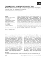
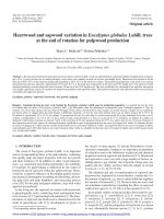
Ki67 levels in patients with N0 grade II tumours
The St. Gallen low-risk criteria, excluding the Ki67 status, were satisfied in 134 cases, who were thus deemed
to have no indication for potential adjuvant chemotherapy. Of these 134 cases, 108 had a Ki67low status and 26
a Ki67hi status (Fig. 1).
The St. Gallen high-risk criteria, excluding the Ki67
status, were satisfied in 93 cases, of whom 31 were
Ki67hi and 62 Ki67low (Fig. 1). In the Ki67low tumours,
the corresponding St. Gallen high-risk criteria were pT2
stage (n = 55), and/or the presence of vascular emboli
(n = 5), and/or HER2-positivity (n = 5) or a triplenegative status (n = 4).
When the Ki67 status was added to the St. Gallen
criteria, 119 cases were considered to have a high risk
of recurrence. This effectively increased the percentage of cases with a potential indication for adjuvant
chemotherapy by 28% compared with that using clinicopathological parameters alone (P < 0.001).
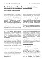
The uPA/PAI-1 status in patients with N0 grade II tumours
Of the 134 cases who satisfied the St. Gallen low-risk
criteria (excluding the Ki67 status), uPA/PAI-1 expression was negative in 47 cases and positive in 87; thus, a
need for adjuvant chemotherapy was indicated in the latter group (Fig. 2). Of the 93 cases who satisfied the St.
Gallen high-risk criteria (excluding the Ki67 status), 56
were positive and 37 were negative for uPA/PAI-1 expression (Fig. 2).
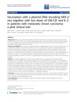
Contribution of Ki67 and uPA/PAI-1 status to risk
assessment in patients with N0 grade II tumours
The St. Gallen low-risk criteria, inclusive of the Ki67 status, were satisfied in 108 cases. Among these cases, Ki67
and uPA/PAI-1 status was concordant in 41 cases (i.e.
Ki67low and uPA/PAI-1 negative) and discordant in 67
cases (i.e. Ki67low and uPA/PAI-1 positive) (Fig. 3).
The St. Gallen high-risk criteria, inclusive of the Ki67
status, were satisfied in 119 cases, with concordant Ki67
and uPA/PAI-1 expression observed in 70 cases. Of
these concordant cases, 42 were Ki67low (uPA/PAI-1
negative) and 28 were Ki67hi (uPA/PAI-1 positive). Of
the 49 cases with discordant Ki67 and uPA/PAI-1 expression, 34 were Ki67low (uPA/PAI-1 positive) and 15
were Ki67hi (uPA/PAI-1 negative).
Inclusion of both the uPA/PAI-1 and Ki67 criteria increased the rate of identification of tumours with a high
risk of recurrence (and thus with a potential indication
for adjuvant chemotherapy) by 20% (P < 0.001) compared with using the St. Gallen criteria alone.
The uPA/PAI-1 status, St. Gallen criteria and multidisciplinary
board decision to perform adjuvant chemotherapy in
patients with N0 grade II breast cancer
Of the 84 cases considered to be at a low risk of recurrence (as defined by the uPA/PAI-1 status), 43 satisfied
the St. Gallen high-risk criteria, with 11 (25%) found to
be Ki67hi. The St. Gallen high-risk criterion in these
cases was a tumour size >2 cm. The multidisciplinary
board proposed chemotherapy for only five cases (1%),
in whom a grade III tumour was identified in the biopsy
materials but not in the surgical specimens.
Table 2 Median uPA/PAI-1 levels in tumour populations
uPA−/PAI-1+
uPA+/PAI-1+
uPA +/PAI-1-
uPA−/PAI-1-
Number of patients
85
92
35
135
Median level of uPA (ng/mg; min-max)
1.7 (0.19–2.9)
4.7 (3.0–12.9)
4.7 (3–7.3)
1.2 (0.0–2.9)
Median level of PAI-1 (ng/mg; min-max)
21.1 (14.0–87.8)
22.6 (14.0–130)
9.56 (5.5–13.8)
6.7 (2.9–10.6)
uPA+: uPA ≥3 ng/mg; uPA-: < 3 ng/mg; PAI-1+: ≥14 ng/mg; PAI-1-: <14 ng/mg
Deluche et al. BMC Cancer (2017) 17:662
Page 6 of 10
Table 3 uPA/PAI-1 and Ki67 levels stratified according to tumour grade
GRADE I
Positive
Negative
Positive vs. negative
uPA/PAI-1
Ki67
uPA+/PAI-1+
uPA +/PAI-1-
uPA−/PAI-1+
uPA−/PAI-1-
Total
P value
< 20%
11 (16)
4 (6)
22 (31)
34 (47)
71
0.5
≥ 20%
1 (33)
0 (0)
0 (0)
2 (67)
3
GRADE II
Positive
Negative
Positive vs. negative
uPA/PAI-1
Ki67
uPA+/PAI-1+
uPA+/PAI-1-
uPA−/PAI-1+
uPA−/PAI-1-
Total
P value
< 20%
42 (25)
15 (9)
43 (25)
70 (41)
170
0.04
≥ 20%
24 (42)
6 (10)
12 (21)
15 (27)
57
GRADE III
Positive
Ki67
uPA+/PAI-1+
Negative
uPA+/PAI-1-
uPA−/PAI-1+
uPA−/PAI-1-
Positive vs. negative
uPA/PAI-1
Total
P value
0.1
< 20%
3 (33)
4 (45)
1 (11)
1 (11)
9
≥ 20%
11 (30)
6 (16)
7 (19)
13 (35)
37
uPA+: uPA ≥3 ng/ml; uPA- < 3 ng/ml; PAI-1+: ≥ 14 ng/ml; PAI-1-: <14 ng/ml. Data are reported as n (%) unless otherwise stated
Of the 143 cases considered to be at a high risk
of recurrence (as defined by the uPA/PAI-1 status),
67 satisfied the St Gallen low-risk criteria. Our
multidisciplinary board decided not to recommend
chemotherapy in 18 of these cases because of problems in interpreting the uPA/PAI-1 data (n = 2),
comorbidities or old age (n = 10), or St. Gallen
low-risk criteria discordant with UPA/PAI-1 positivity (n = 6).
The median follow-up for the entire population was
33 months (range: 1–82 months), which was too short
to assess patient outcomes.
Discussion
To our knowledge, this is the first study to compare recurrence risk as defined by the St. Gallen clinicopathological criteria, Ki67 status and uPA/PAI-1 status in
patients with N0 grade II breast cancer.
Fig. 1 Risk of recurrence and indication for chemotherapy according to the St. Gallen criteria and Ki67 status
Deluche et al. BMC Cancer (2017) 17:662
Page 7 of 10
Fig. 2 Risk of recurrence and indication for chemotherapy according to the St. Gallen criteria and urokinase plasminogen activator (uPA)/plasminogen
activator inhibitor-1 (PAI-1) status
This report emphasises the utility of the uPA/PAI-1
status and St. Gallen criteria, including the Ki67 status,
for identifying tumours at a high risk of recurrence and
thus with a potential indication for chemotherapy. Currently, the use of Ki67 and uPA/PAI-1 as biomarkers remains controversial in adjuvant chemotherapy decisionmaking. The use of Ki67 is supported by the St. Gallen
recommendations [2, 15], and the use of uPA/PAI-1 has
been validated by both The American Society of Clinical
Oncology (ASCO) [11] and The National Institute of
Cancer [18].
Ki67 expression can be used to stratify patients with N0
grade II tumours into two distinct subgroups according to
outcome [4, 19]. For example, Aleskandarany et al.
Fig. 3 Risk of recurrence and indication for chemotherapy according to the St. Gallen criteria, Ki67 status and uPA/PAI-1 status
Deluche et al. BMC Cancer (2017) 17:662
reported that ≥10% Ki67 positivity was a prognostic factor
for progression-free survival (P < 0.001) and overall survival (P < 0.001) [4]. Adjuvant chemotherapy for luminal
B, HER2-negative, N+ tumours was also found to be beneficial in Ki67hi tumours [20].
In the present study, Ki67 analyses were performed in
accordance with the St. Gallen criteria [15], using a
threshold of 20% to define those at risk of relapse; however, this cut-off is not optimal given Ki67’s continuous
distribution. In this context, the percentage of Ki67hi tumours (28%) was almost as high as that reported previously (32%) in a large study conducted by Penault-Lorca
et al. [21]. By using Ki67 in combination with the other
St. Gallen criteria it was possible to distinguish tumours
according to their risk of recurrence, which improved
the rate of identification of high-risk tumours by 19%.
The impact of uPA-PAI-1 staining was previously
demonstrated in a study by the European Organization
for Research and Treatment of Cancer [17], and in a
prospective clinical therapy trial, Chemo N0 [13].
Recently, a long-term 10-year follow-up of Chemo N0
participants confirmed the prognostic value of uPA/PAI1 in N0 breast cancer, especially in grade II tumours
(hazard ratio, 1.94; 95% confidence interval: 1.16–3.24;
P = 0.01) [12]. For this reason, ASCO recently recommended using uPA/PAI-1 expression to guide decisionmaking for adjuvant systemic therapy in patients
diagnosed with ER/PR-positive, HER2-negative, N0 breast
cancer [11]. However, the chemotherapy regimen used in
the chemo N0 trial was CMF, which is not routinely
used in our practice and is less effective than current
treatments with anthracyclines [22]. A prospective
study comparing cyclophosphamide, 5-fluorouracil and an
anthracycline (FEC) with FEC plus docetaxel has yet to be
published [23].
We considered that 63% of the tumours evaluated in
this study had a high risk of recurrence based on their
uPA/PAI-1 level, which potentially increases the indications for adjuvant chemotherapy. These data agree with
those of Saadoun et al., in that incorporation of uPA/
PAI-1 with other makers increased the indications for
adjuvant chemotherapy [24].
Even though measurement of uPA/PAI-1 levels provides good external quality control [13], some technical
and organisational difficulties exist. An assessment of
uPA/PAI-1 expression was not performed in 45% of the
2300 patients during our routine examinations because
of delayed delivery (> 1 h), the initial size of the tumour
(< 1 cm) and/or the need for frozen tissue. Nevertheless,
uPA/PAI-1 remains a feasible, low-cost test.
In the second part of this study, we evaluated the definition of recurrence risk according to uPA/PAI-1 expression combined with the St. Gallen criteria, inclusive
of Ki67. Previous studies have shown that the use of
Page 8 of 10
uPA/PAI-1 combined with a clinicopathological parameter, such as vascular invasion, is extremely helpful for
yielding prognostic data [24]. We identified a correlation
between Ki67 and uPA/PAI-1 status in grade II tumours,
in agreement with Kolben et al. [25], although we lack
an explanation for this correlation.
The efficacy of uPA-PAI-1 testing compared with molecular signatures for recurrence risk assessment is not
clear. The West German Study Group-Plan B trial is the
first study to evaluate the correlation between the Oncotype Dx® recurrence score and uPA/PAI-1 status, which
were prospectively compared as risk indicators in a
phase III trial setting in patients with early stage breast
cancer. A high-risk status, as determined by the recurrence score, was also found to be predictive of a high
risk of recurrence using uPA/PAI-1 measurements, while
the reverse was not found to be true [26].
This study also evaluated adjuvant chemotherapy
decision-making by a multidisciplinary board at our institution. This board currently defines a low risk of recurrence based on the St. Gallen low-risk criteria
(including Ki67 status) in addition to a negative uPA/
PAI-1 status. If either uPA or PAI-1 expression is elevated, the tumour is considered to be at a high risk of
recurrence. Among the study population, 37% of tumours were considered to be at a low risk of recurrence,
with no indication for adjuvant chemotherapy. In five
cases (6%), the board disagreed with the indication for
chemotherapy because of discordance among the uPA/
PAI-1 levels, clinicopathological criteria and Ki67 expression. This rate of indication for adjuvant chemotherapy (i.e. 6%) was similar to that in a previous report [27],
but lower than the rate of 13% reported by Kolben et al.
[25]. However, the cut-off value for Ki67 in those studies
was different from that in the current study (10–15% in
Kolben et al. and Vénat-Bouvet et al. vs. 20% in our
study). In addition, 18 of the 143 tumours (14%) considered at high risk of recurrence did not receive chemotherapy because of comorbidities and/or considerable
discordance among prognostic markers.
While the retrospective design of this study was not
optimal for drawing conclusions regarding the efficacy
of uPA/PAI-1 and Ki67 as markers informing decisionmaking (regarding adjuvant chemotherapy) by a multidisciplinary board, the data suggest a trend. The other
limitation was the variability in the Ki67 staining cut-off
value and its impact on the decision to perform adjuvant
chemotherapy. Specifically, from 2009 [28] to 2015, the
cut-off value has increased from 13.25% to 20–29% [15].
Our results need to be confirmed by a prospective, randomised study to validate uPA/PAI-1 as a risk marker in
patients with an uncertain indication for chemotherapy
according to the current standard assessments; we are
currently awaiting the results of a similar study (NNBC3)
Deluche et al. BMC Cancer (2017) 17:662
[29]. This work highlights the difficulties in evaluating the
precise role and salience of individual risk factors in the
decision to perform chemotherapy [27]. Treatment decisions were based primarily on the current guidelines but
also took other factors into account, such as patient age,
comorbidities, HER2 status and patient preference [30].
Conclusion
Improvements in breast cancer outcomes are attributed
mostly to adjuvant chemotherapy. However, the objective of patient care is not only to prevent recurrence, but
also to improve the patient’s quality of life; this requires
accurate identification of high-risk patients with a clear
justification for chemotherapy. Therefore, the main challenge is determining the individual risk of relapse, particularly in patients with grade II breast cancer. The
combination of the St Gallen criteria (including the Ki67
status) and uPA/PAI-1 status could provide a better estimation of the relapse risk, thereby avoiding unnecessary
adjuvant chemotherapy and improving the quality of life
of these patients.
Taken together, our data show potential discordance
among the markers used to stratify the risk of recurrence, even when each marker is validated independently. A prospective study is needed to validate the use
of a combination of these markers for risk assessment.
In the future, genomic analyses may be combined
with prognostic markers to better guide decisionmaking regarding adjuvant systemic therapy in defined patient subsets.
Abbreviations
ASCO: American Society of Clinical Oncology; CMF: Cyclophosphamide, methotrexate
and 5-fluorouracil; ER: Oestrogen receptor; HER2: Human epidermal growth factor
receptor 2; LoE: Level of evidence; N0: Node-negative or micrometastatic; PAI1: Plasminogen activator inhibitor-1; PR: Progesterone receptor; uPA: Urokinase
plasminogen activator
Acknowledgements
We thank Sylvie Gautier for her assistance with editing the language of this
manuscript. The English in this document has been checked by at least two
professional editors, both native speakers of English.
Funding
This research was not supported by grant funding from the public, commercial,
or not-for-profit sectors.
Availability of data and materials
The data that support the findings of this study are the property of Limoges
University Hospital and cannot be shared in a public repository, as the participants
have not consented to the public sharing of their data.
Authors’ contributions
ED was involved in proposing the research question, performing the statistical
analyses, interpreting the results and drafting the manuscript. SL assisted with
the statistical analyses and drafting and revising of the manuscript. LVB, VF, JM,
NS, IJ and YA helped collect, assemble and interpret the data and revise the
manuscript. NTM was involved in developing the research idea and study
design, collection, assembly and interpretation of the data, and drafting and
revising of the manuscript. All authors read and approved the final manuscript.
Page 9 of 10
Ethics approval and consent to participate
Clinical data were collected in accordance with French bioethics laws regarding
patient information and consent. The use of retrospective and prospective data
from the BRTS (Regional Solid Tumour dataBase) was validated on 04/28/2016
by the Limoges University Hospital ethics committee (president, Dr. Terrier)
under number 200–2016-14. BRTS is a regional database of all clinicopathological
information collected from patients followed for breast cancer. Patients also
provided written (signed) informed consent for the use of their data (obtained
from biological materials) and for the collection of biological materials.
Consent for publication
Not applicable.
Competing interests
The authors declare that they have no competing interests.
Publisher’s Note
Springer Nature remains neutral with regard to jurisdictional claims in
published maps and institutional affiliations.
Author details
1
Department of Medical Oncology, University Hospital, 2 avenue Martin
Luther King, F-87042 Limoges, France. 2Department of Pathology, University
Hospital, F-87042 Limoges, France. 3Department of Gynaecology, Mother and
Child Hospital, F-87042 Limoges, France. 4Department of Radiotherapy,
University Hospital, F-87042 Limoges, France. 5Department of Senology,
Mother and Child Hospital, F-87042 Limoges, France.
Received: 6 March 2017 Accepted: 13 September 2017
References
1. Goldhirsch A, Wood WC, Gelber RD, Coates AS, Thürlimann B, Senn H-J, et al.
Progress and promise: highlights of the international expert consensus on the
primary therapy of early breast cancer 2007. Ann Oncol. 2007;18:1133–44.
2. Goldhirsch A, Winer EP, Coates AS, Gelber RD, Piccart-Gebhart M,
Thurlimann B, et al. Personalizing the treatment of women with early breast
cancer: highlights of the St Gallen international expert consensus on the
primary therapy of early breast cancer 2013. Ann Oncol. 2013;24:2206–23.
3. Ignatiadis M, Sotiriou C. Understanding the molecular basis of histologic
grade. Pathobiology. 2008;75:104–11.
4. Aleskandarany MA, Rakha EA, Macmillan RD, Powe DG, Ellis IO, Green AR.
MIB1/Ki-67 labelling index can classify grade 2 breast cancer into two
clinically distinct subgroups. Breast Cancer Res Treat. 2011;127:591–9.
5. Buta M, Džodić R, \DJurišić I, Marković I, Vujasinović T, Markićević M, et
al. potential clinical relevance of uPA and PAI-1 levels in node-negative,
postmenopausal breast cancer patients bearing histological grade II
tumors with ER/PR expression, during an early follow-up. Tumor Biol.
2015;36:8193–200.
6. Petit T, Wilt M, Velten M, Millon R, Rodier J-F, Borel C, et al. Comparative
value of tumour grade, hormonal receptors, Ki-67, HER-2 and topoisomerase
II alpha status as predictive markers in breast cancer patients treated with
neoadjuvant anthracycline-based chemotherapy. Eur. J. Cancer Oxf. Engl.
1990;2004(40):205–11.
7. Synnestvedt M, Borgen E, Russnes HG, Kumar NT, Schlichting E, Giercksky K-E,
et al. Combined analysis of vascular invasion, grade, HER2 and Ki67 expression
identifies early breast cancer patients with questionable benefit of systemic
adjuvant therapy. Acta Oncol. 2013;52:91–101.
8. Luporsi E, Andre F, Spyratos F, Martin PM, Jacquemier J, Penault-Llorca F,
et al. Ki-67: level of evidence and methodological considerations for its role
in the clinical management of breast cancer: analytical and critical review.
Breast Cancer Res Treat. 2012;132:895–915.
9. De Azambuja E, Cardoso F, de Castro G Jr, Colozza M, Mano MS, Durbecq V,
et al. Ki-67 as prognostic marker in early breast cancer: a meta-analysis of
published studies involving 12,155 patients. Br J Cancer. 2007;96:1504–13.
10. Sonnenblick A, Francis PA, Azim HA Jr, de Azambuja E, Nordenskjöld B,
Gutiérez J, et al. Final 10-year results of the breast international group 2–98
phase III trial and the role of Ki67 in predicting benefit of adjuvant
docetaxel in patients with oestrogen receptor positive breast cancer. Eur J
Cancer. 2015;51:1481–9.
Deluche et al. BMC Cancer (2017) 17:662
11. Harris LN, Ismaila N, McShane LM, Andre F, Collyar DE, Gonzalez-Angulo AM,
et al. Use of Biomarkers to Guide Decisions on Adjuvant Systemic Therapy
for Women With Early-Stage Invasive Breast Cancer: American Society of Clinical
Oncology Clinical Practice Guideline. J. Clin. Oncol. 2016 [cited 2016 Mar 5];
Available from: />201702091330476471.pdf
12. Harbeck N, Schmitt M, Meisner C, Friedel C, Untch M, Schmidt M, et al.
Ten-year analysis of the prospective multicentre chemo-N0 trial validates
American Society of Clinical Oncology (ASCO)-recommended biomarkers
uPA and PAI-1 for therapy decision making in node-negative breast cancer
patients. Eur J Cancer. 2013;49:1825–35.
13. Jänicke F, Prechtl A, Thomssen C, Harbeck N, Meisner C, Untch M, et al.
Randomized adjuvant chemotherapy trial in high-risk, lymph
node-negative breast cancer patients identified by urokinase-type
plasminogen activator and plasminogen activator inhibitor type 1. J
Natl Cancer Inst. 2001;93:913–20.
14. Weaver DL, Ashikaga T, Krag DN, Skelly JM, Anderson SJ, Harlow SP, et al.
Effect of occult metastases on survival in node-negative breast cancer. N
Engl J Med. 2011;364:412–21.
15. Coates AS, Winer EP, Goldhirsch A, Gelber RD, Gnant M, Piccart-Gebhart M,
et al. Tailoring therapies—improving the management of early breast cancer:
St Gallen international expert consensus on the primary therapy of early breast
cancer 2015. Ann Oncol. 2015;26:1533–46.
16. Schmitt M, Pache L, Ulm K, Harbeck N, Höfler H, Graeff H, et al. Urokinase
(uPA) and its inhibitor PAI-1 are strong and independent prognostic factors
in node-negative breast cancer. Breast Cancer Res Treat. 1993;24:195–208.
17. Look MP, van Putten WLJ, Duffy MJ, Harbeck N, Christensen IJ, Thomssen C,
et al. Pooled analysis of prognostic impact of urokinase-type plasminogen
activator and its inhibitor PAI-1 in 8377 breast cancer patients. J Natl Cancer
Inst. 2002;94:116–28.
18. Luporsi E, Bellocq J-P, Barrière J, Bonastre J, Chetritt J, Le Corroller A-G, et al.
uPA/PAI-1, Oncotype DX™, MammaPrint®. Valeurs pronostique et prédictive
pour une utilité clinique dans la prise en charge du cancer du sein. Bull. Cancer
(Paris). 2015;102:719–29.
19. Wishart GC, Rakha E, Green A, Ellis I, Ali HR, Provenzano E, et al. Inclusion of
KI67 significantly improves performance of the PREDICT prognostication
and prediction model for early breast cancer. BMC Cancer. 2014;14:908.
20. Criscitiello C, Disalvatore D, De Laurentiis M, Gelao L, Fumagalli L, Locatelli M,
et al. High Ki-67 score is indicative of a greater benefit from adjuvant chemotherapy
when added to endocrine therapy in luminal B HER2 negative and node-positive
breast cancer. Breast Edinb Scotl. 2014;23:69–75.
21. Penault-Llorca F, André F, Sagan C, Lacroix-Triki M, Denoux Y, Verriele V, et
al. Ki67 expression and docetaxel efficacy in patients with estrogen
receptor-positive breast cancer. J. Clin. Oncol. Off. J. Am. Soc. Clin. Oncologia.
2009;27:2809–15.
22. Early Breast Cancer Trialists’ Collaborative Group. Effects of chemotherapy
and hormonal therapy for early breast cancer on recurrence and 15-year
survival: an overview of the randomised trials. Lancet Lond. Engl. 2005;365:
1687–717.
23. Harbeck N, Schmitt M, Vetter M, Krol J, Paepke D, Uhlig M, et al. Prospective
biomarker trials chemo N0 and NNBC-3 Europe validate the clinical utility of
invasion markers uPA and PAI-1 in node-negative breast cancer. Breast Care.
2008;3:11–5.
24. Saadoun H, Lamy P-J, Thezenas S, Pouderoux S, Bibeau F, Montels F, et al.
Prognostic impact of the inclusion of uPA/PAI-1 tumor levels in the current
adjuvant treatment decision-making for early breast cancer. Future Oncol.
2014;10:195–209.
25. Kolben T, Augustin D, Armbrust R, Kolben TM, Degenhardt T, Burgmann M,
et al. Impact of guideline-based use of uPA/PAI-1 on patient outcome in
intermediate-risk early breast cancer. Breast Cancer Res Treat. 2016;155:109–15.
26. Gluz O, Kreipe H, Degenhardt T, Kates R, Christgen M, Liedtke C, et al.
S4–3: Prospective Comparison of Risk Assessment Tools in Early Breast
Cancer (Recurrence Score, uPA/PAI-1, Central Grade, and Luminal
Subtypes): Final Correlation Analysis from the Phase III WSG-Plan B Trial.
Cancer Res. 2011;71:S4–3–3.
27. Vénat-Bouvet L, Fermeaux V, Leobon S, Saidi N, Monteil J, Mollard J, et al.
Adjuvant chemotherapy in node-negative breast cancer: UPA/PAI-1 determinations
for 163 cases. Anticancer Res. 2014;34:1213–7.
28. Cheang MC, Chia SK, Voduc D, Gao D, Leung S, Snider J, et al. Ki67 index,
HER2 status, and prognosis of patients with luminal B breast cancer. J Natl
Cancer Inst. 2009;101:736–50.
Page 10 of 10
29. Kantelhardt EJ, Vetter M, Schmidt M, Veyret C, Augustin D, Hanf V, et al.
Prospective evaluation of prognostic factors uPA/PAI-1 in node-negative
breast cancer: phase III NNBC3-Europe trial (AGO, GBG, EORTC-PBG)
comparing 6×FEC versus 3×FEC/3×docetaxel. BMC Cancer. 2011;11:140.
30. Wishart GC, Azzato EM, Greenberg DC, Rashbass J, Kearins O, Lawrence G,
et al. PREDICT: a new UK prognostic model that predicts survival following
surgery for invasive breast cancer. Breast Cancer Res. 2010;12:R1.
Submit your next manuscript to BioMed Central
and we will help you at every step:
• We accept pre-submission inquiries
• Our selector tool helps you to find the most relevant journal
• We provide round the clock customer support
• Convenient online submission
• Thorough peer review
• Inclusion in PubMed and all major indexing services
• Maximum visibility for your research
Submit your manuscript at
www.biomedcentral.com/submit