NAT10 is upregulated in hepatocellular carcinoma and enhances mutant p53 activity
Bạn đang xem bản rút gọn của tài liệu. Xem và tải ngay bản đầy đủ của tài liệu tại đây (6.08 MB, 10 trang )
Li et al. BMC Cancer (2017) 17:605
DOI 10.1186/s12885-017-3570-4
RESEARCH ARTICLE
Open Access
NAT10 is upregulated in hepatocellular
carcinoma and enhances mutant p53
activity
Qijiong Li1†, Xiaofeng Liu2†, Kemin Jin3, Min Lu4, Chunfeng Zhang2, Xiaojuan Du2 and Baocai Xing3*
Abstract
Background: N-acetyltransferase 10 (NAT10) is a histone acetyltransferase which is involved in a wide range of
cellular processes. Recent evidences indicate that NAT10 is involved in the development of human cancers.
Previous study showed that NAT10 acetylates the tumor suppressor p53 and regulates p53 activation. As Tp53
gene is frequently mutated in hepatocellular carcinoma (HCC) and associates with the occurrence and development
of HCC, the relationship between NAT10 and HCC was investigated in this study.
Methods: Immunohistochemistry (IHC) and western blot analysis were performed to evaluate the NAT10
expression in HCC. Immunoprecipitation experiments were performed to verify the interaction of NAT10 with
mutant p53 and Mdm2. RNA interference and Western blot were applied to determine the effect of NAT10 on
mutant p53. Cell growth curve was used to examine the effect of NAT10 on HCC cell proliferation.
Results: NAT10 was upregulated in HCC and increased NAT10 expression was correlated with poor overall
survival of the patients. NAT10 protein levels were significantly correlated with p53 levels in human HCC tissues.
Furthermore, NAT10 increased mutant p53 levels by counteracting Mdm2 action in HCC cells and promoted
proliferation in cells carrying p53 mutation.
Conclusion: Increased NAT10 expression levels are associated with shortened patient survival and correlated with
mutant p53 levels. NAT10 upregulates mutant p53 level and might enhance its tumorigenic activity. Hence, we
propose that NAT10 is a potential prognostic and therapeutic candidate for p53-mutated HCC.
Keywords: NAT10, Hepatocellular carcinoma, Prognosis, Mutant p53, Stability
Background
Hepatocellular carcinoma (HCC) is one of the most
prevalent malignancies throughout the world and has
been the third leading-cause of cancer-related death
worldwide [1]. The mechanisms involved in the development and progression of HCC remain poorly understood. In recent years, the relationship between the
somatic mutations and HCC was further elucidated by
the identification of crucial genes and pathways in HCC
[2]. Wnt/β-catenin was found to be the most frequently
* Correspondence:
†
Equal contributors
3
Hepatopancreatobiliary Surgery Department I, Key Laboratory of
Carcinogenesis and Translational Research, Ministry of Education, Peking
University School of Oncology, Beijing Cancer Hospital and Institute, 52
Fucheng Road, Haidian District, Beijing 100142, China
Full list of author information is available at the end of the article
mutated pathway, while the p53 pathway was considered
to be the second most frequently mutated pathway in
HCC, with the occurrence about 21% in HCC [3–5].
Nevertheless, the mechanism of how genetic changes
lead to pathological and physiological changes is still
unclear. Therefore, the further exploration of the
mechanisms of how genetic mutations lead to hepatic
tumorigenesis require further study.
p53, a key tumor repressor, plays a vital role in various
critical cellular processes, including DNA repair, cell
cycle regulation, apoptosis induction, etc. [6, 7]. TP53
mutations were frequently observed in human cancers
and various studies have indicated that some mutant
p53 proteins facilitate tumorigenesis [8, 9]. Mutant p53
exhibits a wide range of distinct change of genetic
structures which lead to altered heat stability, and lose
© The Author(s). 2017 Open Access This article is distributed under the terms of the Creative Commons Attribution 4.0
International License ( which permits unrestricted use, distribution, and
reproduction in any medium, provided you give appropriate credit to the original author(s) and the source, provide a link to
the Creative Commons license, and indicate if changes were made. The Creative Commons Public Domain Dedication waiver
( applies to the data made available in this article, unless otherwise stated.
Li et al. BMC Cancer (2017) 17:605
the ability to bind p53-responding elements and transactivate downstream genes [10]. Moreover, mutant p53
exhibits characteristic of oncogene such as loss of cell
growth control and gain functions in promoting
tumorigenesis [11–13]. Previous studies found that the
Tp53 gene mutations were frequently observed in
HCC, and these mutations were correlated with stage and
prognosis of tumor [4, 5]. Recent study has shown that
compared with HCC patients without detectable p53
mutations, patients carrying Tp53 mutations suffer poor
prognosis of higher recurrence rate and shorter overall
survival [14]. Given the pivotal role of mutant p53 in
tumorigenesis, some strategies to target p53 mutations
have been developed in order to treat HCC [15–19]. Accordingly, these findings indicate that mutant p53 is a
relatively key role in the pathogenesis of HCC. Therefore,
further study to understand the modulations and functional changes of mutant p53 in HCC is essential.
N-acetyltransferase 10 (NAT10; named as hALP as
well), is a member of the Gcn5-related N-acetyltransferase
family of histone acetyltransferases (HATs). Previous study
showed that truncated recombinant NAT10 (amino acids
164–834) acetylates calf thymus histones in vitro [20].
NAT10 is located in the nucleolus and involved in the
regulation of telomerase activity, rRNA transcription, and
cytokinesis [21–24]. The NAT10 inhibitor, remodelin, can
be used to ameliorate laminopathies through correcting
nuclear architecture and attenuating senescence [25]. Recent reports demonstrated that NAT10 enhances p53 activity through acetylating p53 and counteracting Mdm2
action in response to DNA damage [26]. Given its pivotal
role in p53 activation, the aim of this study was to investigate whether NAT10 can regulate mutant p53 activity.
Methods
Cell culture and transfections
The hepatoma cell lines Huh7 (mutant p53 Y220C) was
obtained from COBIOER BIOSCIENCES CO., LTD
(COBIOER, Nanjing, China) and HepG2 (wild-type p53),
MHCC-97H (R249S), MHCC-97 L (R249S) and the normal hepatic cell line LO2 (wild-type p53) were gifts from
Prof. Curtis C. Harris. Liver cancer cells were cultured
and maintained in Dulbecco’s modified Eagle’s medium
supplemented with 10% fetal bovine serum at 37 °C in a
humidified atmosphere containing 5% CO2. Cells were
transfected with plasmid DNA or siRNA duplexes by
using Lipofectamine® 2000 (Invitrogen, CA, USA) according to the manufacturer’s protocol. For silencing
NAT10 expression, a small interfering RNA (siRNA) targeting NAT10 (sequence: 5′-CAGCACCACUGCUGAG
AAUAAGA-3′), Mdm2 (sequence: 5′-AAGCCAUUGC
UUUUGAAGUUA-3′) was synthesized, together with
the control siRNA (5′-ACUACCGUUGUUAUAGGUG3′; Shanghai GenePharma Co., Ltd).
Page 2 of 10
Plasmids and antibodies
FLAG-tagged NAT10 and NAT10 mutants were cloned
into pCI-neo. Anti-p53 (DO-1), anti-actin (C-11), antiMdm2 (SMP14) and anti-p21 (817) antibodies were
purchased from Santa Cruz Biotechnology, Inc. AntiFlag (F3165) antibody was purchased from Sigma.
Anti-NAT10 antibody was a gift from Dr. B. Zhang.
Preparation of cellular extracts
Preparation of cellular extracts was performed as described previously [26]. In Brief, cells were harvested
and washed with PBS. Then, cells were lysed in ice-cold
H lysis buffer A (10 mM Tris-HCl pH 7.4, 10 mM KCl,
2 mM MgCl2, 0.05% Triton™ X-100, 1 mM DTT, 1 mM
EDTA and fresh proteinase inhibitors). Next, the nuclear
pellet was collected after centrifugation for 10 min at
2000 rpm, and the supernatant was collected as the
cytoplasmic extract. The crude nuclear pellet was suspended in T Lysis Buffer (20 mM HEPES pH 7.9,
420 mM NaCl, 0.2 mM EDTA, 1.5 mM MgCl2, 0.5 mM
DTT, and 10% glycerol with protease inhibitor mixture)
and swollen at 4 °C for 30 min. The homogenate was
centrifuged for 15 min at 12000 rpm. Nuclear and cytoplasmic fractions were subjected to Western blot using
the indicated antibodies.
Immunoblotting
Total proteins were extracted and immunoblotting was
performed as the standard procedures. Then, the immunoreactivity was detected with ECL Western blot Detection Reagent (GE Healthcare).
Immunoprecipitation assay
Immunoprecipitation assay was performed as described
previously [27]. In brief, Huh7 cellular lysates were prepared in lysis buffer A (25 mM Tris-HCl pH 7.5,
100 mM KCl, 1 mM DTT, 2 mM EDTA, 0.5 mM phenylmethylsulfonyl fluoride, and 0.1% Nonidet P-40).
Cellular extracts were obtained by centrifugation for
30 min at 12000 rpm. Specific antibodies were incubated with 15 ul protein A and G beads (Amersham
Biosciences) in IPP500 (500 mM NaCl, 10 mM TrisHCl pH 8.0, 0.1% Nonidet P-40). Coupled beads were
incubated with cellular extracts for 2 h at 4 °C. After
extensive washes, the precipitated proteins were subjected to Western blotting.
Cell growth assay
Cell growth curve was analyzed using the Cell Counting
Kit-8 (CCK-8, Dojindo) according to the manufacturer’s
directions. Briefly, Huh7 or MHCC-97 L cells were
transfected with the indicated siRNAs (500 cells per
well) and grew in 10% serum containing media. Cell
numbers were estimated at day 0, 1, 2, 3, 4 and 5. The
Li et al. BMC Cancer (2017) 17:605
Page 3 of 10
growth curve shows the mean ± standard deviation from
three technical replicates.
Patients and tumor tissues
Human HCC tissues and adjacent noncancerous tissues
for western blotting were obtained from 19 patients with
HCC who underwent tumor resection at the Beijing
Cancer Hospital. After resection, specimens were rinsed
thoroughly in ice-cold normal saline and stored in liquid
nitrogen.
Sections were obtained from 119 formalin-fixed,
paraffin-embedded human HCC tissues and corresponding non-cancerous tissues of the same patient
undergoing surgical resection without prior neoadjuvant therapy between January 2003 and October 2006
in the Beijing Cancer Hospital. The clinico-pathological
patient characteristics are summarized in Table 1.
were incubated with rabbit anti-NAT10 polyclonal antibody at 4 °C overnight and then with HRP-conjugated
goat anti-rabbit IgG (Zhongshan Golden bridge Biotechnology, Beijing, China) at 37 °C for 30 min. Immunocomplexes were visualized by using 3,3-diaminobezidine
(DAKO, CA, USA). Slides were counterstained with light
hematoxylin, dehydrated, and cover-slipped.
Histological slides were assessed by two independent
observers, including an experienced pathologist, blinded
to all clinical, pathological, and outcome information.
The score discrepancies were discussed to achieve a
consensus. Immunostaining was categorized into four
groups: negative (0 score), 0%–10% positive cells; faintly
positive (1 score), 10%–25% positive cells; moderately
positive (2 scores), 25–50% positive cells; and highly
positive (3 scores), ≥50% positive cells.
Statistical analysis
Immunohistochemistry assay
Sections (4 μm thick) were dewaxed in xylene and
gradually rehydrated gradually. After antigen retrieval,
endogenous peroxidases were blocked with 3% hydrogen
peroxide. Then, the sections were incubated with 10%
goat serum for 30 min at room temperature. Sections
Table 1 Correlations between NAT10 expression in HCCs and
the clinicopathologic factors
NAT10 expression level (Score)
Age (years)
Tumor Size (cm)
Serum AFP (ng/ml)
Tumor Number
Lymph node metastasis
Tumor encapsulation
Vascular invasion
Edmondson-Steiner grade
0
1
2
3
Total
p
0.598
<=60
9
6
15
59
89
>60
2
1
8
19
30
<5
10
6
13
33
62
>=5
1
1
10
45
57
<=20
4
5
10
27
46
>20
7
2
13
51
73
=1
10
7
20
60
97
>1
1
0
3
18
22
No
11
7
21
75
114
Yes
0
0
2
3
5
No
8
2
13
35
58
Yes
3
5
10
43
61
No
11
7
18
52
88
Yes
0
0
5
26
31
ES = 1 ~ 2
8
6
19
55
88
ES = 3 ~ 4
3
1
4
23
31
All statistical analyses were carried out using SPSS version 17 (SPSS Inc., Chicago, IL, USA) and GraphPad
Prism (GraphPad Software). All data are shown as
mean ± standard deviation. To compare the experimental
groups, Student’s t-test and one-way analysis of variance
were used. Associations between NAT10 immunohistochemical staining and clinico-pathological variables were
analyzed using the Mann-Whitney U test. Survival was
estimated using the Kaplan–Meier method, and the difference between the survival curves was analyzed using
the log-rank test. Univariate and multivariate survival
analyses were performed using the Cox proportional
hazards model.
0.005
Results
0.266
NAT10 is upregulated in HCC patients and is correlated
with shorter survival
0.287
0.579
0.195
0.033
0.597
NAT10 expression was determined in 119 HCCs samples by immunohistochemistry
as described in the Methods. The correlations between the expression levels
of NAT10 and clinico-pathological factors of HCCs were evaluated by the
Mann-Whitney U test. We concluded that NAT10 expression was correlated
with tumor size and vascular invasion (p < 0.05). However, NAT10 expression
was not correlated with other factors such as age, α-fetoprotein (AFP) levels,
capsular formation, tumor number, margin status, and Edmondson-Steiner
grade
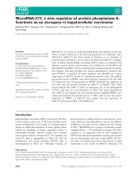
To determine the significance of NAT10 in hepatocellular carcinomas, we performed immunoblotting on human HCC tissues and their matched noncancerous
tissues. Fourteen of 19 (73.7%) tumor samples showed
increased NAT10 protein levels compared with their respective paired noncancerous tissue (Fig. 1a). These
data indicated a positive correlation of NAT10 expression with HCC.
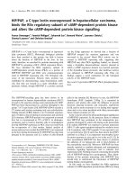
Next, we investigated the correlation of NAT10 expression with HCC progression. Immunohistochemical
staining was performed to evaluate NAT10 expression
on primary human tumors from a large cohort of HCC
patients (n = 119). Among these 119 patients, all biopsy
specimens contained both tumors and matched nontumorous tissues. Consistent with our previous study,
NAT10 was expressed in the nuclei of human HCC
tumor cells (Fig. 2a). For further evaluation of the expression level of NAT10, the staining level was graded
and scored from 0 to 3. According to the staining score,
Li et al. BMC Cancer (2017) 17:605
Page 4 of 10
Fig. 1 N-acetyltransferase (NAT10) is upregulated in human hepatocellular carcinoma (HCC). Immunoblotting revealed higher NAT10 protein in
14 of 19 tumor samples than in the respective matched pericancerous tissues (T, tumor; P, pericancerous tissue). Glyceraldehyde-3-phosphate
dehydrogenase (GAPDH) was used as a loading control
all patients was subgrouped as weak expression (staining
score 0–1) versus strong expression (staining score 2–3).
Strong expression of NAT10 was detected in 101 of 119
cases (84.8%) of HCC tumor tissues, whereas NAT10 expression was not detected in their benign counterparts
(Fig. 2b and c). Thus, NAT10 expression was significantly upregulated in HCC tumor tissues compared with
their non-tumorous counterparts.
The correlation between NAT10 expression and
clinico-pathological variables was analyzed using SPSS
version 17. As shown in Table 1, NAT10 expression was
significantly correlated with vascular invasion (p < 0.05).
However, NAT10 expression did not correlate significantly with age, α-fetoprotein (AFP) levels, capsular formation, tumor number, margin status and EdmondsonSteiner grade. In addition, as indicated by Kaplan-Meier
analysis, high level expression of NAT10 was associated
with shorter overall survival (OS; Fig. 2d) in our cohort
(p < 0.01). According to this result, we further investigated whether NAT10 expression can affect the prognosis of HCC patients independently. Univariate analysis
by Cox-regression revealed that 5 prognostic factors
affecting OS: NAT10 expression level, tumor size,
tumor number, microvascular invasion and lymph node
metastasis. Multivariate analysis by cox-regression revealed that NAT10 expression, tumor number, microvascular invasion, and lymph node metastasis were
independent prognostic factors of OS (Table 2). These
data demonstrated that NAT10 is an independent prognostic factor for HCC patients.
Increased NAT10 expression level is correlated with p53
protein level in HCC
A previous study had demonstrated that NAT10 regulates
p53 activation [26] and that p53 is frequently mutated
in HCC [14]. Therefore, we next investigated whether
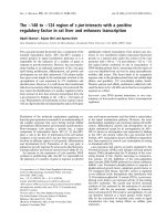
NAT10 regulates mutant p53 activity in HCC. We compared the NAT10 and p53 protein levels from surgically
removed human HCC samples by using immunoblotting.
As shown in Fig. 3a and b, p53 was upregulated in 16 of
19 (84.2%) tumor samples, indicating that these tumor
samples carry p53 mutations. Notably, we found that
NAT10 and p53 levels was positively correlated (r2 = 0.4,
p = 0.03) in the tumor samples with co-upregulation of
NAT10 and p53 (Fig. 3c). NAT10 protein levels were also
positively correlated with p53 in the HCC cell lines (Fig.
3d). Together, the results above indicate that increased
NAT10 expression is correlated with p53 level in HCC.
Li et al. BMC Cancer (2017) 17:605
Page 5 of 10
Fig. 2 Increased NAT10 expression levels are associated with shortened survival of HCC patients. a Representative immunohistochemical staining
of NAT10 in human HCC cells (magnification, ×400). b Representative immunohistochemical staining of NAT10 in adjacent noncancerous tissues
and HCC tissues (magnification, ×200). c Summary of NAT10 expression in human HCC tissues and noncancerous tissues. d Overall survival of
HCC patients with different levels of NAT10 expression by Kaplan-Meier analysis
NAT10 enhances mutant p53 stability
To understand the molecular mechanism by which
NAT10 regulates mutant p53 in HCC, we investigated
whether NAT10 interacts with mutant p53. Extract from
cytoplasm and nucleus were fractionated and subjected
to Western blotting to evaluate NAT10 expression. As
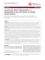
shown in Fig. 4a, NAT10 was detected in the nuclear
extracts. Immunofluorescence staining showed that
NAT10 was partially colocalized with p53 in the nucleoli (Fig. 4b). Co-immunoprecipitation confirmed that
NAT10 bound to mutant p53 in the HCC cell line
Huh7 carrying the p53 mutation (Fig. 4c). These findings indicate that NAT10 interacts with mutant p53.
Given the fact that NAT10 regulates p53 activity and
in light of our findings that NAT10 level is correlated
with mutant p53 level in HCC, we hypothesized that
NAT10 also regulates mutant p53 stability. In agreement
with our hypothesis, depletion of NAT10 decreased mutant p53 levels; however, no alteration of p21 levels was
observed (Fig. 4d). This result was consistent with
previous reports that mutant p53 loses the ability to activate wild-type p53 target genes [28, 29]. Furthermore,
knockdown of NAT10 increased ubiquitination of mutant p53 (Fig. 4e), indicating that NAT10 regulates
mutant p53 ubiquitination and stability. Recent reports
suggest that mutant p53 is still under the regulation of
Mdm2 [30, 31] and NAT10 modulates Mdm2 activity.
Thus, we next analyzed whether NAT10 regulates p53
stability by counteracting Mdm2 action. As shown in
Fig. 4f, ectopic expressed Mdm2 could enhance mutant
p53 ubiquitination, indicating that Mdm2 could still target mutant p53 to decompose. Importantly, coexpression
of NAT10 counteracted the Mdm2-induced ubiquitination of mutant p53 (Fig. 4f ). Moreover, the deletion mutant NAT10-D5, which lost the ability to inhibits Mdm2
activity, failed to do so (Fig. 4f, lane 3 vs. lane 4). In
addition, NAT10 had no effect on mutant p53 stability
in Mdm2-depleted cells (Fig. 4g). The interaction between Mdm2 and NAT10 was further verified by coimmunoprecipitation (Fig. 4h). Taken together, these
Li et al. BMC Cancer (2017) 17:605
Page 6 of 10
Table 2 Univariate and multivariate analyses of factors associated with prognosis in 119 HCCs
Clinicopathological characteristics
N
Age
<=60
89
>60
30
Tumor size (cm)
<5
62
>=5
57
Serum AFP, ng/ml
<=20
46
>20
73
Tumor number
1
97
>1
22
Tumor encapsulation
No
58
Yes
61
Microvascular invasion
No
88
Yes
31
Lymph node metastasis
No
114
Yes
5
Edmondson-Steiner grade
ES = 1 ~ 2
88
ES = 3 ~ 4
31
NAT 10 expression (weak v.s. strong)
0–1
18
2–3
101
Univariable analysis
Multivariable analysis
RR (95% CI)
p
RR (95% CI)
p
0.828 (0.456–1.506)
0.537
0.800 (0.424–1.510)
0.492
1.976 (1.183–3.301)
0.009
1.153 (0.628–2.116)
0.646
1.876 (1.080–3.258)
0.026
1.853 (1.002–3.427)
0.049
2.840 (1.605–5.025)
<0.001
2.409 (1.292–4.491)
0.006
0.594 (0.357–0.987)
0.044
0.593 (0.342–1.029)
0.063
3.585 (2.140–6.006
<0.001
1.928 (1.100–3.379)
0.022
10.727 (3.909–29.439)
<0.001
5.862 (1.854–18.538)
0.003
1.366 (0.786–2.373)
0.269
1.068 (0.569–2.004)
0.838
6.203 (1.922–20.017)
0.002
5.201 (1.492–18.138)
0.010
NAT10 expression was determined by immunohistochemical staining as described in the Methods. Clinico-pathological factors were recorded as mentioned above,
and the overall survival of patients was acquired by postoperative follow-up. The univariate analysis suggested that tumor size, tumor number, vascular invasion,
lymph node metastasis, and NAT10 expression levels were associated with the overall survival of HCC patients. Then, we employed multivariate Cox regression
analysis to identify factors that were independently correlated with patient survival. Tumor size was eliminated, and the remaining factors, including vascular
invasion, tumor numbers, lymph node metastasis, and strong expression of NAT10, were identified as independent prognosis risk factors
data indicated that NAT10 regulates mutant p53 stability
through counteracting Mdm2 action. Given that mutant
p53 often displays acquisition of the ability to potentiates
cell proliferation [32], we investigated whether increased
NAT10 in cells with mutant p53 could be advantageous
to cell proliferation, in contrast to cells with wild-type
p53. As expected, downregulation of NAT10 in Huh7
cells resulted in decreased cell proliferation (Fig. 4i). The
results were confirmed in MHCC-97 L cells which also
carry mutant p53. Overexpression of NAT10 in MHCC97 L cells enhanced cell proliferation, while ectopic
expression of NAT10 had little effect on cell proliferation in p53-depleted MHCC-97 L cells (Fig. 4j). Thus,
NAT10 promotes cell proliferation in cells expressing
mutant p53. Together, our findings demonstrated that
NAT10 regulates mutant p53 and promotes cell proliferation in cells carrying mutant p53.
Discussion
NAT10 has been observed to function in a variety of
cellular processes which are vital to cell growth and proliferation [33, 34]. Besides, it has been indicated that
NAT10 involves in affecting the nuclear architecture for
the recent work found that NAT10 is the target of the
small molecule “Remodelin” which can be used to treat
premature aging syndromes by correcting nuclear architecture [25]. Moreover, NAT10 is downregulated in human colorectal cancer [26]. Thus, functional studies of
NAT10 will be helpful for the further study of the development and occurrence of cancer. The present study
provides experimental evidences that NAT10 is overexpressed in HCC and that NAT10 level is positively correlated with tumor stage. Significantly, shortened OS
was observed in correlation with strong NAT10 expression. Multivariate Cox regression analysis further confirmed that NAT10 expression is an independent
prognostic factor of HCC. The results from our study
cohort suggest that NAT10 may be a tumor promotive
factor in HCC occurrence and development. Therefore,
we propose that protein quantification of NAT10 in
HCC by immunoblotting or immunostaining could be
used in combination with pathological examination to
predict the biological behaviors of HCC. The combination might be useful in the optimization of personalized
treatment.
Previously, we have demonstrated that NAT10 is
downregulated in colorectal cancer samples and that
NAT10 inhibits cell proliferation and colony formation
Li et al. BMC Cancer (2017) 17:605
Page 7 of 10
Fig. 3 Expression of NAT10 increases in parallel with p53 in human HCC tissues. a NAT10 was upregulated in HCC tissues. Proteins extracted from
19 pairs of freshly frozen HCC tissues and paired adjacent non-cancerous tissues were subjected to western blotting with anti-NAT10 and anti-p53
antibodies. GAPDH was used as a loading control (T, cancer tissue; P, pericancerous tissue). b Summary of NAT10 and p53 expression in human HCC
tissues and noncancerous tissues (T, cancer tissue; P, pericancerous tissue). c The positive correlation between the amounts of p53 protein and of
NAT10 protein was tested with a Pearson correlation test. d NAT10 and p53 expression in HCC cell lines. Cell extracts were prepared from different
human HCC cell lines as indicated. Proteins from the extracts were subjected to western blotting for the evaluation of NAT10 and p53 levels.
Beta-actin was evaluated as a loading control
in cells expressing wild-type p53, indicating that NAT10
could inhibit tumorigenesis through regulating p53 [26].
Here, we report that NAT10 also promotes cell proliferation in cells expressing mutant p53 and increased
NAT10 expression correlates with p53 levels in HCC.
It is noteworthy that most tumors were observed overexpression of mutant p53, including HCCs [9]. Nevertheless, the underlying mechanisms remain unclear.
Recent evidences indicated that the downregulated
Mdm2 may be one of the causes for overexpression of
mutant p53 [30]. Our results suggest that NAT10 increases mutant p53 stability and promotes cell growth
in Huh-7 cells carrying mutant p53. Further, we observed that NAT10 inhibits Mdm2-mediated p53 ubiquitination in Huh-7 cells, indicating that NAT10
regulates both wild-type p53 and mutant p53 stability
through counteracting Mdm2 actin. Besides, another
chaperone-associated E3 ligase, CHIP was believed to
play a vital role for mutant p53 degradation [35]. Thus,
it’s unknown whether NAT10 regulates CHIP activity
in mutant p53 degradation.
Depletion of NAT10 promotes cell proliferation in
cells with wild-type p53 background but decreases cell
growth in Huh-7 cells carrying mutant p53. Importantly,
NAT10 is overexpressed in HCCs and overexpression of
NAT10 is correlated with shortened survival. Moreover,
previous study reported that NAT10 plays an important
role in the growth of a subtype of epithelial ovarian
cancer with poor prognosis [36]. Therefore, the role of
NAT10 in tumorigenesis and cancer progression may
vary in different types of tumors. Further investigations,
especially animal experiments, are highly necessary to
understand the pathophysiological role of NAT10 in
tumor initiation and progression.
Most tumors especially HCC have observable genetic
changes. Mutated or functional deficient p53 was one of
the most prevalent events observed and mutations of
p53 are mainly missense point mutations in the DNAbinding domain [37, 38]. Such mutations abrogate transcription of p53 target genes, thereby disrupting the
tumor-suppressing activities of p53. Additionally, the
proteins generated by mutated Tp53 gene acquire
Li et al. BMC Cancer (2017) 17:605
Fig. 4 (See legend on next page.)
Page 8 of 10
Li et al. BMC Cancer (2017) 17:605
Page 9 of 10
(See figure on previous page.)
Fig. 4 NAT10 stabilizes mutant p53 by counteracting Mdm2 action. a LO2, HepG2, MHCC-97H and MHCC-97 L cells were harvested and fractionated.
Fractions were then immunoblotted with the indicated antibodies. (C, cytoplasmic; N, nuclear) b HCC cells were seeded on coverslips and stained
with anti-NAT10 and anti-p53 antibodies. Nuclei were stained with DAPI. Fluorescence images were photographed under confocal microscopy.
c Huh7 cell lysates were immunoprecipitated with control IgG or anti-NAT10 antibodies. The immunoprecipitates were subsequently immunoblotted
with the indicated antibodies. d Huh7 cells were transfected with the indicated siRNAs. Seventy-two hours later, the total proteins were
analyzed by western blotting for the indicated proteins. e Huh7 cells were transfected with the indicated siRNAs and treated with MG132 for 4 h
before harvest. The whole cell lysates were analyzed by western blotting for the indicated antibodies. f Huh7 cells were transfected with the
indicated plasmids. Forty-eight hours later, cells were harvested after MG132 treatment, and the whole cell lysates were analyzed by western
blotting for the indicated antibodies. (NAT10 GE: NAT10 mutant lacking acetyltransferase activity; NAT10 D5: NAT10 mutant lacking ubiquitin
ligase activity) g Huh7 cells were transfected with the indicated plasmids. Forty-eight hours later, cells were harvested and lysed. The whole
cell lysates were analyzed by western blotting for the indicated antibodies. h Huh7 cell lysates were immunoprecipitated with control IgG or
anti-NAT10 antibodies. The immunoprecipitates were subsequently immunoblotted with the indicated antibodies. i Huh7 cells transfected
with the indicated siRNAs were plated in 96-well plates, and cell proliferation was then quantified at the indicated time points. j MHCC-97 L
cells transfected with the indicated siRNAs or vectors were plated in 96-well plates, and cell proliferation was then quantified at the indicated
time points as described in Methods
oncogenic functions by endowing cells with proliferation
and growth advantage [39]. In vivo experiments have revealed that tumors in mice with mutant p53 had the
characteristic of higher malignancy, rapid development
and more invasive compared with wild type or null p53
mice [40, 41]. Thus, novel strategies are being developed
aiming at tumors with mutant p53 [17, 42]. Abrogation
of histone deacetylase HDAC6-binding could cause the
heat-shock proteins disassociated from mutant p53,
with the result that mutant p53 is easier to be degraded
by Mdm2. Thus, HDAC inhibitors such as SAHA has
the potential in promoting mutant p53 degradation and
removing mutant p53 [43, 44]. Small molecule activators of Sirt1 have also been used to reduce mutant p53
levels [45]. NAT10 is upregulated in HCC, and it
enhances p53 stability, indicating that NAT10 might be
a potential target in HCC therapy. Abrogation of the
interaction between NAT10 and p53 would be beneficial for tumor therapy of hepatic cancers carrying p53
mutations. These possibilities need to be examined in
future studies.
Conclusions
Our study demonstrated that NAT10 is upregulated in
HCC and that increased NAT10 expression levels are
associated with shortened patient survival. Moreover,
NAT10 interacts with mutant p53 and increases its stability, resulting in increased cell proliferation in HCC
cells. These results indicate that NAT10 is a potential
therapeutic candidate for p53-mutated HCC.
Abbreviations
CHIP: Carboxyl terminus of Hsc70-interacting protein; HADC: Histone
deacetylase; HCC: Hepatocellular carcinoma; Hsp90: Heat shock protein 90;
IHC: Immunohistochemistry; Mdm2: Murine double minute2; NAT10: Nacetyltransferase 10; OS: Overall survival; PBS: Phosphate buffered solution;
SAHA: Suberoylanilide hydroxamic acid
Acknowledgements
We would like to thank all participants for their support in this study.
Funding
This work was supported by grants from the National Natural Science
Foundation of China (Grant No. 81371868 and 81,672,735).
The funding body had no role in the design of the study and collection,
analysis, and interpretation of data and in writing the manuscript.
Availability of data and materials
The datasets used and/or analyzed during the current study available from
the corresponding author on reasonable request.
Authors’ contributions
QJL and XFL carried out the experimental procedure and data collecting,
analyzing, literature reviewing and participated in writing the manuscript.
KMJ provided the follow up data of the patients, contributed in interpretation
of the data and were involved in the revision of the manuscript. ML, CFZ and
XJD carried out the pathological diagnose and classification works, performed
the statistical analysis, participated in the design of the study and were involved
in the revision of the manuscript. BCX conceived of the study, and participated
in its design and coordination and helped to draft the manuscript. All authors
read and approved the final manuscript.
Ethics approval and consent to participate
This study was approved by the ethics committee of Peking University
School of Oncology (Beijing Cancer Hospital and Institute), and was
performed in accordance with the Helsinki Declaration of 1975, as revised
in 1983. All the patients enrolled were comprehensively informed, and
written informed consent to participate in this research and publish the
data were obtained.
Consent for publication
Not Applicable.
Competing interests
The authors declare that they have no competing interests.
Publisher’s Note
Springer Nature remains neutral with regard to jurisdictional claims in
published maps and institutional affiliations.
Author details
1
Department of Hepatobiliary Oncology, Sun Yat-Sen University Cancer
Center, 651 Dongfeng Road East, Guangzhou, Guangdong 510060, China.
2
Department of Cell Biology, School of Basic Medical Sciences, Peking
University Health Science Center, Beijing 100191, China.
3
Hepatopancreatobiliary Surgery Department I, Key Laboratory of
Carcinogenesis and Translational Research, Ministry of Education, Peking
University School of Oncology, Beijing Cancer Hospital and Institute, 52
Fucheng Road, Haidian District, Beijing 100142, China. 4Department of
Pathology, School of Basic Medical Sciences, Peking University Health
Science Center, Beijing 100191, China.
Li et al. BMC Cancer (2017) 17:605
Received: 14 October 2016 Accepted: 21 August 2017
References
1. Parkin DM. Global cancer statistics in the year 2000. Lancet Oncol.
2001;2(9):533–43.
2. Guichard C, Amaddeo G, Imbeaud S, Ladeiro Y, Pelletier L, Maad IB,
Calderaro J, Bioulac-Sage P, Letexier M, Degos F, et al. Integrated analysis
of somatic mutations and focal copy-number changes identifies key genes
and pathways in hepatocellular carcinoma. Nat Genet. 2012;44(6):694–8.
3. Martin J, Dufour JF. Tumor suppressor and hepatocellular carcinoma.
World J Gastroenterol. 2008;14(11):1720–33.
4. Sell S. Mouse models to study the interaction of risk factors for human liver
cancer. Cancer Res. 2003;63(22):7553–62.
5. Aravalli RN, Steer CJ, Cressman EN. Molecular mechanisms of hepatocellular
carcinoma. Hepatology. 2008;48(6):2047–63.
6. Vousden KH, Prives C. Blinded by the light: the growing complexity of p53.
Cell. 2009;137(3):413–31.
7. Kruse JP, Gu W. Modes of p53 regulation. Cell. 2009;137(4):609–22.
8. Leroy B, Fournier JL, Ishioka C, Monti P, Inga A, Fronza G, Soussi T. The TP53
website: an integrative resource centre for the TP53 mutation database and
TP53 mutant analysis. Nucleic Acids Res. 2013;41(Database issue):D962–9.
9. Kandoth C, McLellan MD, Vandin F, Ye K, Niu B, Lu C, Xie M, Zhang Q,
McMichael JF, Wyczalkowski MA, et al. Mutational landscape and
significance across 12 major cancer types. Nature. 2013;502(7471):333–9.
10. Soussi T, Wiman KG. Shaping genetic alterations in human cancer: the p53
mutation paradigm. Cancer Cell. 2007;12(4):303–12.
11. Freed-Pastor WA, Mizuno H, Zhao X, Langerod A, Moon SH, RodriguezBarrueco R, Barsotti A, Chicas A, Li W, Polotskaia A, et al. Mutant p53
disrupts mammary tissue architecture via the mevalonate pathway. Cell.
2012;148(1–2):244–58.
12. Weissmueller S, Manchado E, Saborowski M, Morris JP, Wagenblast E, Davis CA,
Moon SH, Pfister NT, Tschaharganeh DF, Kitzing T, et al. Mutant p53 drives
pancreatic cancer metastasis through cell-autonomous PDGF receptor beta
signaling. Cell. 2014;157(2):382–94.
13. Morton JP, Timpson P, Karim SA, Ridgway RA, Athineos D, Doyle B,
Jamieson NB, Oien KA, Lowy AM, Brunton VG, et al. Mutant p53 drives
metastasis and overcomes growth arrest/senescence in pancreatic cancer.
Proc Natl Acad Sci U S A. 2010;107(1):246–51.
14. Liu J, Ma Q, Zhang M, Wang X, Zhang D, Li W, Wang F, Wu E. Alterations of
TP53 are associated with a poor outcome for patients with hepatocellular
carcinoma: evidence from a systematic review and meta-analysis. Eur J Cancer.
2012;48(15):2328–38.
15. Bykov VJ, Issaeva N, Shilov A, Hultcrantz M, Pugacheva E, Chumakov P,
Bergman J, Wiman KG, Selivanova G. Restoration of the tumor suppressor
function to mutant p53 by a low-molecular-weight compound. Nat Med.
2002;8(3):282–8.
16. Issaeva N, Bozko P, Enge M, Protopopova M, Verhoef LG, Masucci M,
Pramanik A, Selivanova G. Small molecule RITA binds to p53, blocks
p53-HDM-2 interaction and activates p53 function in tumors. Nat Med.
2004;10(12):1321–8.
17. Yu X, Vazquez A, Levine AJ, Carpizo DR. Allele-specific p53 mutant reactivation.
Cancer Cell. 2012;21(5):614–25.
18. He XX, Zhang YN, Yan JW, Yan JJ, Wu Q, Song YH. CP-31398 inhibits the
growth of p53-mutated liver cancer cells in vitro and in vivo. Tumour Biol.
2016;37(1):807–15.
19. He X, Liu F, Yan J, Zhang Y, Shang H, Dou Q, Zhao Q, Song Y. Trans-splicing
repair of mutant p53 suppresses the growth of hepatocellular carcinoma
cells in vitro and in vivo. Sci Rep. 2015;5:8705.
20. Lv J, Liu H, Wang Q, Tang Z, Hou L, Zhang B. Molecular cloning of a novel
human gene encoding histone acetyltransferase-like protein involved
in transcriptional activation of hTERT. Biochem Biophys Res Commun.
2003;311(2):506–13.
21. Kong R, Zhang L, Hu L, Peng Q, Han W, Du X, Ke Y. hALP, a novel transcriptional
U three protein (t-UTP), activates RNA polymerase I transcription by binding and
acetylating the upstream binding factor (UBF). J Biol Chem. 2011;286(9):7139–48.
22. Shen Q, Zheng X, McNutt MA, Guang L, Sun Y, Wang J, Gong Y, Hou L,
Zhang B. NAT10, a nucleolar protein, localizes to the midbody and regulates
cytokinesis and acetylation of microtubules. Exp Cell Res. 2009;315(10):1653–67.
Page 10 of 10
23. Fu D, Collins K. Purification of human telomerase complexes identifies
factors involved in telomerase biogenesis and telomere length regulation.
Mol Cell. 2007;28(5):773–85.
24. Chi YH, Haller K, Peloponese JM Jr, Jeang KT. Histone acetyltransferase hALP
and nuclear membrane protein hsSUN1 function in de-condensation of
mitotic chromosomes. J Biol Chem. 2007;282(37):27447–58.
25. Larrieu D, Britton S, Demir M, Rodriguez R, Jackson SP. Chemical inhibition of
NAT10 corrects defects of laminopathic cells. Science. 2014;344(6183):527–32.
26. Liu X, Tan Y, Zhang C, Zhang Y, Zhang L, Ren P, Deng H, Luo J, Ke Y, Du X.
NAT10 regulates p53 activation through acetylating p53 at K120 and
ubiquitinating Mdm2. EMBO Rep. 2016;17(3):349–66.
27. Pluk H, Soffner J, Luhrmann R, van Venrooij WJ. cDNA cloning and
characterization of the human U3 small nucleolar ribonucleoprotein
complex-associated 55-kilodalton protein. Mol Cell Biol. 1998;18(1):488–98.
28. Yan W, Chen X. Identification of GRO1 as a critical determinant for mutant
p53 gain of function. J Biol Chem. 2009;284(18):12178–87.
29. Yan W, Liu G, Scoumanne A, Chen X. Suppression of inhibitor of
differentiation 2, a target of mutant p53, is required for gain-of-function
mutations. Cancer Res. 2008;68(16):6789–96.
30. Terzian T, Suh YA, Iwakuma T, Post SM, Neumann M, Lang GA, Van Pelt CS,
Lozano G. The inherent instability of mutant p53 is alleviated by Mdm2 or
p16INK4a loss. Genes Dev. 2008;22(10):1337–44.
31. Lukashchuk N, Vousden KH. Ubiquitination and degradation of mutant p53.
Mol Cell Biol. 2007;27(23):8284–95.
32. Muller PA, Vousden KH. Mutant p53 in cancer: new functions and therapeutic
opportunities. Cancer Cell. 2014;25(3):304–17.
33. Ito S, Horikawa S, Suzuki T, Kawauchi H, Tanaka Y. Human NAT10 is an
ATP-dependent RNA acetyltransferase responsible for N4-acetylcytidine
formation in 18 S ribosomal RNA (rRNA). J Biol Chem. 2014;289(52):35724–30.
34. Liu H, Ling Y, Gong Y, Sun Y, Hou L, Zhang B. DNA damage induces Nacetyltransferase NAT10 gene expression through transcriptional activation.
Mol Cell Biochem. 2007;300(1–2):249–58.
35. Esser C, Scheffner M, Hohfeld J. The chaperone-associated ubiquitin ligase
CHIP is able to target p53 for proteasomal degradation. J Biol Chem.
2005;280(29):27443–8.
36. Tan TZ, Miow QH, Huang RY, Wong MK, Ye J, Lau JA, Wu MC, Bin Abdul
Hadi LH, Soong R, Choolani M, et al. Functional genomics identifies five
distinct molecular subtypes with clinical relevance and pathways for growth
control in epithelial ovarian cancer. EMBO Mol Med. 2013;5(7):1051–66.
37. Muller PA, Vousden KH. p53 mutations in cancer. Nat Cell Biol. 2013;15(1):2–8.
38. Freed-Pastor WA, Prives C. Mutant p53: one name, many proteins. Genes Dev.
2012;26(12):1268–86.
39. Dittmer D, Pati S, Zambetti G, Chu S, Teresky AK, Moore M, Finlay C,
Levine AJ. Gain of function mutations in p53. Nat Genet. 1993;4(1):42–6.
40. Lang GA, Iwakuma T, Suh YA, Liu G, Rao VA, Parant JM, Valentin-Vega YA,
Terzian T, Caldwell LC, Strong LC, et al. Gain of function of a p53 hot spot
mutation in a mouse model of Li-Fraumeni syndrome. Cell. 2004;119(6):861–72.
41. Doyle B, Morton JP, Delaney DW, Ridgway RA, Wilkins JA, Sansom OJ. p53
mutation and loss have different effects on tumourigenesis in a novel mouse
model of pleomorphic rhabdomyosarcoma. J Pathol. 2010;222(2):129–37.
42. Cheok CF, Verma CS, Baselga J, Lane DP. Translating p53 into the clinic.
Nat Rev Clin Oncol. 2011;8(1):25–37.
43. Li D, Marchenko ND, Moll UM. SAHA shows preferential cytotoxicity in
mutant p53 cancer cells by destabilizing mutant p53 through inhibition of
the HDAC6-Hsp90 chaperone axis. Cell Death Differ. 2011;18(12):1904–13.
44. Yan W, Liu S, Xu E, Zhang J, Zhang Y, Chen X. Histone deacetylase inhibitors
suppress mutant p53 transcription via histone deacetylase 8. Oncogene.
2013;32(5):599–609.
45. Yi YW, Kang HJ, Kim HJ, Kong Y, Brown ML, Bae I. Targeting mutant p53 by
a SIRT1 activator YK-3-237 inhibits the proliferation of triple-negative breast
cancer cells. Oncotarget. 2013;4(7):984–94.