Proteomics reveals a therapeutic vulnerability via the combined blockade of APE1 and autophagy in lung cancer A549 cells
Bạn đang xem bản rút gọn của tài liệu. Xem và tải ngay bản đầy đủ của tài liệu tại đây (2.71 MB, 11 trang )
Pan et al. BMC Cancer
(2020) 20:634
/>
RESEARCH ARTICLE
Open Access
Proteomics reveals a therapeutic
vulnerability via the combined blockade of
APE1 and autophagy in lung cancer A549
cells
Shu-Ting Pan1†, Ji Zhou2†, Fang Yang2, Shu-Feng Zhou3* and Tao Ren4*
Abstract
Background: Drug resistance is a major cause of therapeutic failure that is often associated with elevated autophagy
and apurinic/apyrimidinic endonuclease 1 (APE1) expression. Herein, we investigated the role of APE1 and autophagy
in A549 cells treated with cisplatin.
Methods: SILAC proteomics was applied to obtain a panoramic view of cisplatin treatment in KRASG12S-mutant A549
cells. Quantity analysis of cellular apoptosis and autophagy was based on flow cytometry. Western blotting was used to
examine the expression levels of apoptosis- and autophagy-related proteins, as well as those of APE1. Knockdown of
APE1 was achieved by RNA interference. Immunoprecipitation was further employed to reveal the molecular
interaction of APE1, p53, and LC3 when A549 cells were exposed to cisplatin.
Results: SILAC proteomics revealed that 72 canonical pathways, including base excision repair (BER) and autophagy
signalling pathways, were regulated after cisplatin treatment in A549 cells. Cisplatin markedly induced autophagy and
apoptosis in A549 cells, accompanied by remarkable APE1 increase. Suppression of autophagy enhanced the inhibition
effect of cisplatin on cell growth, proliferation, and colony formation; however, APE1 inhibition enhanced the
expression of LC3-I/II, suggesting that APE1 and autophagy are compensatory for cell survival to evade the anticancer
action of cisplatin. Immunoprecipitation results revealed the triple complex of APE1-p53-LC3 in response to cisplatin
plus CQ in A549 cells. Dual inhibition of APE1 and autophagy significantly enhanced cisplatin-induced apoptosis, which
eventually overcame drug resistance in cisplatin-resistant A549 cells.
Conclusions: Dual inhibition of APE1 and autophagy greatly enhances apoptosis in parental KRASG12S-mutant A549
cells and cisplatin-resistant A549 cells via regulation of APE1-p53-LC3 complex assembly, providing therapeutic
vulnerability to overcome cisplatin resistance in the context of KRASG12S-mutant lung cancer.
Keywords: Cisplatin, APE1, Autophagy, Chemotherapy, Non-small cell lung cancer, Apoptosis
* Correspondence: ; ;
†
Shu-Ting Pan and Ji Zhou contributed equally to this work.
3
Department of Pharmaceutical Sciences, College of Pharmacy, University of
South Florida, 12901 Bruce B. Downs Boulevard, Tampa, Florida 33612, USA
4
Oncology Department, The First Affiliated Hospital, Chengdu Medical
College, 278 Baoguang St, Xindu Distr, Chengdu 610500, Sichuan, China
Full list of author information is available at the end of the article
© The Author(s). 2020 Open Access This article is licensed under a Creative Commons Attribution 4.0 International License,
which permits use, sharing, adaptation, distribution and reproduction in any medium or format, as long as you give
appropriate credit to the original author(s) and the source, provide a link to the Creative Commons licence, and indicate if
changes were made. The images or other third party material in this article are included in the article's Creative Commons
licence, unless indicated otherwise in a credit line to the material. If material is not included in the article's Creative Commons
licence and your intended use is not permitted by statutory regulation or exceeds the permitted use, you will need to obtain
permission directly from the copyright holder. To view a copy of this licence, visit />The Creative Commons Public Domain Dedication waiver ( applies to the
data made available in this article, unless otherwise stated in a credit line to the data.
Pan et al. BMC Cancer
(2020) 20:634
Background
Lung cancer is the leading cause of cancer-related death
and remains a major clinical challenge with increasing incidence and mortality [1, 2]. Due to drug resistance, recurrence, and metastasis, the treatment efficacy of lung
cancer remains unsatisfactory. A better understanding of
the aetiology, pathogenesis, and molecular targets is required to develop novel therapeutic modalities. Somatic
gene mutations, including KRAS, EGFR, and TP53 mutations, is a major driver of lung cancer initiation [3]. Accumulating evidence has shown that not all gene mutations
occur equally. In particular, compelling evidence suggests
that RAS mutants function in an allele-specific manner,
justifying the acquirement of a RAS allele-specific approach for RAS-driven cancer therapy [4–6]. Given the
feature of allele specificity and the pivotal role of RAS in
cellular events, including cell growth, cell survival, cell
senescence, and cell death, novel strategies in a RAS
allele-dependent manner are still required.
Autophagy is a cell survival-promoting mechanism following harsh stimuli and has been deeply implicated in
cancer development and therapy [7–9]. Recently, targeting
autophagy has been in the spotlight for cancer therapy via
pharmacological inhibition alone or combination with
other therapeutics [10, 11], providing insight into lung
cancer therapy development. Cisplatin is one of the most
frequently administered chemotherapeutic drugs for many
solid tumours, including lung cancer. Mechanically, cisplatin kills cancer cells via interference with DNA synthesis and repair, subsequently inducing cell apoptosis [12].
However, there is limited clinical efficacy for cisplatinbased therapy because of drug resistance [13]. Several key
factors contribute to cisplatin resistance, including autophagy [14] and apurinic/apyrimidinic endonuclease 1
(APE1) [15]. APE1 is a multifunctional protein with two
major activities, DNA repair and transcriptional regulation
[16]. Importantly, APE1 is often overexpressed in many
tumours, contributing to disease progression, chemoresistance and a poor prognosis [15, 17–20]. Our previous
study found that APE1 is highly expressed in non-small
cell lung cancer (NSCLC). Moreover, APE1 is a prognostic
risk factor indicated by a poor overall survival [15, 19].
Herein, targeting APE1 might represent a therapeutic vulnerability for lung cancer, particularly, cisplatin-resistant
lung cancer.
Thus, based on the aforementioned details, we hypothesized that APE1 and autophagy may contribute to lung
cancer progression and drug resistance and that combined blockade of APE1 and autophagy enhances the
therapeutic effect of cisplatin and overcomes cisplatin
resistance in lung cancer. In the present study, we applied quantitative proteomics to identify the proteomic
responses to cisplatin treatment in KRASG12S-mutant
A549 cells. Both APE1 and autophagy were involved in
Page 2 of 11
the cellular responses to cisplatin exposure. In A549
cells and cisplatin-resistant A549 cells, cisplatin-induced
apoptosis was significantly enhanced via the combination of autophagy inhibition by chloroquine (CQ) and
APE1 knockdown by siRNA with the involvement of
p53 activation.
Methods
Chemicals and reagents
CDDP was purchased from Selleckchem Inc. (Houston,
TX, USA). 13C6-L-lysine, L-lysine, 13C615N4-L-arginine, Larginine, Dulbecco’s modified Eagle’s medium (DMEM)/
F12 for SILAC, APE1 siRNA, dimethyl sulfoxide (DMSO),
2-(4,5-dimethylthiazol-2-yl)-2,5-diphenyltetrazolium
bromide (MTT), bovine serum albumin, and Dulbecco’s
phosphate-buffered saline (PBS) were obtained from
Sigma-Aldrich (St. Louis, MO, USA). 6-Diamidino-2-phenylindole (DAPI), Opti-minimal Essential Medium
(MEM), Lipofectamine 2000, and the negative control
siRNA were purchased from Invitrogen Inc. (Carlsbad,
CA, USA). The Annexin V-phycoerythrin (PE) apoptosis
detection kit was purchased from BD Biosciences Inc.
(San Jose, CA, USA). The Cyto-ID® Autophagy detection
kit was obtained from Enzo Life Sciences Inc. (Farmingdale, NY, USA). The Western blotting substrate, Pierce™
bicinchoninic acid (BCA) protein assay kit, skim milk, and
radioimmunoprecipitation assay buffer (RIPA) were purchased from Thermo Fisher Scientific Inc. (Hudson, NH,
USA). The polyvinylidene difluoride (PVDF) membrane
was obtained from Bio-Rad Inc. (Hercules, CA, USA). The
antibody against human β-actin was obtained from Santa
Cruz Biotechnology Inc. (Dallas, TX, USA). The
remaining primary antibodies for signalling proteins related to apoptosis and autophagy were purchased from
Cell Signaling Technology Inc. (Beverly, MA, USA).
Cell line and cell culture
The human lung cancer cell line A549 (KRASG12S) was
obtained from Chinese Academy of Science Cellbank
(Shanghai, China) and was cultured in RPMI1640
medium supplemented with 10% heat-inactivated foetal
bovine serum (FBS). The cells were maintained at 37 °C
in a 5% CO2/95% air humidified incubator.
Cell viability determination
The MTT assay was used to evaluate cell viability.
Briefly, cells were seeded in 96-well plates at a density of
7.0 × 10³ cells/well. After 24 h. of incubation, the cells
were treated for 48 h. The absorbance was measured
using a Synergy™H4 Hybrid microplate reader (BioTek,
Winooski, VT, USA) at wavelengths of 560 nm (MTT
formazan) and 670 nm (background).
Pan et al. BMC Cancer
(2020) 20:634
Page 3 of 11
Quantitative proteomics
Western blotting assay
Quantitative proteomic experiments were performed
using a stable isotope labelling by amino acids in cell
culture (SILAC)-based approach to identify the molecular targets of CDDP in the treatment of A549 cells as
previously described [21]. Briefly, A549 cells were cultured in DMEM/F12 medium (for SILAC) with (heavy)
or without (light) stable isotope-labelled amino acids
(13C6 L-lysine and 13C615N4 L-arginine) and 10% dialyzed FBS. After treatment with CDDP (5 μM) for 24 h.,
the cell samples were harvested, lysed, and quantified.
Next, an equal amount of heavy and light protein samples were combined to reach a total volume of 50 μL
containing 400 μg of protein, and the combined protein
sample was digested and desalted. Next, the peptide
mixtures (5 μL) were subjected to the hybrid linear ion
trap. The peptide SILAC ratio was calculated using MaxQuant version 1.2.0.13. The proteins were identified
using Scaffold 4.3.2, and the pathway was analysed using
ingenuity pathway analysis (IPA) from QIAGEN Inc.
The protein expression level was examined using Western blotting. Protein samples were extracted using RIPA
buffer, the protein concentrations were measured using
the BCA kit, and an equal amount of protein was separated by SDS-PAGE. The corresponding primary and
secondary antibodies were applied to evaluate the expression levels of targeted proteins. Visualization was
performed using the Bio-Rad ChemiDoc™ XRS system,
and the blot bands were analysed using Image Lab 3.0.
RNA interference
Small interfering RNA-mediated gene silencing was performed to investigate the role of APE1 in cisplatininduced apoptosis and autophagy in A549 cells according to the manufacturer’s instructions. A549 cells were
transfected with the negative control siRNA and APE1siRNA using Lipofectamine 2000. The protein samples
were collected and kept at − 80 °C for further analysis.
Immunoprecipitation
Quantification of cellular apoptosis
Cell apoptosis was evaluated using the Annexin V-PE
apoptosis detection kit as previously described [21].
Briefly, the cells were collected after treatment and resuspended in 1× binding buffer with 5 μL of Annexin VPE and 5 μL of 7-amino-actinomycin D (7-AAD) at 1 ×
105 cells/mL in a total volume of 150 μL. The cells were
gently mixed and incubated in the dark for 15 min at
room temperature. The binding buffer (100 μL) was then
added to each tube, and the number of apoptotic cells
was quantified using flow cytometry and collecting 10,
000 events for analysis.
Quantification of cellular autophagy
Cell autophagy was examined using flow cytometry as
previously described [21]. Briefly, the cells were collected
after treatment and resuspended in 250 μL of assay buffer containing 5% FBS, and Cyto-ID® Green stain solution (250 μL) was added to each tube and mixed gently.
After 20 min of incubation at room temperature in the
dark, the cells were collected by centrifugation, washed
once and analysed using the green (FL1) channel of flow
cytometry.
The interaction between APE1 and p53 was examined using
immunoprecipitation as previously described [22]. After 24 h.
of treatment, A549 cells were lysed in pre-chilled cell lysis
buffer [50 mM Tris-HCl (pH 7.4), 150 mM NaCl, 1 mM
EDTA, 1% NP40, protease inhibitors] for 5 min. The lysates
were precleared with 20 μL of Proteins A/G (Invitrogen;
Thermo Fisher Scientific, Inc.) at 4 °C for 45 min, followed
by incubation with APE1 or p53 antibody overnight at 4 °C.
Following immunoprecipitation, the samples were incubated
with protein G for 3 h. at 4 °C. Thereafter, the samples were
washed with lysis buffer five times to remove any unprecipitated proteins before boiling in SDS buffer for 5 min.
The elution was analysed for precipitated APE1 or p53 protein using Western blotting analysis. Normal rabbit IgG antibody was used as a negative control. The antibodies used
were as follows: APE1 (1:500), p53 (1:500), and normal rabbit
IgG (1:1000).
Statistical analysis
The data were expressed as means ± standard deviation
(SD). One-way analysis of variance (ANOVA) followed
by Tukey’s multiple comparison procedure was used for
comparisons of multiple groups. The value of P<0.05
was considered statistically significant. The assays were
performed at least three times independently.
Confocal fluorescence microscopy
Results
Confocal microscopy was performed to evaluate the cellular autophagy level in A549 cells after treatment with
5 μM CDDP, 10 μM CQ, and 5 μM CDDP + 10 μM CQ
using the Cyto-ID autophagy detection kits as previously
described [21]. The fluorescence was assessed using TCS
SP2 laser scanning confocal microscopy (LSCM).
Overview of the proteomic response to cisplatin
treatment in A549 cells
Until now, a lack of effective therapeutics persists for
KRAS mutation-driven lung cancer. Compelling evidence has shown that RAS mutations vary and has
spurred the development of new therapeutic
Pan et al. BMC Cancer
(2020) 20:634
vulnerabilities in a RAS allele-specific manner. To explore possible therapeutic targets, we applied SILACbased proteomics to reveal the full spectrum of the molecular interactome in A549 cells in the context of the
KRASG12S mutant following cisplatin exposure. We evaluated the proteomic responses to cisplatin (5 μM) treatment and identified at least 3262 protein molecules
responding to cisplatin treatment, including APE1, p53,
LC3-I/II, and many other functional proteins involved in
DNA damage repair, cell proliferation, cell cycle, cellular
metabolism, apoptosis, and autophagy. Subsequent IPA
analysis revealed 1013 cellular functional proteins (450
proteins were upregulated; 563 proteins were downregulated) and 72 canonical signalling pathways that are involved in cell cycle control of chromosomal replication,
RNA signalling, the BER pathway, DNA double-strand
break repair by non-homologous end joining, ILK signalling, mismatch repair, mTOR signalling, ATM signalling,
EGF signalling, telomere extension by telomerase, the
spliceosomal cycle, the role of CHK protein in cell cycle
checkpoint control, glycolysis I, gluconeogenesis I, DNA
methylation and transcriptional repression signalling, the
NRF2-mediated oxidative stress response, apoptosis, and
autophagy (see Additional file 2: Supplementary Fig.
32A-B). As shown in Supplementary Fig. 32C, among
the proteins in the BER pathway, APE1 expression was
increased. Moreover, autophagy participated in the cellular responses to cisplatin treatment in A549 cells, as evident from the alteration in the expression of MAP1LC3
(also named as LC3) after cisplatin treatment (see Additional file 2: Supplementary Fig. 32D). Collectively, we
speculated that both BER and autophagy pathways are
involved in cisplatin-stimulated cellular responses in
KRASG12S-mutant A549 cells. Thus, we subsequently investigated their roles in responses to cisplatin treatment.
Cisplatin induces autophagy and apoptosis and increases
APE1 expression
As observed above regarding the proteomic responses to
cisplatin treatment, autophagy and apoptosis were involved. Thus, we tested cisplatin-induced autophagy and
apoptosis in A549 cells. Cisplatin decreased the number
of viable A549 cells concentration-dependent manner
(Fig. 1a). Cisplatin was reported to induce autophagy
and apoptosis in various cancers, including lung cancer
[23–25], and autophagy is related to chemo-resistance,
providing a cell-protective mechanism that can promote
tumour cell survival following different stresses, including chemotherapeutic treatment [26]. We showed that
cisplatin treatment led to concentration- and timedependent increases in autophagy in KRASG12S-mutant
A549 cells and markedly increased the expression of
LC3-I/II (Fig. 1b, d, f and g). Accompanying the autophagy phenomenon, the dose and timescale experiments
Page 4 of 11
showed that cisplatin treatment also induced remarkable
apoptosis in A549 cells (Fig. 1c and e). Activation by the
cleavage of PARP and caspase 3 only occurred at a high
concentration of cisplatin (10 μM; Fig. 1f-g). Notably,
the proteomic study showed that APE1 is involved in
the responses to cisplatin treatment, and we previously
showed that high APE1 expression in patients with
NSCLC was positively correlated with poor overall survival, implying that APE1 is a prognostic risk factor [15,
19]. In the dose and timescale experiments, the expression of APE1 was dramatically increased following cisplatin treatment in A549 cells (Fig. 1f-g). Together, the
results showed that cisplatin-induced autophagy and
APE1 expression could counteract the apoptotic effect
in KRASG12S-mutant A549 cells, suggesting the inhibition of autophagy or APE1 can enhance the cell-killing
effect of cisplatin.
Inhibition of autophagy enhances cisplatin-induced
apoptosis
Recently, compelling evidence has shown that the inhibition of autophagic flux effectively enhances the tumoursuppressive effect of MAPK signalling inhibitors in the
treatment of cancer [10, 11]. Chloroquine (CQ), originally used as an anti-malarial drug, has been in the spotlight as an autophagy inhibitor and a novel
chemotherapeutic agent [10, 27]. CQ can diffuse through
cell membranes and accumulate in cellular lysosomes,
repressing autophagosome fusion with lysosomes [28,
29]. Thus, we tested the effect of CQ on cell growth and
colony formation alone or in combination with cisplatin
at low concentration (0.5 μM) in A549 cells. First, we
showed that CQ decreased A549 cell viability at a high
dose with an IC50 value at 38.46 μM (Fig. 2a). Next, we
tested the effect of CQ on cell growth and viability alone
and in combination with cisplatin. Treatment of A549
cells with 10 μM CQ did not affect cell growth compared
with the control; however, 0.5 μM cisplatin remarkably
suppressed cell growth after 1 week (Fig. 2b). Notably,
combinatorial treatment of cells with CQ and cisplatin
inhibited cell growth (Fig. 2b). Furthermore, cotreatment of A549 cells with CQ and cisplatin showed a
higher inhibitory effect on cell colony formation than
CQ or cisplatin treatment alone (Fig. 2c). Similarly,
treatment of A549 cells with cisplatin alone increased
the expression of LC3-I/II, whereas combinatorial treatment of cells with CQ and cisplatin decreased the expression of LC3-I/II (Fig. 2d), suggesting that inhibition
of autophagic flux enhances the effect of cisplatin. Indeed, co-treatment of A549 cells with CQ and cisplatin
enhanced cisplatin-induced apoptosis, whereas no effect
of CQ on A549 cell apoptosis was observed (Fig. 3a-b).
Intriguingly, APE1 expression was increased with both
cisplatin treatment alone and in combination with CQ
Pan et al. BMC Cancer
(2020) 20:634
Page 5 of 11
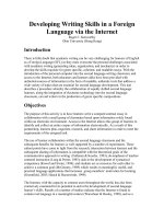
Fig. 1 Cisplatin induces autophagy and apoptosis. a Cisplatin markedly inhibited A549 cell proliferation in a dose-dependent fashion. The IC50 value was
10.26 μM. b A549 cells were treated with cisplatin at 0.5, 5, and 10 μM for 24 h. Flow cytometry was used to determine cisplatin-induced autophagy. c A549
cells were treated with cisplatin at 0.5, 5, and 10 μM for 24 h. Flow cytometry was used to determine cisplatin-induced apoptosis. d Cisplatin-induced
autophagy was performed in a time-dependent manner. A549 cells were treated with cisplatin at 5 μM for 1, 3, 6, 12 and 24 h. e A549 cells were treated
with 5 μM cisplatin for 1, 3, 6, 12, and 24 h. Flow cytometry was used to determine apoptosis. f The expression levels of apoptosis- and autophagy-related
proteins, as well as those of APE1, were examined using Western blotting after treatment with 0.5, 5, and 10 μM cisplatin for 24 h (see Additional file 1:
Supplementary Figure 1-3). g The expression levels of apoptosis- and autophagy-related proteins, as well as APE1, were examined using Western blotting
after treatment with 5 μM cisplatin for 1, 3, 6, 12, and 24 h (see Additional file 1: Supplementary Figure 4-7).
(Fig. 3b), suggesting APE1 counteracts the effect of dual
treatment of cisplatin and CQ. Taken together, these
data showed that inhibition of autophagic flux enhances
the cisplatin-induced cell growth-suppressive effect and
apoptosis.
Inhibition of APE1 stimulates autophagy in A549 cells
We first tested the effect of the mono-inhibition of
APE1 on autophagy in A549 cells, which show high
APE1 expression [30]. SiRNA-mediated knockdown or
chemical inhibition of APE1 increased the expression of
LC3-II (Fig. 4a-c). Confocal microscopic examination
also showed that knockdown of APE1 increased autophagy compared with control siRNA (Fig. 4d). However, in
the presence of cisplatin and CQ treatment alone or together, knockdown of APE1 not only reduced the expression level of LC3-I/II but also prevented nuclei
accumulation (Fig. 4d). Additionally, the merged images
showed co-localization of APE1 and LC3-I/II in A549
cells (Fig. 4d). Next, we performed immunoprecipitation
to examine the possible interactions between LC3-I/II
and key proteins involved in DNA damage, including
APE1 and p53. Immunoprecipitation showed complex
formation of APE1-p53-LC3 in response to cisplatin and
CQ alone or in combination (Fig. 4e). Combinatorial
treatment enhanced the formation of APE1-p53 but decreased LC3-II (Fig. 4E). Collectively, the data suggested
that suppression of APE1 induces LC3-II expression,
and autophagy plays an important role in cell survival in
response to APE1 deficiency with the involvement of the
interaction between APE1, p53, and LC3.
Combined blockade of APE1 and autophagy promotes
cisplatin-induced apoptosis in A549 cells
As demonstrated above, inhibition of autophagy or
APE1 alone did not show an effective cell-killing effect
in A549 cells because of the compensatory effect of autophagy and APE1. Thus, we speculated that combined
blockade of autophagy and APE1 would further enhance
the cell-killing effect of cisplatin in A549 cells. We applied a low concentration of cisplatin (2.5 μM), but the
results showed no effect on autophagy in A549 cells
Pan et al. BMC Cancer
(2020) 20:634
Page 6 of 11
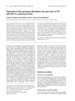
Fig. 2 Inhibition of autophagy enhances the inhibitory effect of cisplatin in A549 cells. a Inhibition of autophagy by CQ markedly decreased A549
cell viability. The IC50 value was 38.46 μM. b CQ (10 μM) enhanced the inhibitory effect of 0.5 μM cisplatin on cell proliferation. c CQ enhanced the
inhibitory effect of cisplatin on cell colony formation. d CQ suppressed the expression level of cisplatin-induced autophagy (see Additional file 1:
Supplementary Figure 8-9).
(Fig. 5a). Knockdown of APE1 attenuated autophagy in
the presence of cisplatin or CQ treatment alone or in
combination; however, the depletion of APE1 enhanced
apoptosis following treatment with cisplatin alone or in
combination with CQ. Notably, the combinatorial treatment of CQ and cisplatin exerted the most effective
apoptotic effect in the presence of APE1 knockdown
(Fig. 5b). The protein expression level of LC3 and activation by the cleavage of PARP and caspase 3 reflected the
autophagy and apoptosis (Fig. 5c). Notably, emerging
data found that cisplatin stimulates p53 activity [31].
Consistent with the results, our data showed that cisplatin treatment alone or in combination with CQ increased p53 expression, as well as p-p53 (ser15)
expression (Fig. 5c), suggesting that p53 is involved in
the response to cisplatin treatment in A549 cells. Taken
together, these results suggest that combined blockade
of APE1 and autophagy enhances cisplatin-induced
apoptosis with the involvement of p53 activation in
A549 cells.
Dual repression of APE1 and autophagy reverses cisplatin
resistance in cisplatin-resistant A549 cells
Acquired cisplatin resistance is the major cause of
chemotherapy failure in the treatment of lung cancer;
thus, we speculated that dual inhibition of APE1 and autophagic flux would overcome cisplatin resistance. We
tested the effects on cell proliferation and apoptosis in
Pan et al. BMC Cancer
(2020) 20:634
Page 7 of 11
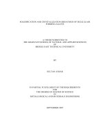
Fig. 3 Inhibition of autophagy enhances cisplatin-induced apoptosis in A549 cells. a Inhibition of autophagy by CQ markedly enhanced cisplatininduced apoptosis in A549 cells. b Western blotting showed that CQ enhanced cisplatin-induced apoptosis in A549 cells (see Additional file 1:
Supplementary Figure 10-12).
acquired cisplatin-resistant A549 cells (parental A549
cells were exposed to cisplatin to develop the acquired
resistant cell line, named A549/CDDP [32]) following
dual inhibition of both the APE1 and autophagy. The
cell viability was decreased in A549/CDDP cells receiving combinatorial treatment with cisplatin and CQ or
cisplatin and APE1 siRNA compared with the vehicle or
mono-treatment (Fig. 5d). Moreover, the most inhibitory
effect on cell viability was observed in A549/CDDP cells
receiving the combinatorial treatment with cisplatin,
CQ, and APE1 knockdown (Fig. 5d). Next, the A549/
CDDP cells were treated with a high concentration of
cisplatin (20 μM) alone or in combination. Co-treatment
with cisplatin and CQ resulted in increased apoptosis
compared with mono-treatment in A549/CDDP cells,
but co-treatment with APE1-SiRNA and cisplatin or CQ
showed no enhancement in apoptosis compared with
mono-treatment (Fig. 5e). The most potent apoptotic effect was observed in A549/CDDP cells treated with cisplatin, CQ, and APE1-SiRNA together. Knockdown of
APE1 markedly sensitized A549/CDDP cells to the cisplatin/CQ combinatorial treatment (Fig. 5e). Protein expression also showed that the activation of p53 and
PARP and caspase 3 cleavage occurred in A549/CDDP
cells treated with cisplatin, CQ, and APE1-SiRNA together (Fig. 5f). No significant cleavage of PARP and caspase 3 was observed in the absence of APE1-SiRNA (Fig.
5F), suggesting the enhancing role of APE1 knockdown
in apoptosis in cisplatin-resistant cells exposed to an autophagy inhibitor. Together, the data suggest that the
combined blockade of APE1 and autophagy may be an
effective strategy to overcome cisplatin resistance.
Discussion
Lung cancer is the leading cause of cancer death, and a
lack of efficacious therapeutics exists. Cisplatin, the most
important chemotherapeutic drug in lung cancer therapy, has shown limited clinical efficacy due to drug resistance. Autophagy and other key cellular events,
including the DNA damage repair response, are involved
in chemo-drug resistance. Notably, there is increasing
attention on the genetic context dependence in lung
cancer therapy. Compelling evidence has shown that
oncogenic RAS mutations vary, although they all promote cancer cell proliferation [4–6]. Zhong et al. found
that inhibition of RAS-AKT-mTOR signalling and blockage of late stage autophagy could synergistically enhance
the cytotoxicity of a tumour suppressor gene ARHI [33].
Specific RAS alleles exhibit differential biochemical features, displaying preferential signalling output and
favouring differential downstream effectors that are subject to differential feedforward and feedback regulations.
Therefore, individualized therapeutics are advocated in
cancer therapy.
In this study, we first applied SILAC proteomics to
obtain a panoramic view of cisplatin treatment in
KRASG12S-mutant A549 cells. At least 3262 protein molecules responded to cisplatin treatment and included
APE1, p53, and LC3-I/II, which are involved in DNA
damage repair, cell proliferation, apoptosis, and autophagy. Subsequent IPA analysis revealed 72 canonical signalling pathways including the BER pathway, DNA
double-strand break repair, and autophagy pathways.
Autophagy is a well-known cell-protective mechanism
related to tumor progression, drug-resistance, and
Pan et al. BMC Cancer
(2020) 20:634
Page 8 of 11
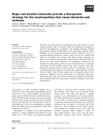
Fig. 4 APE1 knockdown induces autophagy. a Si-RNA-mediated APE1 knockdown increased LC3-I/II expression (see Additional file 1: Supplementary
Figure 13-14). b and c Inhibition of APE1 via E3330 or AT-101 increased LC3-I/II expression in A549 cells (see Additional file 1: Supplementary Figure 1516). d Confocal microscopy showed that depletion of APE1 affected LC-3I/II localization. e Immunoprecipitation showed the interaction between APE1,
LC3, and p53 in A549 cells (see Additional file 1: Supplementary Figure 17-20).
survive [8], and blockade of RAS/RAF/MEK/ERK signalling flux promotes autophagy [10], suggesting that inhibition of autophagy is beneficial. Kinsey et al. [10] and
Bryant et al. [11] have shown synergistic antitumor effects of autophagy inhibition and MAPK inhibition in
RAS-driven cancers, including pancreatic ductal adenocarcinoma, melanoma, and colorectal cancer, in preclinical settings [10, 11]. This combined blockade of
autophagy with other therapeutics revealed a novel
therapeutic vulnerability to treat RAS-driven cancers, including lung cancer. Based on our previous research
concerning BER pathway in platinum-resistance of lung
cancer [30] and present proteomic results, we herein
aimed to investigate whether BER and autophagy have
interaction upon cisplatin treatment in lung cancer cells.
By methods of flow cytometry, fluorescence microscopy,
Western blotting and RNA interference, we found that
cisplatin markedly induced autophagy and apoptosis in
A549 cells, accompanied by remarkable increase of DNA
repair protein APE1. Suppression of autophagy enhanced the inhibition effect of cisplatin on cell growth,
proliferation, and colony formation. The combination
treatment of CQ, an autophagy inhibitor, with cisplatin
dramatically enhanced cisplatin-induced apoptosis.
Moreover, APE1 is a major contributor to cisplatin resistance in lung cancer [15]. In the present study, knockdown of APE1 enhanced cisplatin-induced apoptosis in
both A549 cells and cisplatin-resistant A549 cells. Noteworthy, APE1 knockdown significantly synergized the
apoptosis-inducing effect of cisplatin plus CQ. This dual
inhibition of APE1 and autophagy could minimize the
curative concentration of cisplatin in cisplatin-resistant
A549 cells. The lower concentration of cisplatin was
beneficial in reducing the side effects of chemotherapy
that commonly occur in clinical settings. Besides, the
specific targeting of autophagy without affecting other
Pan et al. BMC Cancer
(2020) 20:634
Page 9 of 11
Fig. 5 Combined blockade of autophagy and APE1 increases chemosensitivity and overcomes cisplatin resistance. a and b Knockdown of APE1
enhanced cisplatin-induced apoptosis in combination with autophagy inhibition by CQ. c Western blotting showed the enhancing effect of APE1
silencing on cisplatin-induced apoptosis in combination with autophagy inhibition by CQ (see Additional file 1: Supplementary Figure 21-26). d
Knockdown of APE1 enhanced cisplatin sensitivity in combination with autophagy inhibition in cisplatin-resistant A549 cells. e Knockdown of
APE1 enhanced cisplatin-induced apoptosis in combination with autophagy inhibition in cisplatin-resistant A549 cells. f Western blotting showed
the enhancing effect of APE1 silencing on cisplatin-induced apoptosis in combination with autophagy inhibition in cisplatin-resistant A549 cells
(see Additional file 1: Supplementary Figure 27-31).
cellular processes has drawn a great attention of researchers. Mutations in the RAS pathway are often associated with the high levels of autophagy that are
required to maintain cancer cell metabolism [34, 35].
The optimal dosage of autophagy inhibitors and timing
of inhibition are vital parameters for maximal therapeutic efficiency. Hopefully, Levy et al. reported that the
treatment of CQ as an autophagy inhibitor in some cancer patients showed no adverse toxicity for extended
time periods [36]. This demonstrates that long-term
treatment with lysosomal autophagy inhibitors is feasible. Provided that cancer cells are more dependent than
normal tissues on autophagy, even a drug that causes
some normal tissue toxicity can have a valuable therapeutic window for an effective cancer treatment [8]. In
inducible Atg7-knockout mice, the growth of KRASdriven lung tumors was significantly inhibited before any
signs of neurotoxicity [37], indicating that therapeutic
window for autophagy inhibition exists in some cancers.
Additionally, our previous data found that promoting
p53 intracellular stability by interfering with APE1 is a
possible mechanism in genistein-induced apoptosis [38].
In the present study, we applied immunoprecipitation to
explore the possible interactions between LC3-I/II and
key proteins involved in DNA damage, including APE1
and p53. An interesting triple complex comprising
APE1-p53-LC3 was formed in response to cisplatin plus
CQ in A549 cells. Taken together, our results suggest
that dual inhibition of APE1 and autophagy could enhance chemo-sensitivity and overcome cisplatin
Pan et al. BMC Cancer
(2020) 20:634
Page 10 of 11
resistance by boosting apoptosis via the modulation of
APE1-p53-LC3 complex assembly in a KRASG12S context. Whether the current findings regarding the cellular
events recapitulate other RAS mutations in lung cancer
or KRASG12S mutation in other cancer types in response
to cisplatin treatment is unknown and warrants further
investigation to better tailor specific therapeutic vulnerabilities for lung cancer treatment [3].
was supported by National Natural Science Foundation of China (81860480)
and Youth Science Fund Project of Science and Technology Department of
Jiangxi Province (20181BAB215022). The sponsor reviewed and approved the
study protocol and the final version of the manuscript. All the analytic decisions were made by the authors, and the final version of the manuscript was
approved by all authors.
Conclusions
In summary, this study revealed a proteomic response to
cisplatin in KRASG12S-mutant A549 cells. APE1, p53,
and LC3-I/II were identified to be involved in DNA
damage repair, cell proliferation, apoptosis, and autophagy. Dual inhibition of APE1 and autophagy synergistically enhanced cisplatin-induced apoptosis via the
regulation of APE1-p53-LC3 complex assembly. This
novel combination strategy is of great potential to overcome cisplatin resistance in the context of KRASG12Smutant lung cancer.
Ethics approval and consent to participate
This study was approved by the Ethics Committee of The First Affiliated
Hospital of Chengdu Medical College (the approval number is
2019CYFYHEC-MS-02). No ethics approval was required for the use of human
cell lines in this study. Not applicable about data from any individual person
and animal.
Supplementary information
Supplementary information accompanies this paper at />1186/s12885-020-07111-w.
Additional file 1: Figure S1-S31. Original gels and blot images. Image
Lab 3.0 software (Bio-Rad, USA) was used to analyse the blots. The
cropping of the blots was labelled with the symbol of “↓”. Corresponding
uncropped full-length blots are presented in Supplementary Figure [1–
31].
Additional file 2: Figure 32. SILAC-based proteomics identifies cellular
response molecules and some related signalling pathways in cells. A
Twenty-seven cranial signalling pathways. B Hot-point picture. C BER signalling pathway. D Autophagy signalling.
Abbreviations
APE1: Apurinic/apyrimidinic endonuclease 1; NSCLC: Non-small cell lung
cancer; CDDP: Cisplatin; CQ: Chloroquine; DMSO: Dimethyl sulfoxide;
DMEM: Dulbecco’s modified Eagle’s medium; PBS: Dulbecco’s phosphatebuffered saline; FBS: Foetal bovine serum; SILAC: Stable isotope labelling by
amino acids in cell culture; LSCM: Laser scanning confocal microscope;
DAPI: 6-diamidino-2-phenylindole; MTT: 2-(4,5-dimethylthiazol-2-yl)-2,5diphenyltetrazolium bromide; RIPA: Radioimmunoprecipitation assay buffer;
PVDF: Polyvinylidene difluoride; IPA: INGENUITY Pathway Analysis
Acknowledgements
We thank Prof. Dong Wang (from Daping Hospital, Army Medical University)
for his kind advice concerning the design of the study and for improving the
language and data interpretation of the manuscript.
Authors’ contributions
STP performed most of the experiments. JZ and FY performed some of the
experiments and prepared Figs. 1, 2, 3, 4 and 5. SFZ contributed to the study
design and revised the manuscript. TR sponsored and performed the study
and was a major contributor to the writing of the manuscript. All the
authors read and approved the final manuscript.
Funding
The collection, analysis, and interpretation of data in this study were
supported by grants from the Science & Technology Department of Sichuan
Province (2020YJ0451), Introduction Foundation of High-level Talents of The
First Affiliated Hospital, Chengdu Medical College (CYFY-GQ22), Health Commission of Sichuan Province (17PJ586). The manuscript writing and editing
Availability of data and materials
The datasets used in the current study are available from the corresponding
author on reasonable request.
Consent for publication
Not Applicable.
Competing interests
The authors declare no competing interests.
Author details
1
Department of Oral and Maxillofacial Surgery, The First Affiliated Hospital of
Nanchang University, 17 Yongwai Main St, Nanchang 330006, Jiangxi, China.
2
Health Management Centre, The First Affiliated Hospital, Chengdu Medical
College, 278 Baoguang St, Xindu Distr, Chengdu 610500, Sichuan, China.
3
Department of Pharmaceutical Sciences, College of Pharmacy, University of
South Florida, 12901 Bruce B. Downs Boulevard, Tampa, Florida 33612, USA.
4
Oncology Department, The First Affiliated Hospital, Chengdu Medical
College, 278 Baoguang St, Xindu Distr, Chengdu 610500, Sichuan, China.
Received: 13 March 2020 Accepted: 25 June 2020
References
1. Bray F, Ferlay J, Soerjomataram I, Siegel RL, Torre LA, Jemal A. Global cancer
statistics 2018: GLOBOCAN estimates of incidence and mortality worldwide
for 36 cancers in 185 countries. CA Cancer J Clin. 2018;68(6):394–424.
2. Feng RM, Zong YN, Cao SM, Xu RH. Current cancer situation in China: good
or bad news from the 2018 global Cancer statistics? Cancer Commun
(Lond). 2019;39(1):22. />3. Rosell R, Bivona TG, Karachaliou N. Genetics and biomarkers in
personalisation of lung cancer treatment. Lancet. 2013;382(9893):720–31.
4. Haigis KM. KRAS alleles: the devil is in the detail. Trends Cancer. 2017;3(10):
686–97.
5. Hobbs GA, Der CJ. RAS mutations are not created equal. Cancer Discov.
2019;9(6):696–8.
6. Gao Y, Chang MT, McKay D, Na N, Zhou B, Yaeger R, Torres NM, Muniz K,
Drosten M, Barbacid M, et al. Allele-specific mechanisms of activation of
MEK1 mutants determine their properties. Cancer Discov. 2018;8(5):648–61.
7. White E. The role for autophagy in cancer. J Clin Invest. 2015;125(1):42–6.
8. Levy JMM, Towers CG, Thorburn A. Targeting autophagy in cancer. Nat Rev
Cancer. 2017;17(9):528–42.
9. Garcia-Cano J, Ambroise G, Pascual-Serra R, Carrion MC, Serrano-Oviedo L,
Ortega-Muelas M, Cimas FJ, Sabater S, Ruiz-Hidalgo MJ, Sanchez Perez I,
et al. Exploiting the potential of autophagy in cisplatin therapy: a new
strategy to overcome resistance. Oncotarget. 2015;6(17):15551–65.
10. Kinsey CG, Camolotto SA, Boespflug AM, Guillen KP, Foth M, Truong A,
Schuman SS, Shea JE, Seipp MT, Yap JT, et al. Protective autophagy elicited
by RAF-->MEK-->ERK inhibition suggests a treatment strategy for RAS-driven
cancers. Nat Med. 2019;25(4):620–7.
11. Bryant KL, Stalnecker CA, Zeitouni D, Klomp JE, Peng S, Tikunov AP, Gunda
V, Pierobon M, Waters AM, George SD, et al. Combination of ERK and
autophagy inhibition as a treatment approach for pancreatic cancer. Nat
Med. 2019;25(4):628–40.
12. Dasari S, Tchounwou PB. Cisplatin in cancer therapy: molecular mechanisms
of action. Eur J Pharmacol. 2014;740:364–78.
Pan et al. BMC Cancer
(2020) 20:634
13. Hill DP, Harper A, Malcolm J, McAndrews MS, Mockus SM, Patterson SE,
Reynolds T, Baker EJ, Bult CJ, Chesler EJ, et al. Cisplatin-resistant triplenegative breast cancer subtypes: multiple mechanisms of resistance. BMC
Cancer. 2019;19(1):1039. />14. Wang J, Wu GS. Role of autophagy in cisplatin resistance in ovarian cancer
cells. J Biol Chem. 2014;289(24):17163–73.
15. Wang D, Xiang DB, Yang XQ, Chen LS, Li MX, Zhong ZY, Zhang YS. APE1
overexpression is associated with cisplatin resistance in non-small cell lung
cancer and targeted inhibition of APE1 enhances the activity of cisplatin in
A549 cells. Lung Cancer. 2009;66(3):298–304.
16. Li M, Wilson DM. Human apurinic/apyrimidinic endonuclease 1. Antioxid
Redox Signal. 2014;20(4):678–707.
17. Wen X, Lu R, Xie S, Zheng H, Wang H, Wang Y, Sun J, Gao X, Guo L. APE1
overexpression promotes the progression of ovarian cancer and serves as a
potential therapeutic target. Cancer Biomark. 2016;17(3):313–22.
18. Xiao X, Yang Y, Ren Y, Zou D, Zhang K, Wu Y. rs1760944 Polymorphism in
the APE1 Region is Associated with Risk and Prognosis of Osteosarcoma in
the Chinese Han Population. Sci Rep. 2017;7(1):9331. />s41598-017-09750-9.
19. Peng Y, Li Z, Zhang S, Xiong Y, Cun Y, Qian C, Li M, Ren T, Xia L, Cheng Y,
et al. Association of DNA base excision repair genes (OGG1, APE1 and
XRCC1) polymorphisms with outcome to platinum-based chemotherapy in
advanced nonsmall-cell lung cancer patients. Int J Cancer. 2014;135(11):
2687–96.
20. Juhnke M, Heumann A, Chirico V, Hoflmayer D, Menz A, Hinsch A, HubeMagg C, Kluth M, Lang DS, Moller-Koop C, et al. Apurinic/apyrimidinic
endonuclease 1 (APE1/Ref-1) overexpression is an independent prognostic
marker in prostate cancer without TMPRSS2:ERG fusion. Mol Carcinog. 2017;
56(9):2135–45.
21. He SJ, Shu LP, Zhou ZW, Yang T, Duan W, Zhang X, He ZX, Zhou SF. Inhibition
of Aurora kinases induces apoptosis and autophagy via AURKB/p70S6K/RPL15
axis in human leukemia cells. Cancer Lett. 2016;382(2):215–30.
22. Zhai D, Cui C, Xie L, Cai L, Yu J. Sterol regulatory element-binding protein 1
cooperates with c-Myc to promote epithelial-mesenchymal transition in
colorectal cancer. Oncol Lett. 2018;15(4):5959–65.
23. Zhang Z, Shao Z, Xiong L, Yang S. Inhibition of autophagy enhances
cisplatin-induced apoptosis in the MG63 human osteosarcoma cell line.
Oncol Lett. 2015;10(5):2941–6.
24. Shi S, Tan P, Yan B, Gao R, Zhao J, Wang J, Guo J, Li N, Ma Z. ER stress and
autophagy are involved in the apoptosis induced by cisplatin in human
lung cancer cells. Oncol Rep. 2016;35(5):2606–14.
25. Chen JH, Zhang P, Chen WD, Li DD, Wu XQ, Deng R, Jiao L, Li X, Ji J, Feng
GK, et al. ATM-mediated PTEN phosphorylation promotes PTEN nuclear
translocation and autophagy in response to DNA-damaging agents in
cancer cells. Autophagy. 2015;11(2):239–52.
26. Das CK, Mandal M, Kogel D. Pro-survival autophagy and cancer cell
resistance to therapy. Cancer Metastasis Rev. 2018;37(4):749–66.
27. Hall EA, Ramsey JE, Peng Z, Hayrapetyan D, Shkepu V, O'Rourke B, Geiger W,
Lam K, Verschraegen CF. Novel organometallic chloroquine derivative
inhibits tumor growth. J Cell Biochem. 2018;119(7). />jcb.26787.
28. Circu M, Cardelli J, Barr M, O'Byrne K, Mills G, El-Osta H. Modulating
lysosomal function through lysosome membrane permeabilization or
autophagy suppression restores sensitivity to cisplatin in refractory nonsmall-cell lung cancer cells. PLoS One. 2017;12(9):e0184922. />10.1371/journal.pone.0184922.
29. Zhang Y, Liao Z, Zhang LJ, Xiao HT. The utility of chloroquine in cancer
therapy. Curr Med Res Opin. 2015;31(5):1009–13.
30. Ren T, Shan J, Qing Y, Qian C, Li Q, Lu G, Li M, Li C, Peng Y, Luo H, et al.
Sequential treatment with AT-101 enhances cisplatin chemosensitivity in
human non-small cell lung cancer cells through inhibition of apurinic/
apyrimidinic endonuclease 1-activated IL-6/STAT3 signaling pathway. Drug
Des Devel Ther. 2014;8:2517–29.
31. Hu F, Guo XL, Zhang SS, Zhao QD, Li R, Xu Q, Wei LX. Suppression of p53
potentiates chemosensitivity in nutrient-deprived cholangiocarcinoma cells
via inhibition of autophagy. Oncol Lett. 2017;14(2):1959–66.
32. Kuang P, Zhou C, Li X, Ren S, Li B, Wang Y, Li J, Tang L, Zhang J, Zhao Y.
Proteomics-based identification of secreted protein dihydrodiol
dehydrogenase 2 as a potential biomarker for predicting cisplatin efficacy in
advanced NSCLC patients. Lung Cancer. 2012;77(2):427–32.
Page 11 of 11
33. Zhong C, Shu M, Ye J, Wang X, Liu Z, Zhao W, Zhao B, Zheng Z, Yin Z, Gao
M, et al. Oncogenic Ras is downregulated by ARHI and induces autophagy
by Ras/AKT/mTOR pathway in glioblastoma. BMC Cancer. 2019;19:441.
/>34. Lock R, Roy S, Kenific CM, Su JS, Salas E, Ronen SM, Debnath J. Autophagy
facilitates glycolysis during Ras-mediated oncogenic transformation. Mol
Biol Cell. 2011;22(2):165–78.
35. Guo JY, Chen HY, Mathew R, Fan J, Strohecker AM, Karsli-Uzunbas G,
Kamphorst JJ, Chen G, Lemons JM, Karantza V, et al. Activated Ras requires
autophagy to maintain oxidative metabolism and tumorigenesis. Genes
Dev. 2011;25(5):460–70.
36. Levy JM, Thompson JC, Griesinger AM, Amani V, Donson AM, Birks DK,
Morgan MJ, Mirsky DM, Handler MH, Foreman NK, et al. Autophagy
inhibition improves chemosensitivity in BRAF(V600E) brain tumors. Cancer
Discov. 2014;4(7):773–80.
37. Karsli-Uzunbas G, Guo JY, Price S, Teng X, Laddha SV, Khor S, Kalaany NY,
Jacks T, Chan CS, Rabinowitz JD, et al. Autophagy is required for glucose
homeostasis and lung tumor maintenance. Cancer Discov. 2014;4(8):914–27.
38. Zhu J, Zhang C, Qing Y, Cheng Y, Jiang X, Li M, Yang Z, Wang D. Genistein
induces apoptosis by stabilizing intracellular p53 protein through an APE1mediated pathway. Free Radical Bio Med. 2015;86:209–18.
Publisher’s Note
Springer Nature remains neutral with regard to jurisdictional claims in
published maps and institutional affiliations.