Distinct immunophenotypes and prognostic factors in renal cell carcinoma with sarcomatoid differentiation: A systematic study of 19 immunohistochemical markers in 42 cases
Bạn đang xem bản rút gọn của tài liệu. Xem và tải ngay bản đầy đủ của tài liệu tại đây (1.56 MB, 8 trang )
Yu et al. BMC Cancer (2017) 17:293
DOI 10.1186/s12885-017-3275-8
RESEARCH ARTICLE
Open Access
Distinct immunophenotypes and
prognostic factors in renal cell carcinoma
with sarcomatoid differentiation: a
systematic study of 19
immunohistochemical markers in 42 cases
Wenjuan Yu1, Yuewei Wang3, Yanxia Jiang1, Wei Zhang2* and Yujun Li1*
Abstract
Background: Renal cell carcinoma (RCC) with sarcomatoid differentiation is a relatively rare tumor containing both
carcinoma and sarcomatoid components. However, there has not been a systemic study on immunophenotypes of
renal cell carcinoma with sarcomatoid differentiation, especially using some renal specific immunohistochemical
markers. In this study, we aimed to comprehensively investigate the distinct immunophenotypes of RCC with
sarcomatoid differentiation
to analyze the pathogenesis of sarcomatoid differentiation and identify new prognostic factors in RCC with
sarcomatoid differentiation.
Methods: A total of 42 cases of RCCs with sarcomatoid differentiation were enrolled into the study.
Immunohistochemistry study was performed on tissue microarrays to evaluate the expressions of 19
immunohistochemical markers including a series of epithelial, mesenchymal markers and RCC specific markers.
Kaplan-Meier method was applied to assess the prognostic values of CD10, CAIX, p53 and Bcl-2.
Results: Histologically, 42 cases of RCCs with sarcomatoid differentiation presented with different proportions
of carcinoma and sarcomatoid components. The cohort contained 35 cases of clear cell renal cell carcinoma
(CCRCC) and 7 cases of chromophobe renal cell carcinoma (ChRCC) based on the carcinoma components.
Immunohistochemically, all cases were positive for vimentin, and 80% of cases showed immunostaining for at
least one epithelial marker, such as CK, EMA, CK7 and CK18. Notably, the expression rates of CAIX, CD10 and PAX8
in sarcomatoid cells were 76%, 76% and 64%, respectively. The carcinoma component of the tumors showed
differentient labeling for CAIX, CD10, vimentin, CK7 and CD117 in CCRCC vs ChRCC, but the sarcomatoid
component lost the specificity for these markers (p < 0.05). Patients with positive expressions of CAIX, p53 and
Bcl-2 had a poor prognosis.
(Continued on next page)
* Correspondence: ; ;
2
Department of Pathology, 401 Hospital of People’s Liberation Army, 22
Minjiang Rd, Qingdao 266071, China
1
Department of Pathology, The Affiliated Hospital of Qingdao University, 16
Jiangsu Road, Qingdao 266003, China
Full list of author information is available at the end of the article
© The Author(s). 2017 Open Access This article is distributed under the terms of the Creative Commons Attribution 4.0
International License ( which permits unrestricted use, distribution, and
reproduction in any medium, provided you give appropriate credit to the original author(s) and the source, provide a link to
the Creative Commons license, and indicate if changes were made. The Creative Commons Public Domain Dedication waiver
( applies to the data made available in this article, unless otherwise stated.
Yu et al. BMC Cancer (2017) 17:293
Page 2 of 8
(Continued from previous page)
Conclusions: The sarcomatoid cells in RCC with sarcomatoid differentiation express both epithelial and mesenchymal
markers, supporting their epithelial origin. PAX8, CAIX and CD10 could be used as the reliable and useful markers to
determine the renal origin of sarcomatoid cells such as in fine needle aspiration cases and metastatic RCC with
sarcomatoid differentiation. CAIX, p53 and Bcl-2 might play important roles in the transformation from renal cell
carcinoma to high malignant sarcomatoid differentiation, and these three immunohistochemical markers are adverse
prognostic factors for the survival of patients with RCC with sarcomatoid differentiation.
Keywords: Renal cell carcinoma, Sarcomatoid, Pathogenesis, Immunohistochemistry
Background
Renal cell carcinoma (RCC) with sarcomatoid differentiation is a rare renal carcinoma, also called sarcomatiod
renal cell carcinoma, spindle cell carcinoma and carcinomatous sarcoma, etc. One of its key histological features
is the presence of both carcinoma and sarcomatoid differentiation in the tumor [1]. According to the 2004 and
2016 WHO Classification of Tumors of the Urinary System and Male Genital Organs, RCC with sarcomatoid differentiation is not recognized as a separate and distinct
entity and the sarcomatoid component could arise from
the genetic background of any of the RCC subtypes [2].
RCC with sarcomatoid differentiation presents unique
histological characteristics, invasive behavior and poor
prognosis [3]. Due to the low morbidity and the deficiency
of study data, the pathogenesis of the transformation from
carcinoma to sarcomatoid component along with the
prognostic factors for RCC with sarcomatoid differentiation remains unelucidated. Herein we performed our
immunohistological study using 19 immune markers, including the renal specific markers PAX8 and CAIX in 42
cases of RCCs with sarcomatoid differentiation. Our study
was focused on the analysis of the expression differences
between those two components. This study may shed
some light on revealing the pathogenesis and assessing the
prognosis of the patients with this relatively rare renal
carcinoma.
Methods
Among the 42 cases of RCCs with sarcomatoid differentiation, 35 cases were collected in the Affiliated Hospital
of Qingdao University and 7 cases were collected in 401
Hospital of People’s Liberation Army from November
2003 to January 2015. All cases were re-diagnosed by
two pathologists majored in Genitourinary Pathology.
The samples were made into 2 tissue microarrays and
there were 42 punches in each tissue microarray. Each
tumor was made into 2 punches including carcinoma
and sarcomatoid components separately based on the
observation under microscope. The diameter of the cores
was 2 mm.
All tissues were 4-um-thick, fixed with 4% neutral
formaldehyde, and embedded in paraffin for H&E
staining. Immunohistochemistry was performed on tissue
microarrays comprising of 42 cases containing both
typical renal cell carcinoma cells and sarcomatoid cells.
Primary antibodies used in the study including vimentin (Cell Marque, V9), cytokeratin (CK) (Origene, AE1/
AE3), epithelial membrane antigen (EMA) (Thermo,
E29), cytokeratin-7 (CK7) (Cell Marque, OV-TL12/30),
cytokeratin-18 (CK18) (Origene, UMAB50), high molecular
weight cytokeratin (HMW-CK) (Cell Marque, CKHMW),
and CAIX (Novus, polyclonal), CD10 (Ventana, SP67),
PAX8 (Ventana, MRQ-50), alpha-methylacylCoA racemase
(AMACR) (Zeta,13H4), CD117 (Ventana, 9.7), CD99
(Covance, O13), SMA (Origene, 1A4), CD34 (Leica,
OBEnd/10), S100 (Thermo, 4c4.9), Melanoma (Cell
Marque, HMB45), and Melan A (Cell Marque, A103),
p53 (Ventana, DO-7), and Bcl-2 (Thermo, 8c8). UltraView™
DAB detection kit was purchased from Ventana (Arizona,
America). All immunohistochemistry assays were performed on the Roche BenchMark XT fully automatic
IHC/ISH instrument by optimized protocols. Positive
and negative controls were used in this study.
The positive staining of vimentin, CK, CK7, CK18,
HMW-CK, CD34, AMACR, SMA, Bcl-2, HMB45 and
melan A predominantly appeared in the tumor cell cytoplasm, whereas p53 and PAX8 mainly appeared in the
nuclei. CD99, EGFR and CAIX tended to express on the
cell membrane. EMA, CD10 and CD117 expressed on
the cell membrane or cytoplasm; S100 expressed in the
cytoplasm and nuclei. The positive staining was first
scored as 0, 1, 2, 3 based on the staining intensity (no
staining, faint, mild, strong) and the percentage of positively stained cells was (0%, ≤25%, 26% ~ 75%, >75%), respectively. Two scores then multiplied together, which
were considered as the final scores, grading as – (0–1),
1+ (2–4), 2+ (5–6), 3+ (7–9).
Clinical follow-up and survival analysis
Follow-up data were collected for 33 cases. Survival was
calculated from the date of surgery to the death or the
last follow-up visit. For the analysis, only deaths for RCCs
with sarcomatoid differentiation were considered as
events. Survival curves were derived from Kaplan-Meier
analysis and log-rank test to compare overall survival
Yu et al. BMC Cancer (2017) 17:293
between different groups based on the expressions of
CD10, CAIX, p53 and Bcl-2 since the four markers might
play roles in the proliferation of tumors. Furthermore, in
order to investigate the factors that may affect survival
patterns, the P values for prognostic factors analyses were
adjusted for multiple analyses using Cox regression
model.
Statistical analysis
SPSS 13.0 software was applied to perform statistical
analysis. Statistical significance was tested by chi-square
test (or Fisher’s exact test) to compare each marker’s expression in the carcinoma and sarcomatoid components.
Statistical significance was considered as P value less
than 0.05.
Results
There were 35 clear cell renal cell carcinomas (CCRCCs)
and 7 chromophobe renal cell carcinomas (ChRCCs)
based on the carcinoma component. The sarcomatoid
components of the investigated tumors resembled these
entities as fibrosarcoma, leiomyosarcoma and malignant
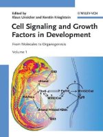
fibrous histiocytoma (Fig. 1a–d).
The immunohistochemistry results of the two components including carcinoma and sarcomatoid cells were
listed in Table 1. The cohorts could be divided into four
groups based on the expression panels of each immunohistochemical markers: carcinoma positive and sarcomatoid
cell positive (C+/S+); carcinoma positive and sarcomatoid
Page 3 of 8
cell negative (C+/S-); carcinoma negative and sarcomatoid
cell positive (C−/S+); carcinoma negative and sarcomatoid
cell negative (C−/S-). On the whole, the expressions of
vimentin, EMA, SMA, and Bcl-2 were significantly
different between carcinoma and sarcomatoid cells
(P < 0.05). No significant difference was revealed in the
expressions of other immunohistochemical markers
between the two components (P > 0.05). The following
is the detailed results.
The expression patterns of vimentin, CK, and EMA in
RCCs with sarcomatoid differentiation
The positive expression rates of vimentin were 52% (22/42)
in carcinoma cells and 100% (42/42) in sarcomatoid cell
(P = 0.000). The main expression panels of vimentin were
C+/S+ (22/42) and C−/S+ (20/42). The positive expression
rates of CK in carcinoma and sarcomatoid cells were 81%
(34/42) and 50% (21/42), respectively (P = 0.005). The
main expression panels of CK were C+/S+ (18/42)
and C+/S- (16/42). The positive expression rates of EMA
were 88% (37/42) and 50% (21/42) in carcinoma and sarcomatoid cells, respectively (P = 0.000). The expression
panels of EMA were C+/S+ (21/42) and C+/S- (16/42).
The expression patterns of CK7, CK18, and HMW-CK in
RCC with sarcomatoid differentiation
CK7 was expressed in carcinoma cells in 11 of 42 cases
(11/42) and sarcomatoid cells in 5 of 42 cases (5/42)
(P = 0.164). The expression panels of CK7 were C+/S-
Fig. 1 The histopathological figures of RCC with sarcomatoid differentiation. a CCRCC together with fibrosarcomatoid cells b ChRCC together with
fibrosarcomatoid cells c Clear cell carcinoma together with leiomyosarcoma-like cells d ChRCC together with malignant fibrous histiocytoma-like
cells. H&E × 100
Yu et al. BMC Cancer (2017) 17:293
Page 4 of 8
Table 1 The immunophenotypes of 42 cases of RCCs with
sarcomatoid differentiation and the expression differences
between carcinoma and sarcomatoid differentiation
Markers
C+/S+ C+/S- C−/S+ C−/S- Total C+ Total S+
Vimentin 22
P value
0
20
0
22 (52%) 42 (100%) 0.000*
CK
18
16
3
5
34 (81%) 21 (50%)
0.005*
EMA
21
16
0
5
37 (88%) 21 (50%)
0.000*
CK7
0
11
5
26
11 (26%) 5 (12%)
0.164
CK18
12
13
7
10
25 (60%) 19 (45%)
0.275
3 (7%)
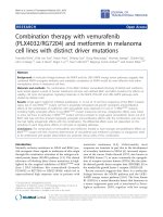
+/S+ (24/42), and C+/S- (10/42). The positive expressions of CAIX, CD10, and PAX8 were showed in
Fig. 2a, b, and c.
HMW-CK 0
0
3
39
0 (0%)
CAIX
20
1
12
9
21 (50%) 32 (76%)
0.023*
0.241
CD10
20
3
12
7
23 (55%) 32 (76%)
0.065
PAX8
24
10
3
4
34 (81%) 27 (64%)
0.141
AMACR
3
8
3
28
11 (26%) 6 (14%)
0.277
CD117
0
7
3
32
7 (17%)
3 (7%)
0.313
CD99
0
3
9
30
3 (7%)
9 (21%)
0.116
SMA
0
0
8
34
0 (0%)
8 (19%)
0.005*
CD34
0
0
0
42
0 (0%)
0 (0%)
−
S100
0
0
0
42
0 (0%)
0 (0%)
−
HMB45
0
0
0
42
0 (0%)
0 (0%)
−
MelanA
0
0
0
42
0 (0%)
0 (0%)
−
P53
8
2
12
20
10 (24%) 20 (48%)
0.040*
Bcl-2
9
16
0
17
25 (60%) 9 (21%)
0.001*
C carcinoma cell, S sarcomatoid cell
*The difference is statistically significant
(11/42) and C−/S+ (5/42). CK7 was not simultaneously
expressed in two types of cells. CK18 was expressed in
carcinoma cells in 25 of 42 cases (25/42) and sarcomatoid cells in 19 of 42 cases (19/42) (P = 0.275). The
main expression panels of CK18 were C+/S+ (12/42)
and C+/S- (13/42). HMW-CK was only expressed in the
sarcomatoid cells in 3 of 42 cases (3/42) (P = 0.241).
The expression patterns of CAIX, CD10, and PAX8 in RCC
with sarcomatoid differentiation
The positive expression rates of CAIX in carcinoma and
sarcomatoid cells were 50% (21/42) and 76% (32/42), respectively (P = 0.023). The major expression panels of
CAIXwere C+/S+ (20/42) and C−/S+ (12/42). Among
them, CAIX was diffusely strong expressed (3+) in
carcinoma cells in 18 of 42 cases and in sarcomatoid
cells in 29 of 42 cases. The positive expression rates of
CD10 in carcinoma and sarcomatoid cells were 55% (23/
42) and 76% (32/42), respectively (P = 0.065). The main
expression panels of CD10 were C+/S+ (20/42) and C−/S
+ (12/42). PAX8 was expressed in carcinoma cells in 34 of
42 cases (34/42) and sarcomatoid cells in 11 of 42 cases
(27/42) (P = 0.141). Notably, among them, 24 cases
showed co-expression of PAX8 in two types of cells
(24/42). The main expression panels of PAX8 were C
The expression patterns of AMACR, CD117, and CD99 in
RCC with sarcomatoid differentiation
The expression panels of AMACR were C+/S+ (3/42),
C+/S- (8/42), and C−/S+ (3/42). The expression patterns showed no difference between carcinoma and
sarcomatoid cells (P = 0.277). CD117 were merely
expressed in carcinoma cells in 7 of 42 cases and sarcomatoid cells in 3 of 42 cases (P = 0.313). In addition, 3
samples presented CD99 expression in carcinoma cells
and 9 cases presented CD99 expression in sarcomatoid
cells (P = 0.116).
The expression patterns of SMA, CD34, S100, HMB45, and
melanA in RCC with sarcomatoid differentiation
SMA was expressed in sarcomatoid cells in 8 cases without positive expression in carcinoma cells (P = 0.005).
But beyond that, CD34, S100, HMB45 and melanA were
not expressed in two types of cells in all cases.
The expression patterns of p53 and Bcl-2 in RCC with
sarcomatoid differentiation
Ten cases showed p53 expression in carcinoma cells and
20 cases showed p53 expression in sarcomatoid cells
(P = 0.04). The main expression panels of p53 were C+/S+
(8/42) and C−/S+ (12/42). The positive expression rates of
Bcl-2 in carcinoma and sarcomatoid cells were 60% (25/
42) and 21% (9/42), respectively (P = 0.001). The
expression panels of Bcl-2 were C+/S+ (9/42) and C+/S(16/42). The positive expression of p53 were showed in
Fig. 2d.
Expression differences of CAIX, CD10, vimentin, CK7 and
CD117 in the carcinoma and sarcomatoid cells between
CCRCC and ChRCC
The expressions of CAIX, CD10, vimentin, CK7 and
CD117 in the carcinoma component between CCRCC
and ChRCC showed significant differences (P < 0.05).
However, no significant difference was revealed in the sarcomatoid component between the two tumors (P > 0.05)
(Table 2).
Clinical follow-up and survival analysis
Follow-up data were collected for 33 cases with the
follow-up time ranged from 1 to 42 months. Among
them, 1 patient died from other disease rather than
tumor. There were another 24 deaths, including 22
CCRCCs and 2 ChRCCs according to the carcinoma elements which occurred in the 1st ~ 30th month after the
surgery for metastasis of tumors to the bone or lung.
Other 8 patients are still alive uneventfully after surgery.
Yu et al. BMC Cancer (2017) 17:293
Page 5 of 8
Fig. 2 Immunohistochemical staining figures of RCC with sarcomatoid differentiation. a The positive expression of CA IX in clear cell carcinoma
together with fibrosarcomatoid cells b The positive expression of CD10 in clear cell carcinoma together with fibrosarcomatoid cells c The positive
expression of PAX8 in ChRCC together with fibrosarcomatoid cells d The positive expression of p53 in fibrosarcomatoid cells and negative
expression in clear cell carcinoma. Immunohistochemistry × 100
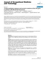
The Kaplan-Meier survival curves comparing overall
survival according to the expressions of CD10, CAIX, p53
and Bcl-2 were presented in Fig. 3. Univariate analysis
showed that the expressions of CAIX (p = 0.000), p53
(p = 0.000) and Bcl-2 (p = 0.001) were all adverse prognostic factors for tumor related survival (Fig. 3). Furthermore, the multiple analyses using the Cox regression
model revealed that CAIX, p53 and Bcl-2 were independent predictive factors for prognosis of RCC with sarcomatoid differentiation (P = 0.001, 0.011, 0.013, respectively).
Discussion
A malignant tumor comprising of malignant epithelial
cells together with mesenchymal cells is called
Table 2 The expression differences of CAIX, CD10, vimentin,
CK7 and CD117 between CCRCC and ChRCCs in the carcinoma
and sarcomatoid differentiation
CCRCC
ChRCC
P value
CCRCC
ChRCC
S+
S+
P value
C+
C+
CAIX
23/35
0/7
0.002*
29/35
7/7
0.567
CD10
21/35
0/7
0.009*
27/35
5/7
1.000
Vimentin
23/35
0/7
0.002*
35/35
7/7
−
CK7
3/35
7/7
0.000*
5/35
0/7
0.569
CD117
3/35
6/7
0.000*
2/35
0/7
1.000
C carcinoma cell, S sarcomatoid cell
*The difference is statistically significant
carcinosarcoma. According to the histological classification of 2016 WHO for the epithelial tumors of kidney, this type of tumor is regarded as transformation
from different types of renal cell carcinoma, rather than
an independent histological entity [2]. However, a large
sample and systematic study on immunophenotypes of
both carcinoma and sarcomatoid cells are still limited,
especially for some renal specific immunohistochemical
markers [4]. Herein we performed immunohistochemistry study to characterize 19 different markers in 42
cases of RCC with sarcomatoid differentiation and analyzed the expression patterns of these markers in both
carcinoma and sarcomatoid cells. We grouped these immunohistochemical markers into 4 subgroups as followed
based on their different physiological functions.
Epithelial markers
In the present study, sarcomatoid cells in 81% cases
expressed at least one epithelial marker. CK and EMA
were expressed in approximately 62% cases. Sarcomatoid
cells in 19 cases expressed CK18. However, 8 cases presented no expression of any tested epithelial marker. 5
cases showed CK7 positive staining in sarcomatoid cells
rather than in clear cell carcinomas. The expressions of
epithelial markers in sarcomatoid cells confirmed that
sarcomatoid cells resulted from different differentiation
orientations at different degrees of epithelial cells. Thus
we recommend a series of cytokeratins with different
Yu et al. BMC Cancer (2017) 17:293
Page 6 of 8
Fig. 3 The Kaplan-Meier survival curves comparing overall survival according to the expressions of CD10, CAIX, p53 and Bcl-2. Univariate and
multiple analyses showed that the positive expressions of CAIX, p53 and Bcl-2 were all adverse prognostic factors for tumor related survival
molecular weights to identify the epithelial expression
patterns of sarcomatoid cells.
Mesenchymal markers
As reported in the literature most sarcomatoid cells
expressed vimentin with the positive rates of 56%–100%
[5], few cases expressed actin and S100 [6]. In the present
study, all 42 cases showed strong positive expression of
vimentin in sarcomatoid cells. Among them, 30 cases
expressed epithelial markers, 8 cases expressed SMA, but
none of them expressed S100. Statistical analysis revealed
the expression of vimentin in carcinoma and sarcomatoid
cells was significantly different, which might be due to the
lack of vimentin expression in 4 ChRCCs. Notably, vimentin was expressed in the sarcomatoid cells of CCRCC,
which is different from that of ChRCC.
RCCs specific markers
CAIX is a valuable marker indicating low endogenous
oxygen level, which catalyzes the formation of carbonic
acid from carbon dioxide and adjusts the pH value in
tumor cells to adapt to the surrounding microenvironment
and facilitate tumor proliferation. CAIX is now considered
as a highly sensitive and specific marker for clear cell
renal cell carcinoma [7]. Immunohistochemical study on
CAIX in RCC with sarcomatoid differentiation has been
rarely reported up till now. We revealed a high frequency
of expression of CAIX in sarcomatoid cells (90%) with extensive and strong staining. Moreover, strong CAIX staining in sarcomatoid cells had been observed regardless the
negative expression of CAIX in carcinoma cells (5
CCRCCs, 6 ChRCCs), indicating its involvement in proliferation of sarcomatoid cells and the transformation of carcinoma to sarcomatoid cells. It is known that CD10 could be
used as a supplementary diagnostic marker for renal cell
carcinoma. 32 cases showed strong CD10 expression in sarcomatoid cells. Among them, CD10 was expressed in both
carcinoma and sarcomatoid cells in 20 cases, whereas 12
cases presented CD10 expression only in sarcomatoid cells.
PAX8 belongs to PAX gene family, which is an important
transcription factor in renal organogenesis and a reliable
marker for primary kidney cancer [7]. Chang et al. [8] had
reported that the expression rates of PAX8 were 69% and
18% in sarcomatoid cells of RCC with sarcomatoid differentiation and sarcomatoid urothelial carcinoma, respectively.
However, it was rarely expressed in renal epithelioid
Yu et al. BMC Cancer (2017) 17:293
angiomyolipoma and leiomyosarcoma, suggesting that it
could be used as a good diagnostic marker for RCC with
sarcomatoid differentiation. The present study revealed that
PAX8 was expressed in carcinoma cells of 34 cases and sarcomatoid cells of 27 cases. Among them, 24 cases showed
extensive expression in both two components, which leads
to an idea that PAX8 could be used as a useful diagnostic
marker for identifying the RCC with sarcomatoid differentiation in fine needle aspiration cases and in metastatic RCC
with sarcomatoid differentiation. AMACR was rarely
expressed in sarcomatoid cells, and only 6 cases showed
strong AMACR expression in our study.
Prognosis and treatment related markers
CAIX, one of the most important tumor-associated carbonic anhydrases (CAs), is strongly induced by hypoxia
in renal carcinoma cells, which could promote the
growth of tumor cells. However, few research endeavors
have been devoted to studying its role in predicting the
outcome of RCC with sarcomatoid differentiation.
Tickoo et al. [9] reported that over expression of CAIX
showed no association with survival in 22 clear cell
RCCs and 12 nonclear cell RCCs with sarcomatoid differentiation. Whereas, the results of the present study
indicated that positive expression of CAIX was related
to the adverse prognosis of 42 cases of RCCs with sarcomatoid differentiation. Therefore, more cases are needed
for further identifying the predictive function of CAIX for
the prognosis of RCCs with sarcomatoid differentiation.
P53 is a tumor suppressor gene, which plays a key role
in carcinogenesis. Our study demonstrated that the expression rate of p53 was 48% in sarcomatoid cells, which
is higher than that in carcinoma cells, suggesting that
p53 might be involved in triggering the development of
high malignant sarcomatoid tumors from renal cell carcinoma. Furthermore, p53 could be a reliable marker for
predicting the prognosis or treatment outcome of RCC
with sarcomatoid differentiation.
Bcl-2 is an apoptosis suppressor gene; therefore its
overexpression is a hallmark of cell proliferation and
suppression of cell apoptosis. Our result showed a significant difference of Bcl-2 expressions between carcinoma cells and sarcomatoid cells, suggesting that Bcl-2
might facilitate the aberrant growth of carcinoma cells
and the transformation of carcinoma cells to high malignant sarcomatoid differentiation via suppression of
apoptosis. In addition, the expression of Bcl-2 was associated with the adverse outcome of RCC with sarcomatoid differentiation and Bcl-2 could be an important
prognostic factor for the overall survival in RCC with
sarcomatoid differentiation.
Additionally, Castillo et al. [10] had reported that sarcomatoid cells in 94.7% of RCC with sarcomatoid differentiation overexpressed CD117, suggesting the idea that
Page 7 of 8
tyrosine kinase inhibitor could be used for the therapy of
CD117 positive RCC with sarcomatoid components patients. Wang et al. [11] did not find any CD117 expression
in sarcomatoid urothelial carcinoma of the upper urinary
tract. Our results showed CD117 only expressed in sarcomatoid cells of 3 cases of RCC and carcinoma cells of 7
ChRCCs with sarcomatoid differentiation. More studies
are needed to discover the possible therapeutic use of
tyrosine kinase inhibitor for RCC with sarcomatoid
differentiation.
Due to the rarity of RCCs with sarcomatoid differentiation, more cases are needed to further confirm the specific
features of the expression panels of these 19 immunomarkers, especially the prognostic values of CAIX, p53 and
Bcl-2 playing in RCCs with sarcomatoid differentiation.
Conclusions
Sarcomatoid cells in RCC with sarcomatoid differentiation
express both epithelial and mesenchymal markers, supporting their epithelial origin. The difference of expression profiles between carcinoma cells and sarcomatoid cells might
be due to the distinct orientation and the degree of differentiation in renal cell carcinoma. CAIX, p53 and bcl-2 might
play important roles in triggering the transformation from
renal cell carcinoma to high malignant sarcomatoid component, and could be beneficial to predicting the prognosis or
treatment outcome of RCC with sarcomatoid differentiation. PAX8, CAIX and CD10 could be used as the reliable
markers to determine the renal origin of sarcomatoid cells,
especially in fine needle aspiration cases and the cases of
metastatic RCC with sarcomatoid differentiation. The clinical therapeutic application of tyrosine kinase inhibitors to
RCC with sarcomatoid differentiation needs to be investigated with more research studies and clinical trials.
Abbreviations
AMACR: Alpha-methylacylCoA racemase; CCRCC: Clear cell renal cell carcinoma;
ChRCC: Chromophobe renal cell carcinoma; RCC: Renal cell carcinoma
Acknowledgements
Not applicable.
Funding
This work was supported by National Natural Science Foundation of China
(81201654) in the design of the study and in writing the manuscript.
Shandong Province Science and Technology Development Plan
(2013GSF11866) and Medical Research Guiding Program of Qingdao
(2014WJZD195) contributed in collection, analysis, and interpretation of data.
Availability of data and materials
All data generated or analysed during this study are included in this
published article.
Authors’ contributions
WY was involved in drafting the manuscript and revising it critically for
important intellectual content. YW made substantial contributions to
acquisition of data. YJ analysis and interpretation of data. WZ was involved
in drafting the manuscript or revising it. YL made substantial contributions
to conception and design of the study. All authors read and approved the
final manuscript.
Yu et al. BMC Cancer (2017) 17:293
Page 8 of 8
Competing interests
The authors declare that they have no competing interests.
Consent for publication
Not applicable.
Ethics approval and consent to participate
The use of human subjects in the study has been approved by the Ethics
Committee of the Affiliated Hospital of Qingdao University. The patients
consented to participate in the study by verbal informed consent. The need
for written consent has been waived by the Ethics Committee of the
Affiliated Hospital of Qingdao University, and the Ethics Committee
approved the verbal informed consent.
Publisher’s Note
Springer Nature remains neutral with regard to jurisdictional claims in
published maps and institutional affiliations.
Author details
1
Department of Pathology, The Affiliated Hospital of Qingdao University, 16
Jiangsu Road, Qingdao 266003, China. 2Department of Pathology, 401
Hospital of People’s Liberation Army, 22 Minjiang Rd, Qingdao 266071, China.
3
Department of Vascular Surgery, The Affiliated Hospital of Qingdao
University, Qingdao 266003, China.
Received: 16 February 2016 Accepted: 11 April 2017
References
1. Eble JN, Sauter G, Epstein J, et al. World Health Organization classification of
tumours of the urinary system and male genital organs. Lyon, France: IARC
Press. 2004.
2. Moch H, Humphrey PA, Ulbright TM, et al. WHO classification of tumors of
the urinary and male genital organs. Lyon, France: IARC Press, 2016.
3. Pamela A, Arnoux V, Long JA, et al. Sarcomatoid renal cell carcinoma:
follow-up of a series of 23 patients. Prog Urol. 2014;24(5):301–6.
4. Lopez-Beltran A, Scarpelli M, Montironi R, et al. 2004 WHO classification of
the renal tumors of the adults. EurUrol. 2006; 49 (5): 798-805.
5. Yan Y, Liu L, Zhou J, et al. Clinicopathologic characteristics and prognostic
factors of sarcomatoid renal cell carcinoma. J Cancer Res Clin Oncol.
2015;141(2):345–52.
6. Gadre SA, Math SK, Elfeel KA, et al. Cytology of a Sarcomatoid renal cell
carcinoma with unusual Coexpression of S-100 protein: a case report,
review of the Literatureand Cytologic-Histologic correlation. Diagn
Cytopathol. 2009;37(3):195–8.
7. Zhang W, Yu W, Xia Y, et al. The expressions of CAIX, PAX2 and PAX8 in
epithelial renal tumors and the clinicopathological significance. Zhonghua
Bing Li Xue Za Zhi. 2013;42:442–5.
8. Chang A, Brimo F, Montgomery EA, et al. Use of PAX8 and GATA3 in
diagnosing sarcomatoid renal cell carcinoma and sarcomatoid urothelial
carcinoma. Hum Pathol. 2013;44(8):1563–8.
9. Tickoo SK, Alden DS, Fine S, et al. Immunohistochemical expression of
hypoxia inducible factor-1alpha and its downstream molecules in
sarcomatoid renal cell carcinoma. J Urol. 2007;177(4):1258–63.
10. Castillo M, Petit A, Mellado B, et al. C-kit expression in sarcomatoid renaI cell
carcinoma: potential therapy with imtinib. J Urol. 2004;171(6 Pt 1):2176–80.
11. Wang X, MacLennan GT, Zhang S, et al. Sarcomatoid carcinoma of the
upper urinary tract: clinical outcome and molecular characterization.
Hum Pathol. 2009;40(2):211–7.
Submit your next manuscript to BioMed Central
and we will help you at every step:
• We accept pre-submission inquiries
• Our selector tool helps you to find the most relevant journal
• We provide round the clock customer support
• Convenient online submission
• Thorough peer review
• Inclusion in PubMed and all major indexing services
• Maximum visibility for your research
Submit your manuscript at
www.biomedcentral.com/submit