Observation of Zn-photoprotoporphyrin red Autofluorescence in human bronchial cancer using color-fluorescence endoscopy
Bạn đang xem bản rút gọn của tài liệu. Xem và tải ngay bản đầy đủ của tài liệu tại đây (626.11 KB, 7 trang )
Ohsaki et al. BMC Cancer (2017) 17:289
DOI 10.1186/s12885-017-3277-6
RESEARCH ARTICLE
Open Access
Observation of Zn-photoprotoporphyrin
red Autofluorescence in human bronchial
cancer using color-fluorescence endoscopy
Yoshinobu Ohsaki1*, Takaaki Sasaki1, Satoshi Endo1, Masahiro Kitada1, Shunsuke Okumura1, Noriko Hirai1,
Yoshihiro Kazebayashi1, Eri Toyoshima1, Yasushi Yamamoto1, Kaneyoshi Takeyama1, Susumu Nakajima2
and Isao Sakata1,3
Abstract
Background: We observed red autofluorescence emanating from bronchial cancer lesions using a sensitive colorfluorescence endoscopy system. We investigated to clarify the origin of the red autofluorescence.
Methods: The wavelengths of the red autofluorescence emanating from lesions were measured in eight patients
using a spectrum analyzer and compared based on pathologic findings. Red autofluorescence at 617.3, 617.4, 619.0,
and 617.1 nm was emitted by normal bronchus, inflamed tissue, tissue exhibiting mild dysplasia, and malignant
lesions, respectively.
Protoporphyrin, uroporphyrin, and coproporphyrin, the major porphyrin derivatives in human blood, were
purchased to determine which porphyrin derivative is the source of red fluorescence when acquired de novo. We
synthesized photoporphyrin, Zn-protoporphyrin and Zn-photoprotoporphyrin from protoporphyrin.
Results: Coproporphyrin and uroporphyrin emitted only weak fluorescence. Fluorescence was emitted by our
synthesized Zn-photoprotoporphyrin at 625.5 nm and by photoprotoporphyrin at 664.0 nm.
Conclusions: From these results, we conclude that Zn-photoprotoporphyrin was the source of the red
autofluorescence observed in bronchial lesions. Zn-protoporphyrin is converted to Zn-photoprotoporphyrin by
radiation with excitation light. Our results suggest that red autofluorescence emanating from Zn-photoprotoporphyrin
in human tissues could interfere with photodynamic diagnosis using porphyrin derivatives such as Photofrin® and
Lazerphyrin® with a sensitive endoscopy system, because color cameras cannot differentiate Zn-photoprotoporphyrin
red fluorescence from that of other porphyrin derivatives.
Keywords: Photodynamic diagnosis, Autofluorescence, Endoscopy, Prophyrin, Zn-photoprotoporphyrin
Background
Components of the human body such as collagen,
nicotinamide-adenine dinucleotide phosphate (NADP),
and flavin-adenine dinucleotide (FAD), emit fluorescence
when irradiated with light of an appropriate excitation
wavelength [1, 2]. Normal human bronchial epithelial
tissue emits green autofluorescence at a wavelength of
ca. 540 nm due to NADP and FAD when excited with
405-nm blue light. This green autofluorescence is less
* Correspondence:
1
Respiratory Center, Asahikawa Medical University, 2-1-1-1 Midorigaoka
Higashi, Asahikawa 078-8510, Japan
Full list of author information is available at the end of the article
intense in cancer lesions due to thickening of the epithelium, reductions in the levels of the source materials,
and absorption of the fluorescence within the lesion.
Therefore, cancerous lesions of the bronchus will be
demonstrated by a reduction in the intensity of green
autofluorescence when the lesions are observed using
autofluorescence endoscopy.
Several endoscopy systems have been developed for
use in early detection of cancer lesions in the human
bronchus. These systems include the LIFE lung [3, 4]
(Xillix, Richmond, Canada), SAFE-3000 [5, 6] (Asahi
Optical, Tokyo, Japan), D-Light AF [7] (Storz, Tuttlingen,
Germany), and AFI (Olympus, Tokyo, Japan). Superior
© The Author(s). 2017 Open Access This article is distributed under the terms of the Creative Commons Attribution 4.0
International License ( which permits unrestricted use, distribution, and
reproduction in any medium, provided you give appropriate credit to the original author(s) and the source, provide a link to
the Creative Commons license, and indicate if changes were made. The Creative Commons Public Domain Dedication waiver
( applies to the data made available in this article, unless otherwise stated.
Ohsaki et al. BMC Cancer (2017) 17:289
Page 2 of 7
rates of early bronchial carcinoma detection using autofluorescence bronchoscopy (AFB) have been reported in
meta-analyses that included data from our study [8, 9]. Although, LIFE lung and SAFE 3000 can detect both red
and green fluorescence, only a decrease in the intensity of
green autofluorescence in the cancer lesion is detectable
using the above-mentioned systems, because their sensitivity is too low to permit visualization of color autofluorescence from human bronchial tissue and because a
black and white charged coupled device (CCD) is used in
the AFI system [10].
We developed a color fluorescence endoscopy system
(PDS-2000 [11, 12]; Hamamatsu Photonics, Hamamatsu,
Japan) to observe autofluorescence emanating from human tissues. This system detects both green autofluorescence from normal human organs as well as red
autofluorescence from the accumulation of administered
porphyrin derivatives. We compared the sensitivity of
detection for bronchial cancers and precancerous lesions
using this system and found that rate of lesion detection
increased significantly, from 54.1 to 89.2%, when AFB
was combined with white-light bronchoscopy [13]. During the above clinical study, we detected red autofluorescence emanating from cancer lesions, contact bleeding
sites, and blood vessels, and we reported that the red to
green autofluorescence ratio (R/G ratio) was significantly
higher in the cancer lesions [13].
The accumulation of de novo porphyrin derivatives in
cancer tissue, including the accumulation of protoporphyrin IX, has been reported [14, 15]. However, previous
reports were based on the results of spectral analyses of
resected tumor and drawn blood samples [16–18]. We
observed red autofluorescence in human cancer lesions,
contact bleeding sites, and the blood vessels of the
bronchial wall using a color AFB system. The wavelength of the observed red autofluorescence differed
from that reported in previous studies. In the present
study, we measured the wavelength of red autofluorescence in order to determine the fluorescent component.
This is the first report describing the origin of red autofluorescence observed in human cancer tissues, blood
vessels, and contact bleeding sites in living patients using
autofluorescence endoscopy.
average wavelength of 405 nm generated by a 300-W
xenon lamp using a band-pass filter is radiated through
the light channel of the fiberscope. The system is connected to an endoscope using an Olympus Endoscopy
System attachment.
Methods
Results
Autofluorescence endoscopy system
Analysis of the wavelength of autofluorescence
emanating from human bronchus
The PDS-2000 fluorescence endoscopy system was
developed by Hamamatsu Photonics and Asahikawa
Medical University [11–13, 19]. The system includes an
intensified color CCD camera, a red-green and blue
(RGB) control unit, a source of ca. 405-nm blue light,
and a blue-light cut filter. The RGB control unit contains an RGB frame memory, image averaging system,
scan converter, and camera control unit. Blue light of an
Analysis of autofluorescence spectra
Eight Asian patients with high risk of bronchial malignancy were enrolled in the present study. Seven patients
had previously treated bronchogenic carcinoma, and one
patient had history of bloody sputum (Table 1).
Bronchial lesions in eight patients were observed using a
bronchofiberscope connected to the PDS-2000 system.
Biopsy samples were taken from lesions exhibiting red
autofluorescence after measurement of the wavelength
emitted from each lesion; samples were also taken from
green autofluorescence–emitting tissue of the adjacent
normal bronchial wall. The wavelength of lesion autofluorescence was analyzed using a PMA-12 modified color
spectrum analyzer (Hamamatsu Photonics). The observation fiber was connected to the PMA-12 and then introduced into the 2-mm channel of the fiberscope. A
band-pass filter cutting ca. 405-nm light was used to attenuate blue excitation light from the 300-W xenon
lamp. The wavelengths of autofluorescence emanating
from the cancer lesions, normal bronchial wall, blood,
and blood vessels were determined. This study was
approved by the Institutional Review Board of the
Asahikawa Medical University (Approval number #237).
Synthesis of porphyrin derivatives
Uroporphyrin, coproporphyrin and protoporphyrin were
purchased from Wako (Osaki, Japan). Photoprotoporphyrin, Zn-protoporphyrin, and Zn-photoprotoporphyrin
were synthesized from protoporphyrin according to
previously described methods [20, 21] (Fig. 1).
Measurement of the wavelengths of fluorescent synthetic
porphyrin derivatives
The wavelength of fluorescence emitted by each of our
synthesized porphyrins was measured under various
conditions and compared with the wavelengths of autofluorescence emanating from the biological specimens.
Bright-green autofluorescence was observed in normal
human bronchial wall tissue examined using AFB with
the PDS-2000 system [13]. Red fluorescing blood vessels
were observed in the normal bronchial wall even by
AFB. A decrease in the intensity of the green autofluorescence was observed in the bronchial carcinoma
lesions.
Ohsaki et al. BMC Cancer (2017) 17:289
Page 3 of 7
Table 1 Patients who were enrolled in the present study
Case
Gender
Age
Smoking history/Pack-Year
Diagnosis
Preceding therapy
1
Male
50–59
Current smoker/45
SqCC
Chemo/Ra
2
Male
80–89
Ex-smoker/14
SqCC
PDT
3
Male
70–79
Ex-smoker/36
SqCC
PDT
4
Male
60–69
Current smoker/26
Bloody Sputum
none
5
Male
70–79
Ex-smoker /180
SqCC
PDT
6
Male
70–79
Ex-smoker /105
SqCC
PDT
7
Male
70–79
Current smoker/83
SqCC recurrence
Chemo/Ra, PDT
8
Male
70–79
Current smoker/50
SCLC/SqCC
Chemo/Ra
SqCC squamous cell carcinoma, SCLC small cell carcinoma, Chemo chemotherapy, Ra radiation, PDT photodynamic therapy
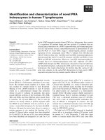
Fig. 1 Chemical structures of porphyrin derivatives examined in the present study. Protoporphyrin (PP-H) is converted to photoprotoporphyrin
(PPP-H) via 1,4-addition of oxygen to the vinyl substitute. Zn-protoporphyrin (Zn-PP) is converted to Zn-photoprotoporphyrin (Zn-PPP) via 1,4addition of oxygen to the vinyl substitute. In vitro reported fluorescence wavelengths are 630 nm for PP-H, 664 nm for PPP-H, 585 nm for Zn-PP,
and 625 nm for Zn-PPP
Ohsaki et al. BMC Cancer (2017) 17:289
Page 4 of 7
A total of 29 lesions exhibiting red fluorescence were
found in 8 patients. Pathologic diagnosis was normal for
5 lesions and indicated inflammation for 13 lesions, mild
dysplasia for 7 lesions, severe dysplasia for 1 lesion, and
squamous cell carcinoma for 3 lesions. In the present
study, we included lesions exhibiting weak red autofluorescence; therefore, our samples included non-cancerous
as well as cancerous lesions. However, it was not difficult
to differentiate cancerous from non-cancerous lesions,
because the intensity of the red autofluorescence differed. Cancerous lesions were characterized by red autofluorescence by AFB [13].
Spectral analyses revealed that the wavelength of the
green autofluorescence emanating from the normal
bronchial wall tissue adjacent to the 29 lesions was
541.7 ± 0.51 nm (average ± SD, Table 2 and Fig. 2). The
average wavelength of the red autofluorescence emanating from the 29 lesions was 617.7 ± 1.31 nm. The
intensity of the green autofluorescence was markedly
reduced in the squamous cell carcinoma lesions. The
cancer lesions appeared red, and spectral analysis of the
red autofluorescence showed an average wavelength of
617.1 ± 0.38 nm (Table 2 and Fig. 3). Red autofluorescence associated with bleeding in the bronchial wall
resulting from contact with the bronchofiberscope and
autofluorescence associated with the blood vessels in the
bronchial wall was also observed. The wavelength of red
autofluorescence was similar between lesions with different pathologic diagnoses. The wavelengths of green and
red autofluorescence according to pathologic diagnosis
are listed in Table 2.
Analysis of the wavelength of fluorescence emitted by
synthetic porphyrin derivatives
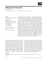
Fig. 2 Spectrogram of green autofluorescence observed in the
normal bronchial wall with a wavelength of ~540 nm. Data were
acquired using a modified PMA-12 (Hamamatsu Photonics, Japan)
photoprotoporphyrin, Zn-protoporphyrin, and Znphotoprotoporphyrin reportedly emit fluorescence at
630, 664, 585, and 625 nm, respectively, when excited with 400-nm light (Fig. 1).
Our synthesized Zn-photoprotoporphyrin and photoprotoporphyrin were dissolved in 5% albumin solution
and excited with 400-nm light. Fluorescence at wavelengths of 587.5, 625.5, and 664.0 nm was observed
(Fig. 4). We added 5% albumin to the solution, because it was reported that the biochemical/biological
environment, which might alter the quantum yield and
lifetime of the fluorophore(s) [22]. However, 5% albumin
To elucidate the source of the red autofluorescence
observed by AFB in the bronchial lesions, we tested
various porphyrin derivatives found in the human body,
which include coproporphyrin, uroporphyrin, and
protoporphyrin, and our synthesized photoprotoporphyrin, Zn-protoporphyrin and Zn-photoprotoporphyrin.
Coproporphyrin and uroporphyrin emitted only weak
fluoresce when excitation light was applied. Protoporphyrin,
Table 2 Wavelengths of green and red autofluorescence
emanating from bronchial lesions in eight patients
(average ± SD)
Pathologic diagnosis
Green autofluorescence
(nm)
Red autofluorescence
(nm)
Normal (n = 5)
541.4 ± 0.00
617.3 ± 0.03
Inflammation (n = 13)
541.7 ± 0.49
617.4 ± 0.82
Mild dysplasia (n = 7)
542.0 ± 0.67
619.0 ± 2.04
Malignanta (n = 4)
541.0 ± 0.00
617.1 ± 0.38
a
Includes three squamous cell carcinoma and one severe dysplasia
Fig. 3 Spectrogram of red autofluorescence observed in squamous
cell carcinoma bronchial lesions with a wavelength of ca. 620 nm.
Data were acquired using a modified PMA-12 (Hamamatsu
Photonics, Japan)
Ohsaki et al. BMC Cancer (2017) 17:289
Fig. 4 Our synthesized Zn-photoprotoporphyrin and
photoprotoporphyrin were dissolved in 5% albumin solution and
excited with 400-nm light. Fluorescence emitted by synthetic
porphyrin derivatives at wavelengths of 587.5, 625.5, and 664.0 nm. We
concluded that the 587.5-nm fluorescence was from albumin, the
625.5-nm fluorescence was from Zn-photoprotoporphyrin, and the
664.0-nm fluorescence was from photoprotoporphyrin
did not seem to alter wavelength of the fluorescence. We
concluded that the 587.5-nm fluorescence was from
albumin, the 625.5-nm fluorescence was from Znphotoprotoporphyrin, and the 664.0-nm fluorescence was
from photoprotoporphyrin. In the present study, our synthesized Zn-protoporphyrin emitted 578-nm fluorescence
(data not shown). These results suggested that Znprotoporphyrin in living patients is converted to Znphotoprotoporphyrin upon excitation with 400-nm light,
and emits 625.5-nm fluorescence [23]. The difference
between the 617.7-nm fluorescence observed in the
human bronchus and the 625.5-nm fluorescence observed
in the above experiment can be attributed to differences
between in vivo and in vitro conditions. Therefore, we
concluded that the source material of the red autofluorescence observed in cancer lesions, blood vessels, and
contact bleeding sites using the PDS-2000 system was
Zn-photoprotoporphyrin. The red fluorescence from
Zn-photoprotoporphyrin could be detected visibly using
the fluorescence endoscopy system in cancer lesions in
which the intensity of green autofluorescence from normal tissue decreased, as well as in blood vessels and contact bleeding sites.
Discussion
Detection of red autofluorescence in cancers of the
bladder, stomach, and lung has been reported. Highperformance liquid chromatography (HPLC) analysis of
tissues from patients with these cancers revealed
substances emitting faint red fluorescence [17, 18]. The
source of this red fluorescence has been attributed to
the de novo accumulation of porphyrins [16, 24].
Page 5 of 7
However, this hypothesis has not been confirmed. We
observed bronchogenic cancer lesions using a color
fluorescence endoscopy system and found an increase in
the R/G ratio in the cancer lesions [13]. Kluftunger et al.
[25] reported increase of R/G ratio greater than 1.5
times the control, fluorescence imaging correctly identified areas of hyperplasia, dysplasia, CIS and invasive
cancer using DMBA-induced hamster cheek pouch
model. In our previous study, R/G ratio in bronchogenic
cancer was significantly greater than those in normal
bronchial wall due to decrease of green fluorescence and
increase of red fluorescence in the cancer lesions [13].
This red fluorescence was also observed in blood vessels
as well as in fresh contact bleeding sites in the bronchial
wall. We found that the wavelength of the red fluorescence was 617.7 nm, and the source of the red fluorescence in the present study was identified as Znphotoprotoporphyrin. Zn-photoprotoporphyrin seems to
be formed from Zn-protoporphyrin following irradiation
with 405-nm blue light. However, it was difficult to extract porphyrin analogues from small biopsy specimen
from the bronchial wall.
De novo protoporphyrin IX has been implicated as a
source of the red autofluorescence associated with cancerous tissues. Moesta et al. reported the emission of red
fluorescence from colorectal cancers [18]. They analyzed
chemical extracts of involved lymph nodes using
reversed-phase HPLC and found a substance emitting
630-nm fluorescence. They concluded that protoporphyrin IX was the source of the red autofluorescence in
these involved lymph nodes. Croce et al. reported naturally occurring porphyrins in a spontaneous tumorbearing mouse model [17]. They reported substantial
levels of protoporphyrin IX in tumor, spleen, liver, and
plasma samples.
Protoporphyrin IX is formed from 5-aminolevulinic
acid; however, its concentration in normal human tissues
is low [26]. In addition, the wavelength of protoporphyrin IX fluorescence is 635 nm when excited with 405nm light [27]. These data suggest that the 617.7-nm
autofluorescence emanating from cancer lesions, blood,
and blood vessels in the present study was from a source
other than protoporphyrin IX. The human body must
therefore naturally contain a substance that emits
strong, red autofluorescence. The present study was
conducted to identify the source of the 617.7-nm red
autofluorescence observed in previous studies.
The major porphyrin derivatives found in normal
human blood are uroporphyrin, coproporphyrin, and
Zn-protoporphyrin. Normal blood levels of porphyrins
are 0–1.0 μg/dl for total porphyrin, <2 μg/dl for coproporphyrin, 16–60 μg/dl for protoporphyrin, <2 μg/dl for
uroporphyrin [28] and 23 μg/dl for Zn-protoporphyrin
[29]. Zn-protoporphyrin reportedly emits fluorescence at
Ohsaki et al. BMC Cancer (2017) 17:289
585 nm, but our synthesized Zn-protoporphyrin examined emitted 578-nm red fluorescence. This wavelength
differed from the 617.7-nm fluorescence observed in
bronchial cancer lesions, blood vessels, and contact
bleeding sites. We then examined our synthesized Znphotoprotoporphyrin and photoprotoporphyrin by
dissolving them in 5% albumin solution to mimic the
conditions of the human body, and fluorescence from
both of these porphyrin derivatives was detected. Znphotoprotoporphyrin and photoprotoporphyrin emitted
fluorescence at 625.5 and 664.0 nm, respectively, and we
therefore concluded that the red fluorescence emanating
from bronchial cancer lesions, blood vessels, and contact
bleeding sites in the present study was associated with
Zn-photoprotoporphyrin. This conclusion is plausible, as
the difference in wavelengths was acceptable, considering
the measurement method and the in vivo and in vitro
conditions. In the human body, Zn-protoporphyrin
(emitting 578-nm red fluorescence) seems to become Znphotoprotoporphyrin (emitting 625.5-nm red fluorescence) following irradiation with 405-nm excitation light
via photooxidation [30]. It is known that cancer lesions
emit bi-phasic red fluorescence during photodynamic
therapy (PDT) forming protoporphyrin photoproducts
[31, 32]. This bi-phasic red fluorescence is emitted by protoporphyrin IX, which emits 636 nm red fluorescence,
and photoprotoporphyrin, which emits 674 nm red fluorescence in case of PDT using 5-ALA [32]. In PDT, protoporphyrin becomes photoprotoporphyrin upon laser
irradiation. Our present report is the first to describe the
origin of red autofluorescence emanating from cancer lesions, blood vessels and fresh contact bleeding sites in living patients.
Autofluorescence endoscopy revealed a decrease in the
intensity of the green fluorescence emanating from normal human tissue. Autofluorescence endoscopy typically
utilizes AFI and D-light AF systems. However, it is difficult to detect the red autofluorescence that emanates
from cancer lesions, blood, and blood vessels using either system. This has led some researchers to conclude
that human blood and blood vessels do not emit autofluorescence or emit only weak autofluorescence associated
with hemoglobin. However, we found that red autofluorescence could be clearly detected using a sensitive autofluorescence endoscopy system such as the PDS-2000.
We have observed red autofluorescence not only in
bronchogenic carcinoma but also in tumors metastasized
from breast, colon, and pancreatic cancers. We developed a new autofluorescence endoscopy system using an
EM-CCD, PDS-TriMode (FLOVEL, Tachikawa, Japan),
based on the PDS-2000 technology. The PDS-TriMode
is a high-vision system, and its sensitivity is greater than
that of the PDS-2000. The PDS-TriMode is capable
of clearly detecting not only decreases in green
Page 6 of 7
autofluorescence but also abnormal red autofluorescence emanating from cancer lesions, blood, and
blood vessels.
Analysis of the wavelength of red fluorescence can provide very important information. When 5-aminolevulinic
acid (5-ALA) is orally administered, levels of protoporphyrin IX (which emits 635-nm red fluorescence when
excited with 405-nm light) increase in cancer tissues
[33, 34]. Photofrin® and Lazerphyrin® have been approved and are currently used in PDT in Japan. In
cancer tissues, Photofrin® and Lazerphyrin® emit 640- and
664-nm red fluorescence, respectively. These drugs are
also used to detect cancerous tissue in photodynamic
diagnosis (PDD). It is obviously difficult to differentiate
617.7-nm red autofluorescence emanating from the blood
from 635-, 640-, and 664-nm red fluorescence using a
sensitive color CCD camera. Attempts to do so could lead
to false results in PDD. Our results indicate that reduction
in the intensity of 617.7-nm red autofluorescence emanating from the blood is necessary for reliable PDD using
porphyrin derivatives and 5-ALA.
Conclusions
We conclude that Zn-photoprotoporphyrin was the
source of the red autofluorescence observed in bronchial lesions. Zn-protoporphyrin is converted to Znphotoprotoporphyrin by radiation with excitation
light. Our results suggest that red autofluorescence
emanating from Zn-photoprotoporphyrin in human
tissues could interfere with photodynamic diagnosis using
porphyrin derivatives such as Photofrin® and Lazerphyrin®
with a sensitive endoscopy system, because color cameras
cannot differentiate Zn-photoprotoporphyrin red fluorescence from that of other porphyrin derivatives.
Abbreviations
5-ALA: 5-aminolevulinic acid; AFB: Autofluorescence bronchoscopy;
CCD: Charged coupled device; FAD: Flavin-adenine dinucleotide;
HPLC: High-performance liquid chromatography; NADP: Nicotinamideadenine dinucleotide phosphate; PDT: Photodynamic therapy; R/G
ratio: Red to green autofluorescence ratio; RGB: Red, green and blue
Acknowledgements
Not applicable.
Funding
Fund from Grant-in-Aid for Scientific Research, Japan Society for the
Promotion of Science supported collection, analysis, and interpretation of
data and in writing the manuscript. Fund from Translational Research
Network Program, Foundation for Biomedical Research and Innovation,
Japan supported development and modification of the fluorescent endoscopic
system.
Availability of data and materials
The datasets used and/or analyzed during the current study available from
the corresponding author on reasonable request.
Authors’ contributions
YO conducted this study as a principal investigator. KT and SN made
substantial contributions to development of fluorescence endoscopy. TS,
Ohsaki et al. BMC Cancer (2017) 17:289
SE, MK, SO, NH, YK, ET, YY, KT, SN and IS made substantial contributions
to acquisition of data, analysis and interpretation of data; and been
involved in drafting the manuscript and revising it critically for important
intellectual content. Especially, IS made contributions to synthesis and
analysis of the porphyrin derivatives. All authors have given final
approval of the version to be published; participated sufficiently in the
work to take public responsibility for appropriate portions of the
content; and agreed to be accountable for all aspects of the work in
ensuring that questions related to the accuracy or integrity of any part
of the work are appropriately investigated and resolved.
Page 7 of 7
13.
14.
15.
16.
Competing interests
The authors declare that they have no competing interests.
17.
Consent for publication
Not applicable.
18.
Ethics approval and consent to participate
This study was approved by the Institutional Review Board of the Asahikawa
Medical University (Approval number #237). All patients were asked and
agreed to participate in this study with written informed consent, and the
study was performed in accordance with the GCP guideline from Japanese
Government.
19.
Publisher’s Note
Springer Nature remains neutral with regard to jurisdictional claims in
published maps and institutional affiliations.
Author details
1
Respiratory Center, Asahikawa Medical University, 2-1-1-1 Midorigaoka
Higashi, Asahikawa 078-8510, Japan. 2Moriyama Memorial Hospital,
Asahimachi 2-1-31, Asahikawa 070-0832, Japan. 3Porphyrin Lab, Okayama
700-0086, Japan.
Received: 4 January 2017 Accepted: 12 April 2017
References
1. Lakowicz JR. Principles of fluorescence spectroscopy. New York: Plemium
Press; 1983.
2. Schomacker KT, Frisoli JK, Compton CC, Flotte TJ, Richter JM, Nishioka NS,
et al. Ultraviolet laser-induced fluorescence of colonic tissue: basic biology
and diagnostic potential. Laser Surg Med. 1992;12:63–78.
3. Palcic B, Lam S, Hung J, MacAulay C. Detection and localization of early
lung cancer by imaging techniques. Chest. 1991;99:742–3.
4. George PJ. Fluorescence bronchoscopy for the early detection of lung
cancer. Thorax. 1999;54:180–3.
5. Kakihana M, Li KK, Okunaka T, Furukawa K, Hirano T, Konaka C, et al.
Early detection of bronchial lesions using system of fluorescence endoscopy
(SAFE) 1000. Diagn Ther Endosc. 1999;5:99–104.
6. Adachi R, Utsui T, Furusawa K. Developement of the autofluorescence
endoscope imaging system. Diagn Ther Endosc. 1999;5:65–70.
7. Leonhard M. New incoherent autofluorescence/fluorescence system for
early detection of lung cancer. Diagn Ther Endosc. 1999;5:113–8.
8. Chen W, Gao X, Tian Q, Chen L. A comparison of autofluorescence
bronchoscopy and white light bronchoscopy in detection of lung cancer
and preneoplastic lesions: a meta-analysis. Lung Cancer. 2010;73:183–8.
9. Sun J, Garfield DH, Lam B, Yan J, Gu A, Shen J, et al. The value of
autofluorescence bronchoscopy combined with white light bronchoscopy
compared with white light alone in the diagnosis of intraepithelial
neoplasia and invasive lung cancer: a meta-analysis. J Thorac Oncol.
2011;6:1336–44.
10. Aihara H, Sumiyama K, Saito S, Tajiri H, Ikegami M. Numerical analysis of the
autofluorescence intensity of neoplastic and non-neoplastic colorectal
lesions by using a novel videoendoscopy system. Gastrointest Endosc.
2009;69:726–33.
11. Ohsaki Y, Nishigaki Y, Takeyama K, Nakanishi K, Ide H, Matsumoto H, et al.
Visualization of cancer using high sensitive fluorodynamic camera and
fiber-optic endoscope. Porphyrins. 2000;9:197–203.
12. Ohsaki Y, Takeyama K, Nakao S, Tanno S, Toyoshima E, Nakanishi K, et al.
Detection of photofrin fluorescence from malignant and premalignant
20.
21.
22.
23.
24.
25.
26.
27.
28.
29.
30.
31.
32.
33.
34.
lesions in the bronchus using a full-color endoscopic fluorescence imaging
system: a preliminary report. Diagn Ther Endosc. 2001;7:187–95.
Nakanishi K, Ohsaki Y, Kurihara M, Nakao S, Fujita Y, Takeyama K, et al.
Color auto-fluorescence from cancer lesions: improved detection of central
type lung cancer. Lung Cancer. 2007;58:214–9.
Ghadially FN, Neish WJP. Porphyrin fluorescence of experimentally
produced squamous cell carcinoma. Nature. 1960;188:1124.
Lycette RM, Leslie RB. Fluorescence of malignant tissue. Lancet. 1965;286:436.
Bottiroli G, Croce AC, Marchesini R, Pignoli E, Tomatis S, Cuzzoni C, et al.
Natural fluorescence of normal and neoplastic human colon: a
comprehensive 'ex vivo' study. Lasers Surg Med. 1995;16:48–60.
Croce AC, Santamaria G, De Simone U, Lucchini F, Freitas I, Bottiroli G.
Naturally-occurring porphyrins in a spontaneous-tumour bearing mouse
model. Photochem Photobiol Sci. 2011;10:1189–95.
Moesta KT, Ebert B, Handke T, Nolte D, Nowak C, Haensch WE, et al.
Protoporphyrin IX occurs naturally in colorectal cancers and their
metastases. Cancer Res. 2001;61:991–9.
Shibukawa K, Miyokawa N, Tokusashi Y, Sasaki T, Osanai S, Ohsaki Y. High
incidence of chromosomal abnormalities at 1p36 and 9p21 in early-stage
central type squamous cell carcinoma and squamous dysplasia of bronchus
detected by autofluorescence bronchoscopy. Oncol Rep. 2009;22:81–7.
Dolphin D, Sivasothy R. The preparation of porphyrin S-411
(dehydrocoproporphyrin) and harderoporphyrin from protoporphyrin IX.
Can J Chem. 1981;59:779–85.
Nakae Y, Fukusaki E-I, Kajiyama S-I, Kobayashi A, Nakajima S, Sakata I.
Syntheses and screening tests of new chlorin derivatives as photosensitizer.
J Photochem Photobiol A. 2005;174:187–93.
Ramanujam N. Fluorescence spectroscopy of neoplastic and non-neoplastic
tissues. Neoplasia. 2000;2:89–117.
Falk JE, Smith KM. Porphyrins and metalloporphyrins. Revised ed. Elsevier
Science; 1975. p. 688–689.
Ghadially FN, Neish WJP, Dawkins HC. Mechanisms involved in the
production of red fluorescence of human and experimental tumors.
J Pathol Bacteriol. 1963;85:77–92.
Kluftinger AM, Davis NL, Quenville NF, Lam S, Hung J, Palcic B. Detection of
squamous cell cancer and pre-cancerous lesions by imaging of tissue
autofluorescence in the hamster cheek pouch model. Surg Oncol.
1992;1:183–8.
Sachar M, Anderson KE, Ma X. Protoporphyrin IX: the good, the bad, and
the ugly. J Pharmacol Exp Ther. 2016;356:267–75.
Nakai Y, Anai S, Onishi S, Masaomi K, Tatsumi Y, Miyake M, et al.
Protoporphyrin IX induced by 5-aminolevulinic acid in bladder cancer cells
in voided urine can be extracorporeally quantified using a
spectrophotometer. Photodiagn Photodyn Ther. 2015;12:282–8.
Porphyrins-blood test. MedlinePlus. />003372.htm
Suga RS, Fischinger AJ, Knoch FW. Establishment of normal values in adults
for zinc protoporphyrin (ZPP) using hematofluorometer: correlation with
normal blood lead values. Am Ind Hyg Assoc J. 1981;42:637–42.
Cox G, Whitten DG. Mechanisms for the photooxidation of protoporphyrin
IX in solution. J Am Chem Soc. 1982;104:516–21.
König K, Schneckenburger H, Rück A, Steiner R. In vivo photoproduct
formation during PDT with ALA-induced endogenous porphyrins.
J Photochem Photobiol B. 1993;18:287–90.
Robinson DJ, de Bruijn HS, van der Veen N, Stringer MR, Brown SB, Star WM.
Fluorescence photobleaching of ALA-induced protoporphyrin IX during
photodynamic therapy of normal hairless mouse skin: the effect of light
dose and irradiance and the resulting biological effect. Photochem
Photobiol. 1998;67:140–9.
Kitada M, Ohsaki Y, Matsuda Y, Hayashi S, Ishibashi K. Photodynamic
diagnosis of malignant pleural diseases using the autofluorescence imaging
system. Ann Thorac Cardiovasc Surg. 2014;20:378–82.
Kitada M, Ohsaki Y, Matsuda Y, Hayashi S, Ishibashi K. Photodynamic
diagnosis of pleural malignant lesions with a combination of 5aminolevulinic acid and intrinsic fluorescence observation systems.
BMC Cancer. 2015;15:174.