Tính hữu ích của siêu âm vùng chậu để chẩn đoán dậy thì sớm trung ương ở trẻ em gái
Bạn đang xem bản rút gọn của tài liệu. Xem và tải ngay bản đầy đủ của tài liệu tại đây (757.92 KB, 7 trang )
Original article
Yu
J, et al.
• Pelvic
ultrasonography for central precocious puberty
Korean
J Pediatr
2015;58(8):294-300
/>pISSN 1738-1061•eISSN 2092-7258
Korean J Pediatr
Usefulness of pelvic ultrasonography for the
diagnosis of central precocious puberty in girls
Jung Yu, MD1, Ha Young Shin, MD1, Sun Hee Lee, MD2, You Sung Kim, MD3, Jae Hyun Kim, MD1
1
Department of Pediatrics, Inje University Ilsan Paik Hospital, Inje University College of Medicine, Goyang, 2Department of Pediatrics, Gil Medical Center, Graduate
School of Medicine, Gacheon University of Medicine and Science, Incheon, 3Department of Radiology, Inje University Ilsan Paik Hospital, Inje University College of
Medicine, Goyang, Korea
Purpose: It is difficult to differentiate between central precocious puberty (CPP) and premature
thelarche (PT) in girls. The aim of this study was to investigate the diagnostic usefulness of pelvic
ultrasonography to distinguish between CPP and PT in girls with early breast development.
Methods: This study included girls with early breast development who visited the clinic between
January 2012 and December 2013. Clinical, laboratory, and pelvic ultrasonographic data were
evaluated. CPP and PT were confirmed using the gonadotropin-releasing hormone stimulation test.
Results: A total of 248 girls aged 7–8 years were included, among whom 186 (75.0%) had CPP and
62 (25.0%) had PT. The uterine length, transverse diameter, fundus, volume, and cross-sectional area
were significantly larger in the CPP group (uterine length, 2.45±0.50 cm vs. 2.63±0.49 cm, P=0.015;
uterine volume, 0.95±0.62 cm3 vs. 1.35±0.76 cm3, P<0.001). However, there were no differences
in the fundus/cervix ratio and ovarian measurements. In receiver operating characteristic analysis, a
uterine volume of at least 1.07 cm3 was the most predictive parameter for CPP with an area under the
curve of 0.670 (95% confidence interval, 0.593–0.747).
Conclusion: Uterine measurements by pelvic ultrasonography in girls with early pubertal development
were significantly larger in the CPP group. However, the diagnostic value of ultrasonographic para
meters was not high because of a considerable overlap of values between the two groups. Therefore,
pelvic ultrasonography in combination with clinical and laboratory tests may be useful to distinguish
between CPP and PT in girls.
Corresponding author: Jae Hyun Kim, MD
Department of Pediatrics, Inje University Ilsan Paik
Hospital, Inje University College of Medicine, 170
Juhwa-ro, Ilsanseo-gu, Goyang 411-706, Korea
Tel: +82-31-910-7942
Fax: +82-31-910-7108
E-mail:
Received: 21 August, 2014
Revised: 12 October, 2014
Accepted: 20 October, 2014
Key words: Precocious puberty, Diagnosis, Pelvis, Ultrasonography, Girls
Introduction
Precocious puberty is defined as the appearance of the secondary sexual characteristics
before the age of 8 years in girls1). Central precocious puberty (CPP) is caused by the
premature activation of the hypothalamic gonadotropin-releasing hormone (GnRH) pulse
generator and is generally idiopathic2). CPP can be associated with diverse problems such
as compromised final adult height and psychological as well as emotional conflicts3-5).
Therefore, an early diagnosis and a proper management are critical6,7).
However, it is difficult to differentiate between CPP and premature thelarche (PT). PT is
featured by an isolated appearance of breast development, that is not progressive and does
not require treatment8). The differentiation between CPP and PT is confirmed by clinical,
radiologic and laboratory tests such as physical examination, evaluation of bone age,
height velocity measurement and GnRH stimulation test1). The laboratory determination of
the peak luteinizing hormone (LH) concentration during the GnRH stimulation test is
considered the most important diagnostic process, although it had some disadvantages in
294
/>
Copyright © 2015 by The Korean Pediatric Society
This is an open-access article distributed under the
terms of the Creative Commons Attribution NonCommercial License ( />licenses/by-nc/3.0/) which permits unrestricted noncommercial use, distribution, and reproduction in any
medium, provided the original work is properly cited.
Korean J Pediatr 2015;58(8):294-300
cluding time-consuming multiple samples resulting in discomfort
to patients and a low sensitivity despite of its high specificity9).
The transabdominal pelvic ultrasonography has been used to
differentiate CPP from PT10). Pelvic ultrasonography is noninva
sive and relatively less time-consuming. Several studies reported
that larger uterine and ovarian measurements were associated
with CPP than PT11-14). However, it is not conclusive to the diag
nostic role of pelvic ultrasonography in patients with early
pubertal signs.
The aim of this study was to investigate the diagnostic use
fulness of pelvic ultrasonography to differentiate between CPP
and PT in girls with early breast development and to determine
the optimal cutoff values of ultrasonographic measurements to
distinguish CPP and PT.
Materials and methods
1. Subjects
Girls with early breast development who were referred for the
evaluation of CPP to our pediatric endocrinology clinic between
January 2012 and December 2013 were included in this study.
Inclusion criteria were as follows: (1) chronological age between
7 and 8 years at the first visit; (2) breast budding before the age of
8 years; (3) breasts with Tanner stage 2 or more on the first
examination in our clinic; (4) advanced bone age by one or more
years; and (5) GnRH stimulation test and pelvic ultrasonography
for the evaluation of CPP. Exclusion criteria were as follows: (1)
peripheral precocious puberty; (2) CPP due to an organic
intracranial lesion; (3) presence of a chronic illness such as
diabetes mellitus and thyroid disorders; and (4) medication which
may affect the hypothalamic-pituitary-ovarian axis. Of 250 girls
who met the inclusion criteria, 248 were enrolled in the study.
Two girls were excluded for following reasons: one girl presented
with CPP after the treatment of anaplastic astrocytoma and the
other girl had a previous history of acute lymphocytic leukemia.
the Greulich-Pyle method16).
A standard GnRH stimulation test was conducted in the early
morning after overnight fasting. Basal serum samples were ob
tained for the measurement of LH, follicular stimulating hormone
(FSH) and estradiol just before the intravenous bolus injection of
100 μg of GnRH (Relefact, Sanofi-Aventis, Frankfurt, Germany).
After the administration of GnRH, blood samples for the deter
mination of LH and FSH concentration were withdrawn at 30, 45,
60, and 90 minutes. Serum LH, FSH and estradiol level were
measured by an electrochemiluminescence immunoassay (Roche
Diagnostics GmbH, Manheim, Germany). Within-run and total
precision of the hormonal assays were ranging from 0.7% to
1.2% and from 1.6% to 2.2% for LH; from 2.5% to 2.8% and from
3.6% to 4.5% for FSH; from 1.7% to3.3% and 2.2% to 4.7% for
estradiol. The limits of detection were 0.1 IU/L for LH, 0.1 IU/L for
FSH and 5.0 pg/mL for estradiol. A peak LH concentration of at
least 5 IU/L during GnRH stimulation test was considered as
CPP7). Subjects with a peak LH concentration of less than 5 IU/L
were classified as PT.
Transabdominal pelvic ultrasonography was obtained with a
micro convex probe 8C (3.5–11.5 MHz, Logiq 9, GE healthcare,
Milwaukee, WI, USA). Ultrasonography was performed with full
bladder filling by a single experienced radiologist (Y.S.K.). Fol
lowing parameters of the uterus were evaluated including the
length, transverse diameter, fundal anteroposterior diameter
(fundus), cervical anteroposterior diameter (cervix) and the pre
sence of endometrial echogenicity. The uterine cross-sectional
area was calculated by multiplying the length by the fundal
anteroposterior diameter. The uterine volume was calculated
according to the ellipse formula (length×transverse diameter×
fundal anteroposterior diameter×0.5233). The ratio of the fundal
to cervical anteroposterior diameter (fundus/cervix ratio) was
calculated. In ovaries, the height, width, and length were evaluat
ed. The circumference of each ovary was calculated according to
the ellipse circumference formula {2.222×[(height)2+(length)2]1/2}.
The volume of each ovary was computed using the same ellipse
formula as that for uterus.
2. Methods
The retrospective review of medical records for this study was
approved by the Institutional Review Board of the Inje University
Ilsan Paik Hospital (IB-1407-029). We reviewed the medical
records of the subjects who met the inclusion criteria. Demogra
phic and clinical parameters around the day of GnRH stimulation
test and pelvic ultrasonography were investigated including
chronological age, bone age, height, body weight, body mass
index (BMI), sexual maturity rate, parental height and hormonal
profiles. The standard deviation score (SDS) of height, body
weight and BMI for the same age and sex were calculated using
the LMS methods proposed in 2007 Korean National Growth
Charts15). The assessment of the bone age was performed using
3. Statistics
Statistical analyses were performed with STATA 12.1 (StataCorp
LP., College Station, TX, USA). Results were expressed as mean±
standard deviation. Student t test was used to compare demographic
and clinical parameters between the CPP and PT group. Logistic
regression analysis was performed to determine the association
between the results of GnRH stimulation test and clinical, laboratory
and ultrasonographic variables. Receiver operating characteristic
(ROC) analyses was used to investigate the predictive ability of
laboratory and ultrasonographic parameters17). The optimal cutoff
values were determined using the Youden index (J), which is
defined by “J= maximum (sensitivity+specificity–1)” 18).
/>
295
Yu J, et al. • Pelvic ultrasonography for central precocious puberty
Sensitivity, specificity, positive predictive value (PPV) and
negative predictive value (NPV) was calculated for each cutoff
value. A P value less than 0.05 was considered significant.
Results
1. Characteristics of the study subjects
Of 248 subjects, 186 (75.0%) were diagnosed with CPP and 62
(25.0%) with PT (Table 1). The chronological age was 8.36±0.44
years in the CPP group and 8.28±0.68 years in the PT group (P=
0.367). The bone age was 10.06±0.77 years in the CPP group and
10.06±0.72 years in the PT group (P=0.886). The advancement of
bone age over chronological age was 1.67±0.66 years in the CPP
group and 1.77±0.60 years in the PT group (P=0.306). The BMI
SDS was significantly higher in the PT group, although the height
SDS showed no significant difference between the two groups.
Laboratory parameters during GnRH stimulation test including
basal LH, peak LH, basal FSH, peak FSH, basal LH/FSH ratio, peak
LH/FSH ratio, estradiol, insulin-like growth factor-I (IGF-I), and
IGF-I SDS were significantly higher in the CPP group (Table 1).
2. Pelvic ultrasonography findings
Measurements of uterine length, uterine transverse diameter,
fundus, uterine volume and uterine cross-sectional area were
significantly higher in the CPP group (Table 2). However, the
fundus/cervix ratio was not different between both groups. An
endometrial echogenicity was observed in one subject with CPP.
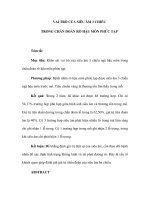
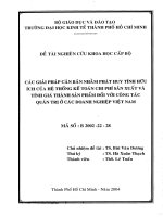
There was no significant difference in both ovarian circum
ferences and volumes between both groups (Fig. 1).
3. Logistic regression analysis and ROC analysis
Univariate logistic regression analysis was carried out to de
termine variables affecting the diagnosis of precocious puberty
during GnRH stimulation test. Basal LH, basal FSH, basal LH to
FSH ratio, IGF-I SDS and BMI SDS were significant parameters.
In ultrasonographic findings were uterine length, uterine
transverse diameter, fundus, uterine volume and uterine crosssectional area predictors of the diagnosis of CPP (Table 3).
ROC curves were constructed based on logistic regression
analyses. The area under the curve (AUC) with 95% confidence
Table 2. Pelvic ultrasonographic findings of the study subjects
Variable
Table 1. Clinical and biochemical characteristics of the study subjects
Characteristic
Premature
thelarche
No. of subjects (%)
62 (25.0)
Central precocious
P value
puberty
186 (75.0)
Premature
thelarche
No. of subjects (%)
62 (25.0)
186 (75.0)
Uterine length (cm)
2.45±0.50
2.63±0.49
0.015
Uterine transverse diameter (cm)
0.76±0.22
0.89±0.24
<0.001
Fundus (cm)
0.89±0.30
1.02±0.28
0.002
Fundus/cervix ratio
1.50±0.59
1.49±0.46
0.809
0.95±0.62
1.35±0.76
<0.001
1.86±0.67
<0.001
8.28±0.68
8.36±0.44
0.367
Uterine volume (cm3)
10.06±0.72
10.04±0.79
0.886
Uterine cross-sectional area (cm2) 1.47±0.52
Bone age–chronological age (yr)
1.77±0.60
1.67±0.66
0.306
Endometrial echogenicity
Height SDS
1.04±0.87
0.89±0.82
0.232
Ovarian circumference (cm)
Body mass index SDS
0.63±1.02
0.22±0.96
0.005
Chronological age (yr)
Bone age (yr)
Central precocious
P value
puberty
0
1
Left
5.61±1.10
5.66±1.07
0.775
Obesity, n (%)
23 (37.1)
41 (22.0)
<0.001
Right
5.66±1.15
5.77±1.14
0.524
Basal LH (IU/L)
0.11±0.27
0.72±0.99
<0.001
Mean
5.63±1.04
5.71±0.99
0.606
Peak LH (IU/L)
3.55±1.12
13.02±8.17
<0.001
Basal FSH (IU/L)
2.29±1.47
3.56±2.06
<0.001
Left
1.61±0.77
1.61±0.82
0.999
Peak FSH (IU/L)
10.95±3.70
13.47±4.72
<0.001
Right
1.52±0.77
1.70±0.91
0.167
0.03±0.06
0.16±0.19
<0.001
Mean
1.57±0.70
1.66±0.78
0.428
Basal LH/FSH ratio
Values are presented as mean±standard deviation unless otherwise indicated.
Peak LH/FSH ratio
0.35±0.15
1.02±0.57
Estradiol (pg/mL)
10.22±12.41
14.51±16.10
0.056
IGF-I
286±71
319±92
0.009
IGF-I SDS
0.56±0.82
0.94±1.09
0.014
IGFBP-3
4978±875
5177±813
0.102
Variable
IGFBP-3 SDS
3.97±1.66
4.35±1.58
0.105
Uterine length (cm)
–0.12±0.79
–0.15±0.69
0.766
Uterine transverse diameter (cm)
Target height SDS
<0.001
Ovarian volume (cm3)
Values are presented as mean±standard deviation unless otherwise indicated.
SDS, standard deviation score; LH, luteinizing hormone; FSH, folliclestimulating hormone; IGF-I, insulin-like growth factor-I; IGFBP-3, insulin-like
growth factor-binding protein-3.
296
/>
Table 3. Logistic regression analysis to predict the positive results of the
gonadotropin-releasing hormone stimulation test
Odds ratio 95% Confidence interval P value
2.14
1.15–3.96
0.013
11.64
2.98–45.40
<0.001
Fundus (cm)
5.86
1.92–17.83
Uterine volume (cm3)
2.52
1.51–4.19
<0.001
0.001
Uterine cross-sectional area (cm2)
2.91
1.70–5.00
<0.001
Korean J Pediatr 2015;58(8):294-300
Fig. 1. Ultrasonographic data of the study subjects with central precocious puberty
(CPP) and premature thelarche (PT).
iterval (CI) for basal LH, basal FSH and basal LH/FSH ratio was
0.766 (0.708–0.825), 0.727 (0.652–0.802), and 0.769 (0.712–
0.825), respectively. There was no significant difference between
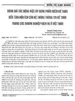
parameters (P=0.329). The AUC (95% CI) for uterine length,
uterine transverse diameter, fundus, uterine volume and uterine
cross-sectional area was 0.588 (0.503–0.673), 0.656 (0.577–
0.734), 0.660 (0.579–0.741), 0.670 (0.593–0.747), and 0.661
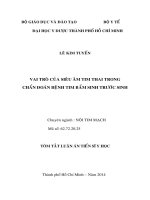
(0.586–0.737), respectively (Fig. 2). The AUC of uterine volume
was the biggest among ultrasonographic parameters (P=0.04).
The optimal cutoff value for each parameter was selected using
the Youden index (J) based on ROC analyses. Sensitivity,
specificity, PPV, and NPV for each cutoff value are shown in
Table 4.
4. Analysis of subjects according to BMI
Further analyses were performed according to the BMI. All
subjects were grouped according to their BMI assigned into two
categories, either the obese group (BMI at least 85th percentile for
age and sex) or the normal weight group (BMI less than 85th
Fig. 2. Receiver operator characteristic curves of pelvic ultrasonographic
measurements for the diagnosis of central precocious puberty with an
area under the curve (95% confidence interval) of 0.588 (0.503–0.673)
for uterine length, 0.656 (0.577–0.734) for uterine transverse diameter,
0.660 (0.579–0.741) for fundus, 0.670 (0.593–0.747) for uterine volume,
and 0.661 (0.586–0.737) for uterine cross-sectional area (The uterine
transverse diameter, fundus, and uterine cross-sectional area are not
shown in this graph.).
/>
297
Yu J, et al. • Pelvic ultrasonography for central precocious puberty
Table 4. Sensitivity, specificity, positive predictive value (PPV), and negative predictive value (NPV) of significant clinical and ultrasonographic
parameters
Variable
Sensitivity (%)
Specificity (%)
PPV (%)
NPV (%)
Basal LH ≥0.1 IU/L
72.0
74.2
89.3
43.9
Basal FSH ≥2.4 IU/L
71.0
66.1
86.3
43.2
Basal LH/FSH ratio ≥0.042
70.4
77.4
90.3
46.6
Uterine length ≥2.2 cm
83.3
33.9
79.1
40.4
Uterine transverse diameter ≥0.76 cm
71.0
56.5
83.0
39.3
Fundus ≥0.88 cm
73.1
61.3
85.0
43.2
Uterine volume ≥1.07 cm3
59.1
71.0
85.9
36.7
Uterine cross-sectional area ≥1.76 cm2
50.0
77.4
86.9
34.0
LH, luteinizing hormone; FSH, follicle-stimulating hormone.
Discussion
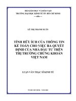
Fig. 3. Receiver operator characteristic curves of pelvic ultrasonographic
measurements for the diagnosis of central precocious puberty in the
normal weight group with an area under the curve (95% confidence
interval) of 0.663 (0.561–0.765) for uterine length, 0.708 (0.617–0.800)
for uterine transverse diameter, 0.727 (0.636–0.817) for fundus, 0.751
(0.666–0.838) for uterine volume, and 0.731 (0.645–0.816) for uterine
cross-sectional area (The uterine transverse diameter, fundus, and
uterine cross-sectional area are not shown in this graph.).
percentile for age and sex). Pelvic ultrasonographic findings were
analyzed between both groups. In the normal weight group, there
were significantly greater values in the uterine length, uterine
transverse diameter, fundus, uterine volume and the uterine
cross-sectional area, although the fundus/cervix ratio, ovarian
circumferences and volumes were not significantly different. In
ROC analysis, the AUC for uterine length was 0.663 (95% CI,
0.561–0.765) with a cutoff value of 2.2 cm. The AUC for uterine
volume was the largest with 0.751 (95% CI, 0.666–0.835) and was
statistically significant (P=0.002) (Fig. 3). The optimal cutoff of
uterine volume by the Youden index (J) was 1.09 cm3 with a
sensitivity of 60.0% and a specificity of 84.6%. In the obese
group, there were no differences in pelvic ultrasonographic
findings between the CPP and PT group.
298
/>
In girls with early pubertal signs, the GnRH stimulation test
was considered the gold standard for the laboratory confirmation
of CPP. However, GnRH stimulation test had several drawbacks
such as the discomfort to patients, a time-consuming procedure
with multiple samples, relatively high costs and a low sensitivity
despite high specificity9). The pelvic ultrasonography is a useful
method to evaluate pelvic organs. It is noninvasive, relatively less
time-consuming and relative inexpensive19).
In this study, we compared the pelvic ultrasonographic para
meters in the CPP and PT group. Significant differences were
observed in uterine length, uterine transverse diameter, fundus,
uterine volume and uterine cross-sectional area volume. Those
measurements were larger in the CPP group. However, the
fundus/cervix ratio showed no difference (Table 2). In the con
sensus statement, uterine length and uterine volume were sug
gested as useful parameters to distinguish CPP from PT. The
cutoff values for uterine length and uterine volume ranged from
3.4 to 4.0 cm and from 1.0 to 3.0 cm3, respectively7). Haber et al.12)
reported a 100% sensitivity and specificity for the cutoff value of
1.8 mL of uterine volume. De Vries et al.11) reported a sensitivity
of 88.8% and specificity of 89.4% for the cutoff value of 2.0-mL
uterine volume and a sensitivity of 80.2% and specificity of
57.8% for the cutoff value of 3.4-cm uterine length. In another
report, the diagnostic cutoff value was 3.74-cm uterine length
and 3.48-mL uterine volume20). In our study, the cutoff value of
uterine length was 2.2 cm and 1.07 cm3 for uterine volume. These
were smaller than those reported in other reports (Table 4). The
sensitivity and specificity at each cutoff point was 83.3% and
33.9% for uterine length and 59.1% and 71.0% for uterine
volume. In the ROC analysis, the AUC of uterine length and
volume was 0.588 (95% CI, 0.503–0.673) and 0.670 (95% CI,
0.593–0.747), indicating low accuracy for a diagnostic test (Fig.
2)21). In the previous article reported on Korean girls, uterine
measurements were similar to our study13). Reasons for the
Korean J Pediatr 2015;58(8):294-300
differences of cutoff values may be ethnic differences, body size
differences, interpersonal varia tions of radiologists and
performance differences among ultrasonographic machines.
The fundus/cervix ratio was reported as an important para
meter of the pubertal uterus. The prepubertal uterus had a tubular
shape and the fundus/cervix ratio was almost 122,23). In puberty,
hormonal influences to the uterus made the fundus prominent
with a fundus/cervix ratio greater than 1. Previous studies re
ported a bigger fundus/cervix ratio in the CPP group11,20). How
ever, in other reports there were no significant differences in the
fundus/cervix ratio between the CPP and PT group13,24). In this
study, the fundus/cervix ratio was 1.49±0.46 in the CPP group
and 1.50±0.59 in the PT group, without significant differences.
Ovarian measurements and morphology were other parameters
to differentiate between CPP and PT. In previous studies, the
average ovarian volume and ovarian area were larger in the CPP
group11,13,14,20,25). An ovarian circumference with a suggested cutoff
of at least 4.5 cm is a good indicator for the pubertal develop
ment11). However, in this study, there was no significant difference
in all ovarian measurements (Table 2).
A subgroup analysis was performed in the obese and normal
weight group. In the normal weight group, uterine measurements
were larger in the CPP group, except for the fundus/cervix ratio.
The optimal cut off was 2.2 cm for the uterine length and 1.09
cm3 for the uterine volume. In the ROC analysis, the AUC of
uterine length and volume were 0.663 (95% CI, 0.561–0.765) and
0.751 (95% CI, 0.666–0.835), indicating a moderate accuracy (Fig.
3)21). In the obese group, there was no significant difference in
uterine and ovarian measurements between the CPP and PT
group. It could be suggested that the uterine growth could be
influenced by the body fat. However, more research is required on
this topic.
The uterine endometrial echogenicity may be of help in the
diagnosis of CPP, although it was highly specific, but less sen
sitive11,26). In our study, endometrial echogenicity was observed in
only one case with advanced CPP, suggesting that the evaluation
in this study was performed in the early phase of puberty. The
color Doppler during the pelvic ultrasonography showed a lower
uterine arterial impedance in CPP patients27). However, no color
Doppler was carried out in this study.
This study has several limitations. The study design was retro
spective. Control subjects were not included without pubertal
development. During the pelvic ultrasonography, color Doppler
was not performed and uterine and ovarian morphology were not
described. However, a large number of subjects with suspicious
precocious puberty were enrolled and the pelvic ultrasonographic
variance was minimized because subjects were in a relatively
narrow range of age. Also only one experienced radiologist per
formed all imaging studies.
In conclusion, uterine measurements in the pelvic ultrasono
graphy of girls with early pubertal development were signifi
cantly larger in the CPP group with laboratory confirmation after
GnRH stimulation test in this study. The uterine volume of at least
1.07 cm3 was the most predictive parameter among pelvic
ultrasonographic findings to diagnose CPP. Pelvic ultrasono
graphy was more efficient to differentiate CPP from PT in the
normal weight group than in the obese group. However, the
diagnostic value of ultrasonographic parameters was not high
because of a considerable overlap between values (Fig. 1). There
fore, the pelvic ultrasonography with an adjunct to clinical and
laboratory parameters is helpful to enhance the diagnostic
precision between CPP and PT. Additionally reference values of
pelvic ultrasonographic parameters among Korean girls accord
ing to chronological age, bone age and pubertal stage is needed.
Conflict of interest
No potential conflict of interest relevant to this article was
reported.
References
1. Lee PA. Central precocious puberty. An overview of diagnosis,
treatment, and outcome. Endocrinol Metab Clin North Am 1999;
28:901-18, xi.
2. Parent AS, Teilmann G, Juul A, Skakkebaek NE, Toppari J,
Bourguignon JP. The timing of normal puberty and the age limits
of sexual precocity: variations around the world, secular trends,
and changes after migration. Endocr Rev 2003;24:668-93.
3. Brauner R, Adan L, Malandry F, Zantleifer D. Adult height in girls
with idiopathic true precocious puberty. J Clin Endocrinol Metab
1994;79:415-20.
4. Yang JH, Han SW, Yeom CW, Park YJ, Choi WS, Seo JY, et al.
Depression and self-concept in girls with perception of pubertal
onset. Ann Pediatr Endocrinol Metab 2013;18:135-40.
5. Mrug S, Elliott M, Gilliland MJ, Grunbaum JA, Tortolero SR,
Cuccaro P, et al. Positive parenting and early puberty in girls: pro
tective effects against aggressive behavior. Arch Pediatr Adolesc
Med 2008;162:781-6.
6. Carel JC, Leger J. Clinical practice: precocious puberty. N Engl J
Med 2008;358:2366-77.
7. Carel JC, Eugster EA, Rogol A, Ghizzoni L, Palmert MR; ESPELWPES GnRH Analogs Consensus Conference Group, et al. Con
sensus statement on the use of gonadotropin-releasing hormone
analogs in children. Pediatrics 2009;123:e752-62.
8. Salardi S, Cacciari E, Mainetti B, Mazzanti L, Pirazzoli P. Outcome
of premature thelarche: relation to puberty and final height. Arch
Dis Child 1998;79:173-4.
9. Pescovitz OH, Hench KD, Barnes KM, Loriaux DL, Cutler GB Jr.
Premature thelarche and central precocious puberty: the relation
ship between clinical presentation and the gonadotropin response
to luteinizing hormone-releasing hormone. J Clin Endocrinol
Metab 1988;67:474-9.
10. Shawker TH, Comite F, Rieth KG, Dwyer AJ, Cutler GB Jr, Loriaux
/>
299
Yu J, et al. • Pelvic ultrasonography for central precocious puberty
DL. Ultrasound evaluation of female isosexual precocious puberty.
J Ultrasound Med 1984;3:309-16.
11. de Vries L, Horev G, Schwartz M, Phillip M. Ultrasonographic and
clinical parameters for early differentiation between precocious
puberty and premature thelarche. Eur J Endocrinol 2006;154:8918.
12. Haber HP, Wollmann HA, Ranke MB. Pelvic ultrasonography: early
differentiation between isolated premature thelarche and central
precocious puberty. Eur J Pediatr 1995;154:182-6.
13. Kang HJ, Nam JS, Cho WK, Cho KS, Park SH, Jung MH, et al.
Pelvic ultrasonography findings in girls with precocious puberty. J
Korean Soc Pediatr Endocrinol 2010;15:126-32.
14. Herter LD, Golendziner E, Flores JA, Moretto M, Di Domenico K,
Becker E Jr, et al. Ovarian and uterine findings in pelvic sono
graphy: comparison between prepubertal girls, girls with isolated
thelarche, and girls with central precocious puberty. J Ultrasound
Med 2002;21:1237-46.
15. Moon JS, Lee SY, Nam CM, Choi JM, Choe BK, Seo JW, et al. 2007
Korean National Growth Charts: review of developmental process
and an outlook. Korean J Pediatr 2008;51:1-25.
16. Greulich WW, Pyle SI. Radiologic atlas of skeletal development of
the hand and wrist. 2nd ed. California: Stanford University Press,
1959.
17. Akobeng AK. Understanding diagnostic tests 3: receiver operating
characteristic curves. Acta Paediatr 2007;96:644-7.
18. Youden WJ. Index for rating diagnostic tests. Cancer 1950;3:32-5.
300
/>
19. Garel L, Dubois J, Grignon A, Filiatrault D, Van Vliet G. US of the
pediatric female pelvis: a clinical perspective. Radiographics
2001;21:1393-407.
20. Badouraki M, Christoforidis A, Economou I, Dimitriadis AS, Katzos
G. Evaluation of pelvic ultrasonography in the diagnosis and
differentiation of various forms of sexual precocity in girls. Ultra
sound Obstet Gynecol 2008;32:819-27.
21. Fluss R, Faraggi D, Reiser B. Estimation of the Youden Index and
its associated cutoff point. Biom J 2005;47:458-72.
22. Haber HP, Mayer EI. Ultrasound evaluation of uterine and ovarian
size from birth to puberty. Pediatr Radiol 1994;24:11-3.
23. Griffin IJ, Cole TJ, Duncan KA, Hollman AS, Donaldson MD. Pelvic
ultrasound measurements in normal girls. Acta Paediatr 1995;
84:536-43.
24. Buzi F, Pilotta A, Dordoni D, Lombardi A, Zaglio S, Adlard P. Pelvic
ultrasonography in normal girls and in girls with pubertal preco
city. Acta Paediatr 1998;87:1138-45.
25. Sathasivam A, Rosenberg HK, Shapiro S, Wang H, Rapaport R.
Pelvic ultrasonography in the evaluation of central precocious pu
berty: comparison with leuprolide stimulation test. J Pediatr 2011;
159:490-5.
26. de Vries L, Phillip M. Role of pelvic ultrasound in girls with pre
cocious puberty. Horm Res Paediatr 2011;75:148-52.
27. Battaglia C, Mancini F, Regnani G, Persico N, Iughetti L, De
Aloysio D. Pelvic ultrasound and color Doppler findings in differ
ent isosexual precocities. Ultrasound Obstet Gynecol 2003;22:27783.