Identification of SEC62 as a potential marker for 3q amplification and cellular migration in dysplastic cervical lesions
Bạn đang xem bản rút gọn của tài liệu. Xem và tải ngay bản đầy đủ của tài liệu tại đây (3.01 MB, 12 trang )
Linxweiler et al. BMC Cancer (2016) 16:676
DOI 10.1186/s12885-016-2739-6
RESEARCH ARTICLE
Open Access
Identification of SEC62 as a potential
marker for 3q amplification and cellular
migration in dysplastic cervical lesions
Maximilian Linxweiler1*, Florian Bochen1,2, Bernhard Schick1, Silke Wemmert1, Basel Al Kadah1, Markus Greiner2,
Andrea Hasenfus3, Rainer-Maria Bohle3, Ingolf Juhasz-Böss4, Erich-Franz Solomayer4 and Zoltan Ferenc Takacs4
Abstract
Background: Chromosome 3 amplification affecting the 3q26 region is a common genomic alteration in cervical
cancer, typically marking the transition of precancerous intraepithelial lesions to an invasive phenotype. Though
potential 3q encoded target genes of this amplification have been identified, a functional correlation of potential
oncogenic function is still missing. In this study, we investigated copy number changes and the expression level of
SEC62 encoded at 3q26.2 as a new potential 3q oncogene in dysplastic cervical lesions and analyzed its role in
cervical cancer cell biology.
Methods: Expression levels of Sec62 and vimentin were analyzed in liquid based cytology specimens from 107
women with varying grades of cervical dysplasia ranging from normal cases to cancer by immunofluorescence
cytology. Additionally, a subset of 20 representative cases was used for FISH analyses targeting SEC62. To further
explore the functional role of Sec62 in cervical cancer, HeLa cells were transfected with a SEC62 plasmid or SEC62
siRNA and analyzed for their proliferation and migration potential using real-time monitoring and trans-well systems
as well as changes in the expression of EMT markers.
Results: FISH analyses of the swabbed cells showed a rising number of SEC62 gains and amplifications correlating
to the grade of dysplasia with the highest incidence in high grade squamous intraepithelial lesions and squamous
cell carcinomas. When analyzing the expression level of Sec62 and vimentin, we found a gradually increasing
expression level of both proteins according to the severity of the dysplasia. In functional analyses, SEC62 silencing
inhibited and SEC62 overexpression stimulated the migration of HeLa cells with only marginal effects on cell
proliferation, the expression level of EMT markers and the cytoskeleton structure.
Conclusions: Our study suggests SEC62 as a target gene of 3q26 amplification and a stimulator of cellular
migration in dysplastic cervical lesions. Hence, SEC62 could serve as a potential marker for 3q amplification,
providing useful information about the dignity and biology of dysplastic cervical lesions.
Keywords: SEC62, 3q amplification, Cervical dysplasia, Cell migration, Epithelial-mesenchymal transition
Abbreviations: ASCUS, Atypical squamous cells of undetermined significance; CIN I/II/III, Cervical intraepithelial
neoplasia grade I/II/III; EGF, Epithelial growth factor; EMT, Epithelial-mesenchymal transition; ER, Endoplasmic
reticulum; FISH, Fluorescence in situ hybridization; HNSCC, Head and neck squamous cell carcinoma;
HSIL, High-grade squamous intraepithelial lesion; IFC, Immunofluorescence cytology; IRS, Immunoreactive score;
LSIL, Low-grade squamous intraepithelial lesion; NILM, Negative for intraepithelial lesion/malignancy;
NSCLC, Non-small cell lung cancer; SCC, Squamous cell carcinoma
* Correspondence:
1
Department of Otorhinolaryngology, Saarland University Medical Center,
Kirrberger Street 100, Building 6, 66421 Homburg/Saar, Germany
Full list of author information is available at the end of the article
© 2016 The Author(s). Open Access This article is distributed under the terms of the Creative Commons Attribution 4.0
International License ( which permits unrestricted use, distribution, and
reproduction in any medium, provided you give appropriate credit to the original author(s) and the source, provide a link to
the Creative Commons license, and indicate if changes were made. The Creative Commons Public Domain Dedication waiver
( applies to the data made available in this article, unless otherwise stated.
Linxweiler et al. BMC Cancer (2016) 16:676
Background
Cervical cancer represents the third most common cancer
in women worldwide and accounts for approximately 8 %
of all female cancer deaths [1]. Over the past decades, the
molecular carcinogenesis of this cancer entity has been
intensively studied. This has not only led to a better understanding of cancer cell biology, but also resulted in new
therapeutic approaches, e.g., the clinical use of Bevacizumab in advanced and recurrent cases of cervical cancer [2].
An amplification of the long arm of chromosome 3 (3q)
has been identified as a characteristic genomic alteration in
more than 75 % of cervical cancer cases [3, 4] and the smallest amplified region was mapped down to 3q26-27 [5, 6].
When screening dysplastic cells of precancerous cervical lesions for this genomic alteration, the frequency of 3q amplification increased with the severity of the dysplasia with an
incidence of 8–35 % in severe dysplasia [7] and 32–90 % in
invasive squamous cell carcinomas [3, 4, 8, 9]. In normal
cervical epithelium as well as mild and moderate dysplasia,
3q amplification was only sporadically found [7]. Thus, 3q
amplification designates the transition from intraepithelial
cervical neoplasia to invasive cancer [3].
Apart from cervical cancer, 3q amplification was identified as a common genomic alteration in other cancers as
well including non-small-cell lung cancer (NSCLC) [10],
esophageal cancer [11], ovarian cancer [12] and head and
neck squamous cell carcinomas (HNSCC) [13, 14]. Consequently, much effort has been spent identifying potential
oncogenes encoded in this region. This has led to the
identification of SEC62 [15], PIK3CA [16], SOX2 [17], TP63
[18], EIF4G, CLAPM1 and FXR1 [19] as candidate
oncogenes, but no functional correlation of potential oncogenic function has been reported for the majority of these
genes. However, for SEC62 encoding for an endoplasmic
reticulum transmembrane protein involved in intracellular
protein transport [20–22], we previously reported that
overexpression of SEC62 increases the migration ability of
different human cancer cells as a basic mechanism of metastasis [15, 23]. These data suggest SEC62 as a migrationstimulating oncogene [24]. Nevertheless, the molecular
mechanism of migration stimulation by the SEC62 gene remains unknown. In this context, a recent proteomic study
demonstrated that stable overexpression of SEC62 in
HEK293 cells induced a rise in vimentin expression [25]
and a morphological change of the actin cytoskeleton. Consequently, it was proposed that the SEC62-induced stimulation of cell migration could be mediated by the induction
of epithelial-mesenchymal transition (EMT).
EMT, a highly conserved biological process leading to
the induction of invasive growth and metastasis formation, has intensively been studied and is described for
multiple cancers, including cervical cancer [26–28]. On
the molecular level, EMT is marked by an increased
expression of vimentin, a reorganization of the actin
Page 2 of 12
cytoskeleton and downregulation of E-cadherin with a
switch to higher levels of N-cadherin [29, 30]. In cervical
cancer, epidermal growth factor (EGF) has been shown
to be a potent inducer of EMT and to be associated with
tumor invasion and lymph node metastases [31, 32].
In this study, we investigated (i) if 3q amplification in
precancerous and cancerous cervical lesions targets
SEC62 as potential 3q encoded oncogene, (ii) if the dysplastic cervical cells show a corresponding overexpression
of the SEC62 gene and (iii) if SEC62 had an oncogenic
function in cultured cervical cancer cells through altering
cell migration, cell proliferation and EMT induction.
Methods
Patient characteristics and liquid-based cytology
In total, 107 female patients were enrolled in this study
who presented at the Department of Gynecology, Obstetrics and Reproductive Medicine of the Saarland University
Medical Center (Homburg/Saar, Germany) between January 2012 and January 2013 in the context of the national
cervical cancer prevention program. From all patients,
liquid-based cytological swab material of the uterine cervix
was used for further analyses. Thereby, we collected subsamples for cytological negative samples, and each of the
histology groups CIN-I (cervical intraepithelial lesion grade
I) through CIN-III (cervical intraepithelial lesion grade III;
each of size 25) as well as a sample of 7 patients with histologic SCC (squamous cell carcinoma). For 82 patients (82/
107; 76.6 %), probe excisions of the uterine cervix were also
available. For patients with a normal cytological swab, we
abstained from an incisional biopsy. Exclusion criteria
included a history of surgical or medicinal treatment of dysplastic cervical lesions, an acute or chronic cervicitis or
colpitis and non representative cytological or histological
material. From each patient, a cytological smear from the
uterine cervix was taken using the Cytobrush Plus (Cooper
Surgical Inc.; Trumbull, CT, USA) in an ambulatory setting.
After wiping off the macroscopically suspect mucosal areas,
brushes were shaken out in the PreservCyt solution
(Hologic Deutschland GmbH; Wiesbaden, Germany). The
cellular suspensions were used for the preparation of
microscope slides using the ThinPrep-system (Hologic
Deutschland GmbH; Wiesbaden, Germany) according to
the manufacturer’s instructions. For cytopathological staging, the microscope slides were stained according to Papanicolaou using a standard protocol. The slides were
classified by two independent examiners with wide experience in valuing cytological smears of the uterine cervix.
The respective cytological diagnoses according to the Bethesda classification system were NILM (negative for
intraepithelial lesion/malignancy, n = 25), ASCUS (atypical
squamous cells of undetermined significance, n = 9), LSIL
(low-grade squamous intraepithelial lesion, n = 25), HSIL
(high-grade squamous intraepithelial lesion, n = 38) and
Linxweiler et al. BMC Cancer (2016) 16:676
SCC (squamous cell carcinoma, n = 10). The Saarland
Medical Association ethics review committee approved the
scientific use of the patient’s tissue and clinical data (index
number 207/10). Written informed consent was obtained
from all patients.
Fluorescence in situ hybridization (FISH) analysis
Prepared microscope slides were pretreated with RNase
A and pepsin, then denatured with 70 % formamide/
2xSSC at 72 °C, dehydrated in a series of cold ethanol
washes and air-dried.
The BAC clone RP11-379 K17 encoding SEC62 (ImaGenes, Berlin, Germany) was biotin labeled using the
BioPrime DNA Labeling System (Invitrogen, Life Technologies, Darmstadt, Germany). As internal control, a
centromeric probe for chromosome 10 (D10Z3) labeled
with digoxigenin by standard nick-translation according
to the manufacturer’s instructions (Roche Diagnostics
GmbH, Mannheim, Germany) was used. After probe
hybridization overnight, the slides were washed two
times in 2× SSC at 42 °C and three times in 50 % formamide/2× SSC at 42 °C. Immunofluorescence detection
of the biotin signals was carried out using StreptavidinFITC and -biotinylated anti-Streptavidin antibodies
(Vector Laboratories, Burlingame, CA, USA). For the
detection of the digoxigenin signals, anti-Dig-Cy3 and
goat-anti-mouse-Cy3 (Jackson ImmunoResearch Laboratories, West Grove, PA, USA) were used. The slides were
mounted in an anti-fade solution containing DAPI (4, 6diamidino-2-phenylindole; Vector Laboratories, Burlingame, CA, USA) and analyzed with the BX61 fluorescent
microscope equipped with a charge-coupled device camera (Olympus, Hamburg, Germany). In total, 200 nonoverlapping, morphologically well-preserved nuclei per
slide were analyzed. Thereby, we selectively evaluated
the number of FISH signals in the morphologically conspicuous nuclei in the CIN-I, CIN-II, CIN-III and SCC
(histological diagnosis) cases. For the “no CIN” cases,
every nucleus was considered. Gains were defined as
three or four signals per probe; five or more signals were
defined as amplification. The specificity of each probe
was determined by hybridizing and enumerating normal
human lymphocytes and metaphase spreads, prepared
according to standard protocols, for cutoff ranges and
an analysis of cross hybridizations by non-stringency of
hybridization conditions.
FISH analyses were performed on cytological specimens in a representative subset of 20 patients with
histological diagnoses of “no CIN” (n = 5; cytological
diagnosis NILM [n = 5]), CIN-I (n = 5; cytological diagnosis ASCUS [n = 1], LSIL [n = 3] and HSIL [n = 1]),
CIN-II (n = 5; cytological diagnosis ASCUS [n = 1] and
HSIL [n = 4]), CIN-III (n = 5, cytological diagnosis HSIL
Page 3 of 12
[n = 4] and SCC [n = 1]) and SCC (n = 5; cytological
diagnosis SCC [n = 5]).
Immunofluorescence cytology (IFC)
To simultaneously analyze Sec62 and vimentin expression
in the swabbed cells, prepared microscope slides were
dried for 30 min at room temperature. The slides were
washed three times in distilled water (aqua dest.) and PBS
pH7.2. Epitope unmasking was performed by incubation
in Target Retrieval Solution (DAKO, Glostrup, Denmark)
at 95 °C for 60 min. After cooling to room temperature
and three PBS pH 7.2 washes, the slides were incubated
with the primary antibody solution (1:100 dilution in
0.1 % BSA/PBS) for 60 min at room temperature. After
another three PBS washes, the slides were incubated with
the secondary antibody solution (1:100 dilution in 0.1 %
BSA/PBS) for 60 min at room temperature, again followed
by three PBS washes. The slides were counterstained with
Hemalaun (1:4 dilution in aqua dest.) and mounted in
DAPI-Fluoroshield -mounting medium (Sigma-Aldrich,
St. Louis, MO, USA).
To detect Sec62, we generated a polyclonal affinitypurified rabbit antibody directed against the COOHterminal undecapeptide of the human Sec62 protein as
previously described [15, 23–25] and detected it with a
goat anti-rabbit secondary antibody conjugated with
fluorescein isothiocyanate (FITC; Dianova, Hamburg,
Germany). The monoclonal Clone 9 vimentin antibody
was labeled with Cy3 (Sigma-Aldrich, St. Louis, MO,
USA). Slides were imaged with the Nikon Eclipse
TE2000-S inverted microscope, the Nikon Digital Sight
DS-5Mc camera and the NIS-Elements AR software version 3.0 (Nikon; Tokyo, Japan).
The fluorescent signals for Sec62 and vimentin were
quantified in morphologically dysplastic cells in relation
to normal cells of the same slide by six independent examiners. The staining intensity was valued as “-1“for a
weaker fluorescent signal in dysplastic cells compared
with normal cells, “0” for no difference in the staining
intensity between dysplastic and normal cells and “+1”,
“+2” or “+3” for a little stronger, moderately stronger or
markedly stronger signals in dysplastic cells compared
with normal cells. If no dysplastic cells were found in
the slide, the staining intensity of two normal cells was
compared to each other. The overall immunoreactive
score (IRS) for Sec62 and vimentin was set as a sum of
the six single scorings (six separate examiners) with a
minimal score of −6 and a maximal score of 18. For all
IFC analyses, we referred to the histological diagnosis
when grouping the patients into the CIN-I, CIN-II, CINIII and SCC group. For the “no CIN” cases we had to
refer to the cytological diagnosis as no probe excision of
the uterine cervix was available for these patients.
Linxweiler et al. BMC Cancer (2016) 16:676
Cell culture and transfections
HeLa cells (DSMZ-No. ACC 57) and MCF-7 cells (DSMZNo. ACC 115) were cultured in DMEM medium (Gibco
Invitrogen, Karlsruhe, Germany) containing 10 % FBS (Biochrom, Berlin, Germany) and 1 % penicillin/streptomycin
(PAA, Pasching, Austria) at 37 °C in a humidified environment with 5 % CO2. Both cell lines were characterized by
the German Collection of Microorganisms and Cell Culture
(DSMZ) using multiplex PCR of minisatellite markers, isoelectric focusing and karyotyping. The cell lines were obtained by the DSMZ in 2015.
For gene silencing, 5.2 × 10⁵ HeLa cells were seeded in
6 cm dishes and transfected with SEC62 siRNA directed
against the 3′ untranslated region (CGUAAAGUGUAUUCUGUACtt; Ambion, TX, USA) or control siRNA (AllStars Neg. control siRNA; Qiagen, Hilden, Germany)
using HiPerFect Transfection Reagent (Qiagen, Hilden,
Germany) according to the manufacturer’s instructions.
After 24 h, the medium was changed and the cells were
transfected again for additional 24 h.
For overexpression studies, 5.2 × 10⁵ HeLa cells were
seeded in 6 cm dishes. After 24 h, the medium was
changed and the cells were transfected with either the
IRES-GFP-SEC62 plasmid (SEC62 plasmid) or the negative control IRES-GFP-LV plasmid (control plasmid) using
X-tremeGENE HP DNA Transfection Reagent (Roche
Diagnostics GmbH, Mannheim, Germany) according to
the manufacturer’s instructions. For both plasmids,
pcDNA3 served as parent plasmid.
Western blot
2 × 10⁵ HeLa cells were lysed in a lysis buffer (aqua dest. +
10 mM NaCl/10 mM Tris(hydroxymethyl)-aminomethan/
3 mM MgCl2/5 % NP-40) and proteins were resolved by
SDS-PAGE and identified by immunoblotting. Antibodies
used were the previously described anti-human Sec62,
monoclonal anti-human β-actin (Sigma-Aldrich Co., St.
Louis, MO, USA), anti-human E-cadherin Clone 24E10
(Cell signaling Technology, Cambridge, UK), anti-human
vimentin Clone V9 (Dako Denmark A/S, Glostrup,
Denmark) and anti-human GAPDH (sc-25778, Santa Cruz
Biotechnology, Dallas, TX, USA) antibody. Secondary
antibodies used were ECL Plex goat anti-rabbit Cy5 or
anti-mouse Cy3 conjugates (GE Healthcare, Munich,
Germany). Blots were imaged with the Typhoon-Trio system and the Image Quant TL software 7.0 (GE Healthcare, Munich, Germany). Sec62, vimentin, and β-actin
levels were quantified and normalized to GAPDH.
Page 4 of 12
micro electrodes covering the well bottoms (E-plates,
Roche Diagnostics GmbH, Mannheim, Germany). The
relative changes are recorded as Cell Index, a dimensionless parameter. 2.5 × 10³ HeLa cells transfected with
either siRNA or plasmids were seeded in a 96- or 16well e-plate (Roche Diagnostics GmbH, Mannheim,
Germany) according to the manufacturer’s instructions.
Cells transfected with siRNA were seeded 24 h after the
second transfection (48 h after the initial siRNA transfection). Cells transfected with plasmids were seeded
24 h after the plasmid transfection. Cell proliferation
was monitored for 96 h and the data was evaluated with
RTCA 2.0 software (Roche Diagnostics GmbH, Mannheim, Germany). All cell proliferation experiments were
repeated fourfold (n = 4) and a triplicate of every cell
population was analyzed in each experiment.
Migration potential analysis
Cell migration was analyzed using CIM-devices and the
xCELLigence DP system (Roche Diagnostics GmbH,
Mannheim, Germany) as a technique of real-time migration monitoring. 2.0 × 10⁴ HeLa cells transfected either
with siRNA or plasmids were seeded 24 h after the final
transfection in the upper chamber of the CIM-device in
culture medium with 5 % FBS. The upper chamber was
then placed on the lower part of the CIM-device containing culture medium either supplemented with 10 % FBS
as a chemoattractant for cell migration or without FBS
(negative control). Cell migration was followed over a time
period of 48 h by changes of the impedance signal in the
CIM-plate system measured on the backside of the membrane. In parallel, cell proliferation was monitored in a 96well e-plate (xCELLigence SP system) or in a 16-well eplate (xCELLigence DP system) as described above.
The BD Falcon FluoroBlok system (BD, Franklin Lakes,
NJ, USA) with 8 μm pore inserts for 24-well plates was
also used to assess migration. 5 × 10⁴ HeLa cells transfected with either siRNA or plasmids were loaded into the
inserts in normal medium containing 5 % FBS. The inserts
were then placed in the wells of a 24-well plate in medium
with either 10 % FBS as a chemoattractant for migration
or without FBS (negative control). After 15 h (39 h after
the last transfection), the cells were fixed with methanol,
the nuclei counterstained with DAPI and the number of
migrated cells was analyzed by a bottom reading fluorescence microscope.
All cell migration experiments were repeated fourfold
(n = 4) and a triplicate of every cell population was analyzed in each experiment.
Real-time cell proliferation analysis
The xCELLigence SP and DP systems (Roche Diagnostics GmbH, Mannheim, Germany) were used for the
real-time analysis of cell proliferation. These systems
measure changes of impendance in special plates with
Immunofluorescence of cultured cells
5 × 10⁵ HeLa cells either transfected with SEC62 siRNA,
a SEC62 plasmid, control siRNA or a control plasmid
were seeded onto polylysine coated coverslips. 24 h later,
Linxweiler et al. BMC Cancer (2016) 16:676
the coverslips were transferred into the wells of a 6-well
plate and covered with PBS at 4 °C for 3 min. All following steps were performed in a light protected environment. The cells were fixed in paraformaldehyde for
20 min at 4 °C. The coverslips were then washed four
times in PBS (+0.1 M glycine/4 mM MgCl2) before incubating with PSS (PBS + 5 % FCS/0.1 % saponine/50 μg/
ml RNAse I) for membrane permeabilization and blocking for 1 h. The coverslips were incubated in primary
antibody diluted in PSS (1:100 for Sec62-, vimentin- and
E-cadherin antibody; 1:250 for Phalloidin-Alexa488 (Life
Technologies, Carlsbad, CA USA)) for 1 h, washed twice
with PSS, and incubated with secondary antibody diluted
1:1000 in PSS (anti-rabbit Alexa488 and anti-mouse
Texas Red; Life Technologies, Carlsbad, CA, USA) before the final three washes in PSS. The coverslips were
air-dried and mounted on microscope slides with DAPIFluoroshield mounting medium. Imaging was performed
as described for IFC.
Statistical analysis
Statistical analysis of IFC and FISH was performed with
a two-sided Mann–Whitney-U-test using the Statistical
Package for the Social Sciences v. 17.0 (IBM, Chicago,
IL, USA) and XLStat Pro (Addinsoft, NY, USA) software.
Normality test and statistical analysis of cell proliferation
and migration was performed with the D’Agostino &
Pearson normality test and a two-sided, paired Student’s
t-test using GraphPad Prism 6.0 h (GraphPad Software,
La Jolla, CA, USA). P-values <0.05 were considered statistically significant (α = 0.05). In the figures, statistically
significant results are marked by * (p ≤ 0.05), ** (p ≤ 0.01)
or *** (p ≤ 0.001). Statistically non-significant results are
marked by “n.s.”.
Results
The incidence of SEC62 gains and amplifications rises
with the grade of dysplasia
To determine whether the copy number of the 3q26
encoded SEC62 gene changes in dysplastic cells of the
uterine cervix, we performed FISH analyses of SEC62 on
cytological specimens in a representative subset of 20 patients. Their histological diagnoses were “no CIN” (n = 5),
CIN-I (n = 5), CIN-II (n = 5), CIN-III (n = 5) and SCC (n
= 5; for the corresponding cytological diagnoses, see
Methods). The centromere region of chromosome 10
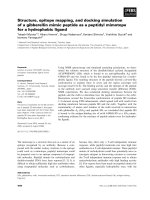
served as an internal control. Gains of the SEC62 gene
were found in 3 % of the counted nuclei in normal cases,
4 % of the nuclei in CIN-I cases, 4 % of the nuclei in CINII cases, 9 % of nuclei in CIN-III cases and 23 % of nuclei
in SCC cases (Fig. 1). Additionally, amplifications of the
SEC62 gene were found in dysplastic nuclei of two CIN-I
cases, one CIN-III case and four SCC cases. Overall, we
observed a rise of SEC62 gains and amplifications
Page 5 of 12
corresponding to the severity of dysplasia with a significantly higher incidence of SEC62 gains in SCC cases compared to all other cases (p = 0.006).
Simultaneous overexpression of Sec62 and vimentin
designates higher grades of cervical dysplasia
To evaluate if the detected SEC62 gains and amplifications
correlate with increased cellular Sec62 protein levels, we
quantified the level of Sec62 in the swabbed cells of all
107 female patients. As an overexpression of the SEC62
gene in HEK293 cells has been reported to induce a rise
in vimentin expression, suggesting that SEC62 mediates
EMT induction [25], we analyzed Sec62 and vimentin
protein levels simultaneously. Therefore, we developed
IFC as a new staining method for liquid-based cytological
swabs. After imaging the immunostained cells, the slides
were used for Papanicolaou staining to evaluate the
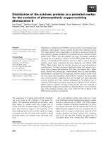
morphology of the swabbed cells. Figure 2 shows representative images for two patients, whose cervical swabs
were staged LSIL (A) and HSIL (B). Figure 3 summarizes
the immunoreactive scores (IRS) for Sec62 and vimentin
delineated for the different histological and cytological
diagnoses for all included patients.
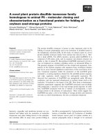
SEC62 and vimentin were overexpressed in dysplastic
cells compared with normal cells on the same slide with
a gradual increase of expression corresponding to the
rising severity of the dysplasia. When comparing the expression level of Sec62 and vimentin in the dysplastic
cells, we found a distinct correlation between the Sec62and vimentin-IRS (r2 = 0.87). To exclude that the dysplastic cells show increased fluorescent signals for all
cytoplasmic proteins due to an altered cellular shape instead of a specific overexpression of the respective genes,
we performed additional IFC stainings for 10 representative cases targeting Sec62 and β-actin (see Additional file
1). Indeed, there was no relevant change of β-actin expression depending on the severity of dysplasia. Therefore, the rise in Sec62 and vimentin protein levels in the
dysplastic cervical cells is likely attributed to a specific
overexpression of both genes.
Altering Sec62 protein levels influences HeLa cell
migration
The IFC analyses indicated that SEC62 overexpression
marks the transition from intraepithelial neoplasia to an
invasive phenotype. To evaluate whether SEC62 has potential oncogenic function, we altered Sec62 levels in
HeLa cells and evaluated changes in cell migration and
proliferation. The experiments were repeated fourfold
(n = 4) and a triplicate of every cell population was analyzed in each experiment.
First, the cells were transfected with SEC62 siRNA,
resulting in decreased Sec62 protein levels to 22 ± 1 %
(mean ± standard error of the mean, SEM) compared with
Linxweiler et al. BMC Cancer (2016) 16:676
Page 6 of 12
Fig. 1 FISH Analysis of dysplastic cervical cells. a Fluorescence in situ hybridization (FISH) analysis with a SEC62- (green) and control chromosome
10 centromere probe (red) with representative images of SEC62 amplifications (left) and gains (right). b The percentage of cells with SEC62 gains is
illustrated by blue bars for the different histomorphological groups (no CIN, CIN-I, CIN-II, CIN-III, SCC). The number of smears showing SEC62
amplifications is indicated by the number in the respective bar. In total, 5 smears per group were investigated with FISH analysis. The
respective standard error is indicated by an error bar. The grey scale bars indicate 10 μm
Fig. 2 Analysis of SEC62 and vimentin expression in swabbed cells by immunofluorescence cytology. Sec62 (left column, green) and vimentin
(middle left column, red) stainings are shown for two representative patients. In the middle right column, both signals are merged and a blue
signal indicating the DAPI-stained nuclei is added. Subsequently, the same smears were stained according to Papanicolaou (right column) for
morphological evaluation of the respective cells and classified according to the Bethesda system as LSIL (a) and HSIL (b). Cytological images are
shown in 100× magnification. The grey scale bars indicate 20 μm
Linxweiler et al. BMC Cancer (2016) 16:676
Page 7 of 12
Fig. 3 SEC62 and vimentin expression in dysplastic cervical lesions. IRS for Sec62 (a) and vimentin (b) immunostaining of uterine cervical smears
from 107 women (n = 25 + 25 + 25 + 25 + 7 for no CIN, CIN-I, CIN-II, CIN-III, SCC). The cytological immunoreactive score (IRS) values are illustrated
for the respective cytomorphological (right) and histomorphological diagnoses (left). Sec62 and vimentin immunoreactivity of morphologically
conspicuous cells was evaluated compared with normal cells of the same smear and valued as weaker (−1), equal (0), slightly more intense (1),
moderately more intense (2) or much more intense (3). For each case, the quantitation of 6 independent examiners was toted up to an overall
IRS ranging from −6 to 18. In (c), the overall IRS for Sec62 was correlated with the overall IRS for vimentin. The strength of squared correlation is
indicated by the squared correlation coefficient (R2)
control siRNA transfected cells. While marginal effects of
SEC62 silencing on cell proliferation were observed (86 ±
3 %, mean ± SEM), there was a crucial reduction in cell migration (27 ± 4 %, mean ± SEM) compared to control cells
using the xCELLigence DP system and the FluoroBlok system for migration monitoring (Figs. 4 and 5).
Next, SEC62 was overexpressed by transfecting the
cells with a SEC62 plasmid resulting in an increase of
Sec62 protein levels to 487 ± 50 % (mean ± SEM)
compared with control cells. This overexpression of
SEC62 led to increased cell migration (171 ± 7 %, mean
± SEM) with no influence on cell proliferation (93 ± 3 %,
mean ± SEM; Figs. 4 and 5). In all transfection experiments, the transfection procedure itself led to a slightly
reduced cell proliferation without however showing relevant differences between the control siRNA and the
SEC62 siRNA transfected cells respectively the control
plasmid and the SEC62 plasmid transfected cells.
Linxweiler et al. BMC Cancer (2016) 16:676
Page 8 of 12
Fig. 4 Real-time cell migration (a) and proliferation (b) analysis of SEC62-overexpressing and Sec62-depleted HeLa cells. a The cell index was measured
as an indicator for migration 15 h after seeding identically pretreated HeLa cells and compared with the respective control cells. b The slope of cell
proliferation curve was measured during the phase of exponential growth (50–74 h after seeding the cells) for HeLa cells transfected with SEC62 siRNA
or a SEC62 plasmid and compared with cells transfected with control siRNA or a control plasmid. The experiments were repeated fourfold (n = 4) and a
triplicate of every cell population was analyzed in each experiment. Cell migration (a) and cell proliferation (b) are presented as a percentage of the
respective controI cells (=100 %) using box and whisker blots. Each box represents the range from the first quartile to the third quartile. The median is
indicated by a line. The whiskers outside the boxes represent the ranges from the minimum to the maximum value of each group
SEC62 overexpression cannot induce EMT in HeLa cells
As SEC62 overexpression in HEK293 cells was reported to
induce a rise in vimentin expression and a reorganization
of the actin cytoskeleton [25], we next investigated if the
SEC62-driven stimulation of HeLa cell migration can be
attributed to an induction of EMT. SEC62 was either
overexpressed by plasmid transfection or downregulated
by siRNA transfection and the effects on cellular vimentin
and E-cadherin levels were analyzed using western blot
and immunofluorescence microscopy. These markers
were chosen, because both are known to change their expression level when cancer cells undergo EMT with an
upregulation of vimentin and a downregulation of Ecadherin levels [33]. As the subcellular F-actin structure
shows structural changes during EMT too [34], we additionally analyzed β-actin as a third EMT marker. There
were no changes in the expression level of vimentin, Ecadherin and β-actin and no changes in the subcellular
structure of the β-actin cytoskeleton (Fig. 6). All differentially pretreated HeLa cell populations contained a moderate expression of vimentin and β-actin independent of the
different treatments and E-cadherin was not detected in
HeLa cells agreement with previous reports [35].
Discussion
Cervical cancer represents the third most common cancer in women worldwide, resulting in approximately
275,000 deaths each year [1]. Despite much effort to develop new diagnostic [36, 37] and therapeutic strategies
[38], the 5-year survival rate has remained at about 70 %
with no significant changes over the past 30 years [39].
3q amplification has been identified as a common genomic alteration in cervical cancer [3, 4], marking the
transition of intraepithelial neoplasia to invasive cancer
[7]. Recently, we observed that SEC62 encoded at 3q26.2
was frequently amplified and overexpressed in NSCLC
tissue specimens [15]. Moreover, a high expression of
SEC62 predicts a poorer clinical outcome for this cancer
entity [24] and crucially influences cell migration, calcium homeostasis and ER stress tolerance of various human tumor cells [23, 24, 40].
In this study, we investigated the potential role of
SEC62 in the carcinogenesis of cervical cancer.
We found (i) that SEC62 is a potential candidate gene
of the amplified 3q region in precancerous and earlystage cancerous cervical lesions, (ii) that SEC62 is overexpressed on the protein level in dysplastic cells of the
uterine cervix compared to normal cells and (iii) that
the ability of cervical cancer cells’ to migrate depends on
their cellular Sec62 protein level.
FISH analyses of representative uterine cervix samples
demonstrated a rise in SEC62 gains and amplifications
corresponding to the grade of dysplasia, with the highest
incidence in invasive cancer cases. Accordingly, we detected an increase in cellular Sec62 protein level correlating to the severity of dysplasia in IFC analyses. These
results agree with previous studies reporting a comparable incidence for the amplification of the entire 3q26
region in precancerous cervical lesions and cervical cancer [41], suggesting that the SEC62 gene harbors an
oncogenic function. However, there are other potential
3q26-encoded oncogenes with a similar pattern of amplification and overexpression in dysplastic cervical lesions including hTERC, LAMP3 and PIK3CA [42–45].
Kuglik et al. reported that gains of the hTERC gene are
specific genomic changes in cytological specimens of the
uterine cervix associated with the progression to a malignant phenotype [42]. Furthermore, a meta-analysis of
Linxweiler et al. BMC Cancer (2016) 16:676
Page 9 of 12
Fig. 5 Cell migration analysis of SEC62-overexpressing and Sec62-depleted HeLa cells using a trans-well system. The cells that have migrated
through the 8 μm sized pores of the insert system were fixed and marked with DAPI (white dots). a Representative images are shown for HeLa
cells transfected either with control siRNA, SEC62 siRNA, a control plasmid or a SEC62 plasmid. b Cellular Sec62 protein level of the different cell
populations was quantified by western blot and normalized to GAPDH. The relative Sec62 expression is indicated below the respective bands as
mean value of 4 identically performed experiments (n = 4) with the respective standard error. The white scale bars indicate 100 μm
12 studies evaluating the diagnostic value of hTERC in
dysplastic cervical lesions found that the detection of
hTERC amplification is a valuable marker for high-grade
cervical lesions and invasive cancer [43]. However, no
functional analyses have been performed to confirm the
potential oncogenic function of hTERC or the molecular
mechanism behind its oncogenic activity. It is also probable that multiple genes in the 3q26 region are responsible for the transition of precancerous cervical lesions
to invasive cancer and that their interplay bridges the
gap from 3q amplification to the molecular cell biology
of cervical cancer carcinogenesis.
As in the first part of our study FISH- and IFC-analyses
indicated a potential oncogenic function of SEC62, we
sought to identify a functional correlate in cancer cell
biology using HeLa cells an in vitro model. Thereby,
SEC62-silencing significantly inhibited cell migration while
conversely, SEC62 overexpression stimulated cell migration.
These results confirmed conclusions of previous studies reporting similar effects of SEC62 gene silencing on
lung cancer, prostate cancer, fibrosarcoma, glioblastoma
and thyroid cancer cell lines [15, 23], as well as effects
of SEC62 overexpression on human embryonic kidney
cells [24]. However, the molecular mechanism of how
SEC62 is able to regulate cell migration remains elusive.
SEC62 encodes for a transmembrane protein of the
endoplasmic reticulum (ER) that is thought to be involved in protein transport across the ER membrane,
including the translocation of the C-terminus of
membrane proteins [20], the membrane insertion and
orientation of moderately hydrophobic signal anchor
proteins [21] and the secretion of small proteins independent of the signal recognition particle pathway [22].
Hence, we speculate that Sec62 might influence the
intracellular transport of proteins that are involved in
cell migration.
Linxweiler et al. BMC Cancer (2016) 16:676
Page 10 of 12
Fig. 6 Influence of SEC62-overexpression and SEC62-silencing in HeLa cells on the expression level of EMT markers. a Immunofluorescence
targeting Sec62 (left column, green), F-actin (middle left column, green) and vimentin (middle right column, red) in HeLa cells transfected with
control siRNA, SEC62 siRNA, a control plasmid or a SEC62 plasmid. The nuclei of the cells are marked with DAPI (blue signal). b Cellular protein
levels of E-cadherin, vimentin, Sec62 and β-actin were quantified in identically pretreated cells and normalized to GAPDH. MCF-7 cells were used
as a positive for E-cadherin expression. The relative expression of vimentin, Sec62 and β-actin is indicated below the respective bands as mean
value of 4 identically performed experiments (n = 4) with the respective standard error. Images in (a) are shown in 60× magnification. The grey
scale bars indicate 20 μm
We previously reported that Sec62 overexpression in
HEK293 cells resulted in an increased vimentin expression
and observed a structural reorganization of the actin cytoskeleton [25]. As increased vimentin expression is a key
marker of EMT [29], SEC62-mediated increase of vimentin expression represents an alternative mechanism of
how SEC62 could influence cell migration. In support of
this hypothesis, IFC- analyses of cervical brush biopsies
demonstrated a distinct correlation between SEC62 and
vimentin expression in our study. However, changes in
Sec62 protein levels in HeLa cells did neither result in detectable changes of the expression of EMT markers nor a
rearrangement of the actin cytoskeleton structure, contrary to our previous findings in HEK293 cells [25]. A possible explanation for these contradictory results could be
that different human cell lines have a varying capability
for EMT induction [46] and cytoskeleton remodeling [47].
Alternatively, it is possible that SEC62 can induce EMT
in vivo but requires unknown accessory factors and thus,
loses this function in an artificial cell culture model.
Irrespective of the underlying molecular mechanism,
the inhibition of cell migration by SEC62 silencing represents a promising approach for a new targeted therapy,
as the molecular effects of SEC62 silencing on cell migration and ER stress tolerance can be mimicked by
trifluoperazine [24], an antipsychotic drug used to treat
schizophrenia patients [48]. In addition to this potential
role of Sec62 as a therapeutic target, the detection of
SEC62 overexpression by IFC could serve as a potential
indicator for 3q26 amplification. As this genomic alteration has a high predictive value for distinguishing CINII/III lesions from normal cases [41] and can predict the
further development of precancerous cervical lesions
[49], Sec62-IFC may provide useful information for the
treatment of women with dysplastic cells in their cervical swab.
Conclusions
Taken together, our study has demonstrated a rising
incidence of SEC62 gains and amplifications in dysplastic cervical lesions as well as an increased cellular
Sec62 protein levels corresponding to the severity of
dysplasia. In functional analyses, we found that SEC62
overexpression promoted an invasive phenotype by
stimulating the cervical cancer cells’ capability to
migrate. Thus, we propose that SEC62 functions as a
migration-stimulating oncogene in the carcinogenesis
of cervical cancer and constitutes not only a potential
marker for 3q26 amplification but also a potential
target for anti-cancer treatment.
Linxweiler et al. BMC Cancer (2016) 16:676
Additional file
Additional file 1: Figure S1. Detection of β-actin (left column) and
Sec62 (middle left column) in swabbed cervical cells. In the middle right
column, both signals are merged and the right column shows the DAPIstained nuclei of the cells. The corresponding PAP-stained smears were
classified as ASCUS, LSIL and HSIL. Cytological images are shown in 20×
magnification. The grey scale bars indicate 50 μm. (TIFF 6165 kb)
Acknowledgements
We thank Ulrike Bechtel, Monika Hoffmann, Alice Kunz and Barbara
Linxweiler for excellent technical assistance and the urology research
laboratory (Saarland University Medical Center; Homburg, Germany) as
well as the staff of Prof. Richard Zimmermann’s laboratory (Saarland
University Medical Center; Homburg, Germany) for their support in
experimental procedures.
Funding
This study was supported by a HOMFOR (Homburger
Forschungsförderungsprogramm) grant to ML. The sponsor had no influence
on the design of the study and collection, analysis, and interpretation of data
and on writing the manuscript.
Availability for publication
All relevant data generated or analyzed during this study are included in this
published article and its supplementary information files. However, if further
data are requested they are available from the corresponding author on
reasonable request.
Authors’ contributions
ML carried out the immunofluorescence cytology experiments, evaluated the
respective staining results, designed the study and drafted the manuscript.
FB performed the cell culture experiments as well as statistical analyses and
participated in drafting the manuscript. BS and BAK participated in the study
design and coordination and helped to draft the manuscript. SW carried out
the FISH analyses and participated in drafting the manuscript. MG, AH and
RMB evaluated the staining results of immunofluorescence cytology
experiments and participated in the coordination of the study as well as
drafting the manuscript. IJB, EFS and ZFT collected the swab samples,
provided clinical data of the included patients, evaluated the staining results
of immunofluorescence cytology analyses and participated in drafting the
manuscript. All authors read and approved the final manuscript.
Authors’ information
Not applicable.
Competing interests
The authors declare that they have no competing interests.
Consent for publication
Written informed consent for the scientific use of tissue samples and clinical
data as well as for the publication of the scientific data was obtained from
all patients.
Ethics approval and consent to participate
The Saarland Medical Association ethics review committee approved the
scientific use of the patient’s tissue and clinical data (reference number 207/10).
Author details
1
Department of Otorhinolaryngology, Saarland University Medical Center,
Kirrberger Street 100, Building 6, 66421 Homburg/Saar, Germany.
2
Department of Medical Biochemistry and Molecular Biology, Saarland
University Medical Center, Kirrberger Street 100, Building 44, Homburg/Saar,
Germany. 3Department of General and Surgical Pathology, Saarland
University Medical Center, Kirrberger Street 100, Building 26, Homburg/Saar,
Germany. 4Department of Gynecology, Obstetrics and Reproductive
Medicine, Saarland University Medical Center, Kirrberger Street 100, Building
9, Homburg/Saar, Germany.
Page 11 of 12
Received: 17 May 2015 Accepted: 8 August 2016
References
1. Jemal A, Bray F, Center MM, Ferlay J, Ward E, Forman D. Global cancer
statistics. CA Cancer J Clin. 2011;61:69–90.
2. Elit LM, Hirte H. Management of advanced or recurrent cervical cancer:
chemotherapy and beyond. Expert Rev Anticancer Ther. 2014;14:319–32.
3. Heselmeyer K, Macville M, Schrock E, Blegen H, Hellstrom AC, Shah K, et al.
Advanced-stage cervical carcinomas are defined by a recurrent pattern of
chromosomal aberrations revealing high genetic instability and a consistent
gain of chromosome arm 3q. Gene Chromosomes Cancer. 1997;19:233–40.
4. Allen DG, White DJ, Hutchins AM, Scurry JP, Tabrizi SN, Garland SM, et al.
Progressive genetic aberrations detected by comparative genomic
hybridization in squamous cell cervical cancer. Br J Cancer. 2000;83:1659–63.
5. Sugita M, Tanaka N, Davidson S, Sekiya S, Varella-Garcia M, West J, et al.
Molecular definition of a small amplification domain within 3q26 in tumors
of cervix, ovary, and lung. Cancer Genet Cytogenet. 2000;117:9–18.
6. Caraway NP, Khanna A, Dawlett M, Guo M, Guo N, Lin E, et al. Gain of the
3q26 region in cervicovaginal liquid-based pap preparations is associated
with squamous intraepithelial lesions and squamous cell carcinoma.
Gynecol Oncol. 2008;110:37–42.
7. Heselmeyer K, Schrock E, du Manoir S, Blegen H, Shah K, Steinbeck R, et al.
Gain of chromosome 3q defines the transition from severe dysplasia to
invasive carcinoma of the uterine cervix. Proc Natl Acad Sci U S A. 1996;93:
479–84.
8. Huang FY, Kwok YK, Lau ET, Tang MH, Ng TY, Ngan HY. Genetic abnormalities
and HPV status in cervical and vulvar squamous cell carcinomas. Cancer Genet
Cytogenet. 2005;157:42–8.
9. Kirchhoff M, Rose H, Petersen BL, Maahr J, Gerdes T, Lundsteen C, et al.
Comparative genomic hybridization reveals a recurrent pattern of
chromosomal aberrations in severe dysplasia/carcinoma in situ of the cervix
and in advanced-stage cervical carcinoma. Genes Chromosomes Cancer.
1999;24:144–50.
10. Dehan E, Ben-Dor A, Liao W, Lipson D, Frimer H, Rienstein S, et al.
Chromosomal aberrations and gene expression profiles in non-small cell
lung cancer. Lung Cancer. 2007;56:175–84.
11. Chang YC, Yeh KT, Liu TC, Chang JG. Molecular cytogenetic characterization
of esophageal cancer detected by comparative genomic hybridization. J
Clin Lab Anal. 2010;24:167–74.
12. Haverty PM, Hon LS, Kaminker JS, Chant J, Zhang Z. High-resolution analysis
of copy number alterations and associated expression changes in ovarian
tumors. BMC Med Genomics. 2009;2:21.
13. Sheu JJ, Lee CH, Ko JY, Tsao GS, Wu CC, Fang CY, et al. Chromosome 3p12.
3-p14.2 and 3q26.2-q26.32 are genomic markers for prognosis of advanced
nasopharyngeal carcinoma. Cancer Epidemiol Biomarkers Prev. 2009;18:
2709–16.
14. Bockmühl U, Schwendel A, Dietel M, Petersen I. Distinct patterns of
chromosomal alterations in high- and low-grade head and neck
squamous cell carcinomas. Cancer Res. 1996;5:5325–9.
15. Linxweiler M, Linxweiler J, Barth M, Benedix J, Jung V, Kim YJ, et al. Sec62
bridges the gap from 3q amplification to molecular cell biology in nonsmall cell lung cancer. Am J Pathol. 2012;180:473–83.
16. Yamamoto H, Shigematsu H, Nomura M, Lockwood WW, Sato M, Okumura
N, et al. PIK3CA mutations and copy number gains in human lung cancers.
Cancer Res. 2008;68:6913–21.
17. McCaughan F, Pole JC, Bankier AT, Konfortov BA, Carroll B, Falzon M, et al.
Progressive 3q amplification consistently targets SOX2 in preinvasive
squamous lung cancer. Am J Respir Crit Care Med. 2010;182:83–91.
18. Massion PP, Taflan PM, Jamshedur Rahman SM, Yildiz P, Shyr Y, Edgerton
ME, et al. Significance of p63 amplification and overexpression in lung
cancer development and prognosis. Cancer Res. 2003;63:7113–21.
19. Comtesse N, Keller A, Diesinger I, Bauer C, Kayser K, Huwer H, et al. Frequent
overexpression of the genes FXR1, CLAPM1 and EIF4G located on amplicon
3q26-27 in squamous cell carcinoma of the lung. Int J Cancer. 2007;120:2538–44.
20. Jung SJ, Kim JE, Reithinger JH, Kim H. The Sec62-Sec63 translocon facilitates
translocation of the C-terminus of membrane proteins. J Cell Sci. 2014;127:
4270–8.
21. Reithinger JH, Kim JE, Kim H. Sec62 protein mediates membrane insertion
and orientation of moderately hydrophobic signal anchor proteins in the
endoplasmic reticulum (ER). J Biol Chem. 2013;288:18058–67.
Linxweiler et al. BMC Cancer (2016) 16:676
22. Lakkaraju AK, Thankappan R, Mary C, Garrison JL, Taunton J, Strub K. Efficient
secretion of small proteins in mammalian cells relies on Sec62-dependent
posttranslational translocation. Mol Biol Cell. 2012;23:2712–22.
23. Greiner M, Kreutzer B, Jung V, Grobholz R, Hasenfus A, Stöhr RF, et al.
Silencing of the SEC62 gene inhibits migratory and invasive potential of
various tumor cells. Int J Cancer. 2011;128:2284–95.
24. Linxweiler M, Schorr S, Schauble N, Jung M, Linxweiler J, Langer F,
et al. Targeting cell migration and the endoplasmic reticulum stress
response with calmodulin antagonists: a clinically tested small molecule
phenocopy of SEC62 gene silencing in human tumor cells. BMC Cancer.
2013;13:574.
25. Linxweiler J, Kollipara L, Zahedi RP, Lampel P, Zimmermann R, Greiner M.
Proteomic insights into non-small cell lung cancer: new ideas for cancer
diagnosis and therapy from a functional viewpoint. EuPA Open Proteomics.
2014;4:25–39.
26. Lopez J, Poitevin A, Mendoza-Martinez V, Perez-Plasencia C, Garcia-Carranca
A. Cancer-initiating cells derived from established cervical cell lines exhibit
stem-cell markers and increased radioresistance. BMC Cancer. 2012;12:48.
27. Myong NH. Loss of E-cadherin and acquisition of vimentin in epithelialmesenchymal transition are noble indicators of uterine cervix cancer
progression. Korean J Pathol. 2012;46:341–8.
28. Koay MH, Crook M, Stewart CJ. Cyclin D1, E-cadherin and beta-catenin
expression in FIGO Stage IA cervical squamous carcinoma: diagnostic value
and evidence for epithelial-mesenchymal transition. Histopathology. 2012;
61:1125–33.
29. Guarino M, Rubino B, Ballabio G. The role of epithelial-mesenchymal transition
in cancer pathology. Pathology. 2007;39:305–18.
30. Scanlon CS, Van Tubergen EA, Inglehart RC, D’Silva NJ. Biomarkers of epithelialmesenchymal transition in squamous cell carcinoma. J Dent Res. 2013;92:114–21.
31. Lee MY, Chou CY, Tang MJ, Shen MR. Epithelial-Mesenchymal transition in
cervical cancer: correlation with tumor progression, epidermal growth factor
receptor overexpression, and Snail up-regulation. Clin Cancer Res. 2008;14:
4743–50.
32. Ha GH, Kim JL, Breuer EKY. TACC3 is essential for EGF-mediated EMT in
cervical cancer. PLoS One. 2013;8, e70353.
33. Sabbah M, Amami S, Redeuilh G, Julein S, Prévost G, Zimber A, Ouelaa R,
Bracke M, De Wever O, Gespach C. Molecular signature and therapeutic
perspective of the epithelial-to-mesenchymal transitions in epithelial
cancers. Drug Resist Updat. 2008;11:123–51.
34. Wu TH, Chiou YW, Chiu WT, Tang MJ, Chen CH, Yeh ML. The F-actin and
adherence-dependent mechanical differentiation of normal epithelial cells
after TGF-ß1-induced EMT (tEMT) using a microplatemeasurement system.
Biomed Microdevices. 2014;16:465–78.
35. Vessey CJ, Wilding J, Folarin N, Hirano S, Takeichi M, Soutter P, et al. Altered
expression and function of E-cadherin in cervical intraepithelial neoplasia
and invasive squamous cell carcinoma. J Pathol. 1995;176:151–9.
36. Reesink-Peters N, Wisman GB, Jéronimo C, Tokumaru CY, Cohen Y, Dong
SM, et al. Detecting cervical cancer by quantitative promoter
hypermethylation assay on cervical scrapings: a feasibility study. Mol Cancer
Res. 2004;2:289–95.
37. Xiaoxia H, Schwarz JK, Lewis Jr JS, Huettner PC, Rader JS, Deasy JO, et al. A
MicroRNA expression signature for cervical cancer prognosis. Cancer Res.
2010;70:1441–8.
38. Duenas-Gonzalez A, Serrano-Olvera A, Cetina L, Coronel J. New molecular
targets against cervical cancer. Int J Women’s Health. 2014;5:1023–31.
39. Siegel R, Ma J, Zou Z, Jemal A. Cancer Statictics, 2014. CA Cancer J Clin.
2014;64:9–29.
40. Greiner M, Kreutzer B, Lang S, Jung V, Cavalié A, Unteregger G, et al. Sec62
protein level is crucial for the ER stress tolerance of prostate cancer. Prostate.
2011;71:1074–83.
41. Wright TC, Compagno J, Romano P, Grazioli V, Verma Y, Kershnar E, et al.
Amplification of the 3q chromosomal region as a specific marker in cervical
cancer. Am J Obstet Gynecol. 2015. doi:10.1016/j.ajog.2015.02.001.
42. Kuglik P, Kasikova K, Smetana J, Vallova V, Lastuvkova A, Moukova L, et al.
Molecular cytogenetic analyses of hTERC (3q26) and MYC (8q24) genes
amplifications in correlation with oncogenic human papillomavirus infection in
Czech patients with cervical intraepithelial neoplasia and cervical carcinomas.
Neoplasma. 2015;62:130–9.
43. Wang X, Liu J, Xi H, Cai L. The significant diagnostic value of human telomerase
RNA component (hTERC) gene detection in high-grade cervical lesions and
invasive cancer. Tumour Biol. 2014;35:6893–900.
Page 12 of 12
44. Kanao H, Enomoto T, Kimura T, Fujita M, Nakashima R, Ueda Y, et al.
Overexpression of LAMP3/TSC403/DC-LAMP promotes metastasis in uterine
cervical cancer. Cancer Res. 2005;65:8640–5.
45. Ma YY, Wei SJ, Lin YC, Lung JC, Chang TC, Whang-Peng J, et al. PIK3CA as
an oncogene in cervical cancer. Oncogene. 2000;19:2739–44.
46. Oyanagi J, Ogawa T, Sato H, Higashi S, Miyazaki K. Epithelial-mesenchymal
transition stimulates human cancer cells to extend microtubule-based
invasive protrusions and suppresses cell growth in collagen gel. PLoS One.
2012;7, e53209.
47. Malicka-Blaszkiewicz M, Filipczak N, Golab K, Juszczynska K, Sebzda T, Gburek J.
Ovocystatin affects actin cytoskeleton organization and induces proapoptotic
acivity. Acta Biochim Pol. 2014;61:753–8.
48. Carpenter Jr WT, Davis JM. Another view of the history of antipsychotic
drug discovery and development. Mol Psychiatry. 2012;17:1168–73.
49. Jalali GR, Herzog TJ, Dziura B, Walat R, Kilpatrick MW. Amplification of the
chromosome 3q26 region shows high negative predictive value for
nonmalignant transformation of LSIL cytologic finding. Am J Obstet Gynecol.
2010;202:581.e1–5.
Submit your next manuscript to BioMed Central
and we will help you at every step:
• We accept pre-submission inquiries
• Our selector tool helps you to find the most relevant journal
• We provide round the clock customer support
• Convenient online submission
• Thorough peer review
• Inclusion in PubMed and all major indexing services
• Maximum visibility for your research
Submit your manuscript at
www.biomedcentral.com/submit