Honokiol inhibits sphere formation and xenograft growth of oral cancer side population cells accompanied with JAK/ STAT signaling pathway suppression and apoptosis induction
Bạn đang xem bản rút gọn của tài liệu. Xem và tải ngay bản đầy đủ của tài liệu tại đây (1.9 MB, 13 trang )
Huang et al. BMC Cancer (2016) 16:245
DOI 10.1186/s12885-016-2265-6
RESEARCH ARTICLE
Open Access
Honokiol inhibits sphere formation and
xenograft growth of oral cancer side
population cells accompanied with JAK/
STAT signaling pathway suppression and
apoptosis induction
Jhy-Shrian Huang1,2†, Chih-Jung Yao1,2,3†, Shuang-En Chuang4, Chi-Tai Yeh5, Liang-Ming Lee6, Ruei-Ming Chen1,7,
Wan-Ju Chao4, Jacqueline Whang-Peng1,2 and Gi-Ming Lai1,2,3,4*
Abstract
Background: Eliminating cancer stem cells (CSCs) has been suggested for prevention of tumor recurrence and
metastasis. Honokiol, an active compound of Magnolia officinalis, had been proposed to be a potential candidate
drug for cancer treatment. We explored its effects on the elimination of oral CSCs both in vitro and in vivo.
Methods: By using the Hoechst side population (SP) technique, CSCs-like SP cells were isolated from human oral
squamous cell carcinoma (OSCC) cell lines, SAS and OECM-1. Effects of honokiol on the apoptosis and signaling
pathways of SP-derived spheres were examined by Annexin V/Propidium iodide staining and Western blotting,
respectively. The in vivo effectiveness was examined by xenograft mouse model and immunohistochemical
tissue staining.
Results: The SP cells possessed higher stemness marker expression (ABCG2, Ep-CAM, Oct-4 and Nestin), clonogenicity,
sphere formation capacity as well as tumorigenicity when compared to the parental cells. Treatment of these SP-derived
spheres with honokiol resulted in apoptosis induction via Bax/Bcl-2 and caspase-3-dependent pathway. This apoptosis
induction was associated with marked suppression of JAK2/STAT3, Akt and Erk signaling pathways in honokiol-treated
SAS spheres. Consistent with its effect on JAK2/STAT3 suppression, honokiol also markedly inhibited IL-6-mediated
migration of SAS cells. Accordingly, honokiol dose-dependently inhibited the growth of SAS SP xenograft and
markedly reduced the immunohistochemical staining of PCNA and endothelial marker CD31 in the xenograft tumor.
Conclusions: Honokiol suppressed the sphere formation and xenograft growth of oral CSC-like cells in association with
apoptosis induction and inhibition of survival/proliferation signaling pathways as well as angiogenesis. These results
suggest its potential as an integrative medicine for combating oral cancer through targeting on CSCs.
Keywords: Honokiol, Cancer stem-like side population, JAK2/STAT3 pathway, Oral cancer
* Correspondence:
†
Equal contributors
1
Comprehensive Cancer Center, Taipei Medical University, Taipei, Taiwan
2
Cancer Center, Wan Fang Hospital, Taipei Medical University, No.111,
Section 3, Hsing-Long Road, Taipei 116, Taiwan
Full list of author information is available at the end of the article
© 2016 Huang et al. Open Access This article is distributed under the terms of the Creative Commons Attribution 4.0
International License ( which permits unrestricted use, distribution, and
reproduction in any medium, provided you give appropriate credit to the original author(s) and the source, provide a link to
the Creative Commons license, and indicate if changes were made. The Creative Commons Public Domain Dedication waiver
( applies to the data made available in this article, unless otherwise stated.
Huang et al. BMC Cancer (2016) 16:245
Background
Oral squamous cell carcinoma (OSCC) is the most common type of head and neck cancer, which is estimated
over 200,000 new cases and 120,000 deaths worldwide
[1]. In Taiwan, OSCC has emerged as one of the major
malignancies with high increasing rate of both incidence
and mortality in the past decade [2]. First-line combination chemotherapy with docetaxel, cisplatin and
5-flurouracil (TPF) nowadays has been the most commonly used induction regimen for the treatment of
advanced diseases (stages III and IV), but the side effects
are severer than single-drug chemotherapy [3, 4]. Despite
the improvements of surgical and radiation techniques,
the 5-year survival rate of oral cancer has remained
unchanged at about 50 % over the past 30 years [5]. Local
recurrence and distant metastases are two critical influencing factors on survival of OSCC. Therefore, it is urgent
to develop more effective agents for the improvement of
clinical outcome.
According to the model of cancer stem cells (CSCs),
increasing evidence suggests that tumor recurrence and
metastases are caused exclusively by a rare subpopulation of tumor-initiating cells with stem cell properties
[6–9]. CSCs exhibit capacities of self-renewal, tumorigenicity and differentiating into non-stem cancer cells
that constitute the bulk of tumors [10, 11]. Thus, targeting the CSCs population has become a novel strategy to
prevent tumor recurrence or metastasis. How to eradicate the existing CSCs to improve the survival of
patients with OSCC after surgery and radio- or chemotherapy becomes a challenging issue.
Isolation of CSCs from solid tumors has been successfully achieved through several methods based on the
properties of CSCs [7, 12]. One common method is the
side population (SP) technique based on the ability of
these cells to efflux a fluorescent DNA-binding dye
Hoechst 33342, as first described by Goodell [13]. The
SP cells are a subset of cells harboring stem cell-like
properties that show a distinct low Hoechst 33342 dye
staining pattern [14]. Some studies demonstrated that
SP cells isolated from various cancer cell lines showed
high expression of stemness markers and the ability to
initiate tumor formation as well as resistance to chemotherapy [14, 15]. Thus, it is postulated that SP cells are
enriched of CSCs and represent an important potential
target for novel anticancer drug development. Several reports had shown that SP cells possessing properties of
CSCs could be isolated from OSCC cell lines [16–18],
however, little is known about the eradication of these
CSCs. Based on our previous studies, natural products
and phytochemicals are the potential source of CSC
targeting agents [19–22].
Honokiol is a bioactive compound purified from the
bark of traditional Chinese herbal medicine Magnolia
Page 2 of 13
species. Evidences from in vitro and animal models had
demonstrated that honokiol possessed a variety of pharmacological effects, such as anti-inflammation, antiangiogenesis, anti-arrhythmic and antioxidant activity
[23, 24]. It had also been shown to exert various protecting effects against hepatotoxicity, neurotoxicity, thrombosis and angiopathy [23]. The anticancer activity of
honokiol had been demonstrated in a variety of cancer
cell lines, including breast, lung, ovary, prostate, gastrointestinal and oral cancer cells as well as in xenograft
animal models [24–26]. Our previous work and the
study by Ponnurangam et al. had demonstrated the eliminating effect of honokiol on the CSCs-like population
in OSCC and colon cancer cells through inhibition of
Wnt/β-catenin [20] and Notch [27] pathway, respectively. In addition to the above stemness-associated pathways, several well-known survival/proliferation pathways
such as JAK/STAT [28], PI3K/Akt [29, 30] and MEK/Erk
[30, 31] had been shown to govern the maintenance and
survival of CSCs. However, the effects of honokiol on
these pathways of CSC are remained to be elucidated.
Hence, it is interesting and worth to investigate
honokiol-mediated elimination of CSCs in association
with inhibition of these pathways.
In this study, we investigated honokiol-mediated
suppression on these survival/proliferation signaling
pathways in CSCs-enriched SP from OSCC cells and examined the in vivo effectiveness by xenograft mouse
model and immunohistochemical tissue staining. As expected, our results showed that honokiol inhibited these
pathways in SP spheres from SAS oral cancer cells and
reduced the growth and immunohistochemical staining
of xenograft tumor.
Methods
Cell lines and sphere culture
Eight human oral squamous cell carcinoma (OSCC) cell
lines (FaDu, KB, OE, OECM-1, SAS, SCC4, SCC25 and
YD10B) were maintained in RPMI 1640 with 10 % FBS
and 1 % penicillin/streptomycin at 370C, 5 % CO2, in a
humidified chamber. After sorting, the side population cells
were seeded at a density of 500 cells/well in 6-well ultralow attachment plates (Corning Life Science, Corning, NY,
USA) with HEscGro medium (Millipore, Billerica, MA,
USA) containing epidermal growth factor (EGF, 10 ng/ml)
plus basic fibroblast growth factor (bFGF, 8 ng/ml) but
without any serum. The spheres were harvested after
14 days of culture for subsequent assays. The non-SP cells
were incubated with serum-containing RPMI medium.
Chemicals and reagents
Honokiol (purity >98 %) was kindly provided by Dr. Jack
L. Arbiser, Emory University, USA. It was dissolved in
dimethyl sulfoxide (DMSO) and further diluted in sterile
Huang et al. BMC Cancer (2016) 16:245
culture medium for in vitro experiments. The final concentrations of DMSO in cell cultures were all less than
0.05 %. The antibodies against Bax (B-9, mouse monoclonal antibody, sc-7480), Bcl-2 (100, mouse monoclonal antibody, sc-509), Erk (K-23, rabbit polyclonal antibody, sc-94),
phospho-Erk (E-4, mouse monoclonal antibody, sc-7383)
and STAT3 (F-2, mouse monoclonal antibody, sc-8019)
were purchased from Santa Cruz Biotechnology Inc. (Santa
Cruz, CA, USA). The antibodies against caspase 3 (5A1E,
rabbit monoclonal antibody, #9664), Akt (5G3, mouse
monoclonal antibody, #2966), phospho-Akt (587 F-11,
mouse monoclonal antibody, #4051), JAK2 (D2E12, rabbit
monoclonal antibody, #3230), phospho-JAK2 (D4A8,
rabbit monoclonal antibody, #8082) and phospho-STAT3
(D3A7, rabbit monoclonal antibody, #9145) were obtained
from Cell Signaling Technology (Beverly, MA, USA).
Identification and purification of side population
The side population (SP) cells were analyzed and sorted by
Hoechst 33342 (Sigma) staining and FACSAria™ III sorter
(BD Biosciences, San Jose, CA, USA). Cells were detached
from dishes with Trypsin-EDTA (Invitrogen, Grand Island,
NY, USA) and suspended at 1 × 10⁶ cells/mL in Hanks balanced salt solution (HBSS) supplemented with 3 % fetal
calf serum and 10 mM HEPES. These cells were then incubated at 37 °C for 90 min with 2.5 μg/mL Hoechst 33342,
either alone or in the presence of 50 μM reserpine (Sigma),
a nonspecific inhibitor of drug-resistance ATP-binding cassette (ABC) pumps. The diminishment of SP cells in the
presence of reserpine was used to define the flow cytometry gate for sorting SP cells. After 90-minute incubation,
the cells were centrifuged for 5 min at 300 x g, 4 °C and resuspended in ice-cold HBSS. The cells were kept on the ice
to inhibit efflux of Hoechst dye and 1 μg/mL propidium
iodide (BD) was then added to discriminate dead cells. Finally, these cells were filtered through a 40 μm cells trainer
(BD) to obtain single suspension cells for the analysis and
sorting on FACSAria III flow cytometer.
In vivo tumorigenicity assay
Dispersed cells were re-suspended in PBS. A 100 μL suspension containing various numbers of SP or non-SP
cells were injected subcutaneously into the right flanks
of 4- to 5-week-old male NOD/SCID mice, obtained
from Taiwan University Animal Center (Taipei, Taiwan).
The animal study protocols were approved by the institutional animal care and use committee of National
Heath Research Institutes, Taiwan. Tumor volume was
measured on a weekly basis by a digital caliper and
calculated using the following formula: 0.52 × L × W2
(L, longest diameter; W, shortest diameter). The experiment was terminated 10 weeks after tumor cells
inoculation and mice were euthanized. The tumor’s
wet weight was then measured.
Page 3 of 13
Sphere formation assay
The spheres were collected by gentle centrifugation,
dissociated with trypsin-EDTA and then mechanically
pipetted. The resulting single cells were re-centrifuged
to remove trypsin-EDTA and re-suspended in SP
medium to allow spheres re-formation. The spheres
were passaged every 5–7 days before they reached a
diameter of 100 μm. For the sphere formation assay, the
SP and non-SP cells were seeded at a low density of 20
cells/μL in the SP medium as described above. Ten days
after plating, the number of spheres (>50 μm) formed
was counted under a microscope.
Colony formation assay
Cells were plated at a density of 500 cells/well on 6well plates and cultured in serum-containing RPMI
media at 37 °C in 5 % CO2 for 2 weeks. The number
of colonies was counted after crystal violet staining
(Sigma).
Reverse transcription polymerase chain reaction (RT-PCR)
Trizol reagent was used to extract the mRNAs from the
SAS SP and parental cells according to the manufacturer’s
recommended protocol. Two μg RNA was added to RTPCR reactions containing primers at a concentration of
0.5 μM. After a 42 °C/60-min reverse transcription step,
25–36 cycles of PCR amplification were performed at 94 °C
for 30 s, 55 °C for 50 s, and 72 °C for 50 s. PCR products
were run on 1.5 % agarose gels for identification. Primers
used were, for ABCG2, forward: 5′-CATCAACTTTC
CGGGGGTGA-3′ and reverse: 5′-TGTGAGATTGACC
AACAGACCA-3′; for EpCAM, forward: 5′-CTGCCA
AATGTTTGGTGATG -3′ and reverse: 5′-ACGCGTTG
TGATCTCCTTCT-3′; for Oct-4, forward: 5′-GGAGAG
CAACTCCGATGG-3′ and reverse: 5′-TTGATGTCCT
GGGACTCCTC-3′; for Nestin, forward: 5′-CTCTGAC
CTGTCAGAAGAAT-3′ and reverse: 5′-GACGCTGAC
ACTTACAGAAT-3′; for GAPDH, forward: 5′-ACCAC
AGTCCATGCCATCAC-3′ and reverse: 5′-TCCACCAC
CCTGTTGCTGTA-3′.
Apoptosis analysis by Annexin V and Propidium iodide
(PI) double staining
The Annexin V-FITC Apoptosis Detection Kit (BD
Biosciences, San Jose, CA, USA) was used. In brief, the
harvested cells were re-suspended in 1x binding buffer
at a density of 1 × 10⁶ cells/mL and cells of each 100 μl
aliquot were stained with Annexin V-PI labeling solution
(containing 5 μl Annexin V-FITC and 5 μl propidium
iodide) at room temperature in the dark for 15 min.
Finally, binding buffer (400 μl) was added and the cells
were analyzed by flow cytometer.
Huang et al. BMC Cancer (2016) 16:245
Page 4 of 13
Western blot analysis
Statistical analysis
The SP-derived spheres were collected and lysed in RIPA
buffer containing protease inhibitors. Protein concentrations were measured by using the BCA protein assay kit
(Thermo Scientific Biosciences, Rockford, IL, USA).
Quantified protein lysates were separated by SDS-PAGE,
transferred onto PVDF membrane (Millipore, Billerica,
MA, USA) and immunoblotted with the primary
antibodies. After incubation with HRP-conjugated
secondary antibody, immunoreactive bands were visualized by enhanced chemiluminescence detection
system (Millipore, Billerica, MA USA). The protein
bands were quantified by AlphaEaseFC™ software.
Quantitative data were shown as mean ± SD. Differences
between control and honokiol-treated groups were
analyzed by Student’s t-test. A p-value of <0.05 was
considered statistically significant in each experiment.
Knockdown of STAT3
STAT3 siRNA was purchased from Cell Signaling
(SignalSilence® Stat3 siRNA II #6582). The mismatch
siRNA oligonucleotide 5′-UCGGCUCUUACGCAUU
CAA-3′ was used as a siRNA control. Cells were transfected with siRNA oligonucleotide using Oligofectamine
reagent according to the manufacturer’s instructions
(Invitrogen, Grand Island, NY, USA) and analyzed 72 h
post-transfection.
Wound healing assay
SAS cells were seeded into a 6-well plate. After growing to confluence, straight scratches were made across
the monolayer by using a white tip along plate cover.
Then, IL-6 (50 ng/ml) or honokiol (5 μM) was added
into wells as indicated and recorded by photography
24 h later.
Xenograft assay
NOD/SCID mice were inoculated subcutaneously with
5 × 10³ SAS SP cells into the flank and allowed to grow.
Mice were randomly divided into four groups (n = 5):
vehicle control (1 % carboxymethyl cellulose, CMC,
Sigma) and honokiol-treated groups at different dose
(20, 40, 80 mg/kg). Three weeks after inoculation, honokiol (diluted in 1 % CMC immediately prior to administration) was given intraperitoneally to mice thrice a
week until week 10. At the end, mice were sacrificed and
the tumors were paraffin embedded for the immunohistochemical staining of PCNA (PC10, mouse monoclonal
antibody, #2586, Cell Signaling Technology, Beverly,
USA) and CD31 (JC/70A, mouse monoclonal antibody,
ab9498, Abcam, Cambridge, UK). The PCNA labeling
index was calculated as the percentage of positively
stained nuclei in a total of 600 cells in 3 different areas.
The vascular density was determined by counting the
number of CD31-positive microvessels per high-power
field (x200) [32].
Results
Identification of SP cells in OSCC cell lines
We examined the existence of SP cells in eight human
OSCC cell lines by staining with Hoechst 33342 dye to
generate a Hoechst blue-red profile. In each cell line, the
percentage of SP cells was markedly diminished by treatment with reserpine, which is an inhibitor of the ABC
pumps responsible for the exclusion of Hoechst dye,
indicating that this population truly represented SP cells.
As depicted in Fig. 1, all the OSCC cell lines contained a
distinct fraction of SP cells, ranging from 1.1 % (YD10B
and SCC25) to 28.1 % (OE) of gated cells.
Side population-derived sphere cells have stem cell
properties
To investigate the CSCs of OSCC cells with different aggressiveness, SP cells from SAS (high malignancy and
metastasis) and OECM-1 (less malignancy) [33] were
chosen and cultured to form spheres according to the
methods described. The spheres derived from SAS and
OECM-1 SP cells appeared to be taking shape on day 4
and were completely formed on day 10. Morphologically,
these spheres grew tightly in clusters in three-dimensional
configuration in contrast to the flattened shape of parental
cells (Fig. 2a). We then examined the expression of stemness markers in these SP-derived spheres and parental
cells by RT-PCR. As shown in Fig. 2b, the mRNA expressions of ABCG2, Ep-CAM, OCT-4 (octamer-binding transcription factor 4) and Nestin was higher in sphere cells
than those in their parental cells. These SP cells also
possessed higher self-renewal ability as they formed much
higher number of spheres in the serum-free SP medium
(Fig. 2c). In parallel with this, the SP cells formed
markedly higher number and larger size of colonies than
the parental cells in serum-containing culture medium
(Fig. 2d).
Comparing the stemness properties of SAS and
OECM-1 sphere cells, we found the elevation of ABCG2
and Oct-4 expressions in SAS spheres were much more
marked than that in OECM-1 spheres (Fig. 2b). This result indicated that the SAS sphere cells were more
CSCs-like than those of OECM-1. Besides, the SAS SP
cells also possessed higher capability of sphere and
colony formation than the OECM-1 SP cells (Fig. 2c
and d). As these stemness characteristics found in vitro
are considered to render the tumorigenicity of SP cells
in vivo, our findings are in consistent with the report by
Huang et al. BMC Cancer (2016) 16:245
Page 5 of 13
Fig. 1 Percentage of side population cells in oral squamous cell carcinoma cells lines. Eight human oral squamous cell carcinoma cell lines were
stained with Hoechst 33342 dye in the presence (bottom) or absence (upper) of 50 μM reserpine and analyzed by flow cytometry. The side
population cells (black triangle), which were disappeared by reserpine, are shown as a percentage of the whole living cell population
Chang et al. that SAS cells are much more tumorigenic
than OECM-1 cells [33, 34].
SAS SP cells show more tumorigenic potential in
xenografts
To further characterize the stemness properties of SAS SP
cells, we examined the tumorigenicity of SAS SP and nonSP cells in vivo. Various numbers of SP (S1-S4) and nonSP (NS1-NS2) cells were subcutaneously inoculated into
NOD/CSID mice. As shown in Fig. 3a, the volume of SP
cell-derived tumors increased in a cell number- and timedependent manner. The SP cells formed tumors in three
out of five mice, even the number of inoculated cells was
as low as 1 × 10³ (Table 1). In addition, the tumor weights
were also measured and found to be increased with the
number of SP cells inoculated (Fig. 3b). In contrast, no
tumors were formed in mice inoculated with non-SP
cells, even the number of inoculated non-SP cells was
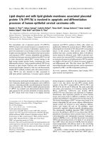
Fig. 2 SP-derived spheres from SAS and OECM-1 cell lines possess the stemness properties. a After cultured in an anchorage-independent manner for
7 days, the spheroidal morphology (phase-contrast images) of SAS (left) and OECM-1 (right) sphere cells were distinct from those of parental cells.
b Marked higher expression of stemness markers in SAS and OECM-1 sphere (“S”) cells compared to parental (“P”) cells. The expression of various
stemness markers was analyzed by RT-PCR and GAPDH was used as a loading control. The intensities of the PCR bands were quantified by densitometry.
The densitometric values indicated at the top of the bands are expressed relative to the value of parental cells after being normalized to actin (#: The
intensity of ABCG2 band of parental SAS cells was undetectable in this PCR condition). Both the SAS and OECM-1 sphere cells had higher capacities in
sphere (c) and colony (d) formation than parental cells. Data are shown as mean ± SD from experiments performed in triplicates. *, p < 0.05; **, p < 0.01
Huang et al. BMC Cancer (2016) 16:245
Page 6 of 13
Table 1 Tumorigenicity of SAS SP and non-SP cells in
NOD/SCID mice
Cell numbers inoculated/mouse
1 × 10³
5 × 10³
1 × 10⁴
5 × 10⁴
1 × 10⁶
5 × 10⁷
SAS SP
3/5
3/5
4/5
5/5
—
—
SAS non-SP
—
—
—
0/5
0/5
2/5
The number of mice with tumor formation/total number of mice inoculated with
SAS SP or non-SP cells was observed for 10 weeks after inoculation. —, not done
results, the tumorigenicity of SAS SP cells was estimated to
be ten-thousand times higher than non-SP cells.
Honokiol inhibits the colony formation and induces
apoptosis in sphere cells
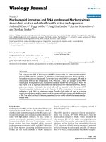
Fig. 3 Side population cells (S1-S4) possess higher tumorigenicity than
non-side population cells (NS1-NS2). a The growth curve of xenograft
tumor. NOD/SCID mice were inoculated subcutaneously with various
cell numbers of SAS SP and non-SP cells, respectively, as indicated.
Tumor volumes were recorded on a weekly basis. **p < 0.01, significant
difference vs. NS1. b The wet weight of tumors measured after
harvested at the end. *p < 0.05; **p < 0.01, significant difference vs. NS1.
c Representative photographs of the tumors harvested at the end
of experiment
up to 1 × 10⁶, and only two out of five mice formed tumors
when 1 × 10⁷ non-SP cells were inoculated (Table 1). The
photograph of representative sizes of SP cells-derived
tumors in each group was shown in Fig. 3c. Based on our
To evaluate the effects of honokiol on the elimination of
CSCs in OSCC, we examined its effects on the colony
formation and apoptosis induction in these sphere cells.
In OECM-1 sphere cells, the number of colonies was
dose-dependently decreased to 70 and 38 % by honokiol
at dose of 5 and 10 μM, respectively. In SAS sphere
cells, the number of colonies was even down to 50 and
22 % by the same doses of honokiol (Fig. 4a). After 48 h
of honokiol treatment, apoptosis was induced in both
OECM-1 and SAS sphere cells in a dose-dependent
manner (Fig. 4b). At a dose of 10 μM, honokiol-induced
apoptosis was up to 52.7 and 56.41 % in OECM-1 and
SAS sphere cells, respectively (Fig. 4b). Moreover, the
honokiol-induced late apoptosis (upper-right quadrant)
was more dominant in SAS sphere (23.9 and 47.8 %)
than in OECM-1 sphere (14.1 and 26 %) cells (Fig. 4b).
Taken together with the result shown in Fig. 4a, the
higher malignant and tumorigenic SAS spheres appeared
to be more sensitive to honokiol-induced anticancer
effects than the OECM-1 sphere cells.
We then examined the changes in levels of the Bcl-2
and Bax proteins that regulate the intrinsic apoptosis
pathway of cancer cells. Both in OECM-1 and SAS
sphere cells, honokiol decreased the anti-apoptotic Bcl-2
while increased the pro-apoptotic Bax protein in a dosedependent manner (Fig. 4c). As expected, this increase
of Bax to Bcl-2 protein ratio led to cleavage/activation of
the key apoptosis co-ordination enzyme, caspase-3, in
both of the two cancer spheres (Fig. 4d). These results
suggest the pivotal role of mitochondria-dependent
(intrinsic) apoptosis in honokiol-mediated elimination of
CSCs in OSCC cells.
Honokiol inhibits the JAK2/STAT3, Akt and Erk signal
pathways in SAS sphere cells
Regarding the profound inhibition of colony formation
and induction of apoptosis shown in Fig. 4, we examined
the survival/proliferation signals such as JAK2/STAT3,
Akt and Erk pathways in honokiol-treated SAS sphere
cells. After 48 h of treatment, honokiol markedly
Huang et al. BMC Cancer (2016) 16:245
Page 7 of 13
a
0 M
5 M
10 M
100
80
**
60
40
**
**
20
0
UR 0.07
UR 0
LR 0.07
LR 0.02
UR 14.1
UR 23.9
LR 20.5
LR 16.0
UR 26
UR 47.8
LR 26.7
LR 8.61
0 M
*
PI
Colony formation (%)
120
SAS
OECM-1
b
OECM-1
OECM-1
SAS
SAS
5 M
10 M
Annexin V-FITC
c
Bax
-actin
-actin
0
5
10
Pro-caspase 3
Cleaved caspase 3
-actin
Pro-caspase 3
Cleaved caspase 3
-actin
SAS spheres
Bax
SAS spheres
Bcl-2
Honokiol ( M)
OECM-1 spheres
Bcl-2
d
OECM-1 spheres
Honokiol ( M)
0
5
10
Fig. 4 Honokiol inhibits colony formation and induces apoptosis via Bax/Bcl-2 and caspase-3-dependent pathway in SP-derived sphere
cells. a Honokiol inhibited colony formation of the SAS and OECM-1 SP-derived sphere cells in a dose-dependent manner. The colony formation data
are expressed as percent of control (without honokiol treatment) cells and shown as mean ± SD. *p < 0.05; **p < 0.01, significant difference vs. control.
b Honokiol induced apoptosis of the SAS and OECM-1 SP-derived spheres in a dose-dependent manner. Apoptosis was determined by Annexin
V-FITC/PI double staining and flow cytometry analysis. The honokiol concentration is shown in the right side of dot plots. The numbers in LR (lower right)
quadrant indicates the percentage of early apoptotic cells. The numbers in UR (upper right) quadrant indicates the percentage of late apoptotic cells.
c Dose-dependent effect of honokiol on the protein levels of Bax and Bcl-2. d Dose-dependent effect of honokiol on cleavage of caspase-3
decreased the levels of phospho-JAK2 (pJAK2) and
phospho-STAT3 (pSTAT3) rather than affecting the total
protein levels of JAK2 and STAT3 (Fig. 5a and b). Honokiol also dose-dependently decreased the phospho-Akt
(pAkt) without affecting the total Akt protein level
(Fig. 5c). Both the phospho-Erk (pErk) and total Erk
were simultaneously reduced by honokiol (Fig. 5d).
These survival/proliferation signaling pathways might be
suppressed through different mechanisms during apoptosis induction by honokiol in the CSC-like sphere cells.
Honokiol suppresses the migration of SAS cells
The JAK2/STAT3 pathway regulates not only the antiapoptotic survival signal but also the motility of cancer
cells [35]. Considering the marked JAK2/STAT3 pathway
inhibition by honokiol, we explored its effect on cell
Huang et al. BMC Cancer (2016) 16:245
Page 8 of 13
Fig. 5 Honokiol inhibits the JAK2/STAT3, Akt and Erk pathways in SP-derived spheres. The SAS SP-derived spheres were incubated with 5 or
10 μM honokiol for 48 h. The protein levels of total and phosphorylated JAK2 (a), STAT3 (b), Akt (c) and Erk (d) were determined by Western blot
and quantified by densitometry. The ratios of pJAK2, pSTAT3, pAkt and pErk to actin were calculated
migration (the wound healing assay) of the highly aggressive SAS cells, using STAT3 siRNA as a positive
control. As shown in Fig. 6a, marked decrease of STAT3
protein expression was observed in the two preparations
of STAT3 siRNA transfected cells (siSTAT3-1, siSTAT3-2).
As expected, the migration of siSTAT3-2-transfected cells
was significantly inhibited as compared to that of the cells
transfected with control siRNA (Fig. 6b). In consistent
with the inhibition on JAK2/STAT3 pathway shown in
Fig. 5, honokiol inhibited SAS cell migration as effective
as the siSTAT3 after 24 h of incubation (Fig. 6b). As the
JAK2/STAT3 pathway in human malignancies could be
triggered by the pro-inflammatory cytokine such as IL-6
[36], we further investigated the inhibitory effect of honokiol on IL-6-mediated cell migration. Notably, we found
honokiol could suppress the migration enhanced by IL-6
as well (Fig. 6b).
Honokiol inhibits the tumor growth of SAS SP xenograft
To confirm the effectiveness in vivo, we examined the
effects of honokiol on the tumor growth of SAS SP
xenograft in SCID mice. The tumor volume was
periodically measured with a metric caliper and the body
weight was also simultaneously measured on a weekly
basis. The tumor volume of control group gradually
increased to 2479 ± 302 mm3 after subcutaneous inoculation with 5 × 10³ SAS SP cells for 10 weeks (Fig. 7a).
At week 10, honokiol decreased the tumor volume to
2024 ± 265, 1555 ± 247 and 879 ± 166 mm3 at doses of
20, 40 and 80 mg/kg, respectively (Fig. 7a). By calculation, the percentage of tumor volume reduction was
32.3 % at dose of 40 mg/kg (p < 0.05) and 64.5 % at dose
of 80 mg/kg (p < 0.01), respectively. The tumors were
excised and weighed at the end of week 10. A dosedependent decrease of tumor weigh was observed in
honokiol-treated groups (Fig. 7b). The tumor weight of
80 mg/kg honokiol-treated group was decreased by
almost 90 % comparing to the control group (p < 0.01).
The changes of body weight were measured weekly after
honokiol treatment. No significant difference between
control and honokiol-treated groups was observed
throughout the experimental protocol (Fig. 7c). Besides,
neither visible sign of toxicity nor any abnormal behavior were observed in honokiol-treated mice.
Huang et al. BMC Cancer (2016) 16:245
Page 9 of 13
Fig. 6 Honokiol suppresses IL-6-mediated migration of SAS cells. a Two preparations of SAS cells were transfected with STAT3 siRNA (siSTAT3-1,
siSTAT3-2) and the control siRNA group was transfected with the mismatch siRNA oligonucleotide. After 72 h, the STAT3 expression was determined
by Western blot. b Wound healing assay. The STAT3 siRNA transfected SAS cells (siSTAT3-2) were seeded into a 6-well plate. After growing to
confluence, straight scratches were made across the monolayer by using a white tip along plate cover. Then, IL-6 (50 ng/ml) or honokiol
(5 μM) was added into wells as indicated and recorded by photography 24 h later. Honokiol inhibited the migration of SAS cells as potent as
STAT3 siRNA. Notably, honokiol also suppressed the migration enhanced by IL-6. In this assay, honokiol (5 μM) and siSTAT3 did not affect the
cell viability of these cells (Additional file 1: Figure S1)
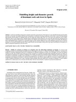
Honokiol decreases the PCNA and CD31 levels in the
tissue of SAS SP xenograft tumor
neovascularization within tumor tissues of the SAS SP
xenograft.
The immunohistochemical examination was performed in
the sections of excised tumors with or without 80 mg/kg
honokiol treatment. In accordance with the reduced
tumor volume and weight, the honokiol-treated tumors
displayed lower PCNA (proliferating cell nuclear antigen)
positive rate (Fig. 8a). The PCNA labeling index in control group was reduced from 74.13 ± 4.1 to 20.87 ± 2.4
(p < 0.001) by honokiol (Fig. 8b). Similar result was also
observed in the staining of angiogenic marker, CD31
(Fig. 8c). As shown in Fig. 8d, the number of CD31positive microvessels (MVD/fields) was significantly reduced from 42.7 ± 3.5 to 17.3 ± 2.1 by treatment with
honokiol (p < 0.01), indicating that honokiol may inhibit
Discussion
The resistance of OSCC to conventional chemotherapy
or radiation therapy might be due to existence of CSCs
[37]. Consequently, agents capable of eliminating this
CSC population are desirable for improving the clinical
outcomes of OSCC treatments. Many preclinical studies
had shown the anticancer activities of honokiol [24]. Recently, our group and Ponnurangam et al., had reported
the elimination of CSC-like population by honokiol in
OSCC and colon cancer cells through Wnt/β-catenin
[20] and notch pathway inhibition [27], respectively.
This study now further demonstrated its inhibitory
Huang et al. BMC Cancer (2016) 16:245
Fig. 7 Honokiol dose-dependently inhibits growth of SAS SP cells
xenograft in NOD/SCID mice. Mice were inoculated subcutaneously
with 5 × 10³ SAS SP cells. Honokiol was administered by intraperitoneal
injection thrice a week. a Tumor volumes were measured once a week.
The tumor growth was dose-dependently inhibited by honokiol.
*p < 0.05; **p < 0.01, significant difference vs. control. b At the
end of week 10, the tumors were harvested and weighed. Honokiol
dose-dependently decreased the tumor weight. Data shown are
mean ± SD (n = 5). **p < 0.01, significant difference vs. control. c The
changes of body weight were measured weekly after honokiol treatment.
No significant difference between control and honokiol-treated groups
was observed
Page 10 of 13
effects on the survival/proliferation signaling such as
JAK2/STAT3, AKT, and ERK in the CSC-like SAS
sphere cells and confirmed the in vivo effectiveness in
xenograft animal model.
Generally, SP has been proposed as a practical method
to enrich and isolate CSCs from many tumor tissues and
cell lines [14]. Several studies had demonstrated that SP
isolated from OSCC cell lines indeed possesses the properties of CSCs and higher tumorigenicity [16–18]. However, Broadley et al. had shown controversial results that
the SP isolated from glioblastoma multiforme cells did
not have enhanced stem-like property and tumor initiating activity over the non-SP cells, suggesting that the
CSCs enriched by SP technique should be further confirmed by animal experiment [38]. In our results, the SP
percentage in OECM-1 (20.5 %) is much higher than
that in SAS (2.9 %) cells. This phenomenon is in accordance with the report by Chiou et al. that OECM-1
expressed higher ABCG2 compared to SAS cells [33].
However, the SAS cells are much more tumorigenic
and metastatic than the OECM-1 cells [34]. Considering this controversy, we performed an animal experiment to confirm that the SAS SP did have much
higher tumorigenicity (approximately ten thousand
times higher) than the non-SP. Therefore, we used SAS
SP xenograft as a model to evaluate the effectiveness of
honokiol.
The effects of honokiol on the increase of Bax to Bcl-2
ratio and subsequent apoptosis induction had been reported in various types of cancer cells [39]. The significance of Bax to Bcl-2 ratio on the progression of several
diseases or malignant tumors had been investigated by
several studies [40]. This ratio may serve as a predictive
marker to evaluate prognosis in patients with rectal carcinomas who have undergone elective colectomy and received post-surgery adjuvant treatment [41]. Our results
further demonstrated the increased Bax to Bcl-2 ratio in
the CSC-like SAS sphere cells after treatment with honokiol, indicating the potential of honokiol to improve
OSCC therapy via apoptosis induction of CSCs. Compared to OECM-1 spheres, the honokiol-induced late
apoptosis was more dominant in SAS sphere cells, suggesting the application of honokiol in the high-grade
and aggressive OSCC might be more useful. Further
clinical investigation is warranted.
Honokiol had been shown to induce apoptosis in various types of cancer cells through inhibition of several
well-known survival/proliferation signaling pathways
such as JAK/STAT, PI3K/Akt and MEK/Erk [42–45]. As
these pathways also govern the CSC maintenance and
survival [28–31], the honokiol-mediated inhibition of
these pathways and apoptosis induction in CSC-like
sphere cells would provide further mechanisms underlying its CSCs elimination potential.
Huang et al. BMC Cancer (2016) 16:245
Page 11 of 13
Fig. 8 Honokiol markedly decreases the immunohistochemical staining of PCNA and CD31 in SAS SP xenograft tumor tissue. Immunohistochemistry
staining of PCNA and CD31 was performed in the paraffin-embedded tissue sections of tumors from mice treated with or without honokiol (80 mg/kg).
a The staining intensity (brown color) of PCNA was markedly lower in honokiol-treated group. b The PCNA labeling indexes of control
and honokiol-treated groups. c The staining intensity of CD31 (endothelial cell marker) was markedly lower in honokiol-treated group. d The number
of CD31 positive microvessel was counted at 200x magnification under a microscope. Significantly reduced microvessel density (MVD)/fields was
observed in honokiol-treated group. Data shown are mean ± SD. **p < 0.01; ***p < 0.001, significant difference vs. control
The STAT3 signaling also mediates IL-6 induced EMT
(epithelial-mesenchymal transition) to promote the metastasis of head and neck tumor cells [46]. The inhibitory effect of honokiol on EMT by targeting STAT3
signaling was recently reported [47]. In line with this, we
also observed inhibitory effects of honokiol on the migration of SAS cells induced by IL-6 and on the STAT3
activity in SAS sphere cells. Furthermore, the contribution of STAT3-mediated EMT on CSC-like phenotype
had also been noted [48, 49]. It is possibile that honokiol
also suppressed the STAT3-EMT-promoted CSC-like
traits in the microenvironment within the xenograft
tumor. Further investigation is needed.
Constitutive activation of the STAT3 is associated with
not only cell proliferation and metastasis but also angiogenesis [50, 51]. It is known that anti-angiogenesis via
STAT3 inactivation also plays an important role in the
honokiol-mediated anticancer activities [52]. In agreement
with this, our immunohistochemical results show that not
only PCNA but also CD31 (endothelial marker) were
markedly suppressed in honokiol-treated xenograft tumor
tissues, indicating that honokiol may be regarded as a useful antiangiogenic agent for the treatment of OSCC.
Conclusions
In conclusion, our results have demonstrated that
honokiol may induce apoptosis and inhibit the survival/
proliferation signaling pathways in oral CSC-like cells.
These effects were associated with the suppressed sphere
formation in vitro and the reduced neovascularization
and growth in xenograft tumors. The clinical development of honokiol as a novel complementary and alternative therapeutics targeting for CSCs to improve the
clinical outcome of OSCC is warranted.
Huang et al. BMC Cancer (2016) 16:245
Page 12 of 13
Availability of data and materials
3.
The effects of Honokiol (5 μM) and siSTAT3 on the
cell viablity of SAS cells in the 24-h wound healing
assay are provided as supplementary information in
Additional file 1: Figure S1.
4.
5.
Additional file
Additional file 1: Figure S1. Honokiol (5 μM) and siSTAT3 did not
affect the cell viability of SAS cells in the 24-h wound healing assay. (A) SAS
cells were treated with honokiol (5 μM) for 24 h in the same culture
condition shown in Fig. 6b for wound healing assay. The cell viability
was then determined by the quantitative staining of cellular proteins by
sulforhodamine B. (B) After transfection with siSTAT3 and analysis for the
expression of STAT3, the SAS cells were seeded into 6-well plate in the
same condition shown in Fig. 6b for wound healing assay. After 24 h of
incubation, the cell viability was then determined by the quantitative
staining of cellular proteins by sulforhodamine B. (TIFF 1240 kb)
Abbreviations
ABC: ATP-binding cassette; CSCs: cancer stem cells; EMT: epithelial-mesenchymal
transition; MVD: microvessel density; OSCC: oral squamous cell carcinoma;
PCNA: proliferating cell nuclear antigen; PI: propidium iodide; SP: side population.
Competing interests
The authors declare that they have no competing interests.
Authors’ contributions
JSH, CJY, SEC and GML conceived this study and wrote the manuscript.
CTY participated in the design of the study and worked with JSH and WJC
to carry out the experiments and analyze the data. LML, RMC and JWP
provided important suggestions for data processing and manuscript editing.
JSH and CJY contributed equally to this paper. All authors read and
approved the final manuscript.
6.
7.
8.
9.
10.
11.
12.
13.
14.
15.
16.
17.
18.
Acknowledgements
This work was supported by National Health Research Institutes (Grant
CA-101-PP-37 and CA-102-PP-37), Wan Fang Hospital, Taipei Medical University
(Grant 103-wf-eva-08), and Health and Welfare Surcharge of tobacco products,
Taiwan (MOHW104-TDU-B-212-124-001). We thank Dr. Jack L. Arbiser, Emory
University, USA for providing bulk honokiol compound. We also thank the staffs
at the Laboratory Animal Center of the National Health Research Institutes
(NHRI, Taiwan) for technical support and Dr. Ying-Ying Shen at the Pathology
Core Laboratory of NHRI for pathology consultation.
Author details
1
Comprehensive Cancer Center, Taipei Medical University, Taipei, Taiwan.
2
Cancer Center, Wan Fang Hospital, Taipei Medical University, No.111,
Section 3, Hsing-Long Road, Taipei 116, Taiwan. 3Department of Internal
Medicine, School of Medicine, College of Medicine, Taipei Medical University,
No.250, Wuxing Street, Taipei 110, Taiwan. 4National Institute of Cancer
Research, National Health Research Institutes, Miaoli County, Taiwan.
5
Department of Surgery, Shuang Ho Hospital, Taipei Medical University,
Taipei, Taiwan. 6Department of Urology, School of Medicine, College of
Medicine, Taipei Medical University, Taipei, Taiwan. 7Graduate Institute of
Medical Sciences, College of Medicine, Taipei Medical University, Taipei,
Taiwan.
Received: 23 April 2015 Accepted: 10 March 2016
19.
20.
21.
22.
23.
24.
25.
26.
References
1. Jemal A, Bray F, Center MM, Ferlay J, Ward E, Forman D. Global cancer
statistics. CA Cancer J Clin. 2011;61:69–90.
2. Liu SA, Tsai WC, Wong YK, Lin JC, Poon CK, Chao SY, et al. Nutritional factors
and survival of patients with oral cancer. Head Neck. 2006;28:998–1007.
27.
Vermorken JB, Remenar E, van Herpen C, Gorlia T, Mesia R, Degardin M,
et al. Cisplatin, fluorouracil, and docetaxel in unresectable head and neck
cancer. N Engl J Med. 2007;357:1695–704.
Haddad R, Colevas AD, Tishler R, Busse P, Goguen L, Sullivan C, et al.
Docetaxel, cisplatin, and 5-fluorouracil-based induction chemotherapy in
patients with locally advanced squamous cell carcinoma of the head and
neck: the Dana Farber Cancer Institute experience. Cancer. 2003;97:412–8.
Chien HT, Liao CT, Huang SF, Chen IH, Liu TY, Jou YS, et al. Clinical
significance of genome-wide minimally deleted regions in oral squamous
cell carcinomas. Genes Chromosomes Cancer. 2011;50:358–69.
Sinha N, Mukhopadhyay S, Das DN, Panda PK, Bhutia SK. Relevance of
cancer initiating/stem cells in carcinogenesis and therapy resistance in oral
cancer. Oral Oncol. 2013;49:854–62.
Monroe MM, Anderson EC, Clayburgh DR, Wong MH. Cancer stem cells in
head and neck squamous cell carcinoma. J Oncol. 2011;2011:762780.
Singh A, Settleman J. EMT, cancer stem cells and drug resistance: an
emerging axis of evil in the war on cancer. Oncogene. 2010;29:4741–51.
Kong D, Li Y, Wang Z, Sarkar FH. Cancer Stem Cells and Epithelial-toMesenchymal Transition (EMT)-Phenotypic Cells: Are They Cousins or Twins?
Cancers (Basel). 2011;3:716–29.
O’Brien CA, Kreso A, Jamieson CH. Cancer stem cells and self-renewal.
Clin Cancer Res. 2010;16:3113–20.
Gonzalez-Moles MA, Scully C, Ruiz-Avila I, Plaza-Campillo JJ. The cancer stem
cell hypothesis applied to oral carcinoma. Oral Oncol. 2013;49:738–46.
Satpute PS, Hazarey V, Ahmed R, Yadav L. Cancer stem cells in head and neck
squamous cell carcinoma: a review. Asian Pac J Cancer Prev. 2013;14:5579–87.
Goodell MA, Brose K, Paradis G, Conner AS, Mulligan RC. Isolation and
functional properties of murine hematopoietic stem cells that are
replicating in vivo. J Exp Med. 1996;183:1797–806.
Wu C, Alman BA. Side population cells in human cancers. Cancer Lett.
2008;268:1–9.
Moserle L, Ghisi M, Amadori A, Indraccolo S. Side population and cancer
stem cells: therapeutic implications. Cancer Lett. 2010;288:1–9.
Zhang Q, Shi S, Yen Y, Brown J, Ta JQ, Le AD. A subpopulation of CD133(+)
cancer stem-like cells characterized in human oral squamous cell carcinoma
confer resistance to chemotherapy. Cancer Lett. 2010;289:151–60.
Zhang P, Zhang Y, Mao L, Zhang Z, Chen W. Side population in oral
squamous cell carcinoma possesses tumor stem cell phenotypes. Cancer
Lett. 2009;277:227–34.
Yanamoto S, Kawasaki G, Yamada S, Yoshitomi I, Kawano T, Yonezawa H,
et al. Isolation and characterization of cancer stem-like side population cells
in human oral cancer cells. Oral Oncol. 2011;47:855–60.
Yao CJ, Han TY, Shih PH, Yi TY, Lai IC, Chang KH, et al. Elimination of cancer
stem-like side population in human glioblastoma cells accompanied with
stemness gene suppression by Korean herbal recipe MSC500. Integr Cancer
Ther. 2014;13:541–54.
Yao CJ, Lai GM, Yeh CT, Lai MT, Shih PH, Chao WJ, et al. Honokiol Eliminates
Human Oral Cancer Stem-Like Cells Accompanied with Suppression of Wnt/
beta -Catenin Signaling and Apoptosis Induction. Evid Based Complement
Alternat Med. 2013;2013:146136.
Yao CJ, Yeh CT, Lee LM, Chuang SE, Yeh CF, Chao WJ, et al. Elimination of
cancer stem-like “side population” cells in hepatoma cell lines by chinese
herbal mixture “tien-hsien liquid”. Evid Based Complement Alternat Med.
2012;2012:617085.
Lai IC, Shih PH, Yao CJ, Yeh CT, Wang-Peng J, Lui TN, et al. Elimination of
cancer stem-like cells and potentiation of temozolomide sensitivity by
Honokiol in glioblastoma multiforme cells. PLoS One. 2015;10:e0114830.
Fried LE, Arbiser JL. Honokiol, a multifunctional antiangiogenic and
antitumor agent. Antioxid Redox Signal. 2009;11:1139–48.
Arora S, Singh S, Piazza GA, Contreras CM, Panyam J, Singh AP. Honokiol: a
novel natural agent for cancer prevention and therapy. Curr Mol Med.
2012;12:1244–52.
Kim DW, Ko SM, Jeon YJ, Noh YW, Choi NJ, Cho SD, et al. Anti-proliferative
effect of honokiol in oral squamous cancer through the regulation of
specificity protein 1. Int J Oncol. 2013;43:1103–10.
Chen XR, Lu R, Dan HX, Liao G, Zhou M, Li XY, et al. Honokiol: a promising
small molecular weight natural agent for the growth inhibition of oral
squamous cell carcinoma cells. Int J Oral Sci. 2011;3:34–42.
Ponnurangam S, Mammen JM, Ramalingam S, He Z, Zhang Y, Umar S, et al.
Honokiol in combination with radiation targets notch signaling to inhibit
colon cancer stem cells. Mol Cancer Ther. 2012;11:963–72.
Huang et al. BMC Cancer (2016) 16:245
28. Zhou J, Wulfkuhle J, Zhang H, Gu P, Yang Y, Deng J, et al. Activation of the
PTEN/mTOR/STAT3 pathway in breast cancer stem-like cells is required for
viability and maintenance. Proc Natl Acad Sci U S A. 2007;104:16158–63.
29. Bleau AM, Hambardzumyan D, Ozawa T, Fomchenko EI, Huse JT, Brennan CW,
et al. PTEN/PI3K/Akt pathway regulates the side population phenotype and
ABCG2 activity in glioma tumor stem-like cells. Cell Stem Cell. 2009;4:226–35.
30. Sunayama J, Matsuda K, Sato A, Tachibana K, Suzuki K, Narita Y, et al.
Crosstalk between the PI3K/mTOR and MEK/ERK pathways involved in the
maintenance of self-renewal and tumorigenicity of glioblastoma stem-like
cells. Stem Cells. 2010;28:1930–9.
31. Hepburn AC, Veeratterapillay R, Williamson SC, El-Sherif A, Sahay N, Thomas HD,
et al. Side population in human non-muscle invasive bladder cancer enriches
for cancer stem cells that are maintained by MAPK signalling. PLoS One.
2012;7:e50690.
32. Weidner N, Semple JP, Welch WR, Folkman J. Tumor angiogenesis and
metastasis–correlation in invasive breast carcinoma. N Engl J Med.
1991;324:1–8.
33. Chiou SH, Yu CC, Huang CY, Lin SC, Liu CJ, Tsai TH, et al. Positive
correlations of Oct-4 and Nanog in oral cancer stem-like cells and highgrade oral squamous cell carcinoma. Clin Cancer Res. 2008;14:4085–95.
34. Chang KW, Liu CJ, Chu TH, Cheng HW, Hung PS, Hu WY, et al. Association
between high miR-211 microRNA expression and the poor prognosis of oral
carcinoma. J Dent Res. 2008;87:1063–8.
35. Gao SP, Bromberg JF. Touched and moved by STAT3. Sci STKE. 2006;2006:pe30.
36. Sansone P, Bromberg J. Targeting the interleukin-6/Jak/stat pathway in
human malignancies. J Clin Oncol. 2012;30:1005–14.
37. Felthaus O, Ettl T, Gosau M, Driemel O, Brockhoff G, Reck A, et al. Cancer
stem cell-like cells from a single cell of oral squamous carcinoma cell lines.
Biochem Biophys Res Commun. 2011;407:28–33.
38. Broadley KW, Hunn MK, Farrand KJ, Price KM, Grasso C, Miller RJ, et al. Side
population is not necessary or sufficient for a cancer stem cell phenotype in
glioblastoma multiforme. Stem Cells. 2011;29:452–61.
39. Xu HL, Tang W, Du GH, Kokudo N. Targeting apoptosis pathways in cancer
with magnolol and honokiol, bioactive constituents of the bark of Magnolia
officinalis. Drug Discov Ther. 2011;5:202–10.
40. Salakou S, Kardamakis D, Tsamandas AC, Zolota V, Apostolakis E, Tzelepi V,
et al. Increased Bax/Bcl-2 ratio up-regulates caspase-3 and increases
apoptosis in the thymus of patients with myasthenia gravis. In Vivo.
2007;21:123–32.
41. Scopa CD, Vagianos C, Kardamakis D, Kourelis TG, Kalofonos HP, Tsamandas AC.
bcl-2/bax ratio as a predictive marker for therapeutic response to radiotherapy
in patients with rectal cancer. Appl Immunohistochem Mol Morphol.
2001;9:329–34.
42. Rajendran P, Li F, Shanmugam MK, Vali S, Abbasi T, Kapoor S, et al. Honokiol
inhibits signal transducer and activator of transcription-3 signaling,
proliferation, and survival of hepatocellular carcinoma cells via the protein
tyrosine phosphatase SHP-1. J Cell Physiol. 2012;227:2184–95.
43. Park EJ, Min HY, Chung HJ, Hong JY, Kang YJ, Hung TM, et al. Downregulation of c-Src/EGFR-mediated signaling activation is involved in the
honokiol-induced cell cycle arrest and apoptosis in MDA-MB-231 human
breast cancer cells. Cancer Lett. 2009;277:133–40.
44. Wang X, Beitler JJ, Wang H, Lee MJ, Huang W, Koenig L, et al. Honokiol
enhances paclitaxel efficacy in multi-drug resistant human cancer model
through the induction of apoptosis. PLoS One. 2014;9:e86369.
45. Biliran Jr H, Wang Y, Banerjee S, Xu H, Heng H, Thakur A, et al.
Overexpression of cyclin D1 promotes tumor cell growth and confers
resistance to cisplatin-mediated apoptosis in an elastase-myc transgeneexpressing pancreatic tumor cell line. Clin Cancer Res. 2005;11:6075–86.
46. Lathia JD, Heddleston JM, Venere M, Rich JN. Deadly teamwork: neural
cancer stem cells and the tumor microenvironment. Cell Stem Cell.
2011;8:482–5.
47. Avtanski DB, Nagalingam A, Bonner MY, Arbiser JL, Saxena NK, Sharma D.
Honokiol inhibits epithelial-mesenchymal transition in breast cancer cells by
targeting signal transducer and activator of transcription 3/Zeb1/E-cadherin
axis. Mol Oncol. 2014;8:565–80.
48. Xie G, Yao Q, Liu Y, Du S, Liu A, Guo Z, et al. IL-6-induced epithelialmesenchymal transition promotes the generation of breast cancer stem-like
cells analogous to mammosphere cultures. Int J Oncol. 2012;40:1171–9.
49. Chung SS, Aroh C, Vadgama JV. Constitutive activation of STAT3 signaling
regulates hTERT and promotes stem cell-like traits in human breast cancer
cells. PLoS One. 2013;8:e83971.
Page 13 of 13
50. Wei D, Le X, Zheng L, Wang L, Frey JA, Gao AC, et al. Stat3 activation
regulates the expression of vascular endothelial growth factor and human
pancreatic cancer angiogenesis and metastasis. Oncogene. 2003;22:319–29.
51. Xiao H, Bid HK, Jou D, Wu X, Yu W, Li C, et al. A Novel Small Molecular
STAT3 Inhibitor, LY5, Inhibits Cell Viability, Cell Migration, and Angiogenesis
in Medulloblastoma Cells. J Biol Chem. 2015;290:3418–29.
52. Liu SH, Wang KB, Lan KH, Lee WJ, Pan HC, Wu SM, et al. Calpain/SHP-1
interaction by honokiol dampening peritoneal dissemination of gastric
cancer in nu/nu mice. PLoS One. 2012;7:e43711.
Submit your next manuscript to BioMed Central
and we will help you at every step:
• We accept pre-submission inquiries
• Our selector tool helps you to find the most relevant journal
• We provide round the clock customer support
• Convenient online submission
• Thorough peer review
• Inclusion in PubMed and all major indexing services
• Maximum visibility for your research
Submit your manuscript at
www.biomedcentral.com/submit