MEK5 overexpression is associated with the occurrence and development of colorectal cancer
Bạn đang xem bản rút gọn của tài liệu. Xem và tải ngay bản đầy đủ của tài liệu tại đây (2.53 MB, 12 trang )
Diao et al. BMC Cancer (2016) 16:302
DOI 10.1186/s12885-016-2327-9
RESEARCH ARTICLE
Open Access
MEK5 overexpression is associated with the
occurrence and development of colorectal
cancer
Dechang Diao1*, Lei Wang2,3, Jin Wan1, Zhiqiang Chen1, Junsheng Peng3, Huanliang Liu2,4, Xinlin Chen5,
Wei Wang1 and Liaonan Zou1
Abstract
Background: Mitogen/extracellular signal-regulated kinase kinase-5 (MEK5) has been confirmed to play a pivotal
role in tumor carcinogenesis and progression. However, few studies have investigated the role of MEK5 in
colorectal cancer (CRC).
Methods: MEK5 expression was determined by immunohistochemistry (IHC) in tissue microarrays (TMAs)
containing 2 groups of tissues, and western blotting was used to confirm MEK5 expression in 8 cases of primary
CRC tissues and paired normal mucosa. RNA interference was used to verify the biological function of MEK5 gene
in the development of CRC.
Results: IHC revealed the expression of MEK5 was higher in tumor tissues (38.1 %), compared with adjacent normal
tissue (8.3 %). Western blot showed that, MEK5 expression was upregulated in CRC tumor tissues compared with
normal tissue. Analysis of clinical pathology parameters indicated MEK5 overexpression was significantly correlated
with the depth of invasion, lymph node metastasis, distant metastasis and histological grade. Survival analysis
revealed that MEK5 overexpression negatively correlated with cancer-free survival (hazard ratio 1.64, P = 0.017). RNA
interference-mediated knockdown of MEK5 in SW480 colon cancer cells decreased their proliferation, division,
migration and invasiveness in vitro and slowed down tumors growth in mice engrafted with the cells.
Conclusion: MEK5 plays an important role in CRC progression and may be a potential molecular target for the
treatment of CRC.
Keywords: MEK5, Colorectal cancer, Univariate analyses, RNA interference, Tumor growth
Background
Colorectal cancer (CRC) is a common malignant disease
and remains one of the leading causes of cancer mortality
worldwide [1]. With the development of China’s economy,
the incidence of CRC in China is increasing and now
causes a substantial cancer burden in China, particularly
in the more developed areas such as Guangdong and
Shanghai [2–4]. The carcinogenesis of CRC is often a
multistep process and possibly consequent of a complex
interaction between multiple factors, both endogenous
and environmental stressors [5]. The environmental
* Correspondence:
1
Department of Gastrointestinal Surgery, Guangdong Provincal Hospital of
Traditional Chinese Medicine, Guangdong 510120, China
Full list of author information is available at the end of the article
stressors such as drinking and smoking could lead to activation of many critical molecular pathways, such as
mitogen-activated protein kinases (MAPKs) [6], and the
Wnt/Wingless signaling pathway [7], eliciting a variety of
biological responses.
MAP kinase kinases (MEKs/MAPKKs) represent a family of protein kinases upstream of the MAP kinases, which
play an important role in cell proliferation and apoptosis
[8]. Mitogen/extracellular signal regulated kinase kinase-5
(MEK5), a key kinase of the MEK5-ERK5 pathway, in turn
specifically phosphorylates and activates extracellular
signal-regulated kinase-5 (ERK5) [9], which directly phosphorylates and activates several transcription factors including c-Myc, Sap-1, c-Fos, Fra-1, and myocyte enhancer
factor family members [10, 11], eliciting a variety of
© 2016 Diao et al. Open Access This article is distributed under the terms of the Creative Commons Attribution 4.0
International License ( which permits unrestricted use, distribution, and
reproduction in any medium, provided you give appropriate credit to the original author(s) and the source, provide a link to
the Creative Commons license, and indicate if changes were made. The Creative Commons Public Domain Dedication waiver
( applies to the data made available in this article, unless otherwise stated.
Diao et al. BMC Cancer (2016) 16:302
Page 2 of 12
biological responses to extracellular signals that include
cytokines, growth factors, and various stress stimuli [12].
The MEK5 cDNA encodes a 444-amino acid protein,
which displays approximately 40 % identity to known
MEKs [13]. The alternative splicing of the mRNA produces two isoforms with different N-termini, MEK5α
(50 kDa) and MEK5β (40 kDa) [14]. The expression of the
MEK5β protein is greater than that of MEK5α in terminally differentiated tissues, while MEK5α expression is
greater in mitotically active tissues such as the liver.
MEK5α directly stimulates ERK5 kinase activity, whereas
MEK5β plays a kinase-dead dominant-negative role that
suppresses ERK5 signaling [15]. A growing number of
studies have shown that overexpression of MEK5α is associated with tumorgenesis and malignancies [16, 17] and
the expression ratio of MEK5α to MEK5β is higher in cancer cell lines, while overexpression of MEK5β inhibits
serum-induced DNA synthesis [17]. Therefore, alternative
splicing of MEK5α and MEK5β may play a pivotal role in
ERK5 activation and subsequent carcinogenesis. There are
many studies suggesting that MEK5 plays a critical role in
cancer occurrence and development, such as prostate
cancer [18], breast cancer [19], hepatocellular cancer [20]
and lung cancer [21].
We have previously shown the -163 T > C polymorphism in the MEK5 promoter might affect the risk of developing CRC, and further research indicated that the
possible mechanism of action might be the effect of
-163 T > C variation on the MEK5 expression [22]. Recently, we found that expression of the phosphorylated
MEK5 protein was associated with TNM staging of colorectal cancer [23]. In this study, we further investigated
the biological role of MEK5 in CRC. We analyzed the relationship between the MEK5 expression and clinicopathological parameters of colorectal carcinoma and
assessed the prognostic value of MEK5 in colorectal carcinoma in a large number of patients. Furthermore, we
silenced the MEK5 expression in colon cancer cell line
SW480 and evaluated the influence of MEK5 on the biological behaviors of colon cancer cells.
between January 2000 and November 2006. The cases selected were based on the following criteria: a distinctive
pathological diagnosis of CRC, having undergone primary
and curative resection for CRC, availability of resection tissue, availability of follow-up data, and having not received
preoperative anticancer treatment. These CRC cases included 185 (54.1 %) men and 157 (45.9 %) women, with a
mean age of 59.6 years. The average follow-up time was
71.5 months, and a total of 102 (30.4 %) patients died during the follow-up period. Patients whose cause of death
remained unknown were excluded from our study. Tumor
grades were defined in accordance with the criteria of the
World Health Organization (WHO) (2000). The pathological TNM status of all CRC was defined according to the
criteria of the sixth edition of the TNM classification of the
International Union Against Cancer (2002). In addition,
eight pairs of fresh CRC tissue specimens and normal adjacent colorectal mucosa specimens were obtained from patients with CRC who underwent surgical tissue resection at
the Sixth Affiliated Hospital of Sun Yat-sen University during 2010. All of the CRC samples selected were the samples
that contained at least 70 % carcinoma tissues in the whole
tissue samples with the help of frozen section examination.
Our study was approved by Clinical Ethics Review
Committee at the Sixth Affiliated Hospital of Sun Yat-sen
University (Guangzhou, China), and written informed consent was obtained from all the patients.
Methods
Immunohistochemistry analysis
Patients and tissue specimens
MEK5 expression was examined in the two sets of tissue
microarrays by IHC. The expression in normal mucosa,
adenoma, and carcinoma was compared, and the potential relationship between MEK5 expression with clinicopathological features and prognosis of adenocarcinomas
was also assessed.
The TMAs were sectioned at 4 μm intervals, deparaffinized in xylene, and rehydrated with graded alcohols.
The TMAs were then immersed in 3 % hydrogen peroxide for 10 min to block endogenous peroxidase activity,
and antigen-retrieved by pressure cooking for 3 min in
citrate buffer (pH = 6). The sections were then incubated
In this study, immunohistochemstry analysis was conducted on two groups of paraffin-embedded samples.
The first group included 24 normal colorectal mucosa,
24 adenomas and 84 primary colorectal adenocarcinomas, which were randomly collected from archival
tissues surgically removed at the Sixth Affiliated Hospital
of Sun Yat-sen University, between 2007 and 2010. All
of these samples were pathologically confirmed. The
second group included 342 archival tissues specimens of
CRC, which were histologically and clinically diagnosed,
from the First Affiliated Hospital of Sun Yat-sen University,
Tissue microarray (TMA)
After pathological review, the representative tumor area
in the paraffin block was selected for creation of a tissue
microarray (TMA). Two cylinders 1.0 millimeter in
diameter were taken from each paraffin block of histologically confirmed specimens to construct the TMAs
using Tissue Array (ALPHELYS, MINIPORE). Specifically, the tissue cylinders were taken from the selected region of each donor tissue block and deposited into a
recipient block. Then H&E staining was performed on
the recipient blocks to verify the adequacy of the tumor,
adenomas, and normal tissues.
Diao et al. BMC Cancer (2016) 16:302
with polyclonal antibody MEK5 (Rabbit polycolonal antibody, 1:200, Santa Cruz, H-94: sc-10795), at 4 °C overnight. The sections were sequentially incubated with
secondary antibody for 30 min at room temperature and
stained with DAB. Finally, the sections were counterstained with hematoxylin, dehydrated, and mounted. For
negative controls, blocking solution was added instead
of the primary antibody. All slides were independently
assessed by two pathologists, who were blinded to the
cases.
IHC evaluation
The MEK5 expressions were evaluated semiquantitatively
according to the method described by Mehta et al.[8]. The
cell was stained mainly in cytoplasm, and the intensity
staining was classified as 0 negative; + weak; ++ moderate;
+++ intense. For the study, tumors classified as 0 or + were
considered to have normal expression and tumors classified
as ++ or +++ were considered to have overexpression
(Fig. 1). All samples were anonymized and independently
scored by two trained pathologists. Scoring was performed
blindly and without knowledge of the eventual clinical
parameters. When differences between inter-observers occurred, the slides in question were jointly reexamined by
two investigators.
Cell line and cell culture
The human colon cell line SW480 was purchased from the
Type Culture Collection of Chinese Academy of Sciences
(Shanghai, China). The cancer cells were maintained in
RPMI 1640 medium (Hyclone, USA) supplemented with
10 % fetal bovine serum (FBS) and 100 units/ml penicillin,
and 100 mg/ml streptomycin in flasks at 37 °C in an environment with 5 % CO2. Stock culture of the cell line was
routinely sub-cultured at least once a week with the
medium changed every 2–3 days.
Page 3 of 12
SiRNA mediated MEK5 knockdown
To knockdown MEK5 expression, lentiviral-MEK5-siRNA
vectors targeting human MEK5 and Nonsilencing MEK5
control vector contained the sequences encoding green
fluorescent protein (GFP) were designed and constructed
by Cyagen Biosciences Inc.. The shRNA sequence was designed to target MEK5 as follows: MEK5sh1, 5′-GAGAACCAGGTGCTGGTAATT-3′; MEK5sh2, 5′- GCCCTCCAA
TATGCTAGTAAA-3′; MEK5sh3, 5′- CCGTTCATCGTGCAGTTCAAT -3′. SW480 cells were seeded in six-well
plates at a density of 5 × 10⁵ cells/well and grown overnight
until 70–80 % confluence was achieved to obtain maximum
transfection efficiency. Transfection of the lentivirus for
SW480 cells were performed with Lipofectamine 2000
(Invitrogen, Carlsbad, CA) according to the manufacturer’s
instructions followed by puromycin selection (1 μg/mL) for
6 days. Cells were divided into three groups as follows: the
knockdown (KD) cells were transfected with MEK5 shRNA
lentivirus (MOI 20); the negative control (NC) cells were
transfected with empty lentivirus (MOI 20) and the blank
control (BC) cells were not transfected. The silencing efficiency of MEK5 was assayed by real-time quantitative-PCR
(qPCR) and western blot at 48 h post-transfection.
Western blot analysis
Protein samples (20 μg) were separated by 10 % acrylamide gel using a Bio-Rad Mini-Protein III system
(100 V for 2 h) and then transferred to PVDF membranes in 200 mA for 50 min in transfer buffer. The
membranes were blocked for 90 min with 5 % skimmed
milk powder in 0.05 % TBS-T at room temperature. The
monoclonal antibody against MEK5 was purchased from
BD Transduction Laboratories (San Diego, CA, USA),
and the monoclonal antibody against β-actin was purchased from Santa Cruz Biotechnology. The membranes
were then incubated overnight at 4 °C with primary antibodies in 2 % BSA dissolved in TBS-T (1:500 dilution),
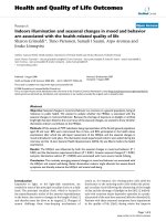
Fig. 1 Immunohistochemical staining of MEK5 protein in normal colorectal mucosa, colorectal adenoma, and CRC. (Left panels, ×100, right panels,
×400). (a) Weak MEK5 expression in normal colorectal mucosa; (b) moderate MEK5 expression in adjacent normal colorectal mucosa; (c) weak MEK5
expression in colorectal adenoma; (d) moderate MEK5 expression in colorectal adenoma; (e) weak MEK5 expression in well differentiated CRC; (f)
strong MEK5 expression in poorly differentiated CRC (mucinous adenocarcinoma). The fig. (g and h) show the MEK5 expression in the occurrence of
CRC: Elevated MEK5 expression in the atypical hyperplasia and tumor cells of CRC tissue compared with those of adjacent normal mucosa
Diao et al. BMC Cancer (2016) 16:302
and the proteins were detected with a Phototopehorseradish peroxidase Western blot detection kit (Cell
Signaling Technology, Inc). Protein expression levels
were normalized to that of β-actin by calculating the
relative expression levels.
RNA extraction and qRT-PCR
Total RNA extraction was carried out using Trizol reagent (Invitrogen) according to the manufacturer’s instruction. Two microgram of total RNA was subjected
to reverse Transcription (RT) using Verso cDNA Ki
(Thermo Scientific). Real-time quantitative PCR was
conducted by Platinum SYBR Green qPCR SuperMix
UDG with ROX kit (Invitrogen) in 20 μl and ABI 7300
real-time PCR thermal cycle instrument (ABI, USA) according to the supplied protocol. The primers for MEK5
were: (F, CTTTAATGCCTCTCCAGCTTCT; R, CCATCATTGAACTGCACGAT). The relative expression
levels were normalized to expression of endogenous
GAPDH. (Primers: F, GGGAAACTGTGGCGTGAT; R,
GAGTGGGTGTCGCTGTTGA).
Cell proliferation assay
Cell Counting Kit-8 (CCK-8; Dojindo) was used in cell proliferation assay. 5 × 10³ cells/well viable cells were seeded in
96-well tissue culture plates in a final volume of 100 μl. At
time points of day 0, day 2, day 3 and day 4, a plate was
subjected to assay by adding 10 μl of CCK-8 solution to
each well, and the plate was further incubated at 37 °C for
4 h, and then the absorbance at 450 nm was calculated.
The experiment was performed in eight replicates.
Cell cycle analysis
Following transfection 48 h later, 1 × 10⁶ cells were collected, washed in PBS, fixed in 70 % ethanol, and kept at 4 °
C overnight. The cells were resuspended to a concentration
of 1 × 10⁶ cells/ml in PBS and incubated with 100 μg/ml
RNase A and 50 mg/ml propidium iodide (PI) at 4 °C for
30 min. The total cellular DNA content was analyzed by
flow cytometry (Becton Dickinson, San Jose, CA).
Page 4 of 12
obtained were then expressed as the rate of wound healing.
The experiments were performed at least in triplicate.
Cell invasion assay
Cell invasion was evaluated by transwell matrigel invasion assay using BD Biocoat Matrigel invasion chambers (BD Biosciences, USA). Briefly, 500 μl of the cell
suspended in serum free RPMI-1640 medium at a concentration of 1 × 10⁵ cells was added to the upper compartment, while the lower compartment contained
750 μl medium with EGF (15 ng/mL) additionally.
After 24 h of incubation, chambers were rinsed and the
Matrigel matrix and noninvading cells on the upper
surface of the membrane were removed using moistened cotton swabs. Afterwards, cells on the lower surface were fixed with methanol and stainedwith 0.1 %
toluidine blue. Membranes were cut out and evaluated
under microscopic by placing on microscope slides.
In vivo tumor model
Six 4-week-old athymic BALB/C nude mice (male,
14–16 g) were purchased from the Laboratory Animal
Center of Southern Medical University (Guangzhou,
China). The animals were housed in SPF under identical
conditions and allowed free access to a standard diet and
tap water with 12-h light and dark cycles, under an experimental protocol approved by the Institutional Animal Care
and Use Committee (IACUC) of Guangdong Provincal
Hospital of Traditional Chinese Medicine. All operations
were performed under clean conditions. KD cells (5 × 10⁶
in 0.1 ml of PBS) were injected subcutaneously into the left
dorsal flank of each mice, while the same number of NC
cells injected subcutaneously into the right dorsal flank.
Tumor mass volume, which was calculated as (length/
2) × (width/2), was measured every two days from day 7 to
day 21. On day 21 the NC tumors all began to fester therefore the six mice were sacrificed and all tumors were harvested. Then MEK5 protein expression in tumors was
detected by western blot analysis as described above. The
experimental procedures were done in accordance with the
ARRIVE guidelines.
Cell migration assay
Cell migration was evaluated by scratch wound assay [24].
In brief, SW480 cells were plated in 6-well plate at concentration of 106/well and cultured overnight to yield confluent
monolayer. Next, the cells were treated with 10 mg/ml mitomycin for 1 h to inhibit proliferation, followed by wounding with 10 ml pipette tip. Remaining cells were washed
twice and then cultured with serum free RPMI-1640
medium. Photographic images were taken from each well
at 0 h, 6 h, 24 h and 48 h. The distance that cells migrated
through the area created by scratching was caculated by
measuring the wound width at the above times and subtracting it from the wound width at the start. The values
Statistical analysis
All of the experimental data were analyzed by using the
statistical software SPSS 17.0. The statistical methods used
included chi-square tests and paired sample’s t tests. The
chi-square test and Fisher’s exact test were used to examine the association between MEK5 expression and various
clinicopathological parameters. Univariate analyses were
conducted using the Kaplan-Meier method, and statistical
significance between survival curves was assessed by the
log-rank test. Univariate Cox proportional hazards regressions were applied to estimate the individual hazard ratios
(HR) for disease-free survival (DFS) and overall survival
Diao et al. BMC Cancer (2016) 16:302
Page 5 of 12
Table 1 MEK5 expression in normal mucosa, adenoma and CRC
tissues
Tissue type
MEK5 expression
All cases
Normal (%)
χ2
MEK5 expression
P value
Over (%)
9.01a
0.011a
Normal
24
22(91.7 %)
2(8.3 %)
1.51b
0.220b
Adenoma
24
19(79.2 %)
5(20.8 %)
2.27c
0.116c
d
0.006d
Carcinoma
84
52(61.9 %)
32(38.1 %)
7.67
The χ and P value of the three groups; the χ and P value of normal
colorectal mucosa V.S. colorectal adenoma; cthe χ2 and P value of colorectal
adenoma V.S. CRC; dthe χ2 and P value of normal colorectal mucosa V.S. CRC
Normal, negative or weak; over, moderate or intense
a
2
b
Table 2 Correlation between MEK5 expression and
clinicopathologic characteristics
2
(OS). The variables that were significant in the univariate
analysis (P < 0.05) were then included into the multivariate
analysis. The HR with 95 % confidence interval (CI) was
measured to estimate the hazard risk of individual factors.
Significant differences between the groups were determined using the unpaired Student’s t-test. All tests were
two-sided, and a p-value less than 0.05 was considered statistically significant.
All cases
Low (%)
High (%)
Female
157
105(66.9)
52(33.1)
Male
185
128(69.2)
57(30.8)
Sex
0.648
Age at diagnosis (years)
0.697
< 60
159
110(69.2)
49(30.8)
≥ 60
183
123(67.2)
60(32.8)
Rectum
160
108(67.5)
52(32.5)
Colon
182
125(68.7)
57(31.3)
Tumor location
0.815
pT (invasion depth)
0.001
T1
5
5(100)
0(0)
T2
57
50(87.7)
7(12.3)
T3
242
157(64.9)
85(35.1)
T4
38
21(55.3)
17(44.7)
Results
N0
205
155(75.6)
50(24.4)
MEK5 expression in CRC tissue and normal colorectal
mucosa samples
N1
87
47(54)
40(46)
N2
50
31(62)
19(38)
M0
312
218(69.9)
94(30.1)
M1
30
15(50)
15(50)
Immunostaining of MEK5 in CRC tissues and normal mucosa was detected as brown-yellow granules in the cytoplasm (Fig. 1). In the first group object of this study, the
MEK5 was overexpressed in 38.1 % of CRC tissues (32 out
of 84); compared with 20.8 % of colorectal adenoma (5
out of 24) and 8.3 % of normal tissues (2 out of 24) (Fig. 1).
Statistical analysis indicated that MEK5 was gradually upregulated from normal mucosa to adenomas, and to
tumor tissues (P = 0.011; Table 1). Furthermore, in some
sections of colorectal adenomas and at the junctions of
tumor and normal mucosa, we found that the MEK5 expression level was notably correlated with progression of
CRC. MEK5 expression was normal in normal colorectal
mucosa and higher in the adjacent atypical hyperplasia of
the mucosa (Fig. 1-g, h).
To confirm the expression levels of MEK5 seen by
immunostaining in the specimens from our TMA, we
examined the expression of MEK5 protein by western
P value
pN (lymph node metastasis)
0.001
pM (distant metastasis)
0.026
TNM stage
0.000
I
48
45(93.8)
3(6.3)
II
147
104(70.7)
43(29.3)
III
117
69(59)
48(41)
IV
30
15(50)
15(50)
Well
28
25(89.3)
3(10.7)
Moderate
284
194(68.3)
90(31.7)
Poorly
30
14(46.7)
16(53.3)
Villous adenocarcinoma
21
16(76.2)
5(23.8)
Tubular adenocarcinoma
277
187(67.5)
90(32.5)
Mucinous adenocarcinoma
29
19(65.5)
10(34.5)
Others
15
11(73.3)
4(26.7)
Differentiation grade
0.002
Pathology type
0.812
Serosal invasion
0.227
Yes
73
54(74)
19(26)
No
269
179(66.5)
90(33.5)
Low, negative or weak; high, moderate or intense
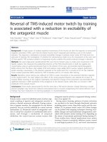
Fig. 2 Western blot analysis of MEK5 protein expression. Western
blot analysis of MEK5 proteins expressed in eight pairs represents
colorectal tumor tissues (T) and their matched adjacent non tumor
tissues (N). Expression level was normalized with β-actin
blot analysis in 8 randomly selected pairs of CRC tissues and their matched not-tumor colorectal tissues.
In 5 of 8 (62.5 %) CRC patients, the total MEK5 protein
Diao et al. BMC Cancer (2016) 16:302
Page 6 of 12
was up-regulated in tumor tissues compared with their adjacent nontumor colorectal mucosa; furthermore, the ratio
of MEK5α to MEKβ was higher in all of the CRC tissues
than in their adjacent normal colorectal mucosa (Fig. 2).
Correlations of MEK5 protein expression and
clinicopathologic parameters
In our study, overexpression of MEK5 protein was observed in 109 of the 342 CRC tissues (31.9 %) in the second
group samples. The relationship between immunohistochemical MEK5 expression in CRC tissues and various
clinicopathologic characteristics is shown in Table 2. The
results demonstrated that high expression of MEK5 was associated with depth of invasion (P = 0.001), lymph node
metastasis (P = 0.001), distant metastasis (P = 0.026), TNM
stage (P < 0.001) and differentiation grade (P = 0.002). There
was no significant association between MEK5 expression
and other clinicopathologic features, including age, sex,
tumor location, pathology type and serosal invasion.
Survival analysis
The mean patient follow-up time was 71.5 months and
the 5-year OS rate of the 342 patients with primary
colorectal cancer was 69.6 %, with 102 deaths observed during the follow-up period. The 5-year DFS
rate was 67.8 %. During the time of follow-up, 82
patients (24.5 %) developed distant metastasis or local
recurrence. According to the univariate analyses,
tumor location, TNM stage and differentiation grade
were significantly associated with patients’ overall survival and disease-free survival (P < 0.05; Tables 3 and
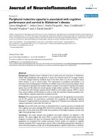
4). Assessment of CRC patient survival also revealed
that overexpression of MEK5 was significantly correlated with short disease-free survival (P < 0.001, Table 3
and Fig. 3-a) and poor overall survival (P = 0.012,
Table 4 and Fig. 3-b).
In order to address potential confounding among variables examined in the univariate analysis, we conducted
multivariate analysis using Cox proportional hazards
model for all of the significant variables in the univariate
analysis. We found that overexpression of MEK5 was an
independent risk factor for poor disease-free survival
(HR: 1.64; 95 % CI: 1.09–2.47; P = 0.017). Of the other
variables, tumor location, TNM stage and differentiation
grade were also found to be independent prognostic predictors for disease-free survival (Table 3). On the other
hand, MEK5 overexpression, tumor location, TNM stage
Table 3 Cox proportional hazards model univariate and multivariate analyses of individual parameters for correlations with diseasefree survival (342 cases)
Univariate analysis
All cases
Multivariate analysis
Mean survival (months)
Sex
P value
HR (95 % CI)
P value
0.011
1.850 (1.23–2.78)
0.003
0.000
2.70 (1.81–4.01)
0.000
0.000
1.88(1.07–3.32)
0.029
1.64 (1.09–2.47)
0.017
0.938
Female
157
93.5
Male
185
92.9
< 60
159
91.0
≥ 60
183
94.7
Age at diagnosis (years)
0.754
Tumor location
Rectum
160
95.2
Colon
182
84.2
I-II
195
120.7
III-IV
147
64.6
TNM stage
Differentiation grade
Well and Moderate
312
96.6
Poorly
30
61.8
Yes
73
98.8
No
269
92.3
Serosal invasion
0.230
MEK5 expression
0.000
Normal expression
233
98.2
Over expression
109
79.3
Diao et al. BMC Cancer (2016) 16:302
Page 7 of 12
Table 4 Cox proportional hazards model univariate and multivariate analyses of individual parameters for correlations with overall
survival (342 cases)
Univariate analysis
All cases
Multivariate analysis
Mean survival (months)
Sex
P value
HR (95 % CI)
P value
0.023
1.76(1.17–2.67)
0.008
0.000
2.75(1.82–4.16)
0.000
0.001
2.40(1.34–4.28)
0.003
1.51(1.01–2.25)
0.045
0.988
Female
157
94.2
Male
185
91.7
Age at diagnosis (years)
0.144
< 60
159
94.9
≥ 60
183
90.6
Rectum
160
96.5
Colon
182
88.8
Tumor location
TNM stage
I-II
195
106.1
III-IV
147
73.0
Well and Moderate
312
96.5
Poorly
30
60.9
Differentiation grade
Serosal invasion
0.411
Yes
73
95.8
No
269
93.1
Normal expression
233
95.2
Over expression
109
84.8
MEK5 expression
0.012
Fig. 3 Survival analysis of primary CRC patients (n = 342). Kaplan-Meier estimates of the DFS (a) and OS (b) according to MEK5 expression in 342 patients.
The DFS and OS were significantly lower in patients with MEK5-high expression when compared with patients who had low MEK5 expression. P values
were calculated using the log-rank test
Diao et al. BMC Cancer (2016) 16:302
Page 8 of 12
and differentiation grade were found to be independent
prognostic predictors for overall survival (Table 4).
lentiviral-MEK5-siRNA-3, NC cells and BC cells with stable
expression were harvested after puromycin selection.
Knockdown of MEK5 expression in SW480 cells
Effect of MEK5 knockdown on the biological behavior of
SW480 Cells in vitro
After 48 h transfection, green fluorescent protein (GFP) expression rates of the KD and NC cells were all more than
80 %, respectively. When compared with the parental NC
cells and BC cells, the three lentiviral-MEK5-siRNA vectors
transfected cells showed obvious decreases in the mRNA
and protein expressions of MEK5. In particular, the silencing efficiency of lentiviral-MEK5-siRNA-3 was the highest,
with the reduction of MEK5 mRNA expression by 86.3 %
(P = 0.025) and protein expression by 69.6 % (Fig. 4) comparing with BC cells. Therefore, the SW480 cells carrying
Cell Counting Kit 8 (CCK-8) assay showed that knockdown of MEK5 expression significantly inhibited the
proliferation of SW480 cell, indicating that MEK5 gene
expression affects the growth of colon cancer cells
(Fig. 5a). The flow cytometery results showed that, in
the NC group 63.43 % of cells were in the G1 phase and
32.77 % of the cells were in S of the cell cycle, and in the
BC group 63.02 % of cells were in the G1 phase and
33.87 % of the cells were in S of the cell cycle, while in KD
Fig. 4 Knockdown of MEK5 gene by MEK5 shRNA lentivirus. a qRT-PCR showed a significant decrease of MEK5 mRNA (by 86.3 %) in the sh3
group vs. BC group. b Western blot assay demonstrated that, normalized by β-actin, MEK5 protein expression was degraded (by 69.6 %) in the
sh3 group vs. BC group. sh1: MEK5sh1; sh2: MEK5sh2; sh3: MEK5sh3; NC, negative control; BC, blank control; KD, knockdown
Diao et al. BMC Cancer (2016) 16:302
Fig. 5 (See legend on next page.)
Page 9 of 12
Diao et al. BMC Cancer (2016) 16:302
Page 10 of 12
(See figure on previous page.)
Fig. 5 Effects of MEK5 knockdown on proliferation, division, migration and invasiveness of SW480 cell in vitro. The proliferation ability of NC group, BC
group and KD group was examined by CCK-8, cell division was examined by flow cytometry, and migrated ability was tested by scratch assay and invasive ability was examined by transwell Matrigel invasion assay. Comparing with NC and BC groups, the proliferation (a), division (b), migration (c) and
invasiveness (d) of siRNA treated cells (KD group) were significantly decreased. NC, negative control; BC, blank control; KD, knockdown
group cells accumulated in G1 71.53 % but reduced to
18.61 % in S phase (Fig. 5b). In scratch wound assay, migration ability of KD group was obviously inhibited than that
of NC group and BC group (Fig. 5c), indicating silence of
MEK5 gene led to a significantly decreased migration ability
of SW480 cells. Transwell matrigel invasion assay showed
that silencing of MEK5 expression significantly inhibited invasion of SW480 cells in vitro (P < 0.01, Fig. 5d).
In vivo studies of SW480 cells xenograft tumor models in
nude mice
To further evaluate the role of reduced MEK5 expression on the tumorigenic phenotype and in particular its
contribution to in vivo tumor growth. SW480 cells infected with non-silencing shRNA and MEK5 shRNA
were injected into 6 mice, (Fig. 6a). The cancer growth
curves in nude mice after injection of MEK5 shRNA
transfected cells and the control cells are shown in
Fig. 6c. The tumor growth speed of the KD cells was obvious slower than that of NC cells (P < 0.05). These results demonstrate that in vivo tumor growth was
inhibited by shRNA-mediated knockdown of MEK5 expression in colon cancer cells. Western blot assay
showed MEK5 protein expression of the xenograft tumors of KD cells was significantly inhibited comparing
with that of NC cells (P < 0.01, Fig. 6d).
Discussion
The occurrence and development of CRC is correlated
with various molecular and genetic incidents. Recent data
have been accumulating to support a key role of MEK5/
ERK5 signaling in carcinogenesis [25], and several studies
have demonstrated that tumor cells can acquire cancerous
capacity by increasing expression of MEK5 to activate
a
b
c
d
e
Fig. 6 Silencing of MEK5 significantly inhibited cancer growth in vivo. a KD cells (5 × 10⁶ in 0.1 ml of PBS) were injected subcutaneously into the
left dorsal flank of each BALB/C nude mice, while the same number of BC cells injected subcutaneously into the right dorsal flank. b Tumor mass
volume was measured every two days from day 7 to day 21. On day 21 the six mice were sacrificed and all tumors were harvested. c Silencing of
MEK5 could significantly inhibited the cancer growth, when compared with BC cells (P < 0.05). d Western blot assay showed MEK5 protein
expression of the xenograft tumors of KD cells was significantly inhibited comparing with that of NC cells. NC, negative control; KD, knockdown
Diao et al. BMC Cancer (2016) 16:302
ERK5. In metastatic prostate cancer, strong MEK5 expression is correlated with bony metastases, and less favorable
prognosis is caused by up-regulated ERK5-induced activator protein-1 (AP-1) activity, a consequent induction of a
high level of matrix metallo-protease-9 (MMP-9) and augmented invasive potential [8]. Dudderidge et al. showed
that the induction of MEK5/ERK5 signalling was associated with activation of the DNA replication licensing
pathway in prostate cancer [26]. Ghosh AK et al. demonstrated that exogenous expression of c-myc promoterbinding protein 1 (MBP-1) induces prostate cancer cell
death by down-regulating the expression of MEK5α and
up-regulating the level of MEK5β [16]. Hui Song et al. observed that MEK5α might be one of the Stat3-regulated
genes and play a critical role in oncogenesis mediated by
aberrantly activated Stat3 signaling in breast carcinomatosis and malignancies [27]. Recently, we found that
pMEK5 expression was correlated with the staging of
CRC and its expression might be helpful for the TNM staging system of CRC [23].
In this study, we examined the expression of MEK5 by
IHC in 24 normal colorectal mucosa, 24 adenomas and 84
primary colorectal adenocarcinomas, and found that
MEK5 was gradually up-regulated in the development of
CRC from normal mucosa, through adenomas, to cancer.
Moreover, we found that the MEK5 expression status was
notably correlated with progression of CRC in the same
pathological section. Elevated MEK5 expression was
found in the adjacent atypical hyperplasia of the mucosa
compared with those of normal colorectal mucosa. In
order to confirm the results seen by immunostaining the
specimens from our TMA, we examined the expression of
MEK5 protein by western blot in 8 randomly selected
pairs of CRC tissues and their matched normal adjacent
mucosa. The results demonstrated that the major CRC tissues examined had higher levels of MEK5 expression than
adjacent normal mucosa. These findings suggest that upregulated expression of MEK5 may provide a selective advantage in CRC tumorigenesis. In the immunostaining of
MEK5 in the 342 cases of CRC, we found that the expression of MEK5 was positively correlated with clinical stage
and differentiation grade. These data suggest that overexpression of MEK5 may facilitate the invasive/metastatic
phenotypes of human colorectal cancer.
Another interesting finding was that, in the western blot
testing of MEK5 in 8 pairs of CRC tissues, we found the
ratios of MEK5α to MEK5β were higher in all CRC tissues
than that in adjacent normal mucosa. It is known that
MEK5α induces cell proliferation by activating its downstream molecules, whereas MEK5β expression is associated with inhibition of cell growth. This result indicated
that the ratio of MEK5α to MEK5β might be more important than the total amount of MEK5 expression in the
activation of MEK5/ERK5 signaling and progression of
Page 11 of 12
carcinogenesis. Activation of MEK5/ERK5 signal has been
demonstrated in cells in response to extracellular signals
that include cytokines, growth factors, and various stress
stimuli. Alternative splicing of MEK5α and MEK5β plays
a significant role in ERK5 activation and subsequently
induce carcinogenesis [13]. We hypothesized that, as a result of long-time extracellular stimulation, the MEK5/
ERK5 pathway in the colorectal mucosa cells was activated
excessively, and eventually induced malignant growth.
Targeting MEK5 kinase activity or blocking the MEK5/
ERK5 pathway may provide an additional means of inhibiting cell migration associated with CRC progression to
metastasis.
MEK5 may have clinical value for prognosis or treatment. Weldon et al. reported that drug-resistant MCF7
cells appeared to have significantly high level of MEK5.
In that report, MEK5 contributed to prevention of cell
apoptosis and chemotherapeutic resistance [28]. In our
study, Kaplan-Meier analysis of the survival curves
showed a significantly worse 5-year DFS and 5-year OS
rate for patients whose tumors overexpressed MEK5.
This suggests that MEK5 protein may be a biomarker
for poor prognosis for CRC patients. In the multivariate
analysis, the result showed that overexpression of MEK5
was an independent predictor of shortened DFS and
poor OS. Therefore, the CRC patients with MEK5 overexpression may require a more powerful adjunctive therapy and intensive follow-up. Whether MEK5 has value
clinically as a biomarker for therapeutic approaches in
patients with colorectal cancer should be followed up
with additional appropriately designed studies.
In order to provide further support that MEK5 contributes to the development and progression of colon
cancer, the colon cancer cell line SW480 was employed
for function experiment. We effectively down-regulated
MEK5 expression in SW480 cells by lentiviral-MEK5shRNA in vitro, and our data indicated that proliferation, cell cycle progression, migration and invasiveness
of stable transfected cells were significantly decreased.
Finally, we showed that down-regulation of MEK5 in
SW480 cells resulted in slower tumor growth in vivo.
Taken together, the function experiments further confirmed that down-regulation of MEK5 could inhibit the
proliferation and aggressiveness of colon cancer cell line
in vitro, and negatively affected development of tumors
in vivo, which were consistent with our data from the
immunohistochemical and western blot analysis using
the clinical CRC samples. In the future study, a larger
sampler size is needed to validate this result.
Conclusion
The overexpression of MEK5 could be used as an effective additional tool in identifying those CRC patients at
Diao et al. BMC Cancer (2016) 16:302
increased risk of tumor invasiveness, metastasis, or differentiation grade, and knock down of MEK5 led to significantly inhibiting the malignant phenotype of colon
cancer cells in vitro an vivo. Moreover, MEK5 could be
an encouraging novel molecular biomarker to predict
the prognosis of CRC patients and may be a potential
molecular target for the treatment of CRC.
Page 12 of 12
9.
10.
11.
12.
Abbreviations
BC: blank control; CI: confidence intervals; CRC: colorectal cancer;
DFS: disease-free survival; ERK5: extracellular signal-regulated kinase-5;
HR: hazards ratio; IHC: immunohistochemistry; KD: knockdown;
MAPK: mitogen-activated protein kinase; MEK5: mitogen/extracellular signal
regulated kinase kinase-5; NC: negative control; OS: overall survival;
TMA: tissue microarray.
13.
Competing interests
The authors declare that they have no competing interests.
16.
Authors’ contributions
LW and DCD designed the study; DCD performed all the experiments and
wrote the paper, JW, ZQC, HLL and JSP performed part of the experiments and
composition of the manuscript. LNZ and WW reviewing and scoring the
degree of immunostaining of sections, XLC was responsible for data collection
and analysis. All authors have read and approved the final manuscript.
17.
14.
15.
18.
19.
Acknowledgments
This study was supported by the Guangdong province natural science
foundation of China S2013040016396 (Dr. Dechang Diao). We thank Liyan
Cui, Megan McLaughlin, Weiling He and Daxiong Wang for their great help
in writing and editing the manuscript.
Author details
1
Department of Gastrointestinal Surgery, Guangdong Provincal Hospital of
Traditional Chinese Medicine, Guangdong 510120, China. 2Institute of
Gastroenterology, Sun Yat-Sen University, Guangzhou 510655, China.
3
Department of Gastrointestinal Surgery, the Sixth Affiliated Hospital, Sun
Yat-sen University, Guangzhou, Guangdong 510655, China. 4Key Laboratory
of Tropical Disease Control (Sun Yat-sen University), Ministry of Education,
Guangzhou, Guangdong 510080, China. 5Department of Preventive Medicine
and Medical Statistics, College of Fundamental Medical Science, Guangzhou
University of Traditional Chinese Medicine, Guangdong 510006, China.
20.
21.
22.
23.
24.
Received: 8 December 2015 Accepted: 20 April 2016
References
1. Siegel R, Naishadham D, Jemal A. Cancer statistics, 2013. CA Cancer J Clin.
2013;63(1):11–30.
2. Li S, Wang J, Lu Y, Fan D. Screening and early diagnosis of colorectal cancer
in China: a 12 year retrospect (1994–2006). J Cancer Res Clin Oncol. 2007;
133(10):679–86.
3. Zhao P, Dai M, Chen W, Li N. Cancer trends in China. Jpn J Clin Oncol. 2010;
40(4):281–5.
4. Xu AG, Yu ZJ, Jiang B, Wang XY, Zhong XH, Liu JH, et al. Colorectal cancer
in Guangdong Province of China: a demographic and anatomic survey.
World J Gastroenterol. 2010;16(8):960–5.
5. Ahmed FE. Gene-gene, gene-environment & multiple interactions in
colorectal cancer. J Environ Sci Health C Environ Carcinog Ecotoxicol Rev.
2006;24(1):1–101.
6. Fang JY, Richardson BC. The MAPK signalling pathways and colorectal
cancer. Lancet Oncol. 2005;6(5):322–7.
7. Chung DC. The genetic basis of colorectal cancer: insights into critical
pathways of tumorigenesis. Gastroenterology. 2000;119(3):854–65.
8. Mehta PB, Jenkins BL, McCarthy L, Thilak L, Robson CN, Neal DE, Leung HY.
MEK5 overexpression is associated with metastatic prostate cancer, and
stimulates proliferation, MMP-9 expression and invasion. Oncogene. 2003;
22(9):1381–9.
25.
26.
27.
28.
Zhou G, Bao ZQ, Dixon JE. Components of a new human protein kinase
signal transduction pathway. J Biol Chem. 1995;270(21):12665–9.
English JM, Pearson G, Baer R, Cobb MH. Identification of substrates and
regulators of the mitogen-activated protein kinase ERK5 using chimeric
protein kinases. J Biol Chem. 1998;273(7):3854–60.
Kato Y, Kravchenko VV, Tapping RI, Han J, Ulevitch RJ, Lee JD. BMK1/ERK5
regulates serum-induced early gene expression through transcription factor
MEF2C. EMBO J. 1997;16(23):7054–66.
Roberts OL, Holmes K, Muller J, Cross DA, Cross MJ. ERK5 and the regulation
of endothelial cell function. Biochem Soc Trans. 2009;37(Pt 6):1254–9.
Wang X, Tournier C. Regulation of cellular functions by the ERK5 signalling
pathway. Cell Signal. 2006;18(6):753–60.
English JM, Vanderbilt CA, Xu S, Marcus S, Cobb MH. Isolation of MEK5 and
differential expression of alternatively spliced forms. J Biol Chem. 1995;
270(48):28897–902.
Seyfried J, Wang X, Kharebava G, Tournier C. A novel mitogen-activated
protein kinase docking site in the N terminus of MEK5alpha organizes the
components of the extracellular signal-regulated kinase 5 signaling
pathway. Mol Cell Biol. 2005;25(22):9820–8.
Ghosh AK, Steele R, Ray RB. c-myc Promoter-binding protein 1 (MBP-1)
regulates prostate cancer cell growth by inhibiting MAPK pathway. J Biol
Chem. 2005;280(14):14325–30.
Cameron SJ, Abe J, Malik S, Che W, Yang J. Differential role of MEK5alpha
and MEK5beta in BMK1/ERK5 activation. J Biol Chem. 2004;279(2):1506–12.
McCracken SR, Ramsay A, Heer R, Mathers ME, Jenkins BL, Edwards J,
Robson CN, Marquez R, Cohen P, Leung HY. Aberrant expression of
extracellular signal-regulated kinase 5 in human prostate cancer. Oncogene.
2008;27(21):2978–88.
Li Z, Li J, Mo B, Hu C, Liu H, Qi H, Wang X, Xu J. Genistein induces cell
apoptosis in MDA-MB-231 breast cancer cells via the mitogen-activated
protein kinase pathway. Toxicol In Vitro. 2008;22(7):1749–53.
Yan F, Wang XM, Pan C. [Expression of ERK5 in multidrug-resistant hepatocellular
carcinoma cell line]. Nan Fang Yi Ke Da Xue Xue Bao. 2009;29(3):483–6.
Winn RA, Van Scoyk M, Hammond M, Rodriguez K, Crossno JJ, Heasley LE,
Nemenoff RA. Antitumorigenic effect of Wnt 7a and Fzd 9 in non-small cell
lung cancer cells is mediated through ERK-5-dependent activation of
peroxisome proliferator-activated receptor gamma. J Biol Chem. 2006;
281(37):26943–50.
Diao D, Wang L, Zhang JX, Chen D, Liu H, Wei Y, Lu J, Peng J, Wang J.
Mitogen/extracellular signal-regulated kinase kinase-5 promoter region
polymorphisms affect the risk of sporadic colorectal cancer in a southern
Chinese population. DNA Cell Biol. 2012;31(3):342–9.
Hu B, Ren DL, Su D, Lin HC, Xian ZY, Wang XY, Zhang JX, Fu XH, Jiang L,
Diao DC, et al. Expression of the phosphorylated MEK5 protein is associated
with TNM staging of colorectal cancer. BMC Cancer. 2012;12(1):127.
Li YW, Wang JX, Yin X, Qiu SJ, Wu H, Liao R, Yi Y, Xiao YS, Zhou J, Zhang BH,
et al. Decreased expression of GATA2 promoted proliferation, migration and
invasion of HepG2 in vitro and correlated with poor prognosis of
hepatocellular carcinoma. PLoS One. 2014;9(1):e87505.
Ramsay AK, McCracken SR, Soofi M, Fleming J, Yu AX, Ahmad I, Morland R,
Machesky L, Nixon C, Edwards DR, et al. ERK5 signalling in prostate cancer
promotes an invasive phenotype. Br J Cancer. 2011;104(4):664–72.
Dudderidge TJ, McCracken SR, Loddo M, Fanshawe TR, Kelly JD, Neal DE,
Leung HY, Williams GH, Stoeber K. Mitogenic growth signalling, DNA
replication licensing, and survival are linked in prostate cancer. Br J Cancer.
2007;96(9):1384–93.
Song H, Jin X, Lin J. Stat3 upregulates MEK5 expression in human breast
cancer cells. Oncogene. 2004;23(50):8301–9.
Weldon CB, Scandurro AB, Rolfe KW, Clayton JL, Elliott S, Butler NN, Melnik LI,
Alam J, McLachlan JA, Jaffe BM, et al. Identification of mitogen-activated
protein kinase kinase as a chemoresistant pathway in MCF-7 cells by using
gene expression microarray. Surgery. 2002;132(2):293–301.