Negative effect of cyclin D1 overexpression on recurrence-free survival in stage II-IIIA lung adenocarcinoma and its expression modulation by vorinostat in vitro
Bạn đang xem bản rút gọn của tài liệu. Xem và tải ngay bản đầy đủ của tài liệu tại đây (2.22 MB, 10 trang )
Lee et al. BMC Cancer (2015) 15:982
DOI 10.1186/s12885-015-2001-7
RESEARCH ARTICLE
Open Access
Negative effect of cyclin D1 overexpression
on recurrence-free survival in stage II-IIIA
lung adenocarcinoma and its expression
modulation by vorinostat in vitro
Eunju Lee1†, DongHao Jin1†, Bo Bin Lee1, Yujin Kim1, Joungho Han2, Young Mog Shim3 and Duk-Hwan Kim1,4*
Abstract
Background: This study was aimed at identifying prognostic biomarkers for stage II-IIIA non-small cell lung cancer
(NSCLC) according to histology and at investigating the effect of vorinostat on the expression of these biomarkers.
Methods: Expression levels of cyclin D1, cyclin A2, cyclin E, and p16 proteins that are involved in the G1-to-S phase
progression of cell cycle were analyzed using immunohistochemistry in formalin-fixed paraffin-embedded tissues
from 372 samples of stage II-IIIA NSCLC. The effect of vorinostat on the expression of these proteins, impacts on cell
cycle, and histone modification was explored in lung cancer cells.
Results: Abnormal expression of cyclin A2, cyclin D1, cyclin E, and p16 was found in 66, 47, 34, and 51 % of 372
cases, respectively. Amongst the four proteins, only cyclin D1 overexpression was significantly associated with poor
recurrence-free survival (adjusted hazard ratio = 1.87; 95 % confidence interval = 1.12 – 2.69, P = 0.02) in
adenocarcinoma but not in squamous cell carcinoma (P = 0.44). Vorinostat inhibited cell cycle progression to the
S-phase and induced down-regulation of cyclin D1 in vitro. The down-regulation of cyclin D1 by vorinostat was
comparable to a siRNA-mediated knockdown of cyclin D1 in A549 cells, but vorinostat in the presence of
benzo[a]pyrene showed a differential effect in different lung cancer cell lines. Cyclin D1 down-regulation by
vorinostat was associated with the accumulation of dimethyl-H3K9 at the promoter of the gene.
Conclusions: The present study suggests that cyclin D1 may be an independent prognostic factor for recurrence-free
survival in stage II-IIIA adenocarcinoma of lung and its expression may be modulated by vorinostat.
Keywords: Lung cancer, Vorinostat, Cyclin D1, Histone modification, Survival
Background
Lung cancer is the leading cause of cancer-related deaths
worldwide and despite significant advances in the diagnosis and treatment of the disease, the current 5-year
survival rate remains low at 15 %. The poor prognosis is
partially due to the high rate of recurrence after surgery,
where the recurrence rate is as high as 20–40 % even for
* Correspondence:
†
Equal contributors
1
Department of Molecular Cell Biology, Sungkyunkwan University School of
Medicine, #300 Chunchun-dong, Jangan-KuKyunggido, Suwon 440-746,
Korea
4
Center for Genome Research, Samsung Biomedical Research Institute, Rm
B155, #50 Ilwon-dong, Kangnam-Ku, Seoul 135-710, Korea
Full list of author information is available at the end of the article
a stage I non-small cell lung cancer (NSCLC) [1, 2]. The
recurrence is the result of local and distant metastasis of
residual cancer cells after surgery. A number of studies
have been conducted to identify specific adjuvant therapy in order to eliminate occult micro-metastases after
curative surgical resection and improve survival. Adjuvant chemotherapy is recommended for some patients
with resected stage II-IIIA NSCLC but controversy continues regarding its need for stage I NSCLC. The role of
adjuvant chemotherapy in patients with stage IB NSCLC
is not well established, and it is recommended only for
certain patient cases [3].
In the last 10 years, adjuvant chemotherapy for patients with completely resected stage II-IIIA NSCLC has
© 2015 Lee et al. Open Access This article is distributed under the terms of the Creative Commons Attribution 4.0
International License ( which permits unrestricted use, distribution, and
reproduction in any medium, provided you give appropriate credit to the original author(s) and the source, provide a link to
the Creative Commons license, and indicate if changes were made. The Creative Commons Public Domain Dedication waiver
( applies to the data made available in this article, unless otherwise stated.
Lee et al. BMC Cancer (2015) 15:982
usually employed platinum-based chemotherapy. After
a history of negative trials over the last few decades,
some progress has been made in overall survival after
platinum-based chemotherapy. Two recent meta-analysis
of randomized controlled trials showed an absolute 5-year
survival benefit of 5 to 10 % irrespective of the associated
drugs such as vinorelbine or etoposide, with the main survival advantage being in the patients with stage II-IIIA
NSCLC [4, 5]. With a better understanding of the biology
of lung cancer in recent years, several groups have proposed novel strategies targeting the epidermal growth factor receptor (EGFR), other receptor and non-receptor
tyrosine kinases, and vascular endothelial growth factor
(VEGF) pathways [3].
A balance between stimulators and inhibitors of cell
proliferation tightly regulates the cell cycle and a
disorganization of the cell cycle leads to an uncontrolled
cellular proliferation of residual cancer cells after curative resection. Chemotherapeutic agents that target and
disrupt different phases of the cell cycle have been developed over the past few years. Among them, histone deacetylase inhibitors (HDACIs) modify the acetylation
state of histone tails and induce cell cycle arrest at both
G1 and G2 phases. Vorinostat, also known as suberoylanilide hydroxamic acid (SAHA), was the first HDACI to
be approved by the United States Food and Drug Administration (FDA) for treatment of refractory cutaneous
T-cell lymphoma [6]. Vorinostat also causes cell growth
inhibition, differentiation, and apoptosis of lung cancer
cells in vitro through various mechanisms [7–10].
To understand the expression pattern and prognostic
significance of key proteins involved in the G1-to-S
phase progression of the cell cycle in stage II-IIIA
NSCLC and to investigate whether vorinostat can modulate expression of these proteins, we analyzed the expression patterns of cyclin A2, cyclin D1, cyclin E, and
p16 proteins in formalin-fixed paraffin-embedded tissues
from 372 patients with stage II-IIIA NSCLC and
assessed the effect of vorinostat on their expression in
lung cancer cells. A serious problem in the treatment of
lung cancer is that some patients continue to smoke
even after a lung cancer diagnosis. The continuous exposure to tobacco smoke may influence the effect of
chemotherapeutic agents [11]. Therefore, we carried out
the in vitro study with and without exposure to benzo[a]pyrene (B[a]P).
Results
Expression patterns of the four proteins
A total of 372 patients with stage II-IIIA NSCLC participated in this study. The clinicopathological characteristics according to histology are described in Table 1.
Representative examples of nuclear immunostaining for
the four proteins are shown in Fig. 1a. A composite
Page 2 of 10
Table 1 Clinicopathological characteristics (N = 372)
Adenoca
(N = 140)
Squamous
(N = 201)
Others
(N = 31)
P-value
59 ± 11
62 ± 8
59 ± 10
0.03
Tumor size (cm)
4.5 ± 2.7
5.3 ± 2.4
6.3 ± 2.8
<0.0001
Pack-years (smoking)a
19 ± 24
37 ± 19
40 ± 35
<0.0001
Male
87
192
27
Female
53
9
4
Variables
Agea
a
Sex
<0.0001c
Smoking status
Never
72
14
7
Former
29
62
5
Current
39
125
19
IIA
44
71
10
IIB
46
54
13
IIIA
50
76
8
Well
25
27
1
Moderate
47
120
1
Poorly
42
40
2
Undifferentiated
2
2
3
No
115
163
26
Yes
25
38
5
No
88
139
22
Yes
52
62
9
No
35
114
16
Yes
105
87
15
<0.0001
Pathologic stage
0.42
Differentiationb
<0.0001c
Adjuvant chemotherapy
0.92
Adjuvant radiotherapy
0.42
Recurrence
<0.0001
Abbreviations: Adenoca, adenocarcinoma; Squamous, squamous cell carcinoma
a
Values indicate mean ± standard deviation
b
Differentiation data are missing for 60 patients
c
Fisher’s exact test
score for each staining was calculated by multiplying the
intensity and percentage scores. Abnormal expression
was defined when a composite score was greater than or
equal to two for cyclin A2, cyclin D1, and cyclin E and
less than two for p16. Abnormal expression was detected
in 66 % of 372 patients for cyclin A2, 47 % for cyclin D1,
34 % for cyclin E, and 51 % for p16. Abnormal expression of these four proteins was compared according to
histology (Fig. 1b): a low prevalence of the overexpression of cyclin A2 (P < 0.0001) and cyclin E (P = 0.003)
was shown in lung adenocarcinoma as compared to
other cell types. As protein-protein interactions play a
fundamental role in many biological processes, a correlation analysis of the expression of these four proteins
Lee et al. BMC Cancer (2015) 15:982
Page 3 of 10
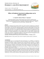
Fig. 1 Expression patterns of four proteins according to histology. a Immunohistochemical staining of 4 proteins was performed in 372 formalin-fixed
paraffin-embedded tissues. Representative positive staining is shown in adenocarcinoma (upper) and squamous cell carcinoma (lower). (Magnification ×200).
b Prevalence of abnormal expression of 4 proteins was compared according to histology. P-values were based on Pearson’s chi-square or Fisher’s exact test.
c-e Correlations among expression levels of 4 proteins were analyzed using Spearman’s rank correlation coefficient in adenocarcinoma (c), squamous cell
carcinoma (d), and other cell types (e). The numbers on either side of plots indicate composite scores that were calculated by multiplying the intensity
score and the proportion score of positive staining tumor cells. Violet color indicates a significant Spearman’s correlation between two proteins (P < 0.05)
was performed according to histology. The expression
levels of cyclin E correlated with those of cyclin A2
(Spearman’s rank correlation coefficient [rs] = 0.38, P <
0.0001) and p16 (rs = 0.32, P = 0.0003) in adenocarcinoma (Fig. 1c). This was also similar to patterns in squamous cell carcinoma (Fig. 1d) and other cell types
(Fig. 1e). However, no correlation was found between
cyclin D1 and other proteins.
analysis showed that adenocarcinoma patients with cyclin D1 overexpression were found to have a 1.87 (95 %
confidence interval = 1.12 – 2.69, P = 0.02) times poorer
RFS than those without (Table 2), after adjusting for age,
tumor size, pathologic stage, and cyclin E expression.
The cyclin A2, cyclin E, and p16 proteins were entered
into the multivariate analysis one by one due to collinearity, but their expression was not associated with RFS
(data not shown).
Survival analysis
The effect of the abnormal expression of individual
proteins on recurrence-free survival (RFS) and overall
survival was analyzed in 372 NSCLCs. Abnormal expression of cyclin A2, cyclin D1, cyclin E, and p16 was not
associated with overall survival (data not shown). In
addition, no association was found between RFS and the
abnormal expression of cyclin A2, cyclin E, and p16
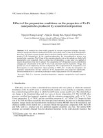
(Additional file 1: Figure S1). However, cyclin D1 overexpression was found to be significantly associated with
poor RFS in adenocarcinoma (P = 0.03; Fig. 2b), not in
squamous cell carcinoma (P = 0.46; Fig. 2c). The median
duration of RFS was 15 months and 25 months for
adenocarcinoma patients with and without cyclin D1
overexpression, respectively. Cox proportional hazards
Vorinostat inhibits cell growth
In our clinical samples, cyclin D1 overexpression was
significantly associated with RFS in stage II-IIIA adenocarcinoma. We used A549, a human lung adenocarcinoma epithelial cell line that expresses relatively high
levels of cyclin D1, as our model to analyze the effect of
vorinostat on cell growth. B[a]P increased cell proliferation, while vorinostat significantly decreased proliferation
in a time- and dose-dependent manner (Fig. 3a and b). In
order to examine the effect of vorinostat on cell growth in
cells exposed to B[a]P as long as possible, we pretreated
A549 cells with 5 μM B[a]P for 9 days and incubated the
cells with 5 μM vorinostat in combination for another
4 days (Fig. 3c). Cell proliferation in the cells exposed to
Lee et al. BMC Cancer (2015) 15:982
Page 4 of 10
Fig. 2 Kaplan-Meier survival curves in stage II-IIIA NSCLC. Recurrence-free survival was compared according to expression status of cyclin D1 in 372
patients (a), 140 patients with adenocarcinomas (b), and 201 patients with squamous cell carcinomas. P–values were based on the log-rank test
B[a]P was also reduced by vorinostat, which showed the
same pattern as the inhibition of cell proliferation by vorinostat in the absence of B[a]P.
Vorinostat induces G1-S arrest in lung cancer cells
Cell cycle was evaluated using flow cytometry in
A549, H460, and H226 cells treated with 5 μM B[a]P
and/or 5 μM vorinostat: vorinostat did have a substantial effect on G1-S cell cycle arrest. The proportion of
S phase cells in the cell lines substantially decreased
as compared to the control by treatment with 5 μM
vorinostat for 1 day. The proportion of S phase cells
in A549 cells decreased from 20 to 7 % by vorinostat
(Fig. 3d). The proportion of S phase cells in A549
cells exposed to 5 μM B[a]P decreased from 23 to
9 % by 5 μM vorinostat (P < 0.05; paired t-test). Vorinostat also blocked cell cycle progression to the S
phase in H460 (large cell carcinoma cell line) and
Lee et al. BMC Cancer (2015) 15:982
Page 5 of 10
Table 2 Cox proportional hazards analysisa for recurrence-free survival
Histology
Cyclin D1 overexpression
HR
Total
No
1.00
(N = 372)
Yes
1.10
Adenocarcinoma
No
1.00
(N = 140)
Yes
1.87
Squamous cell ca
No
1.00
(N = 201)
Yes
0.84
95 % CI
P-value
0.83–1.46
0.51
1.12–2.69
0.02
0.55–1.29
0.44
Abbreviations: HR hazard ratio; CI confidence interval; Squamous cell ca,
squamous cell carcinoma
a
adjusted for age, tumor size, pathologic stage, and cyclin E expression
H226 (squamous cell carcinoma cell line) cells irrespective of exposure to B[a]P (Fig. 3e). These observations suggest that the effect of vorinostat on G1-S
arrest of the cell cycle may not be cell type-specific
in lung cancer.
The effect of vorinostat on cyclin D1 expression is
comparable to cyclin D1 siRNA
The effect of vorinostat on cyclin D1 expression was further analyzed because of our finding that cyclin D1 was
significantly associated with poor RFS in stage II-IIIA lung
adenocarcinoma. Cyclin D1 was found to be downregulated in response to vorinostat in A549, H460, and
H226 cells, but the effect varied according to the cell lines
in presence of B[a]P (Fig. 4a): cyclin D1 down-regulation
by vorinostat was minimal in H226 cells exposed to B[a]P.
To understand if the effect of vorinostat on cyclin D1
down-regulation was comparable to a cyclin D1 knockdown, we treated A549 cells either with vorinostat or cyclin D1 siRNA in absence or presence of B[a]P (Fig. 4b). In
absence of B[a]P, cyclin D1 siRNA and vorinostat showed
similar effects on cyclin D1 down-regulation (lanes 3 and
4, respectively). However, siRNA-mediated knockdown of
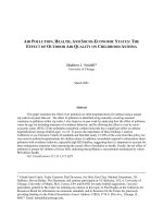
Fig. 3 The effect of vorinostat on cell growth and cell cycle in vitro. a & b A549 cells were cultured with B[a]P or vorinostat at the concentrations
indicated and for the times indicated to analyze their effect on cell growth. c To study the effect of vorinostat on cell growth in A549 cells exposed to
B[a]P as long as possible, A549 cells were first pretreated with 5 μM B[a]P for 9 days (asterisk), and then followed by combination with 5 μM vorinostat
for the times indicated. Viable cells were counted using trypan blue at each experiment, and data are presented as the mean ± standard error (SE) of
triplicate experiments. d A549 cells were cultured with 5 μM vorinostat and/or 5 μM B[a]P as described in the Materials and Methods. After incubation,
the cells were stained with propidium iodide, and cell cycle distributions were analyzed by flow cytometry. e The effect of vorinostat and/or B[a]P on
cell cycle was also analyzed in H460 and H226 lung cancer cell lines in triplicate. The Y-axis indicates the percentage of cells in the S phase of cell cycle,
and error bars indicate one standard deviation. The percentage of cells in the S phase was compared between vorinostat-treated and control cells and
between B[a]P-treated and B[a]P/vorinostat-treated cells. The difference was analyzed using paired Student t-test. The symbols * and ** denote
significant differences at P < 0.05 and P < 0.01, respectively
Lee et al. BMC Cancer (2015) 15:982
Page 6 of 10
Fig. 4 The effect of vorinostat on cyclin D1 expression and histone modification. a Cyclin D1 expression was analyzed in A549, H460, and H226
cells treated with vorinostat and/or B[a]P. Cellular lysate protein (30 μg/lane) was loaded onto a 10 % SDS/PAGE gel, electrophoresed, and
subsequently immunoblotted with a cyclin D1 antibody. b The effect of vorinostat and cyclin D1 siRNA on cyclin D1 down-regulation was compared
in A549 cells. The cells were transfected with 75 nM of cyclin D1 siRNA for 2 days (lanes 2 & 5) or with 5 μM vorinostat for 1 day (lanes 3 & 6), The cyclin
D1 expression in the presence of B[a]P was analyzed after pretreatment of cells with B[a]P for 9 days (lane 4 through lane 6). c & d Dimethylation of
H3K9 histone tail at the promoter of cyclin D1 was analyzed using chromatin immunoprecipitation (ChIP) after incubating A549 cells as described in
the Materials and Methods. The intensities of bands were measured using a densitometer and are presented as relative band intensities compared to
control. Error bars indicate one standard deviation
cyclin D1 (lane 7) was not as effective as vorinostat (lane
8) in A549 cells exposed to B[a]P. Based on this observation, cyclin D1 may be one of the targets of vorinostat in
lung adenocarcinoma cells irrespective of exposure to
B[a]P.
Vorinostat induces histone modification at the promoter
of cyclin D1
To understand the mechanism underlying the cyclin D1
down-regulation in response to vorinostat, we analyzed the
modification of H3K9 histone tail at the promoter of cyclin
D1 using chromatin immune-precipitation (ChIP) in A549
cells. Cyclin D1 promoter contains multiple transcription
factor binding sites, including AP-1, NF- B, E2F, and
Oct-1 (reviewed in ref. [12]). ERK pathway induces cyclin
D1 promoter activity through Ets or AP-1 response elements at cyclin D1 promoter, and benzo[a]pyrene-induced
cell cycle progression occurs through the ERKs/cyclin D1
pathway [13]. Therefore, we analyzed histone modifications at the AP-1 response element region at the cyclin
D1 promoter. The level of dimethyl-H3K9 at the promoter of cyclin D1 was increased in cells treated with vorinostat alone as well as with vorinostat in combination
with B[a]P as compared to the control (Fig. 4c and d): the
quantitative analyses revealed that vorinostat treatment
in A549 cells increased the levels of dimethyl-H3K9 at
the cyclin D1 promoter 2.3 times compared to the control
(P = 0.04, Wilcoxon-rank sum test). These observations
Lee et al. BMC Cancer (2015) 15:982
suggest that cyclin D1 down-regulation may be associated
with the accumulation of dimethyl-H3K9 at the promoter of cyclin D1.
Discussion
Given that adjuvant chemotherapy is effective in some
patients with stage II-IIIA NSCLC, discovery of novel
chemotherapeutic agents has become increasingly important in improving patient survival. Standard adjuvant
chemotherapy in resected NSCLC is usually based on
the use of a cytotoxic agent such as cisplatin. Cisplatin
binds to DNA and forms a spectrum of intra- and interstrand DNA crosslinks, and the resulting cisplatin-DNA
adduct interferes with DNA replication. The benefits of
platinum-based adjuvant therapy remain modest, with
improvements in 5-year survival of 5-10 %, and there
continues to be a need for development of novel adjuvant chemotherapeutic agents. Recently, biomarkerbased adjuvant chemotherapy has been reported as a
model of novel targeted therapy to further improve patient survival after surgery. In this study, the effect of
vorinostat in combination with B[a]P on cell proliferation was tested with the same concentrations (5 μM) for
two agents. And, low dose (1 μM) treatment of vorinostat in A549 cells exposed to B[a]P in vitro also inhibited
cell proliferation to a similar degree (Additional file 2:
Figure S2): the number of viable cells significantly
decreased at 72 h (P = 0.01) and 96 h (P = 0.008, Student
t-test) after treatment with vorinostat compared to the
control. Vorinostat also significantly decreased the number of viable cells in A549 cells exposed to B[a]P (P =
0.009 at 72 h and P = 0.003 at 96 h; Student t-test).
We evaluated vorinostat as a therapeutic candidate
against cyclin D1 in lung cancer cells because vorinostat
is known to suppress cyclin D1 expression in mantle cell
lymphoma cells [14], colon cancer cells [15], renal cancer cells [16], and JB6 mouse epidermal Cl 41 cells [17]
and because cyclin D1 overexpression was significantly
associated with poor RFS in stage II-IIIA lung adenocarcinoma (Fig. 2). Vorinostat suppressed cyclin D1 expression irrespective of exposure to B[a]P in A549 cells. This
effect, however, was different in several other lung cancer cell lines tested according to the presence of B[a]P
(Fig. 4a). Cyclin D1 plays an essential role in the
oncogenic transformation and its overexpression occurs
in approximately 50 % of NSCLC with demonstrated
tumorigenesis seen with human bronchial epithelial cells
exposed to B[a]P. The prognostic significance of cyclin D1
in NSCLC has been reported by a number of groups, with
conflicting results. Some studies reported cyclin D1 overexpression as an independent negative or some a positive
prognostic indicator, whereas others failed to find a significant association of cyclin D1 overexpression with
prognosis [18]. Recently, Zhang et al. [19] performed a
Page 7 of 10
meta-analysis of reported 24 studies with 2731 NSCLC
patients to understand the prognostic significance of cyclin D1 overexpression in NSCLC and found that cyclin D1
overexpression was not associated with overall survival in
NSCLC. We also did not find an association between cyclin D1 overexpression and overall survival in NSCLC (data
not shown). The overexpression of cyclin E is common
(~45 %) in NSCLC, but the impact of its overexpression
on survival remains unclear. The overexpression of cyclin
A is known to be associated with poor overall survival in
NCSLC and p16 is inactivated in approximately 50 % of
NSCLC cases, but there is no convincing evidence to suggest that p16 is a significant prognostic marker in NSCLC.
Only cyclin D1 overexpression in the present study was
significantly associated with poor RFS in stage II-IIIA lung
adenocarcinoma, suggesting that histology may function
as an effect modifier in the relationship between RFS and
protein expression.
Vorinostat leads to cyclin D1 suppression in vitro and
this could be via different mechanisms. For example, vorinostat suppresses cyclin D1 in mantle cell lymphoma cells
by blocking the translation of cyclin D1 via inhibiting the
phosphatidylinositol 3-kinase (PI3K)/Akt/mTOR/eIF4EBP pathway most likely by PI3K inhibition [14]. Vorinostat
also down-regulates cyclin D1 expression by decreasing
histone deacetylase activity [15] or by reducing cyclin D1
mRNA stability [16]. To understand molecular mechanisms underlying cyclin D1 down-regulation by vorinostat
in A549 cells, we analyzed the modification of H3K9 in
response to vorinostat using chromatin immunoprecipitation. Chromatin structure is dynamically altered by reversible modifications of core histones through the activities
of histone acetyltransferases (HATs) and HDACs, and
acetylation of core histones is linked to transcriptional activation. B[a]P is known to induce histone modification in
human cancer cells [20, 21]. Vorinostat is also known to
increase acetylation of histones at the promoter of
p21WAF1 in bladder carcinoma cells [22], multiple myeloma cells [23], and endometrial cancer cells [24]. In this
study, vorinostat increased the levels of dimethyl-H3K9 at
the promoter region of cyclin D1 in A549 cells exposed to
B[a]P, suggesting that vorinostat may down-regulate cyclin
D1 through the modification of the chromatin structure at
the promoter of the gene.
B[a]P is the carcinogenic component of polycyclic aromatic hydrocarbons, one of the main carcinogens in
cigarette smoke, and is regarded as a mediator of lung
cancer. B[a]P is metabolized by cytochrome P450 enzyme
to benzo[a]pyrene-7,8-diol-9,10-epoxide (B[a]PDE), which
is highly cytotoxic, mutagenic, and carcinogenic [25, 26].
B[a]PDE enhances cyclin D1 expression in bronchial epithelial cells, and the increased cyclin D1 promotes malignant transformation of the cells. The PI3K/Akt pathway,
as well as downstream MAPK and p70S6 kinase, are
Lee et al. BMC Cancer (2015) 15:982
known to be involved in B[a]PDE-induced cyclin D1 expression [13]. B[a]P exposure in human embryo lung fibroblasts accelerates the G1-S transition by activating
MAPK and inducing cyclin D. Treatment with antisense
cyclin D1 or antisense CDK4 completely inhibited B[a]Pinduced cell cycle progression at the G1-S checkpoint [27].
In the present study, B[a]P increased the expression of
cyclin D1 in A549 and H226 cells but not in H460 cells
(Fig. 4a). The lack of up-regulation of cyclin D1 in H460
cells treated with B[a]P may result from the presence of
mutant K-ras in these cells.
In this study, vorinostat also repressed the expression
of cyclin E, cyclin A2, and cyclin B1 in A549, H460, and
H226 cells (Additional file 3: Figure S3). But, these proteins were not further evaluated because altered expression of the proteins was not associated with patient
prognosis in stage II-IIIA NSCLC (Additional file 1:
Figure S1). Our study was severely limited by several factors. First, the effect of vorinostat needs to be evaluated
prospectively in patients with stage II-IIIA NSCLC in
which cyclin D1 is overexpressed. Second, vorinostat has
known toxicity and significant side effects as with most
chemotherapeutic agents. To minimize the side effects
of vorinostat and maximize its therapeutic effect, a combination therapy of vorinostat with other drugs such as a
DNMT inhibitor, proteasome inhibitor, or anti-Trail
antibody also needs to be considered. Third, cyclin D1
can also be repressed through other mechanisms such as
autophagy and senescence after vorinostat [28]. Further
study is required to investigate possible mechanisms related to cyclin D1 down-regulation in response to vorinostat and B[a]P.
Conclusions
In summary, the present study suggests that cyclin D1
overexpression may be significantly associated with poor
RFS in stage II-IIIA lung adenocarcinoma and its expression be modulated by vorinostat. We recommend
vorinostat as an adjuvant chemotherapeutic agent for
patients with stage II-IIIA adenocarcinoma in which cyclin D1 is overexpressed.
Page 8 of 10
obtained in accordance with the Declaration of Helsinki.
A written informed consent for the use of formalin-fixed,
paraffin-embedded tissues was obtained from all of the patients before surgery. Information on patient demographics was obtained from an interviewer-administered
questionnaire, and post-operative follow-up for detection
of death or recurrence, which was evaluated as of August
31, 2014, was conducted as previously described [2]. Median duration of follow-up after surgery was 32 months.
Lung cancer staging was determined according to the
guidelines of the American Joint Committee on Cancer
(AJCC) TNM staging system [29].
Cell culture
A549, H460, and H226 lung cancer cell lines were
obtained from the American Type Culture Collection
(Manassas, VA), and the cells were cultured in regular
RPMI-1640 medium (Lonza, Walkerville, MD) supplemented with 10 % fetal bovine serum (Hyclone, Logan,
UT), 1.0 mM of sodium pyruvate (Sigma-Aldrich, St.
Louis, MO) and 1 % HEPES buffer at 37 °C with 5 %
CO2. The cells were tested and authenticated using realtime PCR and capillary sequencing in February 2014.
In vitro growth assay
A549 cells were seeded in six-well plates with 2.0 × 10⁵
in each well. They were treated with vorinostat alone or
in combination with B[a]P. For analysis of dose- and
time-dependent effects of vorinostat and B[a]P on cell
growth, A549 cells were incubated at different concentrations of B[a]P (0, 1, 5, 10 μM) for 10 days, and vorinostat (0, 1, 2, 5 μM) for 4 days. In addition, to analyze the
effect of vorinostat on cell proliferation in cells exposed
to B[a]P, A549 cells were pretreated with 5 μM B[a]P for
9 days and followed by a combination with 5 μM vorinostat for another 1 day. A number of viable cells were
counted using trypan blue or the Vybrant MTT Cell
Proliferation Assay Kit (Life Technologies) according to
the manufacturer’s instructions. The experiments were
performed in triplicate.
Flow cytometry analysis of cell cycle
Methods
Ethics statement
This retrospective study was approved by the Samsung
Medical Center (SMC) Institutional Review Board (IRB # :
2010-07-204). All patients’ records and information were
anonymized and de-identified prior to analysis.
Study population
A total of 372 stage II-IIIA NSCLC patients who received
curative surgical resection between September 1994 and
December 2004 at the Samsung Medical Center in Seoul,
Korea, participated in this study. Tumor samples were
For cell cycle analysis, the cells were cultured in six-well
plates to 70–80 % confluence with culture medium, and
subsequently treated with 5 μM B[a]P for 10 days, or
5 μM vorinostat for 1 day, or 5 μM B[a]P for 9 days
followed by co-incubation with 5 μM vorinostat for an
additional day. After incubation of A549, H460, and
H226 cells with 5 μM vorinostat and/or 5 μM B[a]P, the
cells were harvested and washed in PBS, and then fixed
in 70 % ice-cold ethanol for 24 h. The fixed cells were
stained with propidium iodide (50 mg/ml) containing
5 mg/ml RNase A (Boehringer Ingelheim, Ingelheim,
Germany) and 0.1 % Triton X-100 and incubated for
Lee et al. BMC Cancer (2015) 15:982
15 min in dark. Fluorescence for cell cycle distribution was
detected by the FACScan flow cytometer (Becton-Dickson,
San Jose, CA) and resulting data was analyzed using the
ModFit LT version 3.0 (Verity Software House, Topsham,
ME). At least 10,000 cells were examined for each sample
and the experiments were repeated at least three times.
Page 9 of 10
H3K9 (lys9) and normal IgG (negative control) (Upstate
Biotechnology). The primer sequences for promoter amplification of cyclin D1 were 5′- CTGCCTTCCTACCTT
GACCA -3′ (forward) and 5′- GAAGGGACGTCTAC
ACCCC -3′ (reverse).
Immunohistochemistry
Western blot analysis
Cyclin D1 expression in response to B[a]P and vorinostat
was analyzed by immune-blotting using antibodies against
cyclin D1 (DCS6, #2926, Cell Signaling, Danvers, MA)
and α-tubulin (T6074, Sigma-Aldrich, St. Louis, MO) as a
loading control according to standard procedures.
Reverse transcription polymerase chain reaction (RT-PCR)
Cells were harvested and washed in ice-cold PBS after
culture. Total RNA was isolated using the RNeasy Mini
Kit (Qiagen, Valencia, CA, USA) according to the manufacturer’s instructions. RT-PCR was carried out in a tube
containing 0.5 μg of total RNA and primers specific to
cyclin D1 at a final concentration of 0.5 μM using the
OneStep RT-PCR kit (Qiagen) according to the manufacturer’s protocols. The sequences of primers used for
RT-PCR were as follows: cyclin D1 forward primer
5′-TGTTCGTGGCCTCTAAGATGA-3′, reverse primer
5′-GCTTGACTCCAGAAGGGCTT-3′; GAPDH forward
primer 5′- AACTTTGGCATTGTGGAAGG, reverse primer 5′-TGTGAGGGAGATGCTCAGTG.
Cyclin D1 knockdown using small interfering RNA (siRNA)
To evaluate if the effect of vorinostat on cyclin D1
down-regulation was comparable to a cyclin D1 knockdown in lung cancer cells, A549 cells (2.0 × 10⁵ cells per
well) were seeded in six-well plates, and at 70 %–80 %
confluence the cells were treated with 5 μM vorinostat
for 1 day or with 75 nM of cyclin D1 siRNA (SI
02654547, Qiagen) for 2 days. A549 cells were also pretreated with B[a]P for 9 days and then treated with cyclin
D1 siRNA and/or vorinostat in the same way that was
performed in the absence of B[a]P. Transfection of cyclin
D1 siRNA was performed using FlexiTube siRNA &
HiPerFect Transfection Reagent (Qiagen GmbH, Hilden,
Germany) according to the manufacturer’s protocol.
Chromatin immunoprecipitation
For the analysis of dimethylation of the H3K9 histone tail
at the promoter of cyclin D1, A549 cells were cultured
with 5 μM B[a]P for 10 days, or 5 μM vorinostat for 1 day,
or 5 μM B[a]P for 9 days followed by co-incubation with
5 μM vorinostat for an additional 24 h. After 48 h of
culture, chromatin immunoprecipitation (ChIP) was performed using a ChIP assay kit (Upstate Biotechnology,
Lake Placid, NY) according to the manufacturer’s protocols.
The antibodies used for ChIP were anti-dimethyl-histone
Tissue microarrays (TMAs) of NSCLC and immunohistochemical staining of cyclin A2, cyclin D1, cyclin E, and
p16 were conducted as previously described [30]. The
antibodies used were as follows: cyclin A2 (BF683, Cell
Signaling, Danvers, MA, USA), cyclin D1 (DCS6, Cell
Signaling), cyclin E (HE12, Cell signaling), and p16 (Ab-16,
Neomarker, Fremont, CA). All available slides were
reviewed by two authors (EY Cho, D-H Kim), who were
blinded to all clinicopathological variables, to reduce
inter-observer variability. The expression of individual
proteins was assigned to the extent and intensity of tumor
cells that stained positively in nucleus. Cytoplasmic reactivity, if present, was disregarded. The expression levels
of the four proteins were calculated by multiplying the intensity score (0, none; 1, weak; 2, moderate; 3, strong) with
the proportion score of positive staining tumor cells (0,
absent; 1, 0–10 %; 2, 10–50 %; 3, 50–80 %; 4, > 80 %).
Statistical analysis
In the univariate analysis, Pearson’s chi-square test (or
Fisher’s exact test) and t-test (or Wilcoxon rank-sum
test) were used for analysis of categorical and continuous
variables, respectively. Correlations among expression levels
of the four proteins were compared using Spearman’s rank
correlation coefficients. The effect of protein expression
on recurrence-free survival (RFS) or overall survival
was analyzed by Kaplan-Meier survival curves, and the
significance of differences in survival between the two
groups was evaluated by the log-rank test. Cox proportional hazards regression model was used to estimate
adjusted hazard ratios with 95 % confidence interval for
independent predictor variables. All statistical analyses
were two-sided, with a 5 % type I error rate.
Additional files
Additional file 1: Figure S1. Recurrence-free survival in stage II-IIIA
NSCLC. Recurrence-free survival was compared according to expression
statuses of cyclin A2, cyclin E, and p16. Data were stratified according to
histology. The yellow and blue lines indicate groups with and without
abnormal expression of each protein, respectively. P–values were based
on the log-rank test. (TIFF 2996 kb)
Additional file 2: Figure S2. Effect of low dose vorinostat on cell
proliferation. A549 cells were pre-treated with 5 μM B[a]P for 9 days and
then incubated in combination with 1 μM vorinostat for the indicated
hours. The total number of viable cells at each time point was determined
by MTT assay. The y-axis indicates cell numbers relative to time zero.
Each value represents mean ± standard deviation of three experiments.
(TIFF 1042 kb)
Lee et al. BMC Cancer (2015) 15:982
Additional file 3: Figure S3. The effect of vorinostat on expression of
cyclins. The expression levels of cyclin E, cyclin A2, and cyclin B1 were
analyzed in A549, H460, and H226 cells treated with vorinostat and/or
B[a]P. Western blotting was performed according to standard procedures.
Antibodies against cyclin E (HE12, #4129), cyclin A2 (BF683, #4656), and
cyclin B1 (#4138) were purchased from Cell Signaling Technology.
(TIFF 1815 kb)
Page 10 of 10
9.
10.
11.
Abbreviations
B[a]P: benzo[a]pyrene; CI: confidence interval; HDACIs: histone deacetylase
inhibitors; HR: hazard ratio; NSCLC: non-small cell lung cancer;
RFS: recurrence-free survival; RT-PCR: Reverse transcription polymerase chain
reaction; SAHA: suberoylanilide hydroxamic acid; siRNA: small interfering
RNA; TMAs: Tissue microarrays.
Competing interests
The authors declare that they have no competing interests.
Authors’ contributions
EL and D-HK designed the overall study and drafted the manuscript. EL
performed most of in vitro study. BBL, YK, and DJ contributed to statistical
analysis and immunohistochemistry. JH performed the interpretation of
immunohistochemical staining. All patient surgery was performed by YMS.
All authors read and approved the final manuscript.
Acknowledgements
The authors wish to thank Eun-Kyung Kim and Jin-Hee Lee for data collection
and management, and Hoon Suh for sample collection. This work was
supported by grants from Basic Science Research Program through the
National Research Foundation of Korea (NRF) funded by the Ministry of
Education (2014R1A1A2053325) and from the Korea Health Industry Development
Institute (KHIDI), funded by the Ministry of Health & Welfare (HI14C1979), Republic
of Korea.
Author details
1
Department of Molecular Cell Biology, Sungkyunkwan University School of
Medicine, #300 Chunchun-dong, Jangan-KuKyunggido, Suwon 440-746,
Korea. 2Department of Pathology, Samsung Medical Center, #50 Ilwon-dong,
Kangnam-Ku, Seoul 135-710, Korea. 3Department of Thoracic and
Cardiovascular Surgery, Samsung Medical Center, #50 Ilwon-dong,
Kangnam-Ku, Seoul 135-710, Korea. 4Center for Genome Research, Samsung
Biomedical Research Institute, Rm B155, #50 Ilwon-dong, Kangnam-Ku, Seoul
135-710, Korea.
12.
13.
14.
15.
16.
17.
18.
19.
20.
21.
22.
23.
Received: 27 February 2015 Accepted: 11 December 2015
24.
References
1. Gilbert S, Reid KR, Lam MY, Petsikas D. Who should follow up lung cancer
patients after operation? Ann Thorac Surg. 2000;69:1696–700.
2. Kim JS, Kim JW, Han J, Shim YM, Park J, Kim DH. Cohypermethylation of p16
and FHIT promoters as a prognostic factor of recurrence in surgically
resected stage I non-small cell lung cancer. Cancer Res. 2006;66:4049–54.
3. Heon S, Johnson BE. Adjuvant chemotherapy for surgically resected nonsmall cell lung cancer. J Thorac Cardiovasc Surg. 2012;144:S39–42.
4. Pignon JP, Tribodet H, Scagliotti GV, Douillard JY, Shepherd FA, Stephens RJ,
et al. Lung adjuvant cisplatin evaluation: a pooled analysis by the LACE
Collaborative Group. J Clin Oncol. 2008;26:3552–9.
5. NSCLC Meta-analyses Collaborative Group, Arriagada R, Auperin A, Burdett S,
Higgins JP, Johnson DH, et al. Adjuvant chemotherapy, with or without
postoperative radiotherapy, in operable non-small-cell lung cancer: two
meta-analyses of individual patient data. Lancet. 2010;375:1267–77.
6. Marks PA. Discovery and development of SAHA as an anticancer agent.
Oncogene. 2007;26:1351–6.
7. Bruzzese F, Rocco M, Castelli S, Di Gennaro E, Desideri A, Budillon A.
Synergistic antitumor effect between vorinostat and topotecan in small cell
lung cancer cells is mediated by generation of reactive oxygen species and
DNA damage-induced apoptosis. Mol Cancer Ther. 2009;8:3075–87.
8. Li CT, Hsiao YM, Wu TC, Lin YW, Yeh KT, Ko JL. Vorinostat, SAHA,
represses telomerase activity via epigenetic regulation of telomerase
25.
26.
27.
28.
29.
30.
reverse transcriptase in non-small cell lung cancer cells. J Cell Biochem.
2011;112:3044–53.
Vansteenkiste J, Van Cutsem E, Dumez H, Chen C, Ricker JL, Randolph SS, et
al. Early phase II trial of oral vorinostat in relapsed or refractory breast,
colorectal, or non-small cell lung cancer. Invest New Drugs. 2008;26:483–8.
Komatsu N, Kawamata N, Takeuchi S, Yin D, Chien W, Miller CW, et al. SAHA,
a HDAC inhibitor, has profound anti-growth activity against non-small cell
lung cancer cells. Oncol Rep. 2006;15:187–91.
Mayne ST, Lippman SM. Cigarettes: a smoking gun in cancer
chemoprevention. J Natl Cancer Inst. 2005;97:1319–21.
Guo ZY, Hao XH, Tan FF, Pei X, Shang LM, Jiang XL, et al. The elements of
human cyclin D1 promoter and regulation involved. Clin Epigenetics.
2011;2:63–76.
Ding J, Ning B, Gong W, Wen W, Wu K, Liang J, et al. Cyclin D1 induction by
benzo[a]pyrene-7,8-diol-9,10-epoxide via the phosphatidylinositol 3-inase/
Akt/MAPK- and p70s6k-dependent pathway promotes cell transformation
and tumorigenesis. J Biol Chem. 2009;284:33311–9.
Kawamata N, Chen J, Koeffler HP. Suberoylanilide hydroxamic acid (SAHA;
vorinostat) suppresses translation of cyclin D1 in mantle cell lymphoma
cells. Blood. 2007;110:2667–73.
Jin JS, Tsao TY, Sun PC, Yu CP, Tzao C. SAHA inhibits the growth of colon
tumors by decreasing histone deacetylase and the expression of cyclin D1
and survivin. Pathol Oncol Res. 2012;18:713–20.
Sato A, Asano T, Horiguchi A, Ito K, Sumitomo M, Asano T. Antitumor effect
of suberoylanilide hydroxamic acid and topotecan in renal cancer cells.
Oncol Res. 2011;19:217–23.
Zhang J, Ouyang W, Li J, Zhang D, Yu Y, Wang Y, et al. Suberoylanilide
hydroxamic acid (SAHA) inhibits EGF-induced cell transformation via reduction
of cyclin D1 mRNA stability. Toxicol Appl Pharmacol. 2012;263:218–24.
Zhu CQ, Shih W, Ling CH, Tsao MS. Immunohistochemical markers of
prognosis in non-small cell lung cancer: a review and proposal for a
multiphase approach to marker evaluation. J Clin Pathol. 2006;59:790–800.
Zhang LQ, Jiang F, Xu L, Wang J, Bai JL, Yin R, et al. The role of cyclin D1
expression and patient’s survival in non-small-cell lung cancer: a systematic
review with meta-analysis. Clin Lung Cancer. 2012;13:188–95.
Sadikovic B, Andrews J, Carter D, Robinson J, Rodenhiser DI. Genome-wide
H3K9 histone acetylation profiles are altered in benzopyrene-treated MCF7
breast cancer cells. J Biol Chem. 2008;283:4051–60.
Yan C, Lu J, Zhang G, Gan T, Zeng Q, Shao Z, et al. Benzo[a]pyrene induces
complex H2AX phosphorylation patterns by multiple kinases including ATM,
ATR, and DNA-PK. Toxicol In Vitro. 2011;25:91–9.
Richon VM, Sandhoff TW, Rifkind RA, Rifkind RA, Marks PA. Histone
deacetylase inhibitor selectively induces p21WAF1 expression and geneassociated histone acetylation. Proc Natl Acad Sci U S A. 2000;97:10014–9.
Gui CY, Ngo L, Xu WS, Richon VM, Marks PA. Histone deacetylase (HDAC)
inhibitor activation of p21WAF1 involves changes in promoter-associated
proteins, including HDAC1. Proc Natl Acad Sci U S A. 2004;101:1214–6.
Takai N, Desmond JC, Kumagai T, Gui D, Said JW, Whittaker S, et al. Histone
deacetylase inhibitors have a profound antigrowth activity in endometrial
cancer cells. Clin Cancer Res. 2004;10:1141–9.
Alexandrov K, Cascorbi I, Rojas M, Bouvier G, Kriek E, Bartsch H. CYP1A1 and
GSTM1 genotypes affect benzo[a]pyrene DNA adducts in smokers’ lung:
comparison with aromatic/hydrophobic adduct formation. Carcinogenesis.
2002;23:1969–77.
Smith LE, Denissenko MF, Bennett WP, Li H, Amin S, Tang M, et al. Targeting
of lung cancer mutational hotspots by polycyclic aromatic hydrocarbons.
J Natl Cancer Inst. 2000;92:803–11.
Du HJ, Tang N, Liu BC, You BR, Shen FH, Ye M, et al. Benzo[a]pyreneinduced cell cycle progression is through ERKs/cyclin D1 pathway and
requires the activation of JNKs and p38 mapk in human diploid lung
fibroblasts. Mol Cell Biochem. 2006;287:79–89.
Brown NE, Jeselsohn R, Bihani T, Hu MG, Foltopoulou P, Kuperwasser C, et al.
Cyclin D1 activity regulates autophagy and senescence in the mammary
epithelium. Cancer Res. 2012;72:6477–89.
Edge SB, Byrd DR, Compton CC, Fritz AG, Greene FL, Trotti A. American
Joint Committee on Cancer. In: AJCC Cancer Staging Manual. 7th ed.
New York: Springer; 2010. p. 253–70.
Ko E, Kim Y, Park SE, Cho EY, Han J, Shim YM, et al. Reduced expression of
cyclin D2 is associated with poor recurrence-free survival independent of
cyclin D1 in stage III non-small cell lung cancer. Lung Cancer. 2012;77:401–6.