Stomatin-like protein 2 is overexpressed in epithelial ovarian cancer and predicts poor patient survival
Bạn đang xem bản rút gọn của tài liệu. Xem và tải ngay bản đầy đủ của tài liệu tại đây (2.04 MB, 11 trang )
Sun et al. BMC Cancer (2015) 15:746
DOI 10.1186/s12885-015-1723-x
RESEARCH ARTICLE
Open Access
Stomatin-like protein 2 is overexpressed in
epithelial ovarian cancer and predicts poor
patient survival
Fei Sun1,4†, Wen Ding3†, Jie-Hua He2, Xiao-Jing Wang1, Ze-Biao Ma1 and Yan-Fang Li1*
Abstract
Background: Stomatin-like protein 2 (SLP-2, also known as STOML2) is a stomatin homologue of uncertain
function. SLP-2 overexpression has been suggested to be associated with cancer progression, resulting in adverse
clinical outcomes in patients. Our study aim to investigate SLP-2 expression in epithelial ovarian cancer cells and its
correlation with patient survival.
Methods: SLP-2 mRNA and protein expression levels were analysed in five epithelial ovarian cancer cell lines and
normal ovarian epithelial cells using real-time PCR and western blotting analysis. SLP-2 expression was investigated
in eight matched-pair samples of epithelial ovarian cancer and adjacent noncancerous tissues from the same
patients. Using immunohistochemistry, we examined the protein expression of paraffin-embedded specimens from
140 patients with epithelial ovarian cancer, 20 cases with borderline ovarian tumours, 20 cases with benign ovarian
tumours, and 20 cases with normal ovarian tissues. Statistical analyses were applied to evaluate the clinicopathological
significance of SLP-2 expression.
Results: SLP-2 mRNA and protein expression levels were significantly up-regulated in epithelial ovarian cancer cell
lines and cancer tissues compared with normal ovarian epithelial cells and adjacent noncancerous ovarian tissues.
Immunohistochemistry analysis revealed that the relative overexpression of SLP-2 was detected in 73.6 % (103/140) of
the epithelial ovarian cancer specimens, 45.0 % (9/20) of the borderline ovarian specimens, 30.0 % (6/20) of the benign
ovarian specimens and none of the normal ovarian specimens. SLP-2 protein expression in epithelial ovarian cancer
was significantly correlated with the tumour stage (P < 0.001). Epithelial ovarian cancer patients with higher SLP-2
protein expression levels had shorter progress free survival and overall survival times compared to patients with lower
SLP-2 protein expression levels. Multivariate analyses showed that SLP-2 expression levels were an independent
prognostic factor for survival in epithelial ovarian cancer patients.
Conclusions: SLP-2 mRNA and proteins were overexpressed in epithelial ovarian cancer tissues. SLP-2 protein
overexpression was associated with advanced stage disease. Patients with higher SLP-2 protein expression had shorter
progress free survival and poor overall survival times. Thus, SLP-2 protein expression was an independent prognostic
factor for patients with epithelial ovarian cancer.
Keywords: SLP-2, Epithelial ovarian cancer, Prognosis, Biomarker
* Correspondence:
†
Equal contributors
1
Department of Gynecologic Oncology, Sun Yat-sen University Cancer
Center; State Key Laboratory of Oncology in South China; Collaborative
Innovation Center of Cancer Medicine, 651 Dongfeng Road East, Guangzhou
510060, P.R.China
Full list of author information is available at the end of the article
© 2015 Sun et al. Open Access This article is distributed under the terms of the Creative Commons Attribution 4.0
International License ( which permits unrestricted use, distribution, and
reproduction in any medium, provided you give appropriate credit to the original author(s) and the source, provide a link to
the Creative Commons license, and indicate if changes were made. The Creative Commons Public Domain Dedication waiver
( applies to the data made available in this article, unless otherwise stated.
Sun et al. BMC Cancer (2015) 15:746
Background
Epithelial ovarian cancer accounts for 80 %−90 % of ovarian
cancers and is the leading cause of death in patients with
gynaecologic malignancies [1] . The absence of specific
symptoms and lack of reliable early diagnostic methods has
resulted in the diagnosis of 70 % of patients at an advanced
stage [2]. Despite progress in the development of new
therapeutic methods, the 5-year survival rate of epithelial
ovarian cancer patients has remained at approximately
30 % [3]. Epithelial ovarian cancer is thought to arise from
an accumulation of genetic changes in a manner similar to
other cancers [4]. Therefore, understanding the molecular
mechanisms of the early events of epithelial ovarian cancer
and searching for novel biomarkers involved in the progression of epithelial ovarian cancer is of great value for the
identification of early-stage patients, providing new therapeutic targets, and improving patient survival.
Stomatin-like protein 2 (SLP-2, also known as STOML2)
is a major protein on the mitochondrial inner membrane
and a member of the stomatin superfamily. The relatively
conserved 31-kDa protein has been shown to interact with
prohibitin-1 and−2 [5, 6]. However, human SLP-2 has very
low overall homology compared with other stomatins because SLP-2 lacks the characteristic amino-terminal transmembrane domain. SLP-2 may play an important role in
organizing sphingolipid and cholesterol-rich lipid rafts,
regulating ion channel conductance, and linking other integral membrane proteins to the peripheral cytoskeleton [5].
Previous studies revealed that human SLP-2 is a novel
cancer-related gene of unknown function. The SLP-2 protein was first found to be overexpressed in human
oesophageal cancer. Transecting antisense SLP-2 into the
oesophageal squamous cell carcinoma cell line TE12 reduced cell growth and adhesion. These results suggested
that SLP-2 was a potential oncogene [7, 8]. Further studies
showed that the SLP-2 protein was overexpressed in many
human cancer tissues, including gastric cancer [9], endometrial adenocarcinoma [10], and breast cancer [11]. SLP-2
up-regulation is correlated with the transformation of normal cells into tumour cells by an unknown mechanism.
Thus, SLP-2 expression levels or copy number status may
serve as a useful prognostic factor for cancer patients [10].
However, the expression status of SLP-2 and its clinical significance in epithelial ovarian cancer remain unclear. We
investigated the protein and mRNA expression levels of
SLP-2 in ovarian cancer tissues using immunohistochemistry, western blotting, and RT-PCR to analyse the potential
clinical significance of SLP-2 expression.
Methods
Cell culture
OVCAR3 and Anglne cells were purchased from the
China Center for Type Culture Collection (CCTCC,
Wuhan, China). OVCAR3 cells were grown in RPMI 1640
Page 2 of 11
supplemented with 10 % FBS, and Anglne cells were
grown in Eagle’s minimal essential medium (Eagle’s
MEM) supplemented with 10 % FBS. SKOV3 and
HO8910 cells were purchased from the Shanghai Cell
Bank of the Chinese Academy of Science (Shanghai,
China). SKOV-3 cells were grown in McCoy’s 5A medium
supplemented with 10 % FBS and HO8910 cells were
grown in RPMI 1640 medium (HyClone, Logan, UT,
USA) supplemented with 10 % FBS. A2780 cells (Nanjing
KeyGen Biotech, Nanjing, China) were cultured in high
glucose DMEM supplemented with 10 % FBS. Primary
normal ovarian surface epithelial (NOSE) cells were established according to the method described in previous reports [12].
Tissue samples and patient information
For real-time PCR and western blotting analysis, eight
matched pairs of fresh tumour tissue specimens and adjacent noncancerous tissue samples were obtained from patients with epithelial ovarian cancer immediately after
surgery and immersed at−80 °C until use. The percentages
of tumour purity in these tissues and adjacent sections
used for RNA and protein analyses were established by
routine histopathological analyses. For immunohistochemistry, a total of 140 cancer tissue samples were collected from patients with epithelial ovarian cancer, 20
from patients with borderline ovarian tumours, and 20
from patients with benign ovarian tumours. Additionally,
20 normal ovarian epithelial tissues were collected from
patients with benign uterine tumours who needed a hysterectomy and oophorectomy. All patients received surgery. Most patients (except those who had stage IA and
grade 1 tumors) had post-operation adjuvant chemotherapy with platinum-based regimen. The patient list was obtained from the database of Sun Yat-sen University
Cancer Center. Patient hospital records were reviewed to
obtain demographic data, including age, serum levels of
CA125, diagnosis, volume of ascites, surgical procedures,
tumour stage, pathological reports, post-operation chemotherapy, and results of follow-up. All patient tissue samples were histologically confirmed to be epithelial ovarian
cancers; these patients received treatment at the Sun
Yat-sen University Cancer Center between January 1,
2003, and December 31, 2008. None of the patients
had received prior radiotherapy or chemotherapy.
Eight matched pairs of fresh tumour tissue specimens
and adjacent noncancerous tissue samples were collected
from eight patients with serous epithelial ovarian cancer.
Of these eight patients, three had stage I disease, two had
stage II, and three had stage III; additionally, one patient
had a grade 1–2 tumour, three had grade 2 tumours, two
had grade 3 tumours, and two had grade 2–3 tumours.
Adjacent noncancerous tissue samples were collected
from either the noncancerous stroma of the same ovary
Sun et al. BMC Cancer (2015) 15:746
(Patients 1–5, who had stage I or II tumours) (Fig. 2) or
from the normal stroma of the contra-lateral ovary
(Patients 6–8, who had stage III tumours) (Fig. 2). Of
the 20 patients with borderline tumours, ten had serous tumours, seven had mucinous tumours, two had
mixed tumours, and one had another type. Of the 20 patients with benign tumours, 16 had serous and four had
mucinous tumours. All patients with ovarian cancer received surgery. Most patients (except those who had stage
IA and grade 1 tumours) had post-operation adjuvant
chemotherapy with a platinum-based regimen. Clinical
follow-up data were available until December 31, 2013.
The clinical information on the 140 patients with ovarian
cancer whose tumour tissues were used for immunohistochemistry is summarized in Table 1. Patient’s consent was
waived for this study since every patient at our institute
have signed an informed consent on admission time for
future possible use of the tumour sample for scientific research. Our study was approved by Sun Yat-sen University
Cancer Center IRB (Approval No: B2014-2-26).
Real-time PCR (RT-PCR)
Total RNA samples were extracted from cultured cells
and primary tumour tissues using the TRIzol reagent
(Invitrogen, Carlsbad, CA, USA) in accordance with the
manufacturer’s instructions and treated with RNase-free
DNase. cDNA was synthesized from 2 μg of RNA from
each sample using an iScript™ cDNA Synthesis Kit
(BioRad Laboratories, Hercules, CA, USA). The RT-PCR
cycling conditions incorporated an initial denaturation
at 94 °C for 5 min, followed by 30 denaturation cycles at
94 °C for 30 s, primer annealing at 55 °C for 30 s, primer
extension phase at 72 °C 50 s, and a final extension step
at 72 °C for 7 min. The primers for SLP-2 and
glyceraldehyde-3-phosphate dehydrogenase (GAPDH)
were designed using Primer Express v 2.0 software (Applied Biosystems). The sequences of the primers were as
follows: forward primer 5’-GTGACTCTCGACAATGTAAC-3’ and reverse primer 5’-TGATCTCATAACGGAGGCAG -3’. SLP-2 expression data were normalized
to GAPDH, and all experiments were performed in
triplicate.
Western blotting
The cells were washed twice with ice-cold phosphatebuffered saline (PBS) and lysed on ice in radio immunoprecipitation assay (RIPA) buffer (Cell Signaling Technology,
Danvers, MA) containing complete protease inhibitor cocktail (Roche Applied Science, Mannheim, Germany). Fresh
tissue samples were ground to powder in liquid nitrogen
and lysed with SDS-PAGE sample buffer. All protein
samples (20 μg) were separated on 12 % sodium dodecyl
sulfate–polyacrylamide gels, transferred to polyvinylidene
fluoride (PVDF) membranes (Immobilon P, Millipore,
Page 3 of 11
Bedford, MA) and blocked with 5 % skimmed milk in Trisbuffered saline supplemented with 0.1 % Tween 20 (TBST)
for 1 h at room temperature. After blocking, the membranes were incubated with anti-SLP-2 antibodies (1:1000,
Proteintech, Chicago, IL, USA) at 4 °C overnight. Then, the
membranes were rinsed with TBST and incubated with an
anti-rabbit IgG antibody (Santa Cruz Biotechnology, Santa
Cruz, USA) conjugated to horseradish peroxide for 15 min.
The expression of SLP-2 was detected with the enhanced
chemiluminescence (ECL) prime western blotting detection
reagent (Amersham Bioscience, Switzerland) according to
the manufacturer’s instructions. An anti-ß-actin antibody
(Sigma, St. Louis, MO) were used as a loading control.
Immunohistochemistry
Immunohistochemical analysis was used to study SLP-2
protein expression in 140 epithelial ovarian cancer samples, 20 borderline ovarian tumour samples, 20 benign
ovarian tumour samples and 20 normal ovarian epithelial tissues. Paraffin-embedded specimens were cut into
4-μm-thick sections, de-waxed with xylene and rehydrated.
For antigenic retrieval, the sections were submerged into
EDTA antigenic retrieval buffer and microwaved, and then
treated with 3 % hydrogen peroxide in methanol to quench
endogenous peroxidase activity. Subsequently, the sections
were incubated with 1 % bovine serum albumin to block
nonspecific binding, and then incubated with an anti-SLP-2
rabbit polyclonal antibody (1:1000, Proteintech, Chicago,
IL, USA) overnight at 4 °C. Normal goat serum was used as
the negative control. After washing, the sections were
incubated with a biotinylated anti-rabbit secondary
antibody (Abcam, Cambridge, MA), and then further
incubated with a streptavidin-horseradish peroxidase
complex (Abcam, Cambridge, MA). Finally, the tissue
sections were immersed in 3.30-diaminobenzidine,
counterstained with 10 % Mayer’s hematoxylin, dehydrated and mounted in crystal mount medium.
SLP-2 staining was scored by two independent pathologists. The scores were averaged based on both the intensity
of staining and the proportion of positively stained tumour
cells. The proportion of tumour cells was scored as follows:
0 (<5 % positive tumour cells), 1 (6–25 % positive tumour
cells), 2 (26–50 % positive tumour cells), 3 (51–75 % positive tumour cells), and 4 (>75 % positive tumour cells). The
intensity of staining was graded as follows: 0 (no staining);
1 (weak staining ~ light yellow), 2 (moderate staining ~ yellow brown), and 3 (strong staining ~ brown). The staining
index for SLP-2 expression in epithelial ovarian cancer was
calculated by multiplying the two scores of the proportion
of positive cells and the intensity of staining. Cut-off values
for SLP-2 were based on the median of all products. An optimal cut-off value was identified as follows: a score ≥ 6 was
used to define tumours with high SLP-2 expression and a
score ≤ 4 indicated low SLP-2 expression.
Sun et al. BMC Cancer (2015) 15:746
Page 4 of 11
Table 1 Clinicopathological characteristics of patients with epithelial ovarian cancer and their correlations with SLP-2 expression
Characteristics
Number of
cases (%)
P value
SLP-2 expression (%)
Low or no expression
High expression
Age (years)
0.433
<45
49 (35)
11 (22.4)
38 (77.6)
≥45
91 (65)
26 (29.7)
65 (70.3)
<500U/ml
67 (47.9)
23 (34.3)
44 (65.7)
≥500U/ml
73 (52.1)
15 (20.5)
58 (79.5)
CA125 level (before surgery)
0.067
Tumour size
0.501
<10 cm
58 (41.4)
14 (24.1)
44 (75.8)
≥10 cm
82 (58.6)
24 (29.3)
58 (70.7)
<1000 ml
100 (71.4)
33 (33.0)
67 (67.0)
≥1000 ml
40 (28.6)
5 (12.5)
35 (87.5)
The volume of ascites
0.014
Peritoneal cytology
0.001
Positive
68 (48.6)
10 (14.7)
58 (85.3)
Negative
72 (51.4)
28 (38.9)
44 (61.1)
Serous
80 (57.1)
20 (25.0)
60 (75.0)
Mucinous
14 (10.0)
8 (57.1)
6 (42.9)
Poorly differentiated
36 (25.7)
8 (22.2)
28 (77.8)
10 (7.1)
2 (20.0)
8 (80.0)
Pathological type
a
Others
0.064
Grade of differentiation
0.072
G1
59 (42.1)
11 (18.6)
48 (81.4)
G2
46 (32.9)
13 (28.3)
33 (71.7)
G3
18 (12.9)
9 (50.0)
9 (50.0)
Unknown
17 (12.1)
5 (29.4)
12 (70.6)
I
30 (21.4)
17 (56.7)
13 (43.3)
II+ III + IV
110 (78.6)
21 (19.1)
89 (88.9)
FIGO stage
0.001
Lymph node metastasis
0.688
Positive
21 (15.0)
7 (33.3)
14 (66.7)
Negative
23 (16.4)
5 (21.7)
18 (78.3)
96 (68.6)
26 (27.1)
70 (72.9)
b
Not do RPLND
Cytoreductive surgeryc
0.185
Optimal
107 (76.4)
32 (29.9)
75 (70.1)
Suboptimal
33 (23.6)
6 (18.2)
27 (81.8)
a
Endometrioid adenocarcinoma, two cases; clear cell carcinoma, three cases; mixed epithelial carcinoma, five cases
RPLND, retroperitoneal lymph node dissection, including unilateral or bilateral pelvic lymphadenectomy and /or paraortic lymphadenectomy
Cytoreductive surgery: Optimal, the diameter of the largest residual lesions was < 2 cm;Suboptimal, the diameter of the largest residual lesions was ≥ 2 cm
b
c
Statistical analyses
All statistical analyses were conducted using the SPSS
software package (IBM, standard version 16.0). The relationship between the expression of SLP-2 and clinicopathological characteristics was analysed by Pearson’s χ2
and Fisher’s exact tests. Overall survival (OS) was
defined as the time from surgery to death or to the last
follow-up. Progression-free survival (PFS) was defined as
the length of time after treatment to the onset of recurrence or progression (diagnosed by imaging or clinical
assessment). Kaplan–Meier curves were plotted to assess
the effects of SLP-2 expression levels on PFS and OS,
Sun et al. BMC Cancer (2015) 15:746
and survival curves were compared using a log-rank test.
Multivariate Cox regression analysis was performed for
all clinicopathological variables that were found to be
significant by univariate analysis. In all tests, a two-sided
P-value of less than 0.05 was considered to be statistically significant.
Results
The SLP-2 mRNA and protein were overexpressed in
epithelial ovarian cancer cell lines
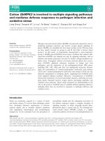
We used real-time RT-PCR and western blotting to investigate the mRNA and protein expression levels of SLP-2
in five epithelial ovarian cancer cell lines (OVCAR3,
Anglne, SKOV-3, HO8910 and A2780) and normal ovarian surface epithelial (NOSE) cells. The mRNA expression
of SLP-2 was at least 4-fold higher in epithelial ovarian
cancer cell lines than in the NOSE cells (Fig. 1a). Moreover, the SLP-2 protein was highly expressed in the epithelial ovarian cancer cell lines and only weakly expressed in
the NOSE cells (Fig. 1b).
Page 5 of 11
The SLP-2 mRNA and protein were overexpressed in
epithelial ovarian cancer tissues
To investigate the SLP-2 mRNA and protein expression
levels in human epithelial ovarian cancer tissues, we
used real-time RT-PCR and western blotting to analyse
eight matched pairs of epithelial ovarian cancer specimens (T) and adjacent noncancerous tissue samples
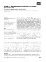
(ANT). SLP-2 mRNA was expressed at higher levels in
all epithelial ovarian cancer tissues compared to adjacent
noncancerous tissues, with the differential expression
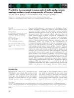
levels ranging from 4.4- to 11.8-fold (Fig. 2a). Additionally, the SLP-2 protein was also up-regulated in epithelial
ovarian cancer tissues compared with the matched noncancerous tissues (Fig. 2b, Fig. 3). SLP-2 was mainly located in the cell membrane and cytoplasm.
SLP-2 protein expression was higher in epithelial ovarian
cancers than in benign and borderline ovarian tumours
To compare the difference in SLP-2 expression between
epithelial ovarian cancer and benign and borderline
ovarian tumours, we examined paraffin-embedded archived samples from 140 cases of epithelial ovarian cancer tissues, 20 borderline ovarian tumour tissues and 20
benign ovarian tumour tissues; additionally, 20 normal
ovarian epithelial tissues were included as the control
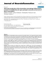
group. SLP-2 protein expression was analysed by immunohistochemical staining. High SLP-2 protein expression
was detected in 72.9 % (102/140) of epithelial ovarian
cancer samples, in 45.0 % (9/20) of borderline ovarian
tumour tissues, in 30.0 % (6/20) of benign ovarian
tumour tissues, and in none (0/20) of the normal ovarian
epithelial tissues (Table 2, Figs. 4 and 5). The 6 cases of
benign tumours with SLP-2 overexpression included 5
serous and 1 mucinous type; The 9 cases of borderline
ovarian tumour with SLP-2 overexpression included 6
serous tumour, 2 mucinous tumour, and 1 mixed
tumour. Thus, SLP-2 protein expression in epithelial
ovarian cancer samples was higher than benign ovarian
tumours and borderline ovarian tumours (both P <
0.001) (Table 2).
SLP-2 overexpression was associated with epithelial
ovarian cancer clinical features
Fig. 1 Overexpression of SLP-2 mRNA and protein in epithelial ovarian
cancer cell lines. SLP-2 mRNA and protein expression in epithelial
ovarian cancer cell lines (OVCAR3, Anglne, SKOV-3, HO8910, and
A2780) and NOSE cells were examined by teal-time PCR (a) and
western blotting (b). Expression levels were normalized against GAPDH
and β-actin, respectively. Error bars represent standard deviation of the
mean (SD) calculated from three parallel experiments. *P < 0.05
Out of the 140 patients with epithelial ovarian cancer, 30
patients had stage I tumours, 23 had stage II tumours,
77 had stage III tumours, and 10 had stage IV tumours.
The median age was 46 years (range, 15 ~ 76 years). All
140 patients received initial treatment, including surgery
and post-operation chemotherapy.
Statistical analysis showed a significant correlation between SLP-2 protein expression and the clinicopathological characteristics of epithelial ovarian cancer, including
tumour stage (P < 0.001), peritoneal cytology (P < 0.001),
and the ascites volume (P = 0.014). In contrast, SLP-2
Sun et al. BMC Cancer (2015) 15:746
Page 6 of 11
Fig. 2 Overexpression of SLP-2 mRNA and protein in epithelial ovarian cancer tissues. a Average T/ANT ratios of SLP-2 mRNA expression in paired
epithelial ovarian cancer tissues (T) and adjacent noncancerous tissues (ANT) were quantified by qPCR and normalized against GAPDH. Error bars
represent the standard deviation of the mean (SD) calculated from three parallel experiments. *P < 0.05. b Representative images of western
blotting analyses of SLP-2 protein expression in eight matched pairs of epithelial ovarian cancer tissues (T) and adjacent noncancerous tissues
(ANT). β-actin was used as the loading control
expression did not correlate with age, CA125 levels,
tumour sizes and other clinicopathological characteristics (Table 1). Logistic multivariate analysis showed that
the SLP-2 protein overexpression level was associated
with the tumour stage (P = 0.049), but was not associated with peritoneal cytology and the ascites volume
(P > 0.05). Patients with late stage disease had higher
SLP-2 protein expression levels compared to patients
with early stage tumours (Table 1).
Relationship between SLP-2 expression and patient
survival
We performed a Kaplan-Meier analysis to investigate the
relationship between SLP-2 expression and the survival
of patients with epithelial ovarian cancer. At the last
clinical follow-up, 86 out of 140 patients were alive and
54 were dead, and the median follow-up time was
52 months (range, 1 ~ 121 months). The median progress free survival (PFS) and overall survival (OS) for all
patients was 33 and 52 months, respectively.
The median PFS of patients with high and low/no
SLP-2 expression was 19 months (range, 1 ~
121 months) and 61 months (range, 1 ~ 108 months),
respectively (Log-rank test χ2 = 14.79,P < 0.001). The
median OS of patients with high and low/no SLP-2
expression was 46 months (range, 4 ~ 121 months)
and 74 months (range, 1 ~ 108 months), respectively
(Log-rank test χ2 = 15.39,P < 0.001). These results
suggested a clear negative correlation between the
level of SLP-2 protein expression and both the PFS
and OS of patients with epithelial ovarian cancer
(both P < 0.01, Fig. 6a).
Fig. 3 Immunohistochemical assay of SLP-2 protein expression in eight pairs of matched epithelial ovarian cancer tissues
Sun et al. BMC Cancer (2015) 15:746
Page 7 of 11
Table 2 SLP-2 protein expression in the epithelial ovarian cancer group and the control groups
Group
1 Epithelial ovarian cancer
χ2
Number
of cases
SLP-2 expression (%)
Low or no expression
High expression
140
38 (27.1)
102 (72.9)
P value
6.803
<0.001a
b
2 Borderline ovarian tumour
20
11 (55)
9 (45)
15.300
<0.001
3 Benign ovarian tumour
20
14 (70)
6 (30)
12.764
<0.001
4 Normal ovarian epithelial tissues
20
20 (100)
0 (0)
41.303
<0.001
c d
a
Comparison between group 1 and group 2
b
Comparison between group 2 and group 3
c
Comparison between group 1 and group 3
d
Comparison between group 1 and group 4
To determine whether SLP-2 protein expression could
serve as an independent prognostic factor, we examined
PFS and OS using the Cox proportional hazards model.
We examined several potential prognosis-related factors,
including age, tumour stage, peritoneal cytology, ascites volume, preoperative CA125 levels, tumour size, histological
type, tumour cell differentiation, lymph node metastasis,
and residual tumours. Univariate analysis revealed that the
tumour stage, peritoneal cytology, ascites volume and SLP2 overexpression were associated with PFS and OS. Further
analysis with a multivariate COX model showed that only
tumour stage (P = 0.04), optimal cytoreductive surgery (P =
0.003) and SLP-2 overexpression (P = 0.023) were independent prognostic factors for poor PFS. Similarly, Cox regression analysis revealed that tumour stage (P = 0.04),
optimal cytoreductive surgery (P = 0.001), pathological type
(P = 0.019) and SLP-2 overexpression (P = 0.009) were also
independent prognostic factors for poor OS.
Next, we performed survival analysis in two subgroups
(serous cancer and poorly differentiated) that possessed
a larger sample size. Univariate analysis revealed that
SLP-2 overexpression was associated with poor PFS (P =
0.022) and OS (P = 0.044) in the 80 patients with serous
cancer (Fig. 6b), while Cox regression analysis showed
that highertumour stage, positive peritoneal cytology,
and SLP-2 overexpression were independent prognostic
factors for both poor PFS (P = 0.05, 0.001, and 0.003, respectively) and OS (P = 0.004, 0.004, and 0.01, respectively). In 36 patients with poorly differentiated cancer,
univariate analysis revealed that SLP-2 overexpression was associated with poor PFS (P = 0.046) and
OS (P = 0.049) (Fig. 6c); Cox regression analysis
showed that SLP-2 overexpression was associated
with OS (P = 0.023), but was not associated with PFS
(P = 0.058). The other factors mentioned above were
not associated with either PFS or OS (P > 0.05).
Validation of the prognostic value of SLP-2 in ovarian
cancer series from publicly available datasets
We evaluated the prognostic value of SLP-2 in ovarian
cancer using online Kaplan-Meier plotter (http://kmplot.
com/analysis/index.php?p=service&cancer=ovar), which
integrates gene expression and clinical data from 12 different data sets from 1648 patients [13]. We found that
higher mean SLP-2 protein expression in 354 patients
was associated with shorter PFS as compared with that
in the 664 patients with lower SLP-2 protein expression
with serous ovarian cancer (HR = 1.33, Logrank P =
0.00038, Fig. 7). These results further suggested that
SLP-2 protein expression is associated with prognosis
and higher SLP-2 protein expression predicts poorer patient’s survival
Discussion
In this study, we showed that the SLP-2 mRNA and protein were overexpressed in epithelial ovarian cancer.
SLP-2 protein overexpression was associated with late
stage disease. The expression of the SLP protein was an
Fig. 4 SLP-2 protein expression in ovarian epithelial cancer tissues and the control group. a normal ovarian epithelial tissues, b benign epithelial
ovarian tumour, c borderline epithelial ovarian tumour, d epithelial ovarian cancer
Sun et al. BMC Cancer (2015) 15:746
Page 8 of 11
Fig. 5 SLP-2 protein expression in epithelial ovarian cancer sections. Representative immunohistochemical images of epithelial ovarian cancer
tissue specimens indicating strong SLP-2 staining (d, e and f) and weak or negative detectable SLP-2 staining in normal ovarian epithelial tissues
(a, b and c). Magnification × 100 (a and d), × 200 (b and e) or × 400 (c and f)
independent prognostic factor in patients with epithelial
ovarian cancer. To the best of our knowledge, this is the
first study on SLP-2 expression in patients with ovarian
cancer.
Studies have shown that SLP-2 is a potential oncogene.
It was first found to be up-regulated in human
oesophageal cancer cells [14, 15]. Knockdown of
STOML2 reduced the growth rate of oesophageal cancer
cells in vitro and in vivo and inhibited cell attachment
[8]. SLP-2 was also found to be over-expressed in other
cancers, including endometrial cancer, lung cancer, laryngeal cancer, and breast cancer [8, 11]. Overexpression of SLP-2 in cancer tissues was associated
with decreased patient survival and was an independent
prognostic factor for lung cancer [16], breast cancer
[11], gastric cancer [9], and glioma [17].
The exact mechanism underlying SLP-2 involvement
in tumourigenesis and development remains unclear.
Wang Y et al. reported that SLP-2 may be involved in
bioenergetics in the mitochondria. Mitochondrial membrane potential (MMP) is an important physiological
parameter that reflects the mitochondria status. MMP
alterations lead to changes in cellular ATP production,
which supplies energy to maintain cell activity. Knockdown of SLP-2 by siRNA in oesophageal squamous cell
carcinoma KYSE 150 cells reduced MMP, decreased the
ATP level, and potently inhibited cell motility and proliferation [18]. Up-regulation of SLP-2 was effectively abrogated by ERK1/2 inhibitors, and the regulation of SLP2 was proposed to be involved in the activation of the
MAPK/ERK pathway [19]. Song L et al. showed that the
invasive ability of glioma cells was reduced by knockdown
of SLP-2 through inhibition of the NF-κB/MMP-9 pathway [17].
We demonstrated that the mRNA and protein levels
of the SLP-2 gene were overexpressed in epithelial ovarian cancer cells. Based on our RT-PCR and western blotting results, SLP-2 mRNA and protein expression levels
were higher in epithelial ovarian cancer cell lines than in
NOSE cells (Figs. 1a and b). Additionally, the SLP-2
mRNA and protein were expressed at higher levels in
fresh epithelial ovarian cancer tissues than in adjacent
noncancerous tissues. Using immunohistochemical staining, we demonstrated that SLP-2 protein expression was
higher in epithelial ovarian cancer cells than in benign and
borderline ovarian tumours. All of these results suggested
that the SLP-2 mRNA and protein were overexpressed in
epithelial ovarian cancers.
Our study demonstrated that SLP-2 overexpression
was associated with disease progression and poor survival outcomes for patients with epithelial ovarian cancer, and thus SLP-2 may be regarded as a potential
prognostic factor. The standard treatment for epithelial
ovarian cancer is surgery, followed by post-operation
chemotherapy. Despite the improvement in surgical
skills and emergence of new chemotherapeutic agents
and methods [2], the overall survival of patients with
epithelial ovarian cancer has remained poor, with a 5year survival rate of approximately 30 % [3]. This is
mainly because approximately 70 % of patients have
late-stage disease at the time of diagnosis, and relapse
occurs in approximately 80 ~ 90 % of the patients [1, 2].
Known prognostic factors that can predict recurrence
and survival include the stage, size of the post-operative
Sun et al. BMC Cancer (2015) 15:746
Fig. 6 (See legend on next page.)
Page 9 of 11
Sun et al. BMC Cancer (2015) 15:746
Page 10 of 11
(See figure on previous page.)
Fig. 6 The level of SLP-2 protein expression affects progression free survival and overall survival. a Kaplan–Meier curves with univariate analysis
(log-rank) for epithelial ovarian cancer patients with high SLP-2 expression (n = 102) versus low or no SLP-2 expression (n = 38) for progression
free survival and overall survival for all histological types. b Kaplan–Meier curves with univariate analysis (log-rank) for epithelial ovarian cancer
patients with high SLP-2 expression (n = 60) versus low or no SLP-2 expression (n = 20) for progression free survival and overall survival for serous
types. c Kaplan–Meier curves with univariate analysis (log-rank) for epithelial ovarian cancer patients with high SLP-2 expression (n = 28) versus
low or no SLP-2 expression (n = 8) for progression free survival and overall survival for poorly differentiated types
residual tumour, and lymph node metastasis. However,
patients with these factors may have different prognoses,
which suggests that other factors may also be present
and affect patient prognosis (i.e., molecular biomarkers).
Thus, it is important to search for new prognostic factors to enable better predictions of patient prognosis
and assist with decisions about treatment options. SLP-2
may be such a prognostic factor. Using immunohistochemical staining, we showed that the SLP-2 protein
overexpression level was associated with the tumour
stage; patients with late stage disease had higher SLP-2
protein expression levels than those with early stage tumours. Further analysis showed that higher SLP-2 protein expression was significantly associated with shorter
PFS time and poorer OS of patients with epithelial ovarian cancer. Multivariate analyses revealed that SLP-2 expression was an independent prognostic factor for
patient survival. Stratified analysis in subgroups also
showed that SLP-2 protein overexpression was an independent prognosis factor for patients with the most
common type of epithelial ovarian cancer (serous cancer). These results suggest that SLP-2 is involved in the
progression of epithelial ovarian cancer and that SLP-2
overexpression is predictive of poor patient survival.
The question as to why the overexpression of SLP-2
leads to poor patient prognosis remains. One possible
reason may be that up-regulated SLP-2 renders cancer
cells resistant to chemotherapy. Post-operation chemotherapy plays an important role in the treatment of ovarian cancer. Patients with tumours overexpressing SLP-2
may exhibit a poorer response to chemotherapy than patients with tumours expressing low levels of SLP-2. Although we did not have clinical data to support this
hypothesis in our study, preclinical studies in the literature may provide us with some suggestions. Tondera D
et al. [20] showed that SLP-2 is required for stressinduced mitochondrial hyperfusion (SIMH); SIMH confers cells with resistance to stressors such as chemotherapeutic agents. In Wang Y et al.’s study, SLP-2 depletion
enhanced the sensitivity to the chemotherapeutic agent
adriamycin in siRNA-transfected oesophageal squamous
cell carcinoma cells YYYY. These results suggest that
SLP-2 is chemotherapy-resistant related and also suggested that SLP-2 is a potential target for enhancing cancer chemotherapy.
Conclusions
In this study, we showed that the SLP-2 mRNA and protein were overexpressed in epithelial ovarian cancer.
SLP-2 protein overexpression was associated with advanced stage disease. Patients with higher SLP-2 protein
expression levels had shorter PFS and poor OS. The expression of the SLP-2 protein was an independent prognostic factor for patients with epithelial ovarian cancer.
Abbreviations
ANT: Adjacent noncancerous tissue; NOSE: Normal ovarian surface epithelial;
OS: Overall survival; PFS: Progress free survival; PVDF: Polyvinylidene fluoride;
SLP-2: Stomatin-like protein 2; TBST: Tris-buffered saline with 0.1 % Tween 20.
Competing interests
The authors declare that they have no competing interests.
Fig. 7 The prognostic value of SLP-2 in ovarian cancer series from
publicly available datasets. Kaplan–Meier curves with univariate
analysis (log-rank) for epithelial ovarian cancer patients with high
SLP-2 expression (n = 354) versus low SLP-2 expression (n = 664) for
progression free survival for serous types
Authors’ contributions
FS performed the western blotting, RNA extraction and real-time PCR, and
drafted the manuscript. WD collected the tissue specimens and patient
information, performed most of the immunohistochemical and statistical
analyses, and edited the manuscript. X-JWparticipated in collecting patient
information. Z-BM performed part of the immunohistochemical analysis.
J-HH participated in the pathological review. Y-FL conceived and designed
the study, guided the editing of it, and gave final approval of the version to
be published. All authors read and approved the final manuscript.
Sun et al. BMC Cancer (2015) 15:746
Acknowledgements
We thank Qi Yang and Bi-cheng Wang for their experimental technique
supports, Lang Wang for the English corrections and Lu Xiao for helpful
discussions.
Author details
1
Department of Gynecologic Oncology, Sun Yat-sen University Cancer
Center; State Key Laboratory of Oncology in South China; Collaborative
Innovation Center of Cancer Medicine, 651 Dongfeng Road East, Guangzhou
510060, P.R.China. 2Department of Pathology, Sun Yat-sen University Cancer
Center; State Key Laboratory of Oncology in South China; Collaborative
Innovation Center of Cancer Medicine, 651 Dongfeng Road East, Guangzhou
510060, P.R.China. 3Department of Obstetrics and Gynecology, Guangzhou
Women and Children’s Medical Center, 9 JinSui Road, 510623 Guangzhou,
P.R. China. 4Present address: Department of Obstetrics and Gynecology,
Nanfang Hospital, Southern Medical University, Guangdong 510515, P.R.
China.
Page 11 of 11
17. Song L, Liu L, Wu Z, Lin C, Dai T, Yu C, et al. Knockdown of stomatin-like
protein 2 (STOML2) reduces the invasive ability of glioma cells through
inhibition of the NF-kappaB/MMP-9 pathway. J Pathol. 2012;226:534–43.
18. Wang Y, Cao W, Yu Z, Liu Z. Downregulation of a mitochondria associated
protein SLP-2 inhibits tumor cell motility, proliferation and enhances cell
sensitivity to chemotherapeutic reagents. Cancer Biol Ther. 2009;8:1651–8.
19. Cao W, Zhang B, Ding F, Zhang W, Sun B, Liu Z. Expression of SLP-2 was
associated with invasion of esophageal squamous cell carcinoma. PLoS
One. 2013;8, e63890.
20. Tondera D, Grandemange S, Jourdain A, Karbowski M, Mattenberger Y,
Herzig S, et al. Slp-2 is required for stress-induced mitochondrial
hyperfusion. Embo J. 2009;28:1589–600.
Received: 28 December 2014 Accepted: 8 October 2015
References
1. Siegel R, Ma J, Zou Z, Jemal A. Cancer statistics, 2014. CA Cancer J Clin.
2014;64(1):9.
2. Lan C, Li Y, Liu J. Intraperitoneal access via direct puncture is an alternative
way to deliver intraperitoneal chemotherapy in ovarian, fallopian tube and
primary peritoneal cancer. Gynecol Oncol. 2009;114(1):42.
3. Anuradha S, Webb PM, Blomfield P, Brand AH, Friedlander M, Leung Y.
Survival of Australian women with invasive epithelial ovarian cancer: a
population-based study. Med J Aust. 2014;201(5):283.
4. Aunoble B, Sanches R, Didier E, Bignon YJ. Major oncogenes and tumor
suppressor genes involved in epithelial ovarian cancer (review). Int J Oncol.
2000;16:567–76.
5. Wang Y, Morrow JS. Identification and characterization of human slp-2, a
novel homologue of stomatin (band 7.2b) present in erythrocytes and other
tissues. J Biol Chem. 2000;275:8062–71.
6. Cote HC, Brumme ZL, Craib KJ, Alexander CS, Wynhoven B, Ting L, et al.
Changes in mitochondrial dna as a marker of nucleoside toxicity in hivinfected patients. N Engl J Med. 2002;346:811–20.
7. Zhang LY, Ding F, Liu ZM, Li WD, Liu ZH, Li YD. Effect of stomatin-like
protein 2 (SLP-2) gene on growth and proliferation esophageal sqaumous
cell carcinoma cell line TE12. Ai Zheng. 2005;24:155–9.
8. Zhang L, Ding F, Cao W, Liu Z, Liu W, Yu Z, et al. Stomatin-like protein 2 is
overexpressed in cancer and involved in regulating cell growth and cell
adhesion in human esophageal squamous cell carcinoma. Clin Cancer Res.
2006;12:1639–46.
9. Liu D, Zhang L, Shen Z, Tan F, Hu Y, Yu J, et al. Increased levels of SLP-2
correlate with poor prognosis in gastric cancer. Gastric Cancer. 2013;16:498–504.
10. Cui Z, Zhang L, Hua Z, Cao W, Feng W, Liu Z. Stomatin-like protein 2 is
overexpressed and related to cell growth in human endometrial
adenocarcinoma. Oncol Rep. 2007;17:829–33.
11. Cao W, Zhang B, Li J, Liu Y, Liu Z, Sun B. SLP-2 overexpression could serve
as a prognostic factor in node positive and HER2 negative breast cancer.
Pathology. 2011;43:713–8.
12. Presneau N, Mes-Masson AM, Ge B, Provencher D, Hudson TJ, Tonin PN.
Patterns of expression of chromosome 17 genes in primary cultures of
normal ovarian surface epithelia and epithelial ovarian cancer cell lines.
Oncogene. 2003;22:1568–79.
13. Gyorffy B, Lanczky A, Szallasi Z. Implementing an online tool for genomewide validation of survival-associated biomarkers in ovarian-cancer using
microarray data of 1287 patients. Endocrine-Related Cancer. 2012;19(2):197–208.
14. Luo A, Kong J, Hu G, Liew CC, Xiong M, Wang X, et al. Discovery of
Ca2+-relevant and differentiation-associated genes down-regulated in
esophageal squamous cell carcinoma using cDNA microarray.
Oncogene. 2004;23:1291–9.
15. Lu J, Liu Z, Xiong M, Wang Q, Wang X, Yang G, et al. Gene expression
profile changes in initiation and progression of squamous cell carcinoma of
esophagus. Int J Cancer. 2001;91:288–94.
16. Chang D, Ma K, Gong M, Cui Y, Liu ZH, Zhou XG, et al. SLP-2 overexpression
is associated with tumour distant metastasis and poor prognosis in
pulmonary squamous cell carcinoma. Biomarkers. 2010;15:104–10.
Submit your next manuscript to BioMed Central
and take full advantage of:
• Convenient online submission
• Thorough peer review
• No space constraints or color figure charges
• Immediate publication on acceptance
• Inclusion in PubMed, CAS, Scopus and Google Scholar
• Research which is freely available for redistribution
Submit your manuscript at
www.biomedcentral.com/submit