MicroRNA-217 functions as a prognosis predictor and inhibits colorectal cancer cell proliferation and invasion via an AEG-1 dependent mechanism
Bạn đang xem bản rút gọn của tài liệu. Xem và tải ngay bản đầy đủ của tài liệu tại đây (2.12 MB, 11 trang )
Wang et al. BMC Cancer (2015) 15:437
DOI 10.1186/s12885-015-1438-z
RESEARCH ARTICLE
Open Access
MicroRNA-217 functions as a prognosis predictor
and inhibits colorectal cancer cell proliferation
and invasion via an AEG-1 dependent mechanism
Bo Wang1†, Zhan-long Shen1*†, Ke-wei Jiang1, Gang Zhao2, Chun-you Wang2, Yi-chao Yan1, Yang Yang1,
Ji-zhun Zhang1, Chao Shen1, Zhi-dong Gao1, Ying-jiang Ye1* and Shan Wang1*
Abstract
Background: Recent studies have indicated the possible function of miR-217 in tumorigenesis. However, the roles
of miR-217 in colorectal cancer (CRC) are still largely unknown.
Methods: We examined the expression of miR-217 and AEG-1 in 50 CRC tissues and the corresponding noncancerous
tissues by qRT-PCR. The clinical significance of miR-217 was analyzed. CRC cell lines with miR-217 upregulation and
AEG-1 silencing were established and the effects on tumor growth in vitro and in vivo were assessed. Dual-luciferase
reporter gene assays were also performed to investigate the interaction between miR-217 and AEG-1.
Results: Our data demonstrated that miR-217 was significantly downregulated in 50 pairs of colorectal cancer
tissues. MiR-217 expression levels were closely correlated with tumor differentiation. Moreover, decreased miR-217
expression was also associated with shorter overall survival of CRC patients. MiR-217 overexpression significantly
inhibited proliferation, colony formation and invasiveness of CRC cells by promoting apoptosis and G0/G1 phase
arrest. Interestingly, ectopic miR-217 expression decreased AEG-1 expression and repressed luciferase reporter
activity associated with the AEG-1 3′-untranslated region (UTR). AEG-1 silencing resulted in similar biological
behavior changes to those associated with miR-217 overexpression. Finally, in a nude mouse xenografted tumor
model, miR-217 overexpression significantly suppressed CRC cell growth.
Conclusions: Our findings suggest that miR-217 has considerable value as a prognostic marker and potential
therapeutic target in CRC.
Keywords: miR-217, AEG-1, colorectal cancer, proliferation, invasion
Background
Colorectal cancer (CRC) is the third most common cancer and the fourth most common cause of cancer deaths
globally, accounting for approximately 1.2 million new
cases and 600,000 deaths each year [1]. There is an urgent need to clarify the mechanisms underlying the
pathogenesis of CRC and to develop novel and effective
methods for its diagnosis and treatment [2, 3]. The identification of tissue-specific biomarkers with prognostic
* Correspondence: ; ;
†
Equal contributors
1
Department of Gastroenterological Surgery, Peking University People’s
Hospital, No.11 Xizhimen South Street, Xicheng District, Beijing 100044,
P.R. China
Full list of author information is available at the end of the article
and therapeutic significance is, therefore, an important
strategy [3, 4].
MicroRNAs (miRNAs) are small noncoding RNAs that
induce degradation or translational repression of target
gene mRNA. Recent evidence suggests that miRNAs are
often aberrantly expressed in various cancers, and are
correlated with prognosis and therapeutic outcomes in
patients. In CRC, a number of miRNAs have been identified as regulators of cell proliferation and invasion, including miR-200a [5], miR-214 [6] and miR-221 [7].
Therefore, more extensive investigations are required to
identify additional relevant miRNAs and to clarify the
roles of these molecules in CRC.
MiR-217 has been reported to play an important role in
carcinogenesis. In pancreatic cancer [8], hepatocellular
© 2015 Wang et al.; licensee BioMed Central. This is an Open Access article distributed under the terms of the Creative
Commons Attribution License ( which permits unrestricted use, distribution, and
reproduction in any medium, provided the original work is properly credited. The Creative Commons Public Domain
Dedication waiver ( applies to the data made available in this article,
unless otherwise stated.
Wang et al. BMC Cancer (2015) 15:437
carcinoma [9], renal cell carcinoma [10] and chronic
myelogenous leukemia [11], miR-217 is downregulated
and functions as a tumor suppressor, while it overexpressed and acts as an oncogene in B-cell lymphomas
[12]. Moreover, miR-217 was demonstrated to modulate
epithelia cell senescence in metabolic disorders [13].
However, the role of miR-217 in CRC remains to be
elucidated. In 2009, Stuckenholz et al. [14] identified a
number of novel genes, including miR-217, involved in
mammalian gastrointestinal development, which were
implicated as potential targets for therapeutic intervention in the management of gastrointestinal disease and
cancer. Based on these findings, we hypothesized that
miR-217 plays a role in human CRC. The present study
compared the expression of miR-217 in CRC tissues
and normal colorectal (CRN) tissues. Furthermore, the
correlations between miR-217 and the clinical characteristics of CRC were analyzed.
Prediction software (TargetScan, microRNA and miRDB)
analysis indicated that astrocyte-elevated gene-1 (AEG-1) is
a potential target of miR-217. AEG-1, also known as metadherin (MTDH) or LYRIC, is induced in primary human
fetal astrocytes infected with HIV-1 or treated with a recombinant HIV-1 envelope glycoprotein (gp120) [15].
AEG-1 has been reported to be significantly overexpressed
and function as a key oncogenic factor in various tumors
such as breast cancer [16], neuroblastoma [17], hepatocellular carcinoma [18], cervical cancer [19] and gastric cancer
[20]. In CRC, ectopic expression of AEG-1 is observed in
CRC tissues and high AEG-1 expression correlates with
poor overall survival of patients [21, 22]. Furthermore, inhibition of AEG-1 expression resulted in suppression of
proliferation and invasiveness of CRC cells, with modulation of MMP2 or AMPK signaling [23–25]. These findings
suggest that AEG-1 promotes CRC. Therefore, in the
current study, we investigated AEG-1 as the target for miR217 to further explore the effects of miR-217/AEG-1 signaling on CRC.
Methods
Tissue samples and cell lines
Tissue samples were obtained from patients undergoing
coloproctectomy according to the National Comprehensive Cancer Network (NCCN) guidelines for colon/rectal
cancer (version 1. 2013). Samples were immediately
snap-frozen and stored at −80 °C until RNA and protein
extraction. All samples were identified as colorectal
adenocarcinoma by two pathologists independently. All
patients provided written informed consent before samples were collected and the study was approved by the
local Research Ethics Committee of Peking University.
The human CRC cell lines SW480, SW620, RKO,
HT29, HCT116, and LoVo were purchased from the
American Type Culture Collection (Manassas, VA,
Page 2 of 11
USA). NCM460 cells were purchased from INCELL
Corporation (San Antonio, TX, USA). The genotypes of
all cell lines were authenticated by DNA fingerprinting. All
cells were cultured in RPMI1640 medium supplemented
with 10 % fetal bovine serum (FBS) (all from Gibco),
100 IU/mL penicillin, and 100 μg/mL streptomycin at 37 °
C under 5 % CO2.
Quantitative real-time reverse transcription polymerase
chain reaction (qRT-PCR)
Reverse transcription was performed with a reverse transcription kit (Takara, Japan). MiRNAs and potential target gene expression levels were measured by qRT-PCR
with the SYBR Green PCR Kit (Takara) using the CFX96
Real-Time PCR Detection System (Bio-Rad, Hercules,
CA, USA). Human U6 RNA or glyceraldehyde-3phosphate dehydrogenase (GAPDH) RNA was amplified
as an internal control. The RNA expression levels were
calculated according to 2-ΔΔCt. MiR-217 expression was
deemed to be high when the expression level was equal
to or above the median of the cohort and low when it
was below the median of the cohort [26]. Primer sequences are shown in Additional file 1: Table S1. The
universal reverse primers provided by Takara were used
for amplification of U6 and miR-217.
Transfection
The miRNAs and siRNAs used in this study were designed and synthesized by RiboBio (Ribobio Co.,
Guangzhou, China). AEG-1 encoding plasmids were obtained from Invitrogen. Transfections with miRNA,
siRNA or AEG-1 plasmids were performed using Lipofectamine 2000 (Invitrogen). CRC cells were seeded into
12-well plates before the transfection. The final concentration of miR-217 mimics or inhibitor or siRNA-AEG1
was 50 nM. The lentiviral miR-217 (LV-miR-217) and
empty lentiviral (LV-miR-NC) vectors were generated by
Genechem Company (Shanghai, China) and were used to
transfect CRC cells according to the manufacturer’s instructions. All oligonucleotide sequences used in this experiment are listed in Additional file 1: Table S2.
Western blot assays
CRC cells were collected at 48 h after treatment with 50
nM miR-217 mimics or inhibitor or siRNA-AEG1 and
corresponding controls. Protein extraction, SDS-PAGE
gel electrophoresis and blotting were performed as previously described [27]. Details of the primary detection
antibodies are shown in Additional file 1: Table S3.
Cell proliferation and colony formation assays
The CCK8 colorimetric assays (Dojindo, Kyushu, Japan)
were performed to estimate the cell proliferation rate according to the manufacturer’s protocol. The cells were
Wang et al. BMC Cancer (2015) 15:437
incubated for 4 h after adding the CCK8 reagents. Proliferation at different time-points was assessed by measuring the absorbance at 450 nm using a microplate reader
(Bio-Rad). The CCK8 assay was repeated three times
with six replicates.
For colony formation assays, transfected cells were
seeded into 6-well plate, incubated for 10 days and then
stained with 0.1 % crystal violet. The colony assay was
repeated three times using duplicate samples.
Page 3 of 11
after the cell inoculation, mice were sacrificed and tumors were excised to measure the volume. All animal
experiments were reviewed and approved by the Animal
Research Committee of the Peking University People’s
Hospital. Care and handling of the animals was performed in accordance with the guidelines of the Institutional and Animal Care and Use Committees.
Statistical analysis
These assays were performed by BD Biosciences flow cytometry as previously reported [28]. For cell cycle assays,
cells were collected and stained using BD cycletestTM plus
DNA reagent kit (BD Biosciences) according to the manufacturer’s instructions. For cell apoptosis analysis, cells
were collected 72 h after transfection, and the assays were
performed with the Alexa FluorR488 annexin V/Dead cell
apoptosis kit (Invitrogen). Data were analyzed with FlowJo
V7 software (Tree Star, Ashland, OR, USA).
Unless otherwise specified, all results were expressed as
mean ± SD and analyzed using the SPSS 20.0 software
(SPSS, Chicago, IL, USA). Differences between groups
were assessed using Student’s t-test and Fisher’s exact
test. The relationship between miR-217 expression and
the clinicopathologic features of CRC was analyzed using
the Pearson χ2 test. The differences between the two patient groups were analyzed by log-rank tests and the
Kaplan–Meier method was used to calculate the overall
survival. P < 0.05 was considered to indicate statistical
significance.
Invasion assay
Results
Transwell assays were performed to evaluate the invasive
ability of CRC cells. Briefly, cells were seeded in the upper
chamber (24-well plates, 8-μm pore size, Corning) with
media containing 0.1 % bovine serum albumin and media
containing 30 % FBS was placed in the lower chamber.
After culture for 48 h, invasive cells at the bottom of the
membrane were stained with 0.1 % crystal violet and were
counted under a microscopic. Invasion assays were repeated three times using duplicate samples.
Clinicopathologic significance of miR-217 in CRC patients
Evaluation of cell cycle distribution and apoptosis
Dual-luciferase assay
MiR-217-binding region of AEG-1 was identified by TargetScan 6.2 ( SW480 cells
were seeded in 96-well plates and cotransfected with total
of 100 ng pMIR-REPORT Luciferase vector (Ribobio Co.)
containing the AEG-1 3′UTR (0–1,500 bp) or mutated sequences plus 50 nM miR-217 mimics or negative control
(NC) mimic according to the manufacturer’s instructions.
After incubation for 48 h, luciferase activity was determined using the dual-luciferase reporter assay system
(Promega, Madison, WI, USA). The relative luciferase activities were determined by normalizing to Renilla Luciferase activities.
CRC xenograft model
Four-week-old female BALB/c-nude mice (Vital River
Laboratories, Beijing, China) were used to investigate
SW480 cell tumorigenicity. A total of 200 μL cell suspensions (containing 1 × 10⁷ SW480 cells) were subcutaneously injected into the right flank of the mice. Tumor
volumes were measured every 4 days and calculated according to V = 0.5 × L (length) × W2 (width). At 32 days
qRT-PCR analysis showed that miR-217 expression was
significantly decreased in CRC tissue samples compared
with the corresponding CRN tissue samples (Fig. 1a).
Furthermore, all six CRC cell lines expressed lower
levels of miR-217 than the normal colorectal cell line
NCM460 (Fig. 1B). The SW480 and SW620 cell lines exhibited the lowest levels of miR-217 expression and were
therefore, selected for use in subsequent studies.
The association of miR-217 expression with CRC
prognosis was also investigated. The analysis of clinical
pathological characteristics showed that low miR-217 expression was significantly associated with poor tumor
differentiation (P = 0.038), but not with patient age, gender, tumor size, TNM stage, lymph node metastasis, or
distant metastasis and vessel infiltration in CRC
(Table 1). Moreover, Kaplan–Meier analysis revealed that
CRC patients with low miR-217 expression had a significantly shorter median survival (19.5 ± 2.9 vs. 32.0 ±
3.8 months, P = 0.032; Fig. 1C) than those with high
miR-217 expression. Furthermore, Cox’s multivariate
analysis showed that miR-217 expression, TNM stage
and distant metastasis were significantly related to overall survival of CRC patients as independent prognostic
factors (Table 2). These results demonstrate that lower
miR-217 expression levels indicate poorer prognosis in
CRC patients.
MiR-217 repressed proliferation of CRC cell lines in vitro
and in vivo
Because miR-217 was expressed at low levels in the CRC
cell lines, miR-217 gain-of-function studies were conducted
Wang et al. BMC Cancer (2015) 15:437
Page 4 of 11
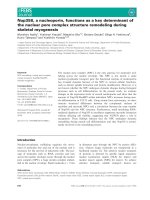
Fig. 1 Determining miR-217 expression in CRC tissues and cell lines and its clinical significance. (a) Relative expression level of miR-217 in human CRC
tissues (n = 50) and CRN tissues (n = 50), examined by qRT-PCR. CRC: colorectal cancer tissues; CRN: matched adjacent noncancerous colorectal tissues.
(b) The relative miR-217 expression level in six CRC cell lines compared with the normal colorectal cell line NCM460. The average gene expression from
NCM460 was appointed as 1. (c) Kaplan-Meier survival curve for CRC patients with miR-217-high (n = 24) and miR-217-low (n = 26) character. P value
was obtained by a log-rank test. *P < 0.05, **P < 0.01
Table 1 The relationship between miR-217 expression and
clinicopathologic characteristics in CRC patients
miR-217 expression
High
(n = 24)
Low
(n = 26)
Total
(n = 50)
P value
≤60
9
11
20
0.729
>60
15
15
30
Parameters
Age (y)
Gender
Female
7
14
21
Male
17
12
29
≤2
11
9
20
>2
13
17
30
0.077
Tumor size (cm)
0.412
using a strategy of transient transfection with miR-217
mimics. As shown in Fig. 2a, after transfection with miR217 mimics, a 19.46-fold and 14.89-fold increase in miR217 expression was observed in SW480 and SW620 cells,
respectively. CCK8 assay showed that the proliferation rate
of SW480 and SW620 cells were both significantly repressed after transfection with miR-217 mimics (Fig. 2b).
Moreover, the colonies formed by cells transfected with
miR-217 mimics were obviously fewer in number and
smaller in size than those formed by the control cells
(Fig. 2c).
These findings were confirmed in a CRC xenograft
model in vivo. Xenografted tumors in mice inoculated
with LV-miR-217-infected SW480 cells grew much more
slowly than those in mice inoculated with the LV-miRNC (Fig. 2d). qRT-PCR analysis showed that miR-217
expression levels were obviously increased in LV-miR217-infected tumors compared with those in the control
Tumor differentiation
Well/moderate
18
12
30
Poor
6
14
20
I + II
12
12
24
III + IV
12
14
26
Positive
11
13
24
Negative
13
13
26
0.038*
Table 2 Multivariate analysis of factors associated with overall
survival in CRC patients
TNM stage
Multivariate analysis
0.786
Lymph node metastasis
0.768
Distant metastasis
Positive
7
7
14
Negative
17
19
36
Positive
10
12
22
Negative
14
14
28
0.860
Vascular infiltration
*Statistically significant (P < 0.05)
0.749
Variable
HR (95 % CI)
P value
Age
1.001 (0.966-1.038)
0.936
Gender
1.296 (0.448-3.749)
0.632
Tumor size
1.583 (0.569-4.404)
0.379
Tumor differentiation
1.646 (0.578-4.683)
0.350
TNM stage
0.132 (0.028-0.623)
0.010*
Lymph node metastasis
0.901 (0.355-2.292)
0.828
Distant metastasis
13.508 (2.770-65.864)
0.001*
miR-217
0.312 (0.111-0.877)
0.027*
AEG-1
1.228 (0.419-3.594)
0.708
HR, hazard ratio; CI, confidence interval
*Statistically significant (P < 0.05)
Wang et al. BMC Cancer (2015) 15:437
Page 5 of 11
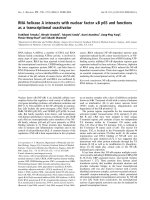
Fig. 2 MiR-217 inhibits the growth of CRC cell lines in vitro and in vivo. (a) The relative expression level of miR-217 when transfected with miR217 mimics and mimics NC measured by qRT-PCR. The average miRNA expression from mimics NC was appointed as 1. (b) The proliferation curve
of SW480 and SW620 cells after transfected with miR-217 mimics by CCK8 assay. (c) Assessment of colony formation when upregulation of miR217 expression. (d) The effects of overexpression of miR-217 on xenograft tumor growth in mice. Tumor growth curves were drew by measuring
the subcutaneous tumor volumes every 4 days. Data are presented as mean ± SD. (e) Relative miR-217 expression level in excised tumors.
*P < 0.05, **P < 0.01
tumors (Fig. 2e). These results indicate that miR-217
suppressed proliferation of CRC cell lines both in vitro
and in vivo.
MiR-217 induced apoptosis and led to cell cycle arrest in
CRC cell lines
To elucidate the mechanism by which miR-217 expression affects cell proliferation, flow cytometry was
employed to analyze the effects of miR-217 overexpression on CRC cell line apoptosis and cell cycle progression. As shown in Fig. 3a, the total apoptosis rate in cells
transfected with miR-217 mimics was significantly increased compared with that in cells transfected with NC
mimics (SW480, 6.16 ± 0.31 % vs. 3.44 ± 0.57 %, P < 0.01;
SW620, 19.93 ± 0.52 % vs. 9.77 ± 0.45 %, P < 0.01). Moreover, for cell cycle analysis, the proportion of miR-217
mimic-transfected cells in the G0/G1 phase increased
compared with that of the controls (SW480, 81.16 ±
0.06 % vs. 72.78 ± 0.71 %, P < 0.01; SW620, 66.94 ± 0.91 %
vs. 56.69 ± 0.70 %, P < 0.01; Fig. 3B). These results demonstrate that ectopic expression of miR-217 promotes apoptosis and G0/G1 phase arrest.
MiR-217 suppressed the CRC cell invasive activity
The effect of miR-217 on cell invasive capability was
investigated in transwell experiments. The numbers
of invading cells on the underside of the membrane
were significantly reduced both in SW480 cells
Wang et al. BMC Cancer (2015) 15:437
Page 6 of 11
Fig. 3 Overexpression of miR-217 enhances apoptosis and promotes G0/G1 phase arrest in CRC cells. (a) Flow cytometry analysis showed that
after transfection with miR-217 mimics and mimics NC, the apoptosis rates of both SW480 and SW620 cells were markedly increased. (b) Cell
cycle distribution assay was also applied using flow cytometry and treated with mimics as mentioned above. The histogram showed that miR-217
induced cell cycle arrest at G0/G1 phase. **P < 0.01
(Fig. 4a, P < 0.01) and SW620 cells (Fig. 4B, P < 0.01)
when transfected with miR-217 mimics compared to
those transfected with NC mimics. These results imply
that miR-217 participates in the regulation of the CRC cell
invasiveness.
MiR-217 was involved in changes in the expression of
molecules associated with invasion, cell cycle and
apoptosis
The expression of the related proteins, MMP-2, MMP-9,
cyclinD1 and Bcl-2 was significantly downregulated in
cells transfected with miR-217 mimics, whereas the
expression of Bax was increased in both SW480 and
SW620 cells (Fig. 4c).
MiR-217 suppressed AEG-1 expression by binding to its 3′
UTR sequence
To clarify the underlying molecular mechanism of the
suppressive effects of miR-217 on the proliferation and
invasive capacity of CRC cells, we used bioinformatics
methods (TargetScan, microRNA and miRDB) to search
for potential target genes of miR-217. All prediction analyses indicated that AEG-1 is a potential target of miR217. We then cloned the 3′UTR of AEG-1 containing
wild-type or mutant seed-sequence-recognizing sites
Wang et al. BMC Cancer (2015) 15:437
Page 7 of 11
Fig. 4 Ectopic miR-217 expression inhibits invasion of SW480 and SW620 cells. (a) Transwell method was applied to assess SW480 cells’ invasion ability
after transfected with miR-217 mimics and negative control. (b) The invasiveness of SW620 cells was evaluated as mentioned above. Statistics analysis
was performed by counting the stained cells that invaded to the lower chamber. All measurements were repeated three times in duplicate. (c)
Western blot analysis showed the expression levels of invasion related molecules MMP2 and MMP9, cell cycle related protein cyclinD1, and apoptosis
associated protein Bax and Bcl-2 after overexpression of miR-217.**P < 0.01
into a dual-luciferase reporter (Fig. 5a). After cotransfection of SW480 cells with miR-217 mimics or NC mimics
and AEG-1-UTR-WT or AEG-1-UTR-MUT plasmids,
luciferase activity was analyzed. Our results showed that
miR-217 significantly decreased the relative luciferase
activity in the reporter containing the wild-type 3′UTR,
whereas the luciferase activity of the mutant was unaffected (Fig. 5b). We further evaluated the expression
levels of AEG-1 mRNA and protein after modulation of
miR-217 expression. As shown in Fig. 5c, transfection of
CRC cells with miR-217 mimics led to a remarkable
downregulation in AEG-1 mRNA and protein levels. In
contrast, treatment with the miR-217 inhibitor caused
an increase in AEG-1 mRNA and protein expression
(Fig. 5d). These data indicate that miR-217 directly targets its predicted AEG-1 seed region.
The correlation between miR-217 and AEG-1 expression
in colorectal tissue samples
We further analyzed the relationship between miR-217
and AEG-1 expression in colorectal tissue samples. First,
we measured the AEG-1 mRNA levels in CRC tissues
and the corresponding adjacent normal tissues by qRTPCR. As shown in Fig. 6a, AEG-1 expression was much
higher in CRC tissues than in the corresponding CRN
tissues (P < 0.01). Interestingly, Pearson correlation analysis revealed an obvious inverse correlation was observed between miR-217 and AEG-1 expression both in
CRC tissues (r = −0.3457, P < 0.05) and in CRN tissues
(r = −0.2944, P < 0.05) (Fig. 6b).
The effect of AEG-1 expression on the survival of CRC
patients
Kaplan–Meier analysis indicated that there were no significant differences in the median survival of CRC patients with either low or high AEG-1 expression
(Additional file 2: Figure S1).
Silencing of AEG-1 inhibits malignant behavior of
colorectal cancer cells
To further investigate the role of AEG-1 in CRC cells,
AEG-1 siRNA was used to knockdown AEG-1 expression. AEG-1 expression was greatly decreased at both
the mRNA and protein levels (Additional file 3: Figure
S2A) after transfection with siRNA-AEG1. Similar to
overexpression of miR-217, AEG-1 silencing markedly
suppressed cell proliferation, colony formation, and invasive capacity, while G0/G1 arrest, and apoptosis were
promoted (Additional file 3: Figure S2b-f ).
Restoration of AEG-1 expression contributed to the
reversal of malignant behavior in SW620 cells
To confirm the role of AEG-1 in the anti-cancer effects
of miR-217 in CRC cells, we restored AEG-1 expression by AEG-1 plasmid transfection after transfection
with miR-217 mimics. As shown in Additional file 4:
Wang et al. BMC Cancer (2015) 15:437
Page 8 of 11
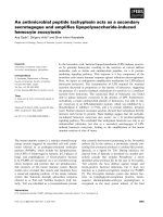
Fig. 5 MiR-217 directly targets AEG-1 in SW480 cells. (a) Predicted binding site of human miR-217 to the 3’UTR of AEG-1 by TargetScan. (Top
panel) The mutation of miR-217 binding site in the 3’UTR of AEG-1. (b) The reporter plasmids containing wild-type or mutant 3’UTR of AEG-1 was
co-transfected with miR-217 mimics or mimics NC. The assay was performed twice in triplicate. The relative luciferase activity was obtained by
Firefly luciferase activity normalized against Renilla luciferase activity. (c) The effects of overexpression of miR-217 on AEG-1 expression at mRNA
level and protein level. (d) The effects of inhibition of miR-217 on AEG-1 expression at mRNA level and protein level. *P < 0.05, **P < 0.01
Figure S3, overexpression of both miR-217 and
AEG-1 in SW620 cells caused no significant effects
on cell proliferation, invasive capacity, cell cycle and
apoptosis.
Discussion
MiRNAs are known to be play a key role in tumorigenesis as a result of their involvement in many cellular processes including cell proliferation, differentiation,
apoptosis and invasion [29, 30]. In the present study, we
focused on miR-217, which is abnormally expressed in a
variety of cancer types [8–10, 12]. To date, the evidence
for aberrant expression of miR-217 in CRC has been obtained in microarray studies. In our study, qRT-PCR
analysis demonstrated that miR-217 was significantly
downregulated in CRC tissue samples and cancer cell
lines. Furthermore, our study revealed, for the first time,
the involvement of miR-217 in tumorigenesis through
targeting AEG-1 targeting and that decreased miR-217
expression correlated with poor prognosis in patients
with CRC. These findings implicate miR-217 as a novel
prognostic marker in CRC.
Wang et al. BMC Cancer (2015) 15:437
Page 9 of 11
Fig. 6 AEG-1 is upregulated in CRC tissues and negatively correlated with the expression level of miR-217 in both CRC and CRN tissue samples.
(a) Upregulation of AEG-1 was observed in CRC tissue samples compared with that in adjacent CRN ones by qRT-PCR. (b) The analysis (Pearson’s
correlation) of relationship between expression levels of AEG-1 and miR-217 in CRC and CRN tissues, respectively. **P < 0.01
Moreover, analysis of clinical data indicated that reduced expression of miR-217 in CRC patients correlated
with poor tumor differentiation. In addition, Cox’s
multivariate analysis indicated that miR-217 expression,
TNM stage and distant metastasis act as an independent
factor in the prediction of overall survival among patients with CRC.
In this study, we also showed, for the first time, that
overexpression of miR-217 significantly repressed CRC
cell proliferation, colony formation, and induced G0/G1
cell cycle arrest and apoptosis. Moreover, our in vivo
studies confirmed that miR-217 overexpression remarkably suppressed CRC xenograft tumor growth in nude
mice. These results imply that miR-217 acts as an inhibitor of colorectal tumorigenesis.
Metastasis, one of the most critical hallmarks of cancer, is the leading cause of cancer-related deaths worldwide, particular in CRC [31, 32]. Accumulating evidence
demonstrates the close correlation of invasive capacity
and metastasis with miRNAs, such as miR-124 in nasopharyngeal carcinoma [33], miR-153 in CRC [34] and
miR-335 in lung cancer [35]. This evidence elucidating
the role of miRNAs in CRC metastasis might represent
the basis of a new therapeutic approach for CRC. The
clinical outcomes in the patients in this study revealed
that the expression level of miR-217 was closely correlated with CRC distant metastasis and also acted as an
independent prognostic factor in patients with CRC. It is
well-known that invasive tumors exist within a complex
microenvironment composed of extracellular matrix
(ECM) proteins, which play important roles in tumor invasion and metastasis [36]. Thus, matrigel invasion assays were performed in our study to mimic this
environment. The results showed that after overexpression of miR-217, the invasion capability of CRC cells
was significantly reduced, indicating the involvement of
miR-217 in CRC invasion and metastasis. Thus, it can
be hypothesized that restoration of miR-217 in CRC
might be a new therapeutic approach in CRC, especially
in CRC with distant metastasis.
The effects of miRNAs are largely dependent on their
regulation of the expression of many cancer-related
genes through post-transcriptional repression [37]. Using
bioinformatics analysis, we found that miR-217 targeted
multiple cancer-related genes that have been reported to
have a close link with cancers, such as KRAS (pancreatic
cancer) [8], E2F3 (hepatocellular carcinoma) [9], and
DACH1 (breast cancer) [38]. Interestingly, in this study,
AEG-1 was predicted to be one of the target genes of
miR-217. AEG-1 expression is frequently increased in
multiple cancers including CRC [21–23] and plays a critical role in oncogenic transformation and angiogenesis,
which are essential to tumor cell development, growth,
and metastatic progression [39–41]. These studies provide important insights and a unique perspective on this
multifunctional oncogene. In the current study, we evaluated the prognostic value of AEG-1 in CRC patients.
Although the survival analysis showed no significant difference between the AEG-1-low and AEG-1-high
groups, the median survival time was longer in the patients with low AEG-1 expression. Moreover, knockdown of AEG-1 repressed cell growth and invasion,
induced G0/G1 arrest and apoptosis, which was similar
to the effects of miR-217 overexpression. Thus, the results of our study indicate that AEG-1 acts as a tumor
promoter in CRC.
We next used dual-luciferase assays to determine
whether miR-217 binds directly to the 3′UTR of AEG-1
mRNA. Ectopic expression of miR-217 resulted in significant AEG-1 downregulation at both the mRNA and
protein levels, whereas miR-217 silencing led to restoration of AEG-1 expression. Furthermore, the expression
level of AEG-1 was inversely correlated with the miR217 expression in both CRC and CRN tissues. Therefore,
the results indicate that decreased AEG-1 expression
represents a mechanism by which miR-217 plays a role
Wang et al. BMC Cancer (2015) 15:437
in the progression of cancer. To further clarify this
point, we performed a rescue experiment which demonstrated that AEG-1 overexpression significantly reversed
miR-217-induced apoptosis, cell cycle arrest, proliferative inhibition and invasive suppression of SW620 cells.
However, the subcutaneous xenograft model in our
study cannot sufficiently represent clinical CRC, especially with regard to metastasis [42]. The present study
demonstrates that miR-217 remarkably represses the invasive ability of CRC cells in vitro; therefore, further investigations in a metastasis model are required to clarify
the effects of miR-217 on invasion and metastasis of
CRC in vivo.
Conclusions
In this study we show that miR-217 is significantly
downregulated in CRC and that decreased miR-217 expression levels indicate poor prognosis of CRC patients.
In addition, our results indicate that miR-217 may suppress the tumorigenesis and aggressiveness of CRC
through directly targeting AEG-1. Importantly, our findings implicate miR-217 as a prognostic marker and potential target for miRNA-based CRC therapy.
Additional files
Additional file 1: Table S1. Primer sequences of genes. Table S2.
Oligonucleotide sequences for transfection. Table S3. Western Blot
primary antibodies.
Additional file 2: Figure S1. The effect of AEG-1 expression level on
survival of CRC patients. Kaplan-Meier survival curve for CRC patients with
AEG-1-high (n = 26) and AEG-1-low (n = 24) character. P value was
obtained by a log-rank test.
Additional file 3: Figure S2. Knockdown of AEG-1 inhibit malignant
biological behavior in SW480 and SW620 cell lines. (A) AEG-1 expression was
downregulated after treated with siRNA-AEG-1 determined by qRT-PCR (left)
and Western blot analysis (right). (B) Inhibition of AEG-1 expression repressed
cell proliferation of SW480 and SW620 cells. (C) Silencing of AEG-1 led to
repression of colony formation. (D) Knockdown of MAP4K4 weakened the
ability of cell invasion. (E) Cell cycle was examined by flow cytometry. Silencing
of MAP4K4 in SW480 and SW620 cells led to G0/G1 arrest. (F) The percentage
of apoptotic cells increased through downregulation of AEG-1 in SW480 and
SW620 cell lines. *P < 0.05, **P < 0.01.
Additional file 4: Figure S3. Rescue of miR-217 ectopic expression
effects by simultaneous overexpression of AEG-1. (A) Cell proliferation
detected in SW620 cells at 1, 2, 3, 4 and 5 days after transfection. (B)
Results of SW620 cell invasion across an 8-μm pore size membrane with
Matrigel. (C) Cell cycle determined in SW620 cells 48 h after transfection
by Propidium-iodide staining flow cytometry. (D) Cell apoptosis detected
by Annexin-V/propidium iodide combined labeling flow cytometry in
SW620 cells 48 h after transfection. *P < 0.05, **P < 0.01.
Abbreviations
CRC: colorectal cancer; CRN: colorectal normal; qRT-PCR: quantitative realtime reverse transcription polymerase chain reaction; UTR: untranslated
region; GAPDH: glyceraldehyde-3-phosphate dehydrogenase; CCK8: cell
counting kit-8; NC: negative control.
Competing interests
The authors declare that we have no competing interests.
Page 10 of 11
Authors' contributions
Conception and design: WB, SZL, WS
Development of methodology: WB, JKW, GZD
Acquisition of data: YY, ZJZ, SC
Analysis and interpretation of data: WB, ZG, WCY
Writing the manuscript: WB, SZL
Administrative, technical or material support: JKW, ZG, WCY, YYC
Study supervision: YYJ, WS
All authors read and approved the final manuscript.
Acknowledgments
This study was supported by grants from the National Natural Science
Foundation of China (81372290, 81372291), Peking University People’s
Hospital Funds (RDB 2013–15).
Author details
1
Department of Gastroenterological Surgery, Peking University People’s
Hospital, No.11 Xizhimen South Street, Xicheng District, Beijing 100044, P.R.
China. 2Pancreatic Disease Institute, Union Hospital, Tongji Medical College,
Huazhong University of Science and Technology, Wuhan, People’s Republic
of China.
Received: 21 October 2014 Accepted: 14 May 2015
References
1. Ferlay J, Shin HR, Bray F, Forman D, Mathers C, Parkin DM. Estimates of
worldwide burden of cancer in 2008: GLOBOCAN 2008. Int J Cancer.
2010;127(12):2893–917.
2. Akagi Y, Kinugasa T, Adachi Y, Shirouzu K. Prognostic significance of isolated
tumor cells in patients with colorectal cancer in recent 10-year studies. Mol
Clin Oncol. 2013;1(4):582–92.
3. Guo Y, Xu F, Lu T, Duan Z, Zhang Z. Interleukin-6 signaling pathway in
targeted therapy for cancer. Cancer Treat Rev. 2012;38(7):904–10.
4. Jemal A, Bray F, Center MM, Ferlay J, Ward E, Forman D. Global cancer
statistics. CA Cancer J Clin. 2011;61(2):69–90.
5. Pichler M, Ress AL, Winter E, Stiegelbauer V, Karbiener M, Schwarzenbacher
D, et al. MiR-200a regulates epithelial to mesenchymal transition-related
gene expression and determines prognosis in colorectal cancer patients. Br
J Cancer. 2014;110(6):1614–21.
6. Chen DL, Wang ZQ, Zeng ZL, Wu WJ, Zhang DS, Luo HY, et al. Identification
of MicroRNA-214 as a negative regulator of colorectal cancer liver metastasis
by way of regulation of fibroblast growth factor receptor 1 expression.
Hepatology. 2014;60(2):598–609.
7. Liu S, Sun X, Wang M, Hou Y, Zhan Y, Jiang Y, Liu Z, Cao X, Chen P, Liu Z
et al.: A microRNA 221- and 222-Mediated Feedback Loop, via PDLIM2,
Maintains Constitutive Activation of NFkappaB and STAT3 in Colorectal
Cancer Cells. Gastroenterology 2014 .
8. Zhao WG, Yu SN, Lu ZH, Ma YH, Gu YM, Chen J. The miR-217 microRNA functions
as a potential tumor suppressor in pancreatic ductal adenocarcinoma by
targeting KRAS. Carcinogenesis. 2010;31(10):1726–33.
9. Su J, Wang Q, Liu Y. miR-217 inhibits invasion of hepatocellular carcinoma cells
through direct suppression of E2F3. Mol Cell Biochem. 2014;392(1–2):289–96.
10. Li H, Zhao J, Zhang JW, Huang QY, Huang JZ, Chi LS, et al. MicroRNA-217,
down-regulated in clear cell renal cell carcinoma and associated with lower
survival, suppresses cell proliferation and migration. Neoplasma. 2013;60(5):511–5.
11. Nishioka C, Ikezoe T, Yang J, Nobumoto A, Tsuda M, Yokoyama A.
Downregulation of miR-217 correlates with resistance of Ph(+) leukemia
cells to ABL tyrosine kinase inhibitors. Cancer Sci. 2014;105(3):297–307.
12. de Yebenes VG, Bartolome-Izquierdo N, Nogales-Cadenas R, Perez-Duran P,
Mur SM, et al. miR-217 is an oncogene that enhances the germinal center
reaction. Blood. 2014;124(2):229–39.
13. MicroRNA 217 Modulates Endothelial Cell Senescence via Silent Information
Regulator 1. Circulation 2009 :1524–1532.
14. Stuckenholz C, Lu L, Thakur P, Kaminski N, Bahary N. FACS-assisted microarray
profiling implicates novel genes and pathways in zebrafish gastrointestinal
tract development. Gastroenterology. 2009;137(4):1321–32.
15. Su ZZ, Kang DC, Chen Y, Pekarskaya O, Chao W, Volsky DJ, et al. Identification
and cloning of human astrocyte genes displaying elevated expression after
infection with HIV-1 or exposure to HIV-1 envelope glycoprotein by rapid
subtraction hybridization. RaSH Oncogene. 2002;21(22):3592–602.
Wang et al. BMC Cancer (2015) 15:437
16. Li J, Yang L, Song L, Xiong H, Wang L, Yan X, et al. Astrocyte elevated gene-1 is
a proliferation promoter in breast cancer via suppressing transcriptional factor
FOXO1. Oncogene. 2009;28(36):3188–96.
17. Lee SG, Jeon HY, Su ZZ, Richards JE, Vozhilla N, Sarkar D, et al. Astrocyte
elevated gene-1 contributes to the pathogenesis of neuroblastoma.
Oncogene. 2009;28(26):2476–84.
18. He XX, Chang Y, Meng FY, Wang MY, Xie QH, Tang F, et al. MicroRNA-375
targets AEG-1 in hepatocellular carcinoma and suppresses liver cancer cell
growth in vitro and in vivo. Oncogene. 2012;31(28):3357–69.
19. Liu X, Wang D, Liu H, Feng Y, Zhu T, Zhang L, et al. Knockdown of astrocyte
elevated gene-1 (AEG-1) in cervical cancer cells decreases their invasiveness,
epithelial to mesenchymal transition, and chemoresistance. Cell Cycle.
2014;13(11):1702–7.
20. Li G, Wang Z, Ye J, Zhang X, Wu H, Peng J, Song W, Chen C, Cai S, He YL
et al.: Uncontrolled inflammation induced by AEG-1 promotes gastric cancer
and is associated with poor prognosis. Cancer Res 2014 .
21. Gnosa S, Shen YM, Wang CJ, Zhang H, Stratmann J, Arbman G, et al.
Expression of AEG-1 mRNA and protein in colorectal cancer patients and
colon cancer cell lines. J Transl Med. 2012;10:109.
22. Song H, Li C, Li R, Geng J. Prognostic significance of AEG-1 expression in
colorectal carcinoma. Int J Colorectal Dis. 2010;25(10):1201–9.
23. Huang S, Wu B, Li D, Zhou W, Deng G, Zhang K, et al. Knockdown of
astrocyte elevated gene-1 inhibits tumor growth and modifies microRNAs
expression profiles in human colorectal cancer cells. Biochem Biophys Res
Commun. 2014;444(3):338–45.
24. Song H, Tian Z, Qin Y, Yao G, Fu S, Geng J. Astrocyte elevated gene-1
activates MMP9 to increase invasiveness of colorectal cancer. Tumour Biol.
2014;35(7):6679–85.
25. Song HT, Qin Y, Yao GD, Tian ZN, Fu SB, Geng JS. Astrocyte elevated gene-1
mediates glycolysis and tumorigenesis in colorectal carcinoma cells via
AMPK signaling. Mediators Inflamm. 2014;2014:287381.
26. Hwang JH, Voortman J, Giovannetti E, Steinberg SM, Leon LG, Kim YT, et al.
Identification of microRNA-21 as a biomarker for chemoresistance and
clinical outcome following adjuvant therapy in resectable pancreatic cancer.
PLoS One. 2010;5(5), e10630.
27. Zhao G, Wang B, Liu Y, Zhang JG, Deng SC, Qin Q, et al. MiRNA-141,
downregulated in pancreatic cancer, inhibits cell proliferation and
invasion by directly targeting MAP4K4. Mol Cancer Ther.
2013;12(11):2569–80.
28. Zhao G, Zhang JG, Liu Y, Qin Q, Wang B, Tian K, et al. miR-148b functions as
a tumor suppressor in pancreatic cancer by targeting AMPKalpha1. Mol
Cancer Ther. 2013;12(1):83–93.
29. Bartel DP. MicroRNAs: genomics, biogenesis, mechanism, and function. Cell.
2004;116(2):281–97.
30. Winter J, Jung S, Keller S, Gregory RI, Diederichs S. Many roads to maturity:
microRNA biogenesis pathways and their regulation. Nat Cell Biol.
2009;11(3):228–34.
31. De Roock W, De Vriendt V, Normanno N, Ciardiello F, Tejpar S. Mutations:
implications for targeted therapies in metastatic colorectal cancer. Lancet
Oncol. 2011;12(6):594–603.
32. Carpizo DR, D'Angelica M. Liver resection for metastatic colorectal cancer in
the presence of extrahepatic disease. Lancet Oncol. 2009;10(8):801–9.
33. Peng XH, Huang HR, Lu J, Liu X, Zhao FP, Zhang B, et al. MiR-124 suppresses
tumor growth and metastasis by targeting Foxq1 in nasopharyngeal
carcinoma. Mol Cancer. 2014;13(1):186.
34. Zhang L, Pickard K, Jenei V, Bullock MD, Bruce A, Mitter R, et al. miR-153
supports colorectal cancer progression via pleiotropic effects that enhance
invasion and chemotherapeutic resistance. Cancer Res. 2013;73(21):6435–47.
35. Gong M, Ma J, Guillemette R, Zhou M, Yang Y, Yang Y, et al. miR-335 inhibits
small cell lung cancer bone metastases via IGF-IR and RANKL pathways. Mol
Cancer Res. 2014;12(1):101–10.
36. Leeman MF, Curran S, Murray GI. New insights into the roles of matrix
metalloproteinases in colorectal cancer development and progression.
J Pathol. 2003;201(4):528–34.
37. Peter ME. Targeting of mRNAs by multiple miRNAs: the next step.
Oncogene. 2010;29(15):2161–4.
38. Zhang Q, Yuan Y, Cui J, Xiao T, Jiang D. MiR-217 Promotes tumor proliferation
in breast cancer via targeting DACH1. J Cancer Educ. 2015;6(2):184–91.
39. Wang YP, Liu IJ, Chiang CP, Wu HC. Astrocyte elevated gene-1 is associated
with metastasis in head and neck squamous cell carcinoma through p65
phosphorylation and upregulation of MMP1. Mol Cancer. 2013;12(1):109.
Page 11 of 11
40. Kochanek DM, Wells DG. CPEB1 regulates the expression of MTDH/AEG-1
and glioblastoma cell migration. Mol Cancer Res. 2013;11(2):149–60.
41. Srivastava J, Siddiq A, Emdad L, Santhekadur PK, Chen D, Gredler R, et al.
Astrocyte elevated gene-1 promotes hepatocarcinogenesis: novel insights
from a mouse model. Hepatology. 2012;56(5):1782–91.
42. Fu X, Guadagni F, Hoffman RM. A metastatic nude-mouse model of human
pancreatic cancer constructed orthotopically with histologically intact
patient specimens. Proc Natl Acad Sci U S A. 1992;89(12):5645–9.
Submit your next manuscript to BioMed Central
and take full advantage of:
• Convenient online submission
• Thorough peer review
• No space constraints or color figure charges
• Immediate publication on acceptance
• Inclusion in PubMed, CAS, Scopus and Google Scholar
• Research which is freely available for redistribution
Submit your manuscript at
www.biomedcentral.com/submit