SNAI2/SLUG and estrogen receptor mRNA expression are inversely correlated and prognostic of patient outcome in metastatic non-small cell lung cancer
Bạn đang xem bản rút gọn của tài liệu. Xem và tải ngay bản đầy đủ của tài liệu tại đây (460.18 KB, 7 trang )
Atmaca et al. BMC Cancer (2015) 15:300
DOI 10.1186/s12885-015-1310-1
RESEARCH ARTICLE
Open Access
SNAI2/SLUG and estrogen receptor mRNA
expression are inversely correlated and
prognostic of patient outcome in metastatic
non-small cell lung cancer
Akin Atmaca1*, Ralph W Wirtz2, Dominique Werner3, Kristina Steinmetz3, Silke Claas2, Wolfgang M Brueckl4,
Elke Jäger1 and Salah-Eddin Al-Batran3
Abstract
Background: Epithelial-mesenchymal transition (EMT) is involved in important malignant features of cancer cells,
like invasion, metastatic potential, anti-apoptotic and stem-cell like phenotypes. Among several transcription factors,
SNAI2/SLUG is supposed to play an essential role for EMT.
Methods: Paraffin embedded tumor samples from 63 patients with metastatic non-small cell lung cancer, enrolled
in a randomized phase II trial, were prospectively collected, 53 samples qualified for further analysis. Automated
RNA extraction from paraffin and RT-quantitative PCR was used for evaluation of SNAI2/SLUG, estrogen receptor 1
(ESR1) and matrix-metalloproteinases (MMP) mRNA expression.
Results: Clinical features like age, gender, performance status, histological subtype and stage were similarly
distributed among SNAI2/SLUG positive and negative patients. SNAI2/SLUG was significantly, inversely correlated
with ESR1 mRNA expression (p < 0.0001). In contrast, MMP2 (p = 0.387), MMP7 (p = 0.396) and MMP9 mRNA
expression (p = 0.366) did not correlate with SNAI2/SLUG. Patients with high SNAI2/SLUG expression (grouped by
median expression) had a worse outcome. Median overall survival in patients with high SNAI2/SLUG expression was
5.7 months versus 11.6 months with low SNAI2/SLUG expression (p = .038). Inversely, patients with high ESR1
expression (grouped by median expression) had an improved median OS with 10.9 months vs. 5.0 months in the
low expression group (p = .032). In multivariate analysis, SNAI2/SLUG2 (p = .022) and ESR1 (p = .017) separately were
independent prognostic factors for survival.
Conclusion: SNAI2/SLUG is prognostic of patients’ outcome. The strong inverse correlation with ESR1 indicates a
significant impact of estrogen receptor pathway regarding these malignant features.
Keywords: SNAI2, SLUG, Estrogen receptor, NSCLC, Metastatic, Prognostic, Survival
Background
Lung cancer is the leading cause of death among all malignant diseases worldwide. In the majority of patients
(about 70%), the disease is diagnosed in an advanced,
non-resectable stage with a very poor outcome. The
prognosis is highly associated with the metastatic
* Correspondence:
1
Department of Hematology and Oncology, Krankenhaus Nordwest,
UCT-University Cancer Center, Steinbacher Hohl 2-26, 60488 Frankfurt am
Main, Germany
Full list of author information is available at the end of the article
behavior of the tumor. Metastatic spread is a complex
process of molecular and phenotypical changes of
tumor cells. In this process the epithelial-mesenchymal
transition (EMT) seems to play a crucial role. During
EMT cells reduce intercellular adhesions, lose polarity
and acquire a fibroblastoid phenotype with high motility
and invasive properties [1]. This process is characterized
by downregulation of E-cadherin and other epithelial
molecules associated with cell adhesion. In parallel, an
up-regulation of mesenchymal proteins, like vimentin
and an increase of secretion of proteolytic enzymes, like
© 2015 Atmaca et al.; licensee BioMed Central. This is an Open Access article distributed under the terms of the Creative
Commons Attribution License ( permits unrestricted use, distribution, and
reproduction in any medium, provided the original work is properly credited. The Creative Commons Public Domain
Dedication waiver ( applies to the data made available in this article,
unless otherwise stated.
Atmaca et al. BMC Cancer (2015) 15:300
matix-metalloproteinases (MMP), can be observed,
contributing to the increase of cell motility, invasiveness and metastatic potential [1,2]. In conclusion, EMT
seems to play a key role in the progression of tumors
towards invasion and metastasis. Among transcription
factors inducing EMT and down regulation of Ecadherin, which represents the hallmark of EMT, the
snail family of zing finger transcription factors, like
SNAIL (SNAI1) and SLUG (SNAI2) play a prominent
role [3]. Overexpression of SNAI2/SLUG can be observed in a variety of different cancers and seems to
be associated with poor outcome [4,5]. In particular,
SNAI2/SLUG is also a negative prognostic factor for
relapse and overall survival in resectable, early stage
lung cancer [6,7]. In breast cancer cell lines, Ye et al.
[8] could show that SNAI2/SLUG is suppressed by
ligand-activation of estrogen receptor α (ERα). Several findings of this study indicate that SNAI2/SLUG
is an estradiol- responsive gene and ERα may play an
important role in EMT in breast cancer.
To further clarify the role of SNAI2/SLUG in lung
cancer and in particular in the advanced setting, this
study was conducted to examine the correlation with
hormone receptor expression as well as different MMP
along with the clinical outcome in Western patients
with metastatic NSCLC, enrolled in a randomized firstline chemotherapy trial.
Methods
Study population
For mRNA analysis, tumor biopsies of patients with
metastatic or advanced NSCLC enrolled in a randomized, multicenter first-line phase II trial and treated
with docetaxel and either cisplatin or oxaliplatin [9]
were used. These samples were prospectively collected
during this study. From a total of 88 randomized patients, tumor samples of 64 patients were available and
of those 53 samples qualified for sufficient mRNA extraction and gene expression analysis.
Patients gave informed consent for the study including sample collection and analysis. Approval of the
local ethic committees was obtained (leading ethics
committee: Landesärztekammer Hessen). Standards of
the International Conference on Harmonization World
Health Organization (WHO) Good Clinical Practice were
followed.
Sample preparation and RNA extraction
Formalin-fixed paraffin-embedded (FFPE) tissue samples obtained before the start of chemotherapy were collected. From each tumor block, a 5-μm section was
stained with hematoxylin–eosin (H&E) and revised by a
pathologist and two consecutive 10-μm sections were
cut on a standard microtome, placed into individual
Page 2 of 7
tubes, and stored at 4°C for ≤1 month until RNA extraction. Fully automated high-throughput RNA extraction
has been carried out similar to methods previously published [10] by using a fully automated XTRACT roboter
and extraction kits (STRATIFYER Molecular Pathology
GmbH, Germany).
Gene expression analysis using quantitative PCR
Expression of SLUG/SNAI2, MMP2, MMP7, MMP9,
estrogen receptor 1 (ESR1) and the normalization
(housekeeping) gene CALM2 were assessed by one-step
RT-quantitative PCR (qPCR). SuperScript ® III Platinum
® One-Step qRT-PCR System with ROX (Invitrogen,
Karlsruhe, Germany) was used according to the manufacturer’s instructions. Experiments were carried out on
a Stratagene Mx3005p (Agilent Technologies, Böblingen, Germany) with 30 min at 50°C, 2 min at 95°C
followed by 40 cycles of 15 s at 95°C and 30 s at 60°C.
The expression of the genes of interest was calculated
by using the ΔCt method. Cycle threshold (Ct) values,
which indicate the (interpolated) number of PCR cycles
until the fluorescence reached its threshold, were
determined. Ct values were normalized by subtracting
the Ct value of the housekeeping gene (CALM2) from
the Ct value of the target gene (ΔCT). RNA results were
then reported as 40- ΔCt values, which would correlate
proportionally to the mRNA expression level of the
target gene. For assessment of DNA contamination in
RNA preparations, a PAEP gene-specific qPCR without
preceding reverse transcription was carried out using the
reagents from the SuperScript III® Platinum® One-Step
qRT-PCR System with ROX and Taq DNA Polymerase. In
samples with a Ct value <35, the DNase I treatments were
repeated to prevent effects on bispecific PCR assays.
Stratagene human QPCR Reference total RNA (Stratagene,
Waldbronn, Germany) was used as positive control for
RTqPCR and human genomic DNA (Roche Diagnostics,
Basel, Switzerland) as positive control for qPCR. All PCR
assays were carried out in triplicate, and the mean of
triplicates was reported.
Statistics
The study was explorative. The median expression of
genes (SNAI2/SLUG, MMP2, MMP7, MMP9 and ESR1)
was used as an objective cut-off to distinguish high from
low expression. Associations between gene expression
values and clinicopathological data were compared and
calculated with chi-square-tests. Progression-free survival (PFS) was measured from the date of assignment
until disease progression or death of any cause. Overall
survival (OS) was measured from date of assignment
until death of any cause. Time-to-event curves were calculated by the Kaplan–Meier method and the log-rank
test was applied. The Cox regression model was used
Atmaca et al. BMC Cancer (2015) 15:300
Page 3 of 7
for the univariate and multivariate analyses. All P values
were two-sided with P values <0.05 indicating statistical
significance. Statistical analyses were performed with
WinSTAT software (Version 2009.1).
Results
SNAI2/SLUG and MMP mRNA expression
SNAI2/SLUG mRNA expression could be evaluated in
49 patients and ranged between 28.01 and 41.70 with a
median of 34.09 (ΔCt). There was no correlation of
SNAI2 expression with clinical characteristics like gender, performance status, stage, histological subtype,
number of metastatic sites or treatment, when patients
were grouped by the median or the 3rd quartile of
SNAI2 expression (Table 1).
ESR1 mRNA expression (n = 53) ranged between
28.72 and 39.2 with a median of 35.71. There was no
significant association between ESR1 mRNA expression
and clinical characteristics, although high ESR1 was
slightly more frequently observed in male vs. female patients (p = .27), and patients with liver (p = .14) and bone
metastases (p = .16) [11].
MMP2 could be evaluated in 51 patients and ranged
between 31.40 and 39.86 (median 36.78). There was no
correlation with clinical characteristics. Similar results
could be obtained for MMP7 expression (n = 52) which
ranged between 30.40 and 42.58 (median 34.98) and
MMP9 expression (n = 45) which ranged between 29.91
and 38.75 (median 33.79).
A highly significant inverse correlation could be observed between SNAI2/SLUG and ESR1 mRNA expression (p < 0.0001). In contrast, MMP2 (p = 0.3868), MMP7
(p = 0.3961) and MMP9 mRNA expression (p = 0.366) did
not correlate with SNAI2/SLUG (Table 2).
Table 1 Patient characteristics
SNAI2 median
Characteristic
Total (n = 49)
Low (%)
High (%)
Median (Range)
66(39–82)
66(39–82)
66(51–75)
Female
23
11
12
Male
26
14
12
Median
1
p-value
Age
Gender
0.778
ECOG PS
0-1
43
23
20
2
6
2
4
Adeno
31
13
18
0.417
Histology
Squamous cell
14
9
5
other
4
3
1
0.345
No. of organs involved
Median
≤1
19
10
9
n.s.
2
18
9
9
n.s.
≥3
12
6
6
n.s.
0.235
Stage
IIIB / IIIA
2
0
2
IV
47
25
22
Arm A (Cisplatin/Docetaxel)
23
13
10
Arm B (Oxaliplatin/Docetaxel)
26
12
14
ESR1 high
25
24
1
ESR1 low
24
1
23
Treatment
0.571
ESR1 status
<0.0001
Atmaca et al. BMC Cancer (2015) 15:300
Page 4 of 7
Table 2 Gene expression correlations
SNAI2-median
Characteristic
Low (%)
High (%)
p-value
<0.0001
ESR1 (n = 49)
high
24
1
low
1
23
high
10
14
low
14
10
MMP2 (n = 48)
0.387
MMP7 (n = 48)
high
14
10
low
11
14
high
13
9
low
9
12
0.396
MMP9 (n = 43)
0.366
Survival analysis
In line with previous works of our group [11], ESR1 expression was identified as a marker of favourable outcome in this patient group (n = 53) with a median OS of
10.9 vs. 5.0 months in ESR1 high vs. low patients, respectively (p = .032, HR 0.51). Grouped by the median
SNAI2/SLUG expression, OS was 5.7 vs. 11.6 months in
the SNAI2/SLUG high vs. low patients (p = .038, HR
0.52) (Figure 1A). When the 3rd quartile of SNAI2/
SLUG expression (37.75) was used for classification into
high vs. low patients, the differences in OS were even
stronger (median OS 4.6 vs 11.5 months, p = .0192, HR
0.45) (Figure 1B).
There was no correlation of different MMPs with survival, median OS was 8.3 vs. 10.5 months for MMP2
high vs. low (p = .431, HR 0.78), 8.3 vs. 9.8 months for
MMP7 high vs. low (p = .967, HR 1.01) and 9.8 vs.
10.1 months for MMP9 high vs. low (p = .341, HR 1.39).
Multivariate analysis
In the multivariate analysis containing the factors gender, age, performance status, histological subtype and
SNAI2/SLUG expression, only SNAI2/SLUG expression was significantly associated with OS (p = .022, HR
0.45) (Table 3). In this patient group, ESR1 was also significantly associated with survival in the multivariate
analysis (p = .015; HR .38), as previously published [11].
Discussion
EMT has emerged as a critical phenomenon in the carcinogenesis. There is growing evidence that EMT occurs
also during lung cancer development. In a series of adenocarcinomas and squamous cell carcinomas, Prudkin and
co-workers [12] showed that EMT phenotype was found
in most of the lung tumors in contrast to dysplastic
lesions or adjacent bronchial epithelium. Shintani et al.
[13] compared tissue specimens of patients with NSCLC
who received preoperative radiochemotherapy. They
observed an EMT marker expression increase in 40% of
patients and this correlated with poor outcome (disease
free-survival).
In stage I NSCLC, EMT markers such as Twist and
SNAI2/SLUG were associated with a worse overall survival and recurrence-free survival [7]. In resected
adenocarcinoma of the lung, Shih et al. [6] could confirm the negative prognostic effect of SNAI2/SLUG expression measured by mRNA on survival and relapse.
They also observed an increase of MMP2-mRNA expression in SNAI2/SLUG overexpressing tumors.
EMT in lung cancer is triggered by multiple intrinsic and extrinsic factors. One of the most important
characterized EMT-inducing oncogenic changes is
the K-RAS mutation [14,15]. Additionally, TGF-β
and hypoxia inducing factor-2α (HIF-2α) are known
EMT inducing intrinsic factors. Among extrinsic factors, hypoxia and inflammatory tumor microenvironment have to be mentioned.
Interestingly, tobacco smoking induced EMT through
HDAC –mediated down regulation of E-cadherin via
up regulation of SNAI2/SLUG [16].
In our study, we could confirm the negative prognostic effect of SNAI2/SLUG expression in advanced lung
cancer, indicating the prognostic effect of EMT. It has
to be noticed that in comparison to previously published data on early stage operable patients, our cohort
consisted of advanced or metastatic stages, so this
phenomenon seems not only be restricted to early stage
lung cancer.
In contrast to findings of Shih et al. and others groups
[17], we could not observe a correlation of SNAI2/
SLUG and MMP 2 in our cohort. Probably this could be
due to the limited patient number in our series. However, a difference in early stage and metastatic cancers
has to be considered as a potential explanation for this
observation.
One important finding of our study was the significant
inverse correlation of SNAI2/SLUG and ESR1. This in
line with results reported from breast cancer, where
SNAI2/SLUG expression was evaluated in different ERpositive and ER negative cell lines [18]. Estrogen
receptor-α directly repressed transcription of SNAI2/
SLUG by the formation of a complex of ligandactivated estrogen receptor-α, histone deacetylase 1
and nuclear receptor corepressor (N-CoR) [8].
This phenomenon can also be observed in ovarian
cancer. Park et al. [19] could show, that E-Cadherin
suppression and SNAI2/SLUG expression is mediated
by estradiol and estrogen receptor alpha in ovarian
cancer cell lines.
Atmaca et al. BMC Cancer (2015) 15:300
Page 5 of 7
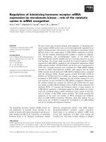
A Overall Survival (SNAi2/Median)
B Overall Survival (SNAI2/Q3)
Figure 1 Kaplan-Meier curves for overall survival (OS) grouped by median SLUG/SNAI2 expression (A) and by 3rd quartile of SLUG/SNAI2
expression (B) for patients with SLUG/SNAI2 high and low tumors. A: median OS (median SLUG/SNAI2 expression): 5.7 vs. 11.6 months, p = 0.038,
HR 0.52. B: median OS (3rd quartile): 4.6 vs. 11.5 months, p = 0.0192, HR 0.45.
Table 3 Multivariate analysis for overall survival
Covariate
P
HR
95% CI (HR)
Histology
0.31
0.64
0.27 - 1.52
Gender
0.28
0.68
0.33 – 1.37
Performance Status
0.96
0.97
0.35 – 2.67
Treatment
0.10
0.54
0.26 – 1.11
Age
0.60
0.80
0.34 – 1.85
SNAI2 expression
0.02
0.45
0.22 – 0.90
Our findings fit into the consistent overall picture
that a distinct subgroup of non-small cell lung cancer
(ESR1 high expression tumors) has certain similarity
and analogy to breast cancer, based on several epidemiologic and observational studies.
First, in a previous study [11] we could show that
ESR1 is an independent prognostic factor in metastatic
NSCLC similar to breast cancer.
Second, the metastatic pattern/ and behavior of
ESR1 positive lung cancer is similar to breast cancer,
where bone metastases are associated with estrogen receptor positivity [11].
Atmaca et al. BMC Cancer (2015) 15:300
Third, SNAI2/SLUG is significantly inversely correlated with ESR1 expression and prognostic in analogy
to breast cancer.
Furthermore, the strong inverse correlation of SNAI2/
SLUG with ESR1 underlines and validates the prognostic relevance of ESR1 in lung cancer. As SNAI2/SLUG is
one of the key factors for E-cadherin suppression and
for EMT, which represents a more aggressive phenotype
of cancer, the results seem reasonable.
With our data we cannot provide a proof that
SNAI2/SLUG expression is directly triggered by the estrogen pathway and we cannot rule out that the strong
inverse correlation of SNAI2/SLUG and ESR1 is determined by an independent pathway. However, the analogy to breast and ovarian cancer suggests that SNAI2/
SLUG is an ER responsive gene in lung cancer as well.
Our results would have two implications in NSCLC
patients. First, SNAI2/SLUG expression adds to the
prognostic factors known in NSCLC, making it meaningful to stratify according to SNAI2/SLUG in future
clinical trials. Additionally, it helps to identify patients
with poor prognosis, who may be candidates for more
aggressive therapies in the future. Second, based on
the strong inverse correlation with ESR1 expression,
SNAI2/SLUG expression and EMT in general should be
studied in response to antihormonal treatment in vitro and
in vivo.
Taken together, our data confirm that, as for breast
cancer, ESR1 expression in lung cancer is associated
with the lower levels of EMT Markers. Therefore, the
results warrant further evaluation of antihormonal
treatment in a subgroup of patients with lung cancer
(ESR1 high lung cancer) in analogy to ER/PR positive
breast cancer.
Conclusion
SNAI2/SLUG is prognostic of patients’ outcome. The
strong inverse correlation with ESR1 indicates a significant impact of estrogen receptor pathway regarding
these malignant features.
Competing interests
The authors declare no potential conflict of interest relevant to this article.
Ralph Wirtz and Silke Claas are employees of STRATIFYER Molecular
Pathology GmbH, Cologne, Germany. Additionally, Ralph Wirtz has stocks
and IP of STRATIFYER.
Authors’ contributions
AA carried out the statistical analysis and drafted the manuscript, RW carried
out the mRNA analysis, DW carried out the statistical analysis and the figure
and table preparation, KS carried out the sample preparation and study
coordination, SC carried out the mRNA analysis, WMB participated in the
study design and in the data interpretation, EJ participated in the design,
coordination of the study and data interpretation, SA designed and
coordinated the study and helped to draft the manuscript. All authors read
and approved the final manuscript.
Page 6 of 7
Acknowledgements
We thank the Institute of Clinical Cancer Research (IKF), Krankenhaus
Nordwest, University Cancer Center Frankfurt for providing a grant
supporting a part of the present study.
Author details
1
Department of Hematology and Oncology, Krankenhaus Nordwest,
UCT-University Cancer Center, Steinbacher Hohl 2-26, 60488 Frankfurt am
Main, Germany. 2STRATIFYER Molecular Pathology GmbH, Werthmannstraße
1, 50935 Cologne, Germany. 3Institute of clinical research (IKF) at
Krankenhaus Nordwest, UCT-University Cancer Center, Steinbacher Hohl 2-26,
60488 Frankfurt am Main, Germany. 4Department of Internal Medicine 3,
Klinikum Nürnberg, Prof.-Ernst-Nathan-Straße 1, 90419 Nuermberg, Germany.
Received: 12 November 2014 Accepted: 1 April 2015
References
1. Thiery JP. Epithelial-mesenchymal transitions in tumour progression. Nat Rev
Cancer. 2002;2:442–54.
2. Savagner PC. Leaving the neighborhood: molecular mechanisms involved
during eipthelial-mesenchymal transition. Bioessays. 2001;23:912–23.
3. de Herreros AG, Peiró S, Nassour M, Savagner P. Snail family regulation and
epithelial mesenchymal transitions in breast cancer progression. J Mammary
Gland Biol Neoplasia. 2010;2:135–47.
4. Cobaleda C, Pérez-Caro M, Vicente-Dueñas C, Sánchez-García I. Function
of the zinc-finger transcription factor SNAI2 in cancer and development.
Annu Rev Genet. 2007;41:41–61.
5. Alves CC, Carneiro F, Hoefler H, Becker KF. Role of the epithelial-mesenchymal
transition regulator Slug in primary human cancers. Front Biosci (Landmark Ed).
2009;14:3035–50.
6. Shih JY, Tsai MF, Chang TH, Chang YL, Yuan A, Yu CJ, et al. Transcription
repressor slug promotes carcinoma invasion and predicts outcome of
patients with lung adenocarcinoma. Clin Cancer Res. 2005;11:8070–8.
7. Jiang W, Pang XG, Wang Q, Shen YX, Chen XK, Xi JJ. Prognostic role of
Twist, Slug, and Foxc2 expression in stage I non-small-cell lung cancer after
curative resection. Clin Lung Cancer. 2012;4:280–7.
8. Ye Y, Xiao Y, Wang W, Yearsley K, Gao JX, Barsky SH. ERalpha suppresses slug
expression directly by transcriptional repression. Biochem J. 2008;416(2):179–87.
9. Atmaca A, Al-Batran SE, Werner D, Pauligk C, Güner T, Koepke A, et al. A
randomized multicenter phase II study with cisplatin-docetaxel versus
oxaliplatin-docetaxel as first-line therapy in patients with advanced or
metastatic non-small cell lung cancer. Br J Cancer. 2013;108(2):265–70.
10. Bohmann K, Hennig G, Rogel U, Poremba C, Mueller BM, Fritz P, et al.
RNA extraction from archival formalin-fixed paraffin-embedded tissue: a
comparison of manual, semiautomated, and fully automated purification
methods. Clin Chem. 2009;55(9):1719–27.
11. Atmaca A, Al-Batran SE, Wirtz RM, Werner D, Zirlik S, Wiest G, et al. The validation
of estrogen receptor 1 mRNA expression as a predictor of outcome in patients
with metastatic non-small cell lung cancer. Int J Cancer. 2014;134(10):2314–21.
12. Prudkin L, Liu DD, Ozburn NC, Sun M, Behrens C, Tang X, et al. Epithelialto-mesenchymal transition in the development and progression of
adenocarcinoma and squamous cell carcinoma of the lung. Mod Pathol.
2009;22:668–78.
13. Shintani Y, Okimura A, Sato K, Nakagiri T, Kadota Y, Inoue M, et al. Epithelial
to mesenchymal transition is a determinant of sensitivity to
chemoradiotherapy in non-small cell lung cancer. Ann Thorac Surg.
2011;92:1794–804.
14. Kim WY, Perera S, Zhou B, Carretero J, Yeh JJ, Heathcote SA, et al. HIF2alpha
cooperates with RAS to promote lung tumorigenesis in mice. J Clin Invest.
2009;119:2160–70.
15. Oft M, Peli J, Rudaz C, Schwarz H, Beug H, Reichmann E. TGF-beta1 and Ha-Ras
collaborate in modulating the phenotypic plasticity and invasiveness of epithelial
tumor cells. Genes Dev. 1996;10:2462–77.
16. Nagathihalli NS, Massion PP, Gonzalez AL, Lu P, Datta PK. Smoking
induces epithelial-to-mesenchymal transition in non-small cell lung
cancer through HDAC-mediated downregulation of E-cadherin.
Mol Cancer Ther. 2012;11(11):2362–72.
17. Qian Q, Wang Q, Zhan P, Peng L, Wei SZ, Shi Y, et al. The role of matrix
metalloproteinase 2 on the survival of patients with non-small cell lung
Atmaca et al. BMC Cancer (2015) 15:300
Page 7 of 7
cancer: a systematic review with meta-analysis. Cancer Invest.
2010;28(6):661–9.
18. Ye Y, Xiao Y, Wang W, Yearsley K, Gao JX, Shetuni B, et al. ERalpha signaling
through slug regulates E-cadherin and EMT. Oncogene. 2010;29(10):1451–62.
19. Park SH, Cheung LW, Wong AS, Leung PC. Estrogen regulates Snail and
Slug in the down-regulation of E-cadherin and induces metastatic
potential of ovarian cancer cells through estrogen receptor alpha.
Mol Endocrinol. 2008;22(9):2085–98.
Submit your next manuscript to BioMed Central
and take full advantage of:
• Convenient online submission
• Thorough peer review
• No space constraints or color figure charges
• Immediate publication on acceptance
• Inclusion in PubMed, CAS, Scopus and Google Scholar
• Research which is freely available for redistribution
Submit your manuscript at
www.biomedcentral.com/submit