Prognostic value of carbonic anhydrase VII expression in colorectal carcinoma
Bạn đang xem bản rút gọn của tài liệu. Xem và tải ngay bản đầy đủ của tài liệu tại đây (1.79 MB, 11 trang )
Yang et al. BMC Cancer (2015) 15:209
DOI 10.1186/s12885-015-1216-y
RESEARCH ARTICLE
Open Access
Prognostic value of carbonic anhydrase VII
expression in colorectal carcinoma
Guang-Zhen Yang1,2†, Liang Hu1*†, Jian Cai3†, Hai-Yang Chen4†, Yu Zhang1, Dan Feng5, Chen-Ye Qi1, Yan-Xia Zhai1,
Hui Gong1, Hao Fu1, Qing-Ping Cai6* and Chun-Fang Gao1*
Abstract
Background: Carbonic anhydrases (CAs) have been implicated in the pathogenesis of human cancers. Carbonic
anhydrase VII (CA7), a member of the CA gene family, was recently demonstrated to be expressed in several human
tissues including colon. Nevertheless, the expression and clinical relevance of CA7 in colorectal carcinoma (CRC) has
not been investigated.
Methods: Real-time PCR, western blot, and immunohistochemistry analyses were used to determine CA7
expression in CRC clinical samples. The correlation of CA7 expression with clinicopathologic features was assessed
in 228 patients from Luoyang, China (training cohort) and validated in 151 patients from Shanghai, China (validation
cohort). Kaplan-Meier and Cox proportional regression analyses were used to estimate the association between CA7
expression and patients’ survival.
Results: CA7 expression was frequently downregulated in CRC tissues at both the mRNA and protein levels. Reduced
expression of CA7 was significantly correlated with poor differentiation, positive lymph node metastasis, advanced
TNM stage and unfavorable clinical outcome not only in the training cohort but also in the validation set. Survival
analysis indicated that patients with lower CA7 expression had a significantly shorter disease-specific survival (DSS)
than those with higher CA7 expression. Importantly, further stage-based analyses revealed that decreased CA7
expression significantly predicted poor DSS and was an independent adverse prognostic indicator for patients
with early stage tumors in both cohorts.
Conclusions: Our results indicate that decreased expression of CA7 correlates with disease progression and
predicts poor prognosis in CRC, especially for patients with early stage tumors.
Keywords: CA7, Colorectal carcinoma, Survival, Prognosis, Early stage
Background
Colorectal carcinoma (CRC) is one of the leading causes
of cancer-related death globally, accounting for more
than 1.2 million new cases and 600,000 deaths per year
[1,2]. Although the survival of patients with CRC has
slowly but steadily improved during the past decades in
the developed countries, mortality rates have continued
increasing in countries including China [3,4]. Due to
post-surgical recurrence and fatal distant metastasis, the
* Correspondence: ; ;
†
Equal contributors
1
Anal-Colorectal Surgery Institute, 150th Hospital of PLA, Luoyang, China
6
Department of Gastrointestine Surgery, Changzheng Hospital, Second
Military Medical University, Shanghai, China
Full list of author information is available at the end of the article
prognosis for CRC patients has shown only limited
improvement despite advances in treatment approaches
over the last few years. Therefore, it is urgent needed to
search for valuable biomarkers to improve prognosis
prediction and clinical outcome of patients with CRC.
Carbonic anhydrases (CAs) are a family of ubiquitously expressed metalloenzymes that catalyze the
reversible conversion of carbon dioxide to bicarbonate
and proton [5]. Previously studies have revealed that
CAs are involved in multiple physiological and pathological processes including gluconeogenesis, lipogenesis, ureagenesis and tumorigenicity [6]. In humans, at
least 15 CA isozymes with different catalytic activity,
subcellular localization and tissue distribution have
been described [5]. Among them, aberrant expression
© 2015 Yang et al.; licensee BioMed Central. This is an Open Access article distributed under the terms of the Creative
Commons Attribution License ( which permits unrestricted use, distribution, and
reproduction in any medium, provided the original work is properly credited. The Creative Commons Public Domain
Dedication waiver ( applies to the data made available in this article,
unless otherwise stated.
Yang et al. BMC Cancer (2015) 15:209
Page 2 of 11
of CA I, II, IX, XII and XIII has been reported in
CRC [7-12]. Recently, CA7, a cytosolic isoform of CAs
with high carbon dioxide hydration activity, was demonstrated to be expressed in several normal tissues
including colon [13]. A previous work from a gene
expression microarray analysis revealed that CA7 was
downregulated in clinically left sided colon tumors
[14]. More recently, a bioinformatics-based study indicated CA7 as an important suppressor gene for the
classification of normal and CRC tissues [15]. In
addition, it has been shown that upregulated expression of CA7 was associated with poor prognosis of
patients with astrocytomas [16].
Despite these, to our knowledge, systematic investigation of the expression and clinical implications of
CA7 in human CRC has not been reported. In the
present study, we examined the expression of CA7 in
CRC clinical samples and assessed the correlation of
CA7 expression with clinicopathologic features and
with patient survival in a training cohort and further
validated our findings in an independent external
cohort. Our data demonstrated that decreased expression of CA7 could serve as an independent predictor
of poor prognosis in CRC, especially for patients who
have early stage tumors.
Table 1 Clinicopathologic features of CRC patients in the
training and validation cohorts
Methods
Lymph node metastasis
Patients and tissue samples
We obtained pathologically confirmed formalin-fixed
paraffin-embedded tissue specimens of 379 stages I–III
CRC patients with typical adenocarcinoma histology. Of
these, 228 received curative surgery in 150th Hospital of
PLA (Luoyang, China) between May 2006 and October
2008 and 151 received curative surgery in Changzheng
Hospital, Second Military Medical University (Shanghai,
China) between July 2006 and April 2008. Distribution
of the continuous variables of the two study cohorts was
listed in Additional file 1. Detailed clinicopathologic
features of CRC patients were listed in Table 1. The
follow-up period was defined as the interval from the
date of surgery to the date of death or last follow-up.
The final date of follow-up was 26 September 2014 for
patients from 150th Hospital of PLA (the Luoyang
cohort) and 11 July 2014 for patients from Changzheng
Hospital (the Shanghai cohort). Disease-specific survival
(DSS) was defined as the interval from the date of
surgery to the date that patient died of CRC. Patients
alive at the end of follow-up were treated as censored
data. Patients were excluded from the study cohorts with
the following exclusion criteria: previously received any
anticancer therapy; impaired heart, lung, liver, or kidney
function; previous malignant disease. TNM staging was
classified according to the American Joint Committee
on Cancer staging manual (seventh edition).
Characteristics
Training cohort
(n = 228)
Validation cohort
(n = 151)
No. of patients (%)
No. of patients (%)
<60
71
(31.1)
43
(28.5)
≥60
157
(68.9)
108
(71.5)
Female
95
(41.7)
61
(40.4)
Male
133
(58.3)
90
(59.6)
Rectum
169
(74.1)
81
(53.6)
Colon
59
(25.9)
70
(46.4)
Well/Moderate
171
(75.0)
114
(75.5)
Poor
57
(25.0)
37
(24.5)
<5
92
(40.4)
57
(37.7)
≥5
136
(59.6)
94
(62.3)
T1-T2
44
(19.3)
14
(9.3)
T3-T4
184
(80.7)
137
(90.7)
Age (years)
Sex
Tumor location
Differentiation grade
Tumor size (cm)
Local invasion
N0
132
(57.9)
84
(55.6)
N1
64
(28.1)
48
(31.8)
N2
32
(14.0)
19
(12.6)
I
36
(15.8)
10
(6.6)
II
96
(42.1)
74
(49.0)
III
96
(42.1)
67
(44.4)
No
124
(54.4)
82
(54.3)
Yes
104
(45.6)
69
(45.7)
TNM stage
Death
Fresh-frozen CRC samples obtained from 84 stages I–
III primary CRC patients who received curative surgery
in 150th Hospital of PLA from April 2013 to September
2013 were used for quantitative polymerase chain reaction (qPCR) and Western blot analysis. Written informed consent was obtained from each patient and
this study was approved by the Ethical Committee of
150th Hospital of PLA and Changzheng Hospital.
Real-Time qPCR analysis
Real-Time qPCR analysis was performed as described
previously [17]. Briefly, total RNAs were isolated from
frozen specimens using TRIzol Reagent (Invitrogen).
Reverse transcription was performed using RevertAid™
Yang et al. BMC Cancer (2015) 15:209
First Strand cDNA Synthesis Kit (Thermo Scientific)
according to the manufacturer’s instructions. qPCR
was performed on ABI Prism 7500 Sequence Detection
System with SYBR Premix Ex Taq™ II (Takara) using
the 2-ΔΔCT method. Gene expression results were normalized by internal control β-actin. The primers used in
this study are as follows: CA7 (NM_005182.2) forward,
5'-CTGCTTTAAGAGGCTGCTCCG-3'; reverse, 5'-CCC
TGGGCAATGGGATACAG-3'; β-actin (NM_001101.3)
forward, 5'-AATCGTGCGTGACATTAAGGAG-3'; reverse, 5’-ACTGTGTTGGCGT ACAGGTCTT-3'. Each
sample was tested in triplicate.
Western blot analysis
Western blotting was performed as described previously
[18]. Briefly, tumor specimens were prepared in lysis
buffer [Tris–HCl (20 mM), pH 7.4, NaCl (150 mM), glycerol (10%), Nonidet P-40 (0.2%), EDTA (1 mM), EGTA
(1 mM), PMSF (1 mM), NaF (10 mM), aprotinin (5 mg/
ml), leupeptin (20 mM), and sodium orthovanadate
(1 mM)] and centrifuged at 12,000 g for 30 min. Protein
concentrations were measured using the BCA assay.
Immunoblotting was performed using a primary antibody specific for CA7 (Abcam, ab103116) and immunocomplexes were incubated with a goat anti-rabbit
fluorescein- conjugated secondary antibody, and then
detected using an Odyssey fluorescence scanner (Li-Cor,
Gene Company). β-actin was used as a loading control
(Santa Cruz Biotechnology, sc-47778).
Immunohistochemistry analysis
Immunohistochemistry of paraffin-embedded tissue sections was performed as described previously [19]. Briefly,
sections were deparaffinized and rehydrated. The endogenous peroxidase activity was blocked with 3% H2O2
for 10 minutes. Antigens were retrieved with citrate buffer (10 mM, pH 6.0) for 15 minutes at 100°C in a microwave oven. After blocking, the sections were incubated
with a primary anti-CA7 antibody (Abcam, ab103116)
with 1:50 dilution at 4°C overnight in a moist chamber
followed by incubated with an anti-rabbit peroxidaseconjugated secondary antibody (Santa Cruz) at room
temperature for 30 minutes. Finally, the visualization signal was developed with diaminobenzidine (Dako) and
the slides were counterstained with hematoxylin.
Stained sections were evaluated in a blinded manner
without prior knowledge of the clinical data using the
German immunoreactive score (IRS) as described previously [20]. Briefly, staining intensity was graded as “0”
(negative), “1” (weak), “2” (moderate) and “3” (strong);
staining extent was graded as “0” (<5%), “1” (5-25%), “2”
(25-50%), “3” (50-75%) or “4” (>75%). Values of the
staining intensity and the staining extent were multiplied
as a final IRS of CA7 expression. Intratumoral CA7
Page 3 of 11
expression was defined as follows: low expression with
the IRS < 3 and high expression with the IRS ≥ 3. Discrepancies in the IRS were resolved by discussing
together with other pathologists to reach a consensus.
Tissue samples of patients from the Luoyang cohort
were used as a training set. Prognostic value of the
expression of CA7 was subsequently validated in the
patients from the Shanghai cohort as an external validation set.
Statistical analysis
Mann–Whitney U test was used to compare CA7 levels
between groups. Pearson chi-square test or Fisher exact
test was used to analyze the relationship between CA7
expression and clinical features. Kaplan-Meier analysis
with log-rank test was used to compare patients’ survival
between subgroups. The effect of each variable on survival was determined by the Cox multivariate regression
analysis. All statistical analyses were carried out using
SPSS PASW Statistics 18.0 software (SPSS, Inc., Chicago,
IL), and p value < 0.05 were considered to be statistically
significant.
Results
1. Downregulation of CA7 in CRC tissues
We first examined the expression levels of CA7 mRNA
in 59 paired human primary CRC tissues and adjacent
normal mucosa tissues using real-time qPCR analysis.
As shown in Figure 1A, CA7 mRNA expression was
markedly decreased in tumor tissues compared with adjacent normal mucosa tissues (50/59, p < 0.001). In
addition, Western blot analysis from an independent set
of 25 paired CRC and adjacent normal specimens demonstrated that protein expression of CA7 was significantly downregulated in tumor tissues compared with
adjacent normal tissues (18/25, p < 0.001, Figure 1B).
To further investigate the phenotypic expression patterns of CA7 protein in CRC tissues, IHC analysis was
performed in 228 specimens of patients from the
training cohort. Representative patterns of CA7 expression (negative, weak, moderate, strong) were shown in
Figure 1C. Positive staining of CA7 was mainly localized in the cytoplasm. Of note, in normal colon, the
strongest immunostaining for CA7 was predominantly
localized in the superficial part of the mucosa, while
its expression in carcinomas was diffuse. Overall,
56.1% (128/228) of the adjacent normal mucosa tissues
presented strong immunostaining, 35.6% (81/228) of
cases showed moderate staining, 8.3% (19/228) showed
weak staining and none showed negative staining of
CA7 protein. In striking contrast, 39.9% (91/228) of
the cancerous tissues investigated showed negative immunoreactivity, 52.2% (119/228) of cases showed weak
staining, 7.0% (16/228) showed moderate staining and
Yang et al. BMC Cancer (2015) 15:209
Page 4 of 11
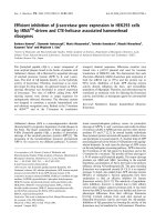
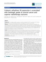
Figure 1 CA7 expression is frequently downregulated in CRC. (A) The expression levels of CA7 mRNA in 59 paired human primary CRC tissues
(T) and adjacent normal tissues (N) were evaluated by real-time qPCR methods. (T vs N, p < 0.001) (B) Protein levels of CA7 in an independent set of
25 paired CRC specimens and adjacent normal tissues were determined by Western blot assay. β-actin was used as a loading control. The relative
protein expression of CA7 was quantified and normalized to β-actin. Each N was arbitrarily designated 1.0. (T: Tumor; N: adjacent normal tissues,
T vs N, p < 0.001) (C) Representative immunohistochemical expression patterns of CA7 in cancerous and adjacent normal mucosa specimens were
shown. (Magnification, left panel, ×100; right panel, ×400) (D) Percentage of patients with different staining intensity of CA7 in the tumor or
adjacent normal tissues in the training cohort (p < 0.001).
only 0.9% (2/228) showed relatively strong staining of
CA7 (p < 0.001, Figure 1D). Consistently, IHC data
from the validation cohort containing 151 CRC patients yielded a similar result (Additional file 2). Thus,
the significant decreased staining signal for CA7 in the
cancerous specimens definitely confirmed that CA7
was frequently downregulated in CRC tissues.
2. Correlation of CA7 expression with clinicopathologic
features
Next, we evaluated the relationship between CA7 expression levels and clinicopathologic characteristics of
CRC patients. Based on the immunoreactive score (IRS)
of intratumoral CA7 expression, patients in the training
cohort were divided into high and low CA7 expression
subgroups with the median IRS value as the cut-off.
As shown in Table 2, low expression of CA7 protein
was significantly correlated with poor differentiation
(p = 0.006), positive lymph node metastasis (p = 0.003),
advanced TNM stage (p = 0.008) and increased death
(p < 0.001). No significant associations were observed between CA7 expression and patient age, sex, tumor location, tumor size or local invasion.
We then applied the same cut-off to dichotomise the
study patients in the validation cohort. Consistently, low
levels of CA7 protein were significantly correlated with
Yang et al. BMC Cancer (2015) 15:209
Page 5 of 11
Table 2 Association between CA7 expression and clinicopathologic characteristics of CRC patients in the training and
validation cohorts
Training cohort (n = 228)
Characteristics
Validation cohort (n = 151)
P valuea
CA7 expression
Low (%)
High (%)
(n = 116)
(n = 112)
Age (years)
P valuea
CA7 expression
Low (%)
High (%)
(n = 89)
(n = 62)
0.267
<60
40
(34.5)
31
(27.7)
≥60
76
(65.5)
81
(72.3)
Sex
0.622
24
(27.0)
19
(30.6)
65
(73.0)
43
(69.4)
0.720
0.490
Female
47
(40.5)
48
(42.9)
38
(42.7)
23
(37.1)
Male
69
(59.5)
64
(57.1)
51
(57.3)
39
(62.9)
Rectum
90
(77.6)
79
(70.5)
47
(52.8)
34
(54.8)
Colon
26
(22.4)
33
(29.5)
42
(47.2)
28
(45.2)
Tumor location
0.224
Differentiation grade
0.806
0.006
0.046
Well/Moderate
78
(67.2)
93
(83.0)
62
(69.7)
52
(83.9)
Poor
38
(32.8)
19
(17.0)
27
(30.3)
10
(16.1)
<5
44
(37.9)
48
(42.9)
32
(36.0)
25
(40.3)
≥5
72
(62.1)
64
(57.1)
57
(64.0)
37
(59.7)
Tumor size (cm)
0.448
Local invasion
0.586
0.141
0.064
T1-T2
18
(15.5)
26
(23.2)
5
(5.6)
9
(14.5)
T3-T4
98
(84.5)
86
(76.8)
84
(94.4)
53
(85.5)
N0
56
(48.3)
76
(67.9)
41
(46.1)
43
(69.4)
N1
36
(31.0)
28
(25.0)
31
(34.8)
17
(27.4)
N2
24
(20.7)
8
(7.1)
17
(19.1)
2
(3.2)
I
13
(11.2)
23
(20.5)
2
(2.2)
8
(12.9)
II
43
(37.1)
53
(47.3)
39
(43.9)
35
(56.5)
III
60
(51.7)
36
(32.2)
48
(53.9)
19
(30.6)
No
47
(40.5)
77
(68.8)
37
(41.6)
45
(72.6)
Yes
69
(59.5)
35
(31.2)
52
(58.4)
17
(27.4)
Lymph node metastasis
0.003
TNM stage
0.003
0.008
Death
0.003
<0.001
<0.001
a
Pearson chi-square test or Fisher exact test was used for comparison between subgroups. Bold type indicates statistical significance.
differentiation grade (p = 0.046), lymph node metastasis
(p = 0.003), TNM stage (p = 0.003) and patient death
(p < 0.001). Collectively, these results indicated that intratumoral CA7 expression was negatively associated with
the progression of CRC.
3. Prognostic values of CA7 expression in CRC patients
Kaplan-Meier survival analyses showed that patients with
low CA7 expression had significantly poorer DSS rates
than those with high CA7 expression in the training
cohort (p < 0.001, Figure 2A). The cumulative 5-year DSS
rate was 73.2% for patients in the high-CA7-expression
group, whereas it was only 44.8% for those in the lowCA7-expression group. Similarly, patients who had tumors with low CA7 expression had a significantly shorter
DSS than those who had tumors with high CA7 expression in the validation cohort (p < 0.001, Figure 2B). In
both of the two cohorts, patients who had advanced
stage (stage III) tumors had a significantly worse prognosis than those who had early stage (stages I-II)
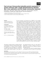
Yang et al. BMC Cancer (2015) 15:209
Page 6 of 11
Figure 2 Kaplan-Meier survival analysis for CRC patients in the training and validation cohorts. (A-B) Kaplan-Meier curves for diseasespecific survival of CRC patients in the training (A) and validation (B) cohorts according to CA7 expression status. Patients were divided into high
and low CA7 expression subgroups with the median IRS value as the cut-off. (C-D) Kaplan-Meier curves for disease-specific survival of CRC patients
in the training (C) and validation (D) cohort according to TNM stage of the disease. The p-value was determined using the log-rank test.
tumors (all p < 0.001, Figure 2C and D). These data
were consistent with the well established adverse prognostic effect of tumor stage [21] and confirmed that
our cohorts were representative and that the survival
analyses were valid. Importantly, reduced expression
of CA7 significantly predicted poor DSS for patients
with early stage tumors both in the training (p =
0.012, Figure 3A) and validation cohorts (p = 0.002,
Figure 3B). In addition, low levels of CA7 protein also
predicted unfavorable DSS for patients with advanced
stage tumors in the training cohort (p = 0.004,
Figure 3C). While, the survival difference was not
statistically significant for patients with advanced
stage tumors in the validation cohort (p = 0.289,
Figure 3D).
The independent prognostic significance of CA7 expression on CRC-specific survival was assessed with a
Cox regression model. The clinicopathologic variables
considered to be potential predictors of survival were
shown in Table 3. Univariate analyses indicated that factors including patient age, tumor differentiation grade,
TNM stage and CA7 expression were predictors of DSS
both in the training and validation cohort. The factors
that significantly correlated with survival in the univariate analysis were further assessed by multivariate
analysis. The results revealed that, besides the patient
age and TNM stage of the disease, decreased CA7
expression was an independent adverse prognostic
factor not only in the training cohort (HR, 2.247;
95% CI, 1.481-3.401, p < 0.001) but also in the validation cohort (HR, 2.058; 95% CI, 1.174-3.610,
p = 0.012).
More importantly, stage-based survival analyses revealed that decreased expression of CA7 also was an
independent predictor of poor prognosis for patients
with early stage tumors in the training cohort (HR,
2.232; 95% CI, 1.220-4.082, p = 0.009) as well as in the
validation cohort (HR, 3.165; 95% CI, 1.326-7.519,
Yang et al. BMC Cancer (2015) 15:209
Page 7 of 11
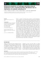
Figure 3 Kaplan-Meier survival analysis for CRC patients with early or advanced stage tumors in the training and validation cohorts.
(A-B) Kaplan-Meier curves for disease-specific survival of CRC patients with early stage tumors in the training (A) and validation (B) cohorts
according to CA7 expression status. (C-D) Kaplan-Meier curves for disease-specific survival of CRC patients with advanced stage tumors in the
training (C) and validation (D) cohorts according to CA7 expression status. The p-value was determined using the log-rank test.
p = 0.009) (Table 4). In addition, low CA7 expression
was also independently associated with poor DSS for
patients with advanced stage tumors in the training
cohort (HR, 2.809; 95% CI, 1.524-5.181, p = 0.001).
However, the prognostic significance of CA7 was not
statistically significant for patients with advanced stage
tumors in the validation cohort. Taken together, these
data demonstrated that decreased CA7 expression was
closely related to poor patient survival, especially for
those with early stage tumors.
Discussion
To date, CAs have been implicated in the tumorigenesis
of several human malignancies including CRC during
the past decades. It has been shown that CA I, II and
XIII are downregulated while CA IX and XII are upregulated in the cancerous tissue compared with the normal
colorectal epithelium [7-12]. In addition, prognostic
implications of individual CA isozymes in CRC have also
been demonstrated. For instance, CA I positive immunostaining has been associated with well or moderate differentiation, lack vascular invasion and favorable clinical
outcome of CRC [7,22], whereas, high expression of CA
IX has been linked with worse prognosis of CRC
patients [23-25]. Thus, distinct tumor-associated CAs
apparently have different expression patterns and prognostic significances in CRC.
Recently, attention has been focus on the CA7 because of its high catalytic activity and relatively limited
tissue distribution [13,26]. It has been shown that CA7
is expressed in several organs including brain, stomach, duodenum, colon, liver, and skeletal muscle. In
addition, the expression of CA7 and its prognostic
significance has been demonstrated in human diffuse
astrocytomas [16]. However, its clinical relevance has
not been assessed in CRC. Although a previous gene
Yang et al. BMC Cancer (2015) 15:209
Page 8 of 11
Table 3 Univariate and multivariate analyses of CA7 expression and disease-specific survival of patients in the training
and validation cohorts
Variables
Categories
Multivariate analysisb
Univariate analysis
c
HR
95% CI
P value
HR
95% CI
P valuec
2.770
1.691-4.537
<0.001
Training Cohort
Age (years)
≥60 / <60
2.263
1.389-3.687
0.001
Sex
Male / female
0.878
0.596-1.293
0.510
Tumor location
Colon / rectum
1.401
0.923-2.125
0.113
Tumor size (cm)
≥5 / <5
1.114
0.753-1.648
0.590
Differentiation grade
Poor / well + moderate
2.626
1.761-3.916
<0.001
2.010
1.337-3.023
0.001
TNM stage
III / I+ II
2.734
1.846-4.048
<0.001
2.402
1.603-3.598
<0.001
Low / high
2.469
1.642-3.717
<0.001
2.247
1.481-3.401
<0.001
Age (years)
≥60 / <60
1.976
1.080-3.614
0.027
2.293
1.247-4.217
0.008
Sex
Male / female
0.737
0.459-1.183
0.207
Tumor location
Colon / rectum
0.961
0.599-1.542
0.868
Tumor size (cm)
≥5 / <5
0.814
0.503-1.317
0.403
a
CA7 expression
Validation Cohort
Differentiation grade
Poor / well + moderate
1.688
1.017-2.803
0.043
TNM stage
III / I+ II
2.828
1.734-4.611
<0.001
2.674
1.615-4.428
<0.001
Low / high
2.632
1.520-4.545
0.001
2.058
1.174-3.610
0.012
a
CA7 expression
Abbreviations: HR hazard ratio, 95% CI 95% confidence interval.
a
For CA7, median values were used as the cut-off point for definition of subgroups (low expression and high expression groups).
b
Multivariate models were adjusted for age, sex, tumor location, tumor size, differentiation grade, and TNM stage.
c
Bold type indicates statistical significance.
Table 4 Multivariate analyses of CA7 expression and disease-specific survival for patients with early or advanced stage
tumors in the training and validation cohorts
Variables
Categories
Early stage
Advanced stage
HR
95% CI
P valueb
HR
95% CI
P valueb
4.062
1.592-10.365
0.003
2.353
1.282-4.319
0.006
2.389
1.419-4.023
0.001
2.809
1.524-5.181
0.001
2.374
1.176-4.794
0.016
2.637
1.414-4.920
0.002
Training Cohort
≥60 / <60
Age (years)
Sex
Male / female
Tumor location
Colon / rectum
Tumor size (cm)
≥5 / <5
Differentiation grade
Poor / well + moderate
a
CA7 expression
Low / high
2.232
1.220-4.082
0.009
Validation Cohort
Age (years)
≥60 / <60
Sex
Male / female
Tumor location
Colon / rectum
Tumor size (cm)
≥5 / <5
Differentiation grade
Poor / well + moderate
CA7 expressiona
Low / high
3.165
1.326-7.519
0.009
Abbreviations: HR hazard ratio, 95% CI 95% confidence interval.
a
For CA7, median values were used as the cut-off point for definition of subgroups (low expression and high expression groups).
b
Bold type indicates statistical significance.
Multivariate models were adjusted for age, sex, tumor location, tumor size and differentiation grade.
Yang et al. BMC Cancer (2015) 15:209
expression profiling study and a recent bioinformaticsbased analysis revealed that CA7 mRNA was downregulated in CRC clinical specimens [14,15], neither of
the two studies conducted validation experiments. To
get a better insight into the phenotypic expression and
prognostic significance of CA7 in CRC, we comprehensively analyzed both colorectal neoplasias and matched
adjacent normal mucosa specimens from the same
patients in two independent study cohorts.
Using qPCR and Western blot analysis, we demonstrated that CA7 was significantly downregulated in
primary CRC samples at both the mRNA and protein
levels. Strikingly, subsequent immunohistochemical analysis of CRC specimens from the training and validation
cohorts showed that 56.1% and 66.9% of the adjacent
normal mucosa sections presented strong CA7 immunoreactivity, whereas only 0.9% and 1.9% of the CRC sections showed relatively strong staining, respectively.
These results definitely confirmed the significant downregulation of CA7 in CRC. Thus, the expression pattern
of CA7 was generally similar with that of CA I, II and
XIII in CRC [8,12]. While, the molecular basis for CA7
downregulation in CRC remains unclear and requires
further investigation.
In the present study, we observed that the strongest
CA7 immunoreaction was localized primarily in the
mature superficial enterocytes but the signals reduced
significantly along with increasing malignancy grades,
indicating an association between CA7 expression and
the differentiation of colorectal epithelium. In fact, several CA isozymes have been proposed to relate to differentiation. Bekku S et al. showed that CA I and II
could be a differentiation marker of human and rat
colonic enterocytes [27]. In addition, Leppilampi M et al.
revealed that high expression of CA IX was associated
with a differentiated phenotype of gastric epithelial cells
[28]. Although our data demonstrated a correlation between CA7 expression and cell differentiation, its potential involvement in differentiation needs to be carefully
determined, for the decrease in CA7 expression in less
differentiated tissues could also be the result of other
factors that lead to dedifferentiation, rather than downregulation of CA7 being a contributing factor in dedifferentiation. Nevertheless, regardless of the mechanism, our
data indicates that CA7 might be useful in the histopathological grading of CRC.
Intriguingly, correlation analyses with clinicopathologic
features from the two independent cohorts unanimously
revealed a significant association between decreased CA7
expression and increased lymph node metastasis, advanced TNM stage and increased patient death, indicating that CA7 might be negatively involved in CRC
progression. Recently, Monti SM and his groups have
demonstrated that, apart from its canonical catalytic
Page 9 of 11
action of carbon dioxide hydration, CA7 has the ability
to protect cells against oxidative damage [29,30]. Given
that oxidative stress has been implicated in the pathogenesis of a wide spectrum of human cancers including CRC
[31-34], the reported protective role of CA7 against oxidative stress might support a tumor-suppressing function
for this enzyme. However, whether or not CA7 plays a
functional role in the tumorigenesis and progression of
CRC remains to be determined. Further studies using
gain-of-function and loss-of-function strategies are warranted to address this issue.
The most interesting findings of this study are from
the survival analysis results. Reduced expression of CA7
was associated with shortened survival for CRC patients
not only in the training cohort but also in the external
validation cohort. In univariate analysis, CA7 protein
emerged as a significant prognostic factor of clinical
outcome. Moreover, in multivariate analysis, it emerged
as a significant independent predictor of survival in
addition to tumor stage and patient age. The present
study indicated that TNM stage also is an important
prognostic factor in CRC, which is in agreement with its
well established adverse prognostic effect [21]. Generally
speaking, CRC patients who had early stage tumors
(stages I-II) have a relatively favorable prognosis than
those who had advanced stage tumors (stages III-IV).
However, a subgroup of patients with early stage tumors
have an increased risk of early recurrence and death.
The potential mechanisms for these aggressive forms of
early stage tumors are complicated; nevertheless, identifying this high-risk subgroup of patients would be of
particular importance in the selection of patients for
appropriate treatment. To further determine the prognostic value of CA7 in the therapeutic decision-making,
we performed survival analyzes stage by stage. Importantly, reduced CA7 expression significantly predicted
poor postoperative prognosis of patients with early stage
tumors in both cohorts. More importantly, stage-based
multivariate analyses from the two cohorts unanimously
confirmed that decreased expression of CA7 also was an
independent unfavorable prognostic indicator for patients with early stage tumors. Although we also observed
a significant association between decreased CA7 expression and poor DSS in the advanced-stage patient group
from the training cohort, its prognostic performance did
not persist in the patients of the same stage category
from the validation cohort (Figure 3D and Table 4). However, additional larger validations will be needed to further assess the prognostic significance of CA7 expression
in patients with advance stage tumors. Of note, in contrast to our results, Bootorabi F et al. reported that high
CA7 expression correlates with poor prognosis of
patients with astrocytomas [16]. The discrepant results
on the prognostic value of CA7 in different malignances
Yang et al. BMC Cancer (2015) 15:209
indicate that its prognostic implication may be tissuedependent and varies with the type of malignancy. The
underlying mechanism for the prognostic importance of
CA7 in CRC is currently unknown and needs to be further investigated. Nevertheless, our results suggest that
determination of the intratumoral CA7 expression status
may help to identify patients with aggressive forms of
CRCs and further guide individualized therapy choices.
The present study remains to be improved on several
aspects. Although we enrolled consecutive patients in
the training cohort, some of them were lost to followup, which may introduce a bias. In addition, disease
recurrence monitoring was incomplete in the two patient cohorts, resulting in the loss of disease-free survival
data, which is also very important for a biomarker validation. Further prospective studies using large cohorts are
necessary to validate the robustness of our findings
before clinical translation.
Conclusions
In summary, this study is the first to demonstrate that
CA7 is frequently downregulated in CRC and that
decreased expression of CA7 is closely related to aggressive clinical features and poor postoperative prognosis of
CRC patients. Our findings from two independent cohorts provide evidence for the potential utility of CA7 as
a prognostic marker for patients with CRC, especially
for those with early stage tumors. In addition, results
from the present work encourage further investigation
of its potential role in CRC pathobiology.
Additional files
Additional file 1: Distribution of the continuous variables of the
two study cohorts.
Additional file 2: Percentage of patients with different staining
intensity of CA7 in the tumor or adjacent normal tissues in the
validation cohort.
Abbreviations
CRC: Colorectal carcinoma; CA7: Carbonic anhydrase VII; DSS: Diseasespecific survival; qPCR: Quantitative polymerase chain reaction;
IHC: Immunohistochemistry; IRS: Immunoreactive score.
Competing interests
The authors declare that they have no competing interests.
Authors’ contributions
GZY and LH performed the immunohistochemistry and interpreted the IHC
results, GZY, LH and JC performed the statistical analysis, JC, HYC, YZ, DF and
YXZ obtained the clinical parameters and survival information, GZY, LH and
HYC performed the Western blot analysis, GZY, CYQ, HG and HF performed
the qPCR analysis, GZY, LH, QPC and CFG conceived of the study, and
participated in its design and coordination. GZY and LH drafted the
manuscript. All authors read and approved the final manuscript.
Acknowledgements
This work was supported by National Natural Science Foundation of China
(81301811).
Page 10 of 11
Author details
1
Anal-Colorectal Surgery Institute, 150th Hospital of PLA, Luoyang, China.
2
Department of Clinical Laboratory, 150th Hospital of PLA, Luoyang, China.
3
Department of Colorectal Surgery, 150th Hospital of PLA, Luoyang, China.
4
Department of Oncology, 150th Hospital of PLA, Luoyang, China.
5
Department of Oncology, Changhai Hospital, Second Military Medical
University, Shanghai, China. 6Department of Gastrointestine Surgery,
Changzheng Hospital, Second Military Medical University, Shanghai, China.
Received: 11 November 2014 Accepted: 18 March 2015
References
1. Jemal A, Bray F, Center MM, Ferlay J, Ward E, Forman D. Global cancer
statistics. CA Cancer J Clin. 2011;61:69–90.
2. Ferlay J, Shin HR, Bray F, Forman D, Mathers C, Parkin DM. Estimates of
worldwide burden of cancer in 2008: GLOBOCAN 2008. Int J Cancer.
2010;127:2893–917.
3. Brenner H, Kloor M, Pox CP. Colorectal cancer. Lancet. 2014;383:1490–502.
4. Guo P, Huang ZL, Yu P, Li K. Trends in cancer mortality in China: an update.
Ann Oncol. 2012;23:2755–62.
5. Alterio V, Di Fiore A, D'Ambrosio K, Supuran CT, De Simone G. Multiple
binding modes of inhibitors to carbonic anhydrases: how to design specific
drugs targeting 15 different isoforms? Chem Rev. 2012;112:4421–68.
6. Supuran CT. Carbonic anhydrases: novel therapeutic applications for
inhibitors and activators. Nat Rev Drug Discov. 2008;7:168–81.
7. Mori M, Staniunas RJ, Barnard GF, Jessup JM, Steele Jr GD, Chen LB. The
significance of carbonic anhydrase expression in human colorectal cancer.
Gastroenterology. 1993;105:820–6.
8. Kivela AJ, Saarnio J, Karttunen TJ, Kivelä J, Parkkila AK, Pastorekova S, et al.
Differential expression of cytoplasmic carbonic anhydrases, CA I and II, and
membrane-associated isozymes, CA IX and XII, in normal mucosa of large
intestine and in colorectal tumors. Dig Dis Sci. 2001;46:2179–86.
9. Saarnio J, Parkkila S, Parkkila AK, Haukipuro K, Pastoreková S, Pastorek J, et al.
Immunohistochemical study of colorectal tumors for expression of a novel
transmembrane carbonic anhydrase, MN/CA IX, with potential value as a
marker of cell proliferation. Am J Pathol. 1998;153:279–85.
10. Niemelä AM, Hynninen P, Mecklin JP, Kuopio T, Kokko A, Aaltonen L, et al.
Carbonic anhydrase IX is highly expressed in hereditary nonpolyposis
colorectal cancer. Cancer Epidemiol Biomarkers Prev. 2007;16:1760–6.
11. Kivelä A, Parkkila S, Saarnio J, Karttunen TJ, Kivelä J, Parkkila AK, et al.
Expression of a novel transmembrane carbonic anhydrase isozyme XII in
normal human gut and colorectal tumors. Am J Pathol. 2000;156:577–84.
12. Kummola L, Hämäläinen JM, Kivelä J, Kivelä AJ, Saarnio J, Karttunen T, et al.
Expression of a novel carbonic anhydrase, CA XIII, in normal and neoplastic
colorectal mucosa. BMC Cancer. 2005;5:41.
13. Bootorabi F, Jänis J, Smith E, Waheed A, Kukkurainen S, Hytönen V, et al.
Analysis of a shortened form of human carbonic anhydrase VII expressed
in vitro compared to the full-length enzyme. Biochimie. 2010;92:1072–80.
14. Birkenkamp-Demtroder K, Olesen SH, Sørensen FB, Laurberg S, Laiho P,
Aaltonen LA, et al. Differential gene expression in colon cancer of the
caecum versus the sigmoid and rectosigmoid. Gut. 2005;54:374–84.
15. Chu CM, Yao CT, Chang YT, Chou HL, Chou YC, Chen KH, et al. Gene
expression profiling of colorectal tumors and normal mucosa by
microarrays meta-analysis using prediction analysis of microarray, artificial
neural network, classification, and regression trees. Dis Markers.
2014;2014:634123.
16. Bootorabi F, Haapasalo J, Smith E, Haapasalo H, Parkkila S. Carbonic
anhydrase VII–a potential prognostic marker in gliomas. Health.
2011;3:6–12.
17. Hu L, Chen L, Yang G, Li L, Sun H, Chang Y, et al. HBx sensitizes cells to
oxidative stress-induced apoptosis by accelerating the loss of Mcl-1 protein
via caspase-3 cascade. Mol Cancer. 2011;10:43.
18. Dong LW, Yang GZ, Pan YF, Chen Y, Tan YX, Dai RY, et al. The oncoprotein
p28GANK establishes a positive feedback loop in β-catenin signaling. Cell
Res. 2011;21:1248–61.
19. Li L, Chen L, Hu L, Liu Y, Sun HY, Tang J, et al. Nuclear factor high-mobility
group box1 mediating the activation of Toll-like receptor 4 signaling in
hepatocytes in the early stage of nonalcoholic fatty liver disease in mice.
Hepatology. 2011;54:1620–30.
Yang et al. BMC Cancer (2015) 15:209
Page 11 of 11
20. Tang L, Tan YX, Jiang BG, Pan YF, Li SX, Yang GZ, et al. The prognostic
significance and therapeutic potential of hedgehog signaling in intrahepatic
cholangiocellular carcinoma. Clin Cancer Res. 2013;19:2014–24.
21. Compton CC. Optimal pathologic staging: defining stage II disease. Clin
Cancer Res. 2007;13:6862s–70.
22. Peng Y, Li X, Wu M, Yang J, Liu M, Zhang W, et al. New prognosis
biomarkers identified by dynamic proteomic analysis of colorectal cancer.
Mol Biosyst. 2012;8:3077–88.
23. Korkeila E, Talvinen K, Jaakkola PM, Minn H, Syrjänen K, Sundström J, et al.
Expression of carbonic anhydrase IX suggests poor outcome in rectal
cancer. Br J Cancer. 2009;24:874–80.
24. Cleven AH, van Engeland M, Wouters BG, de Bruïne AP. Stromal expression
of hypoxia regulated proteins is an adverse prognostic factor in colorectal
carcinomas. Cell Oncol. 2007;29:229–40.
25. Pastorekova S, Parkkila S, Zavada J. Tumor-associated carbonic anhydrases
and their clinical significance. Adv Clin Chem. 2006;42:167–216.
26. Earnhardt JN, Qian M, Tu C, Lakkis MM, Bergenhem NC, Laipis PJ, et al. The
catalytic properties of murine carbonic anhydrase VII. Biochemistry.
1998;37:10837–45.
27. Bekku S, Mochizuki H, Takayama E, Shinomiya N, Fukamachi H, Ichinose M,
et al. Carbonic anhydrase I and II as a differentiation marker of human and
rat colonic enterocytes. Res Exp Med (Berl). 1998;198:175–85.
28. Leppilampi M, Saarnio J, Karttunen TJ, Kivelä J, Pastoreková S, Pastorek J,
et al. Carbonic anhydrase isozymes IX and XII in gastric tumors. World J
Gastroenterol. 2003;9:1398–403.
29. Del Giudice R, Monti DM, Truppo E, Arciello A, Supuran CT, De Simone G,
et al. Human carbonic anhydrase VII protects cells from oxidative damage.
Biol Chem. 2013;394:1343–8.
30. Truppo E, Supuran CT, Sandomenico A, Vullo D, Innocenti A, Di Fiore A,
et al. Carbonic anhydrase VII is S-glutathionylated without loss of catalytic
activity and affinity for sulfonamide inhibitors. Bioorg Med Chem Lett.
2012;22:1560–4.
31. Sosa V, Moliné T, Somoza R, Paciucci R, Kondoh H, LLeonart ME. Oxidative
stress and cancer: an overview. Ageing Res Rev. 2013;12:376–90.
32. Fang J, Seki T, Maeda H. Therapeutic strategies by modulating oxygen stress
in cancer and inflammation. Adv Drug Deliv Rev. 2009;61:290–302.
33. Perše M. Oxidative stress in the pathogenesis of colorectal cancer: cause or
consequence? Biomed Res Int. 2013;2013:725710.
34. Stone WL, Krishnan K, Campbell SE, Palau VE. The role of antioxidants and
pro-oxidants in colon cancer. World J Gastrointest Oncol. 2014;6:55–66.
Submit your next manuscript to BioMed Central
and take full advantage of:
• Convenient online submission
• Thorough peer review
• No space constraints or color figure charges
• Immediate publication on acceptance
• Inclusion in PubMed, CAS, Scopus and Google Scholar
• Research which is freely available for redistribution
Submit your manuscript at
www.biomedcentral.com/submit