Sumoylation of Kif18A plays a role in regulating mitotic progression
Bạn đang xem bản rút gọn của tài liệu. Xem và tải ngay bản đầy đủ của tài liệu tại đây (1.17 MB, 10 trang )
Yang et al. BMC Cancer (2015) 15:197
DOI 10.1186/s12885-015-1226-9
RESEARCH ARTICLE
Open Access
Sumoylation of Kif18A plays a role in regulating
mitotic progression
Feikun Yang1, Yan Chen2 and Wei Dai1,3*
Abstract
Background: Kif18A, the kinesin-8 motor protein, plays an essential role in regulating alignment of bi-oriented
chromosomes at the midzone during mitosis. Kinesin proteins, including Kif18A, are often deregulated in many
types of cancers and are thought to play a critical role in cancer progression. However, little is known about the
post-translational modifications of Kif18A and their effects on its biological activity.
Methods: Kif18A was identified to be a SUMO2 acceptor by using Ni-IDA resin to precipitate proteins from cells
stably expressing His6-SUMO2. To identify the potential lysine residues, multi-site directed mutagenesis together
with transient transfection and Ni-IDA pull-down assay were carried out. The confocal time-lapse imaging and
immunofluorescent staining were used to study the roles of SUMO2 modification on Kif18A’s activity during the
cell cycle.
Results: Kif18A is covalently modified by SUMO2 during the cell cycle, and its sumoylation peaks at metaphase
and then rapidly decreases upon anaphase onset. Mutational analysis identifies multiple lysine residues (K148,
K442, K533, K660 and K683) as potential SUMO acceptors. The functional studies reveal that sumoylation of Kif18A has
little effect on protein stability and subcellular localization. However, compared with the wild-type control, ectopic
expression of SUMO-resistant mutants of Kif18A results in a significant delay of mitotic exit. Confocal microscopy
shows that cells expressing SUMO-resistant Kif18A display a compromised dissociation of BubR1 from kinetochores
after anaphase onset.
Conclusions: Our studies reveal that sumoylation functions as an unidentified form of post-translational modification
that regulates Kif18A activity during mitotic progression.
Keywords: Kif18A, Sumoylation, Cell cycle, Mitosis, Motor protein, Microtubules
Background
Proper equatorial alignment of all condensed chromosomes is an essential cellular process for preserving
chromosomal stability during nuclear division. To this
end, eukaryotic cells have evolved a system in which a set
of conserved proteins monitor completion of chromosomal congression and regulate the dynamics of spindle
microtubules at both spindle poles and kinetochores [1-3].
Increasing evidence indicates that KIF18A, the kinesin-8
molecular motor, plays an important role in regulating
* Correspondence:
1
Department of Environmental Medicine, New York University Langone
Medical Center, 57 Old Forge Road, Tuxedo Park, NY 10987, USA
3
Department of Biochemistry and Molecular Pharmacology, New York
University Langone Medical Center, 57 Old Forge Road, Tuxedo Park, NY
10987, USA
Full list of author information is available at the end of the article
spindle microtubule dynamics and chromosome positioning during mitosis. As a plus-end directed motor, Kif18A
inhibits polymerization dynamics of microtubules, thus
suppressing kinetochore movements [4] and chromosome
oscillations [5]. Depletion of Kif18A results in chromosome congression defects, which is at least partially mediated through destabilizing another plus-end directed
motor protein CENP-E [6]. Mouse genetic study reveals
that ablation of KIF18A causes complete sterility [7].
Kinesin proteins are often deregulated in many types of
cancers and are thought to play a critical role in cancer
progression [8-10]. For example, Kif18A is overexpressed
in human breast cancer at both mRNA and protein levels,
and the degree of Kif18A expression is associated with
tumor grades, metastasis and survival [11]. Kif18A expression is up-regulated in colorectal tumors [12,13]. Ablation
© 2015 Yang et al.; licensee BioMed Central. This is an Open Access article distributed under the terms of the Creative
Commons Attribution License ( which permits unrestricted use, distribution, and
reproduction in any medium, provided the original work is properly credited. The Creative Commons Public Domain
Dedication waiver ( applies to the data made available in this article,
unless otherwise stated.
Yang et al. BMC Cancer (2015) 15:197
Page 2 of 10
in fresh medium for indicated times before harvesting for
various analyses.
of Kif18A reduces cancer cell proliferation, migration and
invasion [12], and promotes cell apoptosis through negative regulation of the PI3K-AKT signaling axis [13]. It has
been also reported that Kif18A can be potentially served
as a biomarker for diagnosing early stages of choloangiocarcinoma [14] and for identifying asbestosis patients at
risk of developing lung cancer [15].
Post-translational modifications play important roles
in regulating the activity of kinesin proteins. For example, kinesin light chain 1 of kinesin-1 is phosporylated
at serine 460 by ERK and this phosporylation regulates
its ability in cargo-binding and trafficking [16]. Kif2A, a
microtubule depolymerase, is phosphorylated by Aurora B
on multiple sites and the phosphorylation is important for
the kinesin to function properly in cytokinesis [17,18].
Moreover, CENP-E, a member of kinesin-7 family, is modified by SUMO-2/3 and the modification is essential for its
kinetochore localization during mitosis [19]. Furthermore,
Kif18A is modified by phosphorylation and ubiquitination
during mitosis and these modifications appear to play an
important role in regulating degradation of Kif18A at anaphase [20-22].
Given that sumoylation plays an essential role in regulating mitotic proteins [23], we asked whether Kif18A was
modified by sumoylation and whether the modification affected its activity in mitosis. We found that Kif18A was
preferentially modified by SUMO2 and that the modification was closely associated with mitotic progression.
Site-directed mutagenesis coupled with ectopic expression revealed that several lysine residues (K148, K442,
K533, K660 and K683) were potential SUMO2 acceptors. Expression of a SUMO-deficient Kif18A mutant, but
not the wild-type counterpart resulted in a significant
delay in mitotic exit. Therefore, our combined study reveals a new type of post-translational mechanism that regulates Kif18A’s function in mitosis.
Small interfering RNAs (siRNAs) of human KIF18A were
synthesized from Dharmacon which corresponded to the
following sequences: 5′ACA GATTCGTGATCTCTTA3′,
which is known to silence human KIF18A [6]. Briefly, cells
seeded at 60% confluency in an antibiotic-free culture
medium were transfected using Lipojet™ (Signagene)
with siRNA duplexes at a final concentration of 200 pM
for 48 hours. Firefly (Photinus pyralis) luciferase siRNAs
(5′UUCCTACGCTGAGTACTTCGA3′, GL-3 from
Dharmacon) were served as negative control.
Methods
Western blot
Cell culture
SDS-PAGE was carried out using the mini gel system from
Bio-Rad. Proteins were transferred to PVDF membranes.
After blocking with TBST containing 5% nonfat dry milk
for 1 h, the membranes were incubated with primary antibodies overnight at 4°C followed by incubation with horseradish peroxidase-conjugated secondary antibodies for 1 h
at room temperature. After thorough washing the membranes with TBST buffer, signals were developed with an
enhanced chemiluminescent system (Pierce).
HeLa and HEK293T cells were cultured in DMEM supplemented with 10% fetal bovine serum (FBS, Invitrogen)
and antibiotics (100 μg/ml of penicillin and 50 μg/ml of
streptomycin sulfate, Invitrogen) at 37°C under 5% CO2.
Cell cycle synchronization
HeLa cells were synchronized at the G1/S boundary by
double-thymidine blocks. Briefly, cells were treated with
2 mM thymidine for 18 h followed by a 9 h release; the
cells were treated with 2 mM thymidine for another
18 h and then released into the cell cycle for various
times. Mitotic shake-off cells were obtained from gentle
tapping of cell culture plates treated with nocodazole
(40 ng/ml) or taxol (40 nM) (Sigma-Aldrich) for 16 h. In
some experiments, mitotic cells were rinsed and cultured
Antibodies
Kif18A antibodies were purchase from Bethyl Laboratories
LLC. Antibodies to HA, Flag and β-actin were purchased from Cell Signaling Technology Inc. Rabbit polyclonal antibodies to BubR1 were developed in the
laboratory. GFP antibodies were purchased from Santa
Cruz Biotechnology. Mouse anti-SUMO2/3 antibodies
were kindly provided by Dr. Michael J. Matunis (Johns
Hopkins University).
Plasmids, mutagenesis, and transfection
Full-length wild-type human KIF18A cDNA with HA-his
tag was subcloned into pcDNA3 plasmid or a GFPexpression plasmid. Potential SUMO targeting lysine mutants were generated using the QuickChange Lightning
Multi Site-directed Mutagenesis kit (Stratagene). Individual mutations were confirmed by DNA sequencing.
SENP-1 and its mutant expression plasmids were kindly
provided by J. Cheng [24]. Plasmid transfection was carried out using Fugene HD according to instructions provided by the supplier (Roche).
RNA interference
Pull-down analysis
HeLa cells transfected with indicated plasmids or stably
expressing His6 -tagged SUMO-2 were lysed in a lysis
buffer [50 mM Na2HPO4/NaH2PO4 (pH 7.4), 300 mM
NaCl, 8 M urea, 0.2% Triton X-100] supplemented with
20 mM imidazole. Ni2+-IDA-agarose resin (Clontech)
Yang et al. BMC Cancer (2015) 15:197
was then added to the cell lysates and incubated at room
temperature for 3 h. The resin was washed 3 times at
room temperature with the lysis buffer supplemented
with 40 mM imidazole. After washing, His6 -tagged proteins were eluted in the lysis buffer containing 300 mM
imidazole. Samples were then blotted with individual
antibodies.
Fluorescence microscopy
Fluorescence microscopy was essentially performed as described [23]. Briefly, HeLa cells seeded on chamber slides
were transfected with indicated expression constructs for
48 h. At the end of transfection, cells were fixed with 4%
paraformaldehyde in PBS for 20 min at room temperature.
After permeabilization using 0.5% Triton X-100 in PBS for
20 min, cells were incubated with 2% bovine serum albumin (BSA) in PBS for 1 h followed by incubation overnight
with the antibody to BubR1. Cells were stained with Alex
Fluor 555-conjugated goat anti-rabbit IgGs (Invitrogen) for
1 h. Cellular DNA was finally stained with 4′,6-diamidino2-phenylindole (DAPI, Molecular Probe, Eugene, OR).
Fluorescence signals were detected on a Leica TCS SP5
confocal microscope.
Statistical analysis
Student’s t test was used to evaluate significance of differences between two groups. A P value <0.05 was considered statistically significant.
Results
To study post-translational modifications of Kif18A and
their potential function in regulating mitotic progression,
synchronized HeLa cells through the double-thymidine
block were released into the cell cycle for various times.
Immunoblotting analysis revealed that Kif18A levels gradually increased during the release, peaking around 10 h
before returning to the basal level (Figure 1A). Intriguingly, as cells entered mitosis as indicated by cyclin B1
levels, a slower mobility band immunoreactive to the
Kif18A antibody was present (Figure 1A, Asterisk). The
slow mobility signal also peaked around 10 h post the
double thymidine release and became barely detectable
1 h after exiting from mitosis. Oscillation of Kif18Aspecific signals suggests a role of the kinesin and its modified form in regulating mitotic progression.
The molecular mass of the slow mobility band of
Kif18A was about 125 kDa, which is 15 kDa larger than
the non-modified form. Given the major size difference
between the basal and modified forms, we speculated
that it might be caused by SUMO modification. To test
this hypothesis, we took advantage of the cell lines stably
expressing His6 -SUMO2 [25]. Cells were arrested at mitosis by nocodazole or taxol for 16 h before harvesting.
Equal amounts of cell lysates were used for Ni-IDA resin
Page 3 of 10
pull-down analysis and the precipitates were blotted for
antibodies against Kif18A and SUMO2. A slower mobility
band immunoreactive to Kif18A antibody was detected in
SUMO2-expressing cells in mitotic cells but not in asynchronized cells. This band was not present in parental
cells arrest at mitosis. These observations suggest that
Kif18A is targeted by SUMO2 at mitosis. Interestingly,
taxol enhanced the Kif18A signal to a greater extent than
that of nocodazole, which is likely due to microtubule
stabilization by taxol that triggers a significant plus-end
accumulation of Kif18A [26]. Unmodified Kif18A was also
detected in the pull-down precipitates, which could be derived from its electrostatic interaction with the Ni-IDA
resin or the proteins binding to the resin.
To further confirm that the slower mobility band is
Kif18A-specific, we transfected His6 -SUMO2 cells with
siRNAs to Kif18A or luciferease. His6 -GFP was used for
co-transfection to monitor transfection and pull-down
efficiency. Transfected cells were treated with or without
taxol for 16 h and equal amounts of cell lysates were
subjected to pull-down analysis. As shown in Figure 2A,
Kif18A siRNAs, but not control siRNAs, almost completely depleted the slow mobility band, indicating that
the signal was Kif18-specific.
SUMO modification is a reversible process, and deconjugation of SUMO from targeted proteins is catalyzed
by sentrin-specific isopeptidases. In vertebrates, six
SUMO-specific isopeptidases, including SENP1, SENP2,
SENP3, SENP5, SENP6, and SENP7, have been reported
[27]. To further confirm Kif18A is modified by SUMO2,
His6 -SUMO2-expressing cells were transiently transfected
with a plasmid construct expressing either FLAG-tagged
wild-type SENP1 or its enzymatically inactive counterpart
(SENP1-Mut). The transfected cells were then treated with
taxol for 16 h. Ni-IDA pull-down precipitates were blotted
for Kif18A, FLAG and SUMO2. As shown in Figure 2B,
expression of FLAG-SENP1 largely eliminated the slow
mobility band that was immunoactive to Kif18A antibody.
However, the mutant SENP1 was not effective in suppressing the signal. SENP1 could also be precipitated by Ni-IDA
resin due to its histidine-rich property. Thus, expression of
both SENP1 and its mutant was confirmed by blotting with
the anti-FLAG antibody (Figure 2B). Combined, these results strongly support the notion that Kif18A is modified
by SUMO2 at mitosis.
To identify the potential lysine residue (s) for sumo
modification, we analyzed Kif18A amino acid sequences
for optimal sumoylation using the criteria available at
Abgent Inc. Five lysines sites (K148, K442, K533, K660
and K683) with the highest scores were subjected to mutagenic analysis. The relative position of these sites to
other domains is shown in Figure 3A. HeLa cells were
co-transfected for 48 h with a SUMO2- construct and a
construct expressing His6 -HA-tagged wild-type Kif18A
Yang et al. BMC Cancer (2015) 15:197
Page 4 of 10
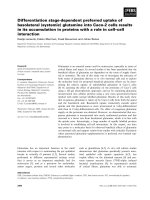
Figure 1 SUMO2 modification of Kif18A at mitosis. (A) HeLa cells were subjected to double thymidine treatment as described in Materials &
Methods. Cell pellets were lysed in 8 M urea. Equal amount of cell lysates were blotted for antibodies against Kif18A, cyclin B1, and β-actin. Kif18A
blots of both short and long exposure are shown. Asterisk (*) indicates the Kif18A related signals. (B) Parental HeLa cells and HeLa cells stably expressing
transfected His6 -SUMO2 were treated with nocodazole (40 ng/mL) or taxol (40 nM) for 16 h and then lysed in 8 M urea. Equal amounts of cell lysates
were used for Ni-IDA pull-down analysis. The precipitates were blotted for Kif18A and SUMO2. Kif18A-S denotes potentially SUMO-modified Kif18A.
(His6-HA-WT) or Kif18A with 5 lysine residues mutated
into arginines (His6-HA-5R). After treatment with Taxol
for 16 h, cells were lysed and equal amounts of cell lysates
were subjected to pull-down analysis using Ni-NDA resin.
Immunoblotting with antibody against the HA tag showed
major bands for both ectopically expressed His6-HA-WT
and its mutant counterpart (Figure 3B). Of great importance is that an extra slower mobility band that was immunoreactive to HA antibody was detected in cells
expressing His6-HA-WT, but not His6-HA-5R. Furthermore, immunoblotting with the antibody to FLAG revealed a specific band migrated at the same position as
the one detected by the HA tag antibody. These results
not only confirmed that Kif18A was modified by SUMO2
at mitosis but also indicated that K148, K442, K533, K660
and/or K683 were potential acceptors for SUMO2. The
molecular difference between SUMO2-modified and unmodified Kif18A was about 15 kDa, which suggests mono
sumoylation. On the other hand, we were unable to identify the single lysine residue for the modification as mutation of any of these lysine residues alone failed to abolish
the signal (see Additional file 1).
Sumoylation plays an important role in regulating stability and subcellular localization of targeted proteins [28,29].
However, this does not seem to be the case for Kif18A as
there was no significant difference in the half-life between
His6-HA-WT and His6-HA-5R (see Additional file 2).
Since Kif18A sumoylation peaked at metaphase and rapidly
decreased thereafter (Figure 1A), we then asked whether
Kif18A sumoylation might be involved in regulating mitotic exit. Kif18A exists in unmodified form in interphase,
and undergoes dynamic phosphorylation/de-phosphorylation during mitosis (Figure 1 and see Additional file 3).
Dephosphorylation of His6 -HA-WT took place within
40 minutes upon nocodazole release (Figure 4A, arrow).
However, compared with His6-HA-WT, sumoylation-
Yang et al. BMC Cancer (2015) 15:197
Page 5 of 10
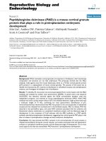
Figure 2 Kif18A is SUMO2-modified. (A) HeLa cells stably expressing His6 -SUMO2 were co-transfected with Kif18A (or control) siRNAs and a
plasmid construct expressing His6 -GFP for 48 h. Transfected cells were then treated with 40 nM taxol for 16 h. At the end of treatment, cells were
lysed in 8 M urea. Equal amounts of cell lysates were subjected to Ni-IDA pull-down analysis. Pull-down proteins were then blotted for Kif18A and
GFP. Kif18A-S denotes SUMO2-modified Kif18A. (B) HeLa cells stably expressing His6 -SUMO2 were transfected with a plasmid expressing FLAG-tagged
SENP-1 or enzymatically defective SENP1 (SENP-1-Mut) for 48 h. Transfected cells were then treated with 40 nM taxol for 16 h. At the end of treatment,
cell pellets were lysed in 8 M urea. Equal amounts of cell lysates were subjected to Ni-IDA pull-down analysis. Pull-down proteins were then blotted for
Kif18A, Flag, and SUMO2. Notably, Flag-SENP1 was precipitated by Ni-IDA resin due to its histidine-rich property.
resistant mutant His6-HA-5R appeared to be dephosphorylated at a slower pace, which was accompanied by slower
decline of cyclin B1 (Figure 4A and see Additional file 4).
These observations suggest that cells expressing His6HA-5R exhibit delayed mitotic exit. To further confirm
that sumoylation of Kif18A plays a role in regulating mitotic progression, HeLa cells ectopically expressing
GFP-tagged Kif18A-WT (GFP-WT) or its mutant
counterpart Kif18A-5R (GFP-5R) were examined via
time-lapse confocal microscopy. We observed that both
GFP-WT and GFP-5R exhibited normal microtubule plusend localization in metaphase cells (Figure 4B), which is
consistent with a previous report that Kif18A strongly accumulates at microtubule plus-end during metaphase but not
in prometaphase [1,4]. However, compared to GFP-WT,
the majority of GFP-5R expressing cells exhibited a
Yang et al. BMC Cancer (2015) 15:197
Page 6 of 10
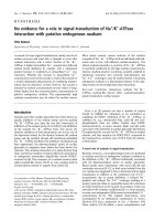
Figure 3 Identification of Kif18A sumoylation sites. (A) Schematic representation of lysine residues of wild-type human Kif18A. Key lysine residues
subjected to mutational analsyis are indicated. HA and His6 tags are fused in-frame at the C-terminus. Three major functional domains of Kif18A are also
shown. (B) HeLa cells were transfected with plasmids expressing His6 -HA-tagged Kif18A (His6 -HA-WT) or the mutant protein with 5 lysine residues
(K148, K442, K533, K660 and K683) replaced with arginines (His6 -HA-5R) for 48 h. Flag-SUMO2 was also used for co-transfection. Transfected cells were
then treated with 40 nM taxol for 16 h. At the end of treatment, cell pellets were lysed in 8 M urea. Equal amounts of cell lysates were subjected
to Ni-IDA pull-down analysis. Pull-down proteins were blotted for HA and Flag signals. Flag blots of both short and long exposure are shown.
prolonged mitotic exit (98 ± 44 min vs. 47 ± 15 min as
shown in Figure 4C).
Our previous work implies that Kif18A may interact
with BubR1, a spindle assembly checkpoint component,
at the mitotic stage because both proteins are associated
with CENP-E [6]. Indeed, GFP-Kif18A accumulated
around the kinetochore region where BubR1 signals
were also detected in metaphase cells ectopically expressing GFP-WT (Figure 5A, upper panel), suggesting a
physical and functional interaction between these two
molecules. To understand the underlying molecular
mechanism responsible for the delayed mitotic exit in
cells expressing GFP-5R, we determined the dissociation
of BubR1 from kinetochore using fluorescence microscopy. HeLa cells transiently transfected with GFP-WT
and its sumo-resistant counterpart for 36 h were fixed
and stained with the antibody to BubR1. As expected,
BubR1 was barely detectable at the kinetochores after
the anaphase onset in control cells or cells expressing
GFP-WT (Figure 5A & B). However, a significant fraction
of GFP-5R-expressing cells displayed persistent kinetochore localization of BubR1 at anaphase and telophase
stages, strongly suggesting that Kif18A sumoylation may
regulate the removal of BubR1 from the kinetochores and
compromise its inactivation. Of note, GFP-5R was also detected in the region where BubR1 signal persisted even
after the apparent anaphase onset (Figure 5A, lower
panel).
Discussion
In this study we report that a fraction of Kif18A is covalently modified by SUMO2 when cells enter the mitotic
stage. Kif18A sumoylation is a transient event as it is
rapidly desumoylated upon the anaphase onset. Kif18A
mutant with lysines 148, 442, 533, 660 and 683 replaced
with arginines largely abolished its sumoylation during
mitosis, strongly suggesting the involvement of these
residues in mediating SUMO modification. Functional
studies reveal that Kif18A sumoylation regulates mitotic
progression as ectopic expression of sumoylationresistant Kif18A mutant significantly delays mitotic exit.
Moreover, sumoylation also plays a role in the removal
of BubR1 from the kinetochores at the anaphase onset,
thus participating in the checkpoint control.
Yang et al. BMC Cancer (2015) 15:197
Page 7 of 10
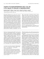
Figure 4 Sumoylation-resistant Kif18A mutant induces a mitotic delay. (A) HeLa cells were co-transfected with a plasmid expressing His6 -HA-WT
or His6 -HA-5R and a plasmid construct expressing His6 -GFP for 36 h. Transfected cells were then treated with nocodazole (40 ng/mL) for 14 h,
after which mitotic cells were collected by shake-off. Mitotic cells were then released into fresh medium. Cells were collected at various times of
release and lysed in 8 M urea. Equal amounts of cell lysates were blotted for HA, cyclin B1, and GFP. Asterisk indicates phosphorylated form of
His6 -HA-Kif18A. Lane A denotes lysates from asynchronized cells to show the unmodified form of His6 -HA-Kif18A. (B) HeLa cells transfected
with GFP-WT or GFP-5R were subjected to time-lapse confocal microscopy analysis. The video-graphic process started when significant plus-end
accumulation of GFP signals was observed. (C) Quantitative analysis of mitotic time of cells as shown in B. Data were summarized from three
independent experiments (WT: n = 13; 5R: n = 13).
Several studies have shown that at the onset of mitosis
many important proteins are SUMO-modified, which is
thought to function in the maintenance of mitotic
chromosome structures [30-32]. It has also been reported
that sumoylation is essential for the proper function of
inner centromeric proteins, as well as components of
outer kinetochore and fibrous corona [19,33,34]. However,
the role of sumoylation in the regulation of kinesin motor
proteins during the cell cycle remains largely unknown.
Kif18A plays an important role in chromosome congression by suppressing chromosome movements [4]. Consistent with previous observations on both endogenous [1,4]
and ectopically expressed venus-tagged Kif18A [21], GFPKif18A localizes along spindle microtubules in prometaphase cells (unpublished observation) and then exhibits as
a comet-like gradient along kinetochore microtubules with
Yang et al. BMC Cancer (2015) 15:197
Page 8 of 10
Figure 5 Expression of sumoylation-resistant Kif18A induces aberrant BubR1 localization in anaphase cells. (A) HeLa cells were transfected
with a plasmid expressing GFP-WT for 48 h, after which cells were fixed and stained with the antibody against BubR1 (red). DNA was stained with
DAPI (blue). Representative metaphase cell images are shown in the upper panel. In the lower panel, HeLa cells transiently expressing GFP-WT or
GFP-5R were fixed and stained with the BubR1 antibody (red). DNA was stained with DAPI (blue). Representative anaphase cell images are shown.
(B) Percentage of anaphase cells with aberrant (signals in anaphase) BubR1 localization were recorded and plotted. Data were summarized from
three independent experiments (WT, n = 85; 5R, n = 94; vector, n = 76). Asterisk indicates statistically significant difference between mutant 5R and
WT (or vector).
the strongest signal detected at the plus-end. Moreover,
after the anaphase onset Kif18A re-distributes to the midzone of the cell, as well as chromatin regions, suggesting
that it may play a role in mid-body formation and cytokinesis. In agreement with previous study [4], expression of
GFP-WT did not cause an obvious mitotic delay or disrupt
chromosome alignment. When cells were transfected with
SUMO-resistant GFP-5R, similar subcellular localization
patterns were observed, indicating that sumoylation does
not affect the plus-end localization of Kif18A. On the other
hand, from both time-lapse microscopy and PFA fixed
samples, we did not see apparent defects in chromosome
alignment between cells expressing GFP-WT and GFP-5R,
indicating that Kif18A sumoylation does not regulate the
capture of microtubules to the kinetochores and the movement of chromosomes during congression. However, GFP5R expressing cells displayed prolonged mitotic exit, suggesting that Kif18A sumoylation may play a role in
regulating segregation of sister centromeres/chromosomes.
Indeed, previous studies have shown that Kif18A directly
regulates kinetochore fiber dynamics, thus controlling the
attachment between kinetochores and microtubules
[2,35,36]. Moreover, Kif18A physically interacts with kinetochore fibrous corona components CENP-E and BubR1
during mitosis [6], consistent with its role in regulating the
dynamic connections between kinetochore and spindle
microtubules.
It is known that BubR1 not only inhibits the activity of
anaphase-promoting complex/cyclosome (APC/C) but
also monitors kinetochore activities that depend on the
kinetochore motor CENP-E [37]. Kif18A sumoylation can
potentially affect the switch rate and velocity of kinetochore/chromosome oscillations at metaphase, thus affecting the tension across spindle poles and delaying mitotic
progression. It has been shown that Kif18A attenuates
centromere movements and increases the proportion of
Yang et al. BMC Cancer (2015) 15:197
time that centromeres spend in a slow velocity state during both directional switches and persistent movements
[4,38]. Expression of GFP-WT at metaphase suppresses
kinetochore oscillatory movements through its motor activity. Moreover, the velocity of poleward anaphase movements is monitored by Kif18A [4]. It will be of interest to
know whether sumoylation regulates the activity of
Kif18A in controlling kinetochore microtubule dynamics.
Kif18A is up-regulated in several types of tumors and
its expression is closely associated with the tumor grade,
metastasis, and survival [11,13,14]. Consistent with its
potential oncogenic role, depletion of Kif18A inhibits
cancer cell growth both in vitro and in vivo [11]. Our
current study shows that Kif18A expression is regulated
in a cell cycle–dependent manner. Kif18A level is highest during mitosis and gradually declined after mitotic
exit. Moreover, Kif18A sumoylation peaks at metaphase,
after which its level is rapidly reduced. Thus, Kif18A
sumoylation appears to be independent of the total protein level because its desumoylation takes place before the
degradation of Kif18A (Figure 1A) [21,22]. Deregulation
in the SUMO pathway is believed to contribute to the
oncogenic transformation by affecting the balance of
sumoylation/desumoylation on various oncoproteins and
tumor suppressors [39-43]. The delayed mitotic exit of
cells expressing SUMO-resistant GFP-5R suggests that
SUMO proteins can be developed as a potential target for
cancer therapy.
Conclusions
Our study demonstrates that post-translational modification via SUMO2 regulates Kif18A activity during mitotic
progression. As de-regulation of Kif18A plays critical
roles in tumor progression, the SUMO regulatory network may be a potential target for cancer intervention.
Additional files
Additional file 1: Identification of potential lysine residues for Kif18A
sumoylation. HeLa cells stably expressing his6-SUMO2 were transiently
transfected with plasmids expressing His6 -HA--tagged Kif18A (His6 -HA-WT)
or the mutant protein with single lysine residue replaced with arginine
(K148R, K442R, K533R, K660R and K683R) or the mutants with all 5 lysine
residues replaced with arginines (5R) for 48 h. Transfected cells were then
treated with 40 nM taxol for 16 h. At the end of treatment, cell pellets were
lysed in 8 M urea. Equal amounts of cell lysates were subjected to Ni-IDA
pull-down analysis. Pull-down proteins were blotted for HA and SUMO2
signals.
Additional file 2: Sumoylation does not affect Kif18A protein stability.
Non-transfected HeLa cells or cells transfected with either His6-HA-WT or
His6-HA-5R for 48 h were randomly but eaqually split into 5 dishes
followed by treatment with 20ug/mL cycloheximide (CHX) for indicated
times. Cells were harvested and lysed in 8 M urea. Equal amount of cell
lysates were blotted for Kif18A, HA and actin signals as indicated.
Additional file 3: Phosphorylation of Kif18A in mitosis. HeLa cells (A)
or HeLa cells transfected with His6-HA-WT plasmids (B) for 48 h were treated
with 40nM taxol for 16 h, and then lysed on ice for 15 min in 1x NEB buffer
Page 9 of 10
for PMP supplemented with 1% triton X-100 and 1 mM MnCl2. After
centrifugation, supernatant with 50ug of proteins was incubated with
400 units of lambda protein phosphatase (PPtase, New England Biolabs
Inc) at 30C for 30 min. Equal amount of protein were blotted for Kif18A,
HA and actin signals.
Additional file 4: Delayed degradation of cyclin B1 in mitotic cells
transfected with His6-HA-5R plasmids. Quantitative analysis of relative
cyclin B1 level as shown in Figure 4A was graphed. Data were summarized
from 3 replicates.
Competing interests
The authors declare that they have no competing interests.
Authors’ contributions
FY participated in the design of the study, carried out all the experiments
and drafted the manuscript. YC participated in data interpretation. WD
participated in designing the studies, as well as in manuscript writing. All
authors read and approved the final manuscript.
Acknowledgements
We thank co-workers in the laboratory and Yinghua Lu from Northwest A&F
University for valuable discussions and assistance during the course of the
study. We also thank Dr. Michael J. Matunis at the Johns Hopkins University
for providing us with antibodies to SUMO-2/3 and Dr. Ronald Hay at University
of Dundee for HeLa cell lines constitutively expressing his6-SUMO-2. We are
grateful to Dr. Jingke Cheng at Shanghai Jiaotong University School of Medicine
for providing us with SENP-1, and SENP-1 mutant expression constructs. This
study was supported in part by US Public Service Awards (to W. D.) (CA090658
and ES019929) and NIEHS Center Grant ES000260.
Author details
Department of Environmental Medicine, New York University Langone
Medical Center, 57 Old Forge Road, Tuxedo Park, NY 10987, USA. 2Center for
Drug Discovery, Northeastern University, 360 Huntington Avenue, Boston,
MA 02115, USA. 3Department of Biochemistry and Molecular Pharmacology,
New York University Langone Medical Center, 57 Old Forge Road, Tuxedo
Park, NY 10987, USA.
1
Received: 14 October 2014 Accepted: 19 March 2015
References
1. Mayr MI, Hummer S, Bormann J, Gruner T, Adio S, Woehlke G, et al. The
human kinesin Kif18A is a motile microtubule depolymerase essential for
chromosome congression. Curr Biol. 2007;17:488–98.
2. Du Y, English CA, Ohi R. The kinesin-8Kif18A dampens microtubule plus-end
dynamics. Curr Biol. 2010;20:374–80.
3. Stumpff J, Du Y, English CA, Maliga Z, Wagenbach M, Asbury CL, et al. A
tethering mechanism controls the processivity and kinetochore-microtubule
plus-end enrichment of the kinesin-8 Kif18A. Mol Cell. 2011;43:764–75.
4. Stumpff J, von Dassow G, Wagenbach M, Asbury C, Wordeman L. The
kinesin-8 motor Kif18A suppresses kinetochore movements to control mitotic
chromosome alignment. Dev Cell. 2008;14:252–62.
5. Gardner MK, Odde DJ, Bloom K. Kinesin-8 molecular motors: putting the
brakes on chromosome oscillations. Trends Cell Biol. 2008;18:307–10.
6. Huang Y, Yao Y, Xu HZ, Wang ZG, Lu L, Dai W. Defects in chromosome
congression and mitotic progression in KIF18A-deficient cells are partly
mediated through impaired functions of CENP-E. Cell Cycle. 2009;8:2643–9.
7. Liu XS, Zhao XD, Wang X, Yao YX, Zhang LL, Shu RZ, et al. Germinal cell
aplasia in Kif18a mutant male mice Due to impaired chromosome congression
and dysregulated BubR1 and CENP-E. Genes Cancer. 2010;1:26–39.
8. Yu Y, Feng YM. The role of kinesin family proteins in tumorigenesis and
progression: potential biomarkers and molecular targets for cancer therapy.
Cancer. 2010;116:5150–60.
9. Rath O, Kozielski F. Kinesins and cancer. Nat Rev Cancer. 2012;12:527–39.
10. Zou JX, Duan Z, Wang J, Sokolov A, Xu J, Chen CZ, et al. Kinesin family
deregulation coordinated by bromodomain protein ANCCA and histone
methyltransferase MLL for breast cancer cell growth, survival and tamoxifen
resistance. Mol Cancer Res. 2014;2:539–49.
Yang et al. BMC Cancer (2015) 15:197
11. Zhang C, Zhu C, Chen H, Li L, Guo L, Jiang W, et al. Kif18A is involved in
human breast carcinogenesis. Carcinogenesis. 2010;31:1676–84.
12. Nagahara M, Nishida N, Iwatsuki M, Ishimaru S, Mimori K, Tanaka F, et al.
Kinesin 18A expression: clinical relevance to colorectal cancer progression.
Int J Cancer. 2011;129:2543–52.
13. Zhu H, Xu W, Zhang H, Liu J, Xu H, Lu S, et al. Targeted deletion of Kif18a
protects from colitis-associated colorectal (CAC) tumors in mice through
impairing Akt phosphorylation. Biochem Biophys Res Commun.
2013;438:97–102.
14. Rucksaken R, Khoontawad J, Roytrakul S, Pinlaor P, Hiraku Y, Wongkham C,
et al. Proteomic analysis to identify plasma orosomucoid 2 and kinesin 18A
as potential biomarkers of cholangiocarcinoma. Cancer Biomark.
2012;12:81–95.
15. Tooker BC, Newman LS, Bowler RP, Karjalainen A, Oksa P, Vainio H, et al.
Proteomic detection of cancer in asbestosis patients using SELDI-TOF
discovered serum protein biomarkers. Biomarkers. 2011;16:181–91.
16. Vagnoni A, Rodriguez L, Manser C, De Vos KJ, Miller CC. Phosphorylation of
kinesin light chain 1 at serine 460 modulates binding and trafficking of
calsyntenin-1. J Cell Sci. 2011;124:1032–42.
17. Knowlton AL, Vorozhko VV, Lan W, Gorbsky GJ, Stukenberg PT. ICIS and
Aurora B coregulate the microtubule depolymerase Kif2a. Curr Biol.
2009;19:758–63.
18. Uehara R, Tsukada Y, Kamasaki T, Poser I, Yoda K, Gerlich DW, et al. Aurora B
and Kif2A control microtubule length for assembly of a functional central
spindle during anaphase. J Cell Biol. 2013;202:623–36.
19. Zhang XD, Goeres J, Zhang H, Yen TJ, Porter AC, Matunis MJ. SUMO-2/3
modification and binding regulate the association of CENP-E with kinetochores
and progression through mitosis. Mol Cell. 2008;29:729–41.
20. Mayr MI. Functional characterization of the mitotic kinesin-like protein
Kif18A, doctorate. Konstanz, Germany: University of Konstanz, Department of
Biology and Konstanz Research School Chemical Biology, University of Konstanz;
2010.
21. Sedgwick GG, Hayward DG, Di Fiore B, Pardo M, Yu L, Pines J, et al.
Mechanisms controlling the temporal degradation of Nek2A and Kif18A by
the APC/C-Cdc20 complex. EMBO J. 2013;32:303–14.
22. Singh SA, Winter D, Kirchner M, Chauhan R, Ahmed S, Ozlu N, et al. Co-regulation
proteomics reveals substrates and mechanisms of APC/C-dependent
degradation. EMBO J. 2014;33:385–99.
23. Yang F, Huang Y, Dai W. Sumoylated BubR1 plays an important role in
chromosome segregation and mitotic timing. Cell Cycle. 2012;11:797–806.
24. Cheng J, Kang X, Zhang S, Yeh ET. SUMO-specific protease 1 is essential for
stabilization of HIF1alpha during hypoxia. Cell. 2007;131:584–95.
25. Tatham MH, Rodriguez MS, Xirodimas DP, Hay RT. Detection of protein
SUMOylation in vivo. Nat Protoc. 2009;4:1363–71.
26. Masuda N, Shimodaira T, Shiu SJ, Tokai-Nishizumi N, Yamamoto T, Ohsugi
M. Microtubule stabilization triggers the plus-end accumulation of Kif18A/
kinesin-8. Cell Struct Funct. 2011;36:261–7.
27. Mukhopadhyay D, Dasso M. Modification in reverse: the SUMO proteases.
Trends Biochem Sci. 2007;32:286–95.
28. Flotho A, Melchior F. Sumoylation: a regulatory protein modification in
health and disease. Annu Rev Biochem. 2013;82:357–85.
29. Bassi C, Ho J, Srikumar T, Dowling RJ, Gorrini C, Miller SJ, et al. Nuclear PTEN
controls DNA repair and sensitivity to genotoxic stress. Science.
2013;341:395–9.
30. Wohlschlegel JA, Johnson ES, Reed SI, Yates 3rd JR. Global analysis of
protein sumoylation in Saccharomyces cerevisiae. J Biol Chem.
2004;279:45662–8.
31. Denison C, Rudner AD, Gerber SA, Bakalarski CE, Moazed D, Gygi SP. A
proteomic strategy for gaining insights into protein sumoylation in yeast.
Mol Cell Proteomics. 2005;4:246–54.
32. Montpetit B, Hazbun TR, Fields S, Hieter P. Sumoylation of the budding
yeast kinetochore protein Ndc10 is required for Ndc10 spindle localization
and regulation of anaphase spindle elongation. J Cell Biol. 2006;174:653–63.
33. Klein UR, Nigg EA. SUMO-dependent regulation of centrin-2. J Cell Sci.
2009;122:3312–21.
34. Yang F, Hu L, Chen C, Yu J, O’Connell CB, Khodjakov A, et al. BubR1 is
modified by sumoylation during mitotic progression. J Biol Chem.
2012;287:4875–82.
35. Garcia MA, Koonrugsa N, Toda T. Spindle-kinetochore attachment requires
the combined action of Kin I-like Klp5/6 and Alp14/Dis1-MAPs in fission
yeast. EMBO J. 2002;21:6015–24.
Page 10 of 10
36. Gupta Jr ML, Carvalho P, Roof DM, Pellman D. Plus end-specific depolymerase
activity of Kip3, a kinesin-8 protein, explains its role in positioning the yeast
mitotic spindle. Nat Cell Biol. 2006;8:913–23.
37. Rao CV, Yamada HY, Yao Y, Dai W. Enhanced genomic instabilities caused
by deregulated microtubule dynamics and chromosome segregation: a
perspective from genetic studies in mice. Carcinogenesis. 2009;30:1469–74.
38. Stumpff J, Wagenbach M, Franck A, Asbury CL, Wordeman L. Kif18A and
chromokinesins confine centromere movements via microtubule growth
suppression and spatial control of kinetochore tension. Dev Cell.
2012;22:1017–29.
39. Muller S, Matunis MJ, Dejean A. Conjugation with the ubiquitin-related
modifier SUMO-1 regulates the partitioning of PML within the nucleus.
EMBO J. 1998;17:61–70.
40. Gostissa M, Hengstermann A, Fogal V, Sandy P, Schwarz SE, Scheffner M,
et al. Activation of p53 by conjugation to the ubiquitin-like protein SUMO-1.
EMBO J. 1999;18:6462–71.
41. Rodriguez MS, Desterro JM, Lain S, Midgley CA, Lane DP, Hay RT. SUMO-1
modification activates the transcriptional response of p53. EMBO J.
1999;18:6455–61.
42. Buschmann T, Fuchs SY, Lee CG, Pan ZQ, Ronai Z. SUMO-1 modification of
Mdm2 prevents its self-ubiquitination and increases Mdm2 ability to ubiquitinate
p53. Cell. 2000;101:753–62.
43. Muller S, Berger M, Lehembre F, Seeler JS, Haupt Y, Dejean A. c-Jun and p53
activity is modulated by SUMO-1 modification. J Biol Chem.
2000;275:13321–9.
Submit your next manuscript to BioMed Central
and take full advantage of:
• Convenient online submission
• Thorough peer review
• No space constraints or color figure charges
• Immediate publication on acceptance
• Inclusion in PubMed, CAS, Scopus and Google Scholar
• Research which is freely available for redistribution
Submit your manuscript at
www.biomedcentral.com/submit