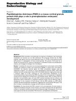
Peptidylarginine deiminase (PAD) is a mouse cortical granule protein that plays a role in preimplantation embryonic development docx
Bạn đang xem bản rút gọn của tài liệu. Xem và tải ngay bản đầy đủ của tài liệu tại đây (3.71 MB, 22 trang )
BioMed Central
Page 1 of 22
(page number not for citation purposes)
Reproductive Biology and
Endocrinology
Open Access
Research
Peptidylarginine deiminase (PAD) is a mouse cortical granule
protein that plays a role in preimplantation embryonic
development
Min Liu
1
, Andrea Oh
1
, Patricia Calarco
2
, Michiyuki Yamada
3
,
Scott A Coonrod
4
and Prue Talbot*
1
Address:
1
Department of Cell Biology and Neuroscience, University of California, Riverside, California 92521, USA,
2
Department of Anatomy and
Medicine, School of Medicine, University of California, San Francisco, California 94143, USA,
3
Graduate School of Integrated Science, Yokohama
City University, Yokohama, 236-0027 Japan and
4
Weill Medical College of Cornell University, New York, NY 10021, USA
Email: Min Liu - ; Andrea Oh - ; Patricia Calarco - ;
Michiyuki Yamada - ; Scott A Coonrod - ; Prue Talbot* -
* Corresponding author
Abstract
Background: While mammalian cortical granules are important in fertilization, their biochemical
composition and functions are not fully understood. We previously showed that the ABL2
antibody, made against zona free mouse blastocysts, binds to a 75-kDa cortical granule protein
(p75) present in a subpopulation of mouse cortical granules. The purpose of this study was to
identify and characterize p75, examine its distribution in unfertilized oocytes and preimplantation
embryos, and investigate its biological role in fertilization.
Results: To identify p75, the protein was immunoprecipitated from ovarian lysates with the ABL2
antibody and analyzed by tandem mass spectrometry (MS/MS). A partial amino acid sequence
(VLIGGSFY) was obtained, searched against the NCBI nonredundant database using two
independent programs, and matched to mouse peptidylarginine deiminase (PAD). When PAD
antibody was used to probe western blots of p75, the antibody detected a single protein band with
a molecular weight of 75 kDa, confirming our mass spectrometric identification of p75.
Immunohistochemistry demonstrated that PAD was present in the cortical granules of unfertilized
oocytes and was released from activated and in vivo fertilized oocytes. After its release, PAD was
observed in the perivitelline space, and some PAD remained associated with the oolemma and
blastomeres' plasma membranes as a peripheral membrane protein until the blastocyst stage of
development. In vitro treatment of 2-cell embryos with the ABL2 antibody or a PAD specific
antibody retarded preimplantation development, suggesting that cortical granule PAD plays a role
after its release in preimplantation cleavage and early embryonic development.
Conclusion: Our data showed that PAD is present in the cortical granules of mouse oocytes, is
released extracellularly during the cortical reaction, and remains associated with the blastomeres'
surfaces as a peripheral membrane protein until the blastocyst stage of development. Our in vitro
study supports the idea that extracellular PAD functions in preimplantation development.
Published: 01 September 2005
Reproductive Biology and Endocrinology 2005, 3:42 doi:10.1186/1477-7827-3-
42
Received: 18 July 2005
Accepted: 01 September 2005
This article is available from: />© 2005 Liu et al; licensee BioMed Central Ltd.
This is an Open Access article distributed under the terms of the Creative Commons Attribution License ( />),
which permits unrestricted use, distribution, and reproduction in any medium, provided the original work is properly cited.
Reproductive Biology and Endocrinology 2005, 3:42 />Page 2 of 22
(page number not for citation purposes)
Background
Mammalian cortical granules are membrane-bound
organelles located in the cortex of unfertilized oocytes
[1,2]. Following gamete membrane fusion, cortical gran-
ules undergo exocytosis, and some of the released compo-
nents block polyspermy by modifying the zona pellucida
[3-14]. In addition, some cortical granule proteins remain
associated with the embryo and appear to regulate embry-
ogenesis, since in vitro culture of 2-cell embryos in the
presence of antibodies specific to these proteins inhibited
embryo cleavage [15-17]. While most cortical granules are
released after fertilization, a subpopulation of Lens culi-
naris agglutinin (LCA)-binding cortical granules are
released around the cleavage furrow during first polar
body extrusion [18]. While the biological significance of
this pre-fertilization release is not yet known, it likely
plays a role in fertilization since it occurs at a specific time
and place and involves a specific population of cortical
granules. These prior studies show that mammalian corti-
cal granules are released both before and after fertilization
and that their functions are probably more complex than
previously realized.
The total number of mammalian cortical granule proteins
has been estimated to be between four and fourteen or
more [10,19,20]. Several specific proteins have been iden-
tified as cortical granule proteins [21]. N-acetylglucosami-
nidase was detected in exudates of ionophore-activated
mouse oocytes using an enzymatic assay and was local-
ized in the cortical granules at the electron microscopic
level [13]. Approximately 90% of oocyte N-acetylglu-
cosaminidase was released following in vivo fertilization
and was shown using competitive inhibitors or anti-N-
acetylglucosaminidase antibodies to be responsible for
the zona block to polyspermy [13]. Ovoperoxidase was
detected in the cortical granules of unfertilized mouse
oocytes at the ultrastructural level using the 3.3'-diami-
nobenzidine (DAB) [7,8]. Following artificial activation,
ovoperoxidase was present on the oocyte's surface, in the
perivitelline space, and in the zona pellucida. Following
fertilization, the enzyme was inferred to harden the zona
pellucida, since both peroxidase inhibitors and tyrosine
analogs prevented hardening [8]. Calreticulin, an endo-
plasmic reticulum protein involved in calcium storage,
was demonstrated in granules in the cortex of hamster
oocytes by indirect immunofluorescence [22]. However, a
subsequent study showed that most of the granules con-
taining calreticulin did not label with the lectin LCA, a
classical marker for mouse oocyte cortical granules [23].
This lead to the conclusion that calreticulin is localized in
a population of granules that is distinct from classical cor-
tical granules.
In addition, several proteins (p32, p56, p62, and p75)
have been localized immunocytochemically in cortical
granules, but their identities have not yet been established
[17,19,20]. p32 was recognized on western blots by a
monoclonal antibody (3E10) made against mouse corti-
cal granule exudates and was localized immunohisto-
chemically to cortical granules in germinal vesicle intact
and metaphase II stage mouse oocytes [19]. Interestingly,
p32 was not detected in 3E10 labeled fertilized oocytes
and preimplantation embryos following the cortical reac-
tion. While the function of p32 is not known, treatment
of unfertilized oocytes with the 3E10 antibody did not
increase polyspermy, indicating that for the experimental
conditions used, p32 did not function in blocking
polyspermy. The polyclonal antibody ABL
2
, which was
made against zona free mouse blastocysts and which
immunoprecipitates a 75-kDa protein from mouse
oocytes, reacts immunocytochemically with cortical gran-
ules [20]. The protein is released following in vitro fertili-
zation and artificial activation [20]. In hamster oocytes, a
pair of cortical granule proteins designated p56 and p62,
was recognized on western blots by the ABL
2
antibody
[16]. These two ABL
2
specific hamster cortical granule pro-
teins are related to sea urchin hyalin since they are also
recognized by the S. purpuratus hyalin specific antibody
IL2 [17]. p56 and p62 are retained in the perivitelline
space and on the oolemma after fertilization. These pro-
teins appear to be involved in early embryogenesis since
in vivo treatment of 2-cell embryos with IL2 or ABL
2
anti-
bodies inhibited blastomere cleavage [16,17]. In vitro
treatment of 2-cell mouse embryos with the ABL
2
anti-
body showed similar inhibition of development [15].
Although experimental and immunohistochemical work
has been done on these cortical granule proteins, they
have not yet been identified biochemically or character-
ized functionally.
The purpose of this study was to identify the mouse corti-
cal granule protein p75, to characterize its distribution in
unfertilized oocytes and preimplantation embryos, and to
examine its function in fertilization. To accomplish this,
p75 was immunoprecipitated from an ovarian lysate, iso-
lated using SDS-PAGE, then analyzed using tandem mass
spectrometry. A partial peptide sequence of the protein
was obtained and used to identify p75 as a member of the
peptidylarginine deiminase (PAD) family of enzymes that
catalyze the conversion of arginine to citrulline [24].
Materials and methods
Chemicals and Supplies
Chemicals used to make all media, polyvinylpyrrolidone
(PVP), bovine serum albumin (BSA), pregnant mare's
serum gonadotropin (PMSG), human chorionic gonado-
tropin (hCG), bovine hyaluronidase, protein A-sepharose
beads, M16 medium, paraformaldehyde, Triton-X 100, α-
D-mannose, N-acetylglucosamine, β-D-galactose, and N-
acetylgalactosamine were purchased from Sigma
Reproductive Biology and Endocrinology 2005, 3:42 />Page 3 of 22
(page number not for citation purposes)
Chemical Company (St. Louis, MO). HEPES buffer, light
mineral oil, slides, and coverslips (#1.5) were purchased
from Fisher (Tustin, CA). Lens culinaris agglutinin (LCA),
streptavidin conjugated to Texas Red, and Vectashield
mounting medium were purchased from Vector Laborato-
ries (Burlingame, CA). SYTOX orange nucleic acid stain
and Alexa-488 conjugated to goat anti-rabbit IgG were
obtained from Molecular Probes (Eugene, OR). PAD V
(N) antibody was made against recombinant human PAD
V and affinity purified on an N-terminal PAD V fragment
(1–262) bound column as previously described [25].
ePAD antibody was made against the N-terminal frag-
ment (1–200) of mouse recombinant ePAD [26].
Animals
NIH Swiss white mice were purchased from Harlan (San
Diego, CA). Mice were housed in a University of Califor-
nia at Riverside vivarium with a 14-hour light and 10-
hour dark cycle and fed water and Purina rodent chow
(Ralston-Purina, St. Louis, MO) ad libitum. Protocols
used in this study were approved by the campus Commit-
tee on Animal Care.
Media and Fixatives
For dissection and oocyte collection, Earle's balanced salt
solution with 28.18 mM of sodium bicarbonate and
24.98 mM of HEPES free acid (EBSS-H), pH 7.4 supple-
mented with 0.3% of polyvinylpyrrolidone (EBSS-H/
0.3% PVP) was made as previously described [27]. For
immunoprecipitation, lysis buffer was made with 150
mM NaCl, 10% NP-40, 0.5% sodium deoxycholate, 0.1%
SDS, 50 mM Tris-HCl, pH 7.5, and a protease inhibitor
cocktail as previously described [13]. For egg activation,
calcium and magnesium free EBSS-H (EBSS
-Ca/Mg
-H) was
used as previously described [19]. High salt-containing
solution was made by increasing the sodium chloride
concentration in EBSS-H/0.3% PVP to 300 mM. For
embryo culture, M16 medium was pregassed in 37°C
humidified incubator (5% CO
2
, 95% air) overnight
before use. For confocal scanning laser microscopy, Dul-
becco's phosphate buffered saline (DPBS), pH 7.4 or
phosphate buffered saline (PBS), pH 7.4 was used. DPBS
was made with 90.9 mM CaCl
2
, 2.68 mM KCl, 1.47 mM
KH
2
PO
4
, 0.49 mM MgCl
2
·6H
2
O, 136.89 mM NaCl, and
8.06 mM Na
2
HPO
4
·7H
2
O. PBS was made as described
previously [25]. For fixation, 4% paraformaldehyde was
made in DPBS, pH 7.4, or in PBS, pH 7.4. Blocking solu-
tion was made in DPBS, pH 7.4 supplemented with 7.5
mg/ml glycine and 3 mg/ml BSA immediately prior to use.
In some cases, blocking solution was made in PBS. 10 mM
citrate buffer pH 7.0 was made with 3.78 g of citric acid
and 2.411 g of sodium citrate in 1 L of H
2
O. To remove
peripheral ABL
2
specific antigen following egg activation,
high salt-containing EBSS-H/0.3% PVP containing 300
mM NaCl was used. For confocal scanning laser micros-
copy, labeling solution was made by supplementing
DPBS, pH 7.4 with 30 mg/ml BSA (DPBS/3% BSA). For
LCA blotting, Tris-buffered saline (TBS), pH 7.6 was used
(147 mM NaCl; 20 mM Tris-base)
Oocyte and Embryo Collection
For epifluorescence microscopy, confocal scanning laser
microscopy, and gel electrophoresis, oocytes and preim-
plantation embryos were collected in EBSS-H/0.3% of
PVP at room temperature. To collect germinal vesicle
intact oocytes, female mice were injected intraperitoneally
with 10 IU of PMSG (Sigma, St Louis, MO). Oocytes were
collected 60 hours later from the ovaries and mechani-
cally denuded of their cumulus cells with a thin-bore glass
pipette. Unfertilized mature metaphase II oocytes were
collected from female mice that were primed with 10 IU
of PMSG at 2200 hours on day 1 followed by 10 IU of
hCG (Sigma, St. Louis, MO) 46 hours later. For egg activa-
tion, oocytes were flushed out from oviducts with collec-
tion medium 16 to 18 hours post the hCG injection. To
collect in vivo fertilized oocytes and preimplantation
embryos, female mice were superovulated by intraperito-
neal injection of 10 IU of PMSG at 1430 hours on day 1
followed by 10 IU of hCG 46 hours later and then placed
in cages containing 2–3 male mice. The following day, fer-
tilized oocytes were collected by flushing the oviduct with
collection medium. Only oocytes with two pronuclei were
used. Two-cell preimplantation embryos were collected
by flushing the oviduct with collection medium 2 days
after mating. Four- and eight-cell preimplantation
embryos were collected by flushing the oviduct or the
uterine horns with collection medium 3 days after the
mating. Blastocysts were collected by flushing the uterine
horns 4 days after mating.
For mature metaphase II oocytes and in vivo fertilized
oocytes, cumulus cells were removed by incubating
oocytes in collection medium containing 100 IU of
hyaluronidase for 5 minutes at room temperature. In
some experiments, zonae pellucidae were removed with
0.25% pronase in collection medium.
Human Peripheral Blood Cell Collection
Human peripheral blood cells were obtained from an
informed and consenting healthy donor. Red blood cells
were removed by sedimentation with dextran 200,000,
and the remaining cells were then subjected to Percoll
density-gradient centrifugation. Layers containing granu-
locytes were collected, and cells were then spread onto
glass slides by cytospinning.
Immunoprecipitation
For immunoprecipitation, all steps were carried out in
lysis buffer unless otherwise specified. Ovaries from adult
female mice were dissected out in EBSS-H and
Reproductive Biology and Endocrinology 2005, 3:42 />Page 4 of 22
(page number not for citation purposes)
homogenized on ice. The homogenate was kept on ice for
one hour then centrifuged at 30,000 g at 4°C for 30 min-
utes to remove any insoluble material. The supernatants
of ovarian homogenate were saved for immunoprecipita-
tion. Homogenates of other tissues were also prepared as
described above. Non-specific binding was reduced by
incubation of the extracts with normal rabbit serum at
4°C with constant agitation for 90 minutes. To remove
any protein-A and Sepharose bead binding proteins
before using ABL
2
, protein A-Sepharose beads were then
added and incubated with the extracts at 4°C with con-
stant agitation for 30 minutes. The beads were pelleted by
a low-speed centrifugation and supernatant was collected.
The clean ovarian extracts were incubated overnight with
ABL
2
at a final concentration of 0.37 mg/ml at 4°C with
constant agitation. Fresh protein A-Sepharose beads were
added and incubated with the ovarian extracts at 4°C for
90 minutes on the next day. Beads were pelleted by a low-
speed centrifugation, and the ovarian extracts were dis-
carded. Beads were rinsed three times for a total of 45
minutes at room temperature, and sample buffer [28] was
added.
Mass Spectrometry
The ABL
2
immunoprecipitate was excised from the silver
stained gel and the sample was sent to W.M. Keck Foun-
dation Biotechnology Resource Laboratory (Yale Univer-
sity, New Haven, CT) for MS/MS identification. The
procedures used at the Keck Laboratory are available on
the website of the facility />.
Briefly, in gel trypsin digestion was performed, and pro-
tein was eluted with 50% acetonitrile and 0.1% formic
acid. The eluted sample was desalted and was then sub-
jected to nanospray MS/MS to obtain amino acid
sequences of the tryptic digest.
Egg Activation
To examine release of PAD from live oocytes using
immunofluorescence microscopy, oocytes were activated
by incubating them in hyaluronidase for 10–15 minutes.
The concentration of hyaluronidase used (approximately
200–250 units) was higher and the length of exposure was
longer than is normally used to remove cumulus cells.
These conditions of hyaluronidase treatment resulted in
activation of most of the oocytes.
To determine if PAD remains associated with the plasma
membrane as a peripheral protein after its release from
cortical granules, zona free unfertilized metaphase II
oocytes were incubated in EBSS
-Ca/Mg
-H supplemented
with 0.3% PVP for 15 min at 37°C, and oocytes were arti-
ficially activated with 2 µM ionomycin for two minutes at
37°C. Control oocytes were incubated with 0.1% of
DMSO for two minutes at 37°C. Activated oocytes were
transferred to fresh EBSS-H supplemented with 0.05%
PVP droplets under light mineral oil and incubated for 15
minutes at 37°C. Oocytes were then incubated in high
salt-containing solution for 2 minutes at room tempera-
ture with constant pipetting to remove exocytosed materi-
als from the oocyte surface. Some control oocytes were
treated as mentioned above.
In Vitro Embryo Culture
Zona intact 2-cell preimplantation embryos were col-
lected as described above in the oocyte and embryo collec-
tion section. Embryos were cultured in 50 µl of M16
supplemented with 0.02% of gentamycin under mineral
oil at 37°C in the incubator (5% CO
2
, 95% air) for three
days. The amount of antibody added to the droplet on day
one as indicated below: 5 µg for polyclonal rabbit IgG,
1:100 dilution for polyclonal guinea pig IgG, 5 µg for anti-
alpha integrin antibody, 5 µg for the antibody ABL
2
, and
1:100 dilution for anti-ePAD antibody. In some experi-
ments, no antibody was added to the droplets. The
embryos were checked everyday and total percentage of
embryos that reached the blastocyst stage was recorded for
each experimental group on day three.
Confocal Scanning Laser and Epifluorescent Microscopy
All procedures for CSLM were carried out at room temper-
ature under light mineral oil unless otherwise specified.
All samples for LCA and ABL
2
labeling were fixed with 4%
paraformaldehyde in DPBS, pH 7.4 for 30 minutes and
most samples for PAD labeling were fixed with 4% para-
formaldehyde in PBS, pH 7.4 for 30 minutes. Following
fixation, samples were washed in blocking solution for a
total of 30 minutes and then permeabilized with 0.1%
Triton X-100 in blocking solution for 5 minutes. All sam-
ples were labeled in labeling solution and each labeling
incubation was followed by several washes in fresh labe-
ling solution for a total of 30 minutes. For ABL
2
labeling,
samples were incubated with a 1:300 dilution (40 µg/ml)
of ABL
2
for 30 minutes followed by 30 minutes of incuba-
tion in goat anti-rabbit IgG conjugated to Alexa 488 with
a 1:300 dilution (6.6 µg/ml). Control samples were incu-
bated with a 1:1000 dilution (28.3 µg/ml) of preimmune
rabbit IgG for 30 minutes followed by goat-anti-rabbit
Alexa 488. For LCA labeling, samples were incubated with
10 µg/ml of biotinylated LCA for 30 minutes followed by
30 minutes of incubation in 5 µg/ml of Texas Red-strepta-
vidin. Control samples were incubated with 10 µg/ml of
LCA that had been preincubated with 100 mM α-methyl-
mannopyranoside for 30 minutes followed by 30 minutes
of incubation with 5 µg/ml of Texas Red-streptavidin. To
double label oocytes or preimplantation embryos, sam-
ples were first incubated with ABL
2
followed by the goat
anti-rabbit IgG conjugated to Alexa 488 then incubated
with LCA followed by Texas Red-streptavidin as described
previously. For PAD labeling, fixed samples were treated
with 10 mM citrate buffer for 15 minutes at 95°C,
Reproductive Biology and Endocrinology 2005, 3:42 />Page 5 of 22
(page number not for citation purposes)
incubated with 2 M Tris-HCl, pH 7.4, for 15 minutes, and
then permeabilized with 0.1% Triton X-100 in PBS for 10
minutes. Samples were blocked with 2% normal goat
serum and 2% BSA in PBS for 60 minutes and incubated
with 1.5 µg/ml of rabbit anti-PAD V overnight. On the fol-
lowing day, samples were incubated in goat anti-rabbit
IgG conjugated to Alexa 488 with a 1:300 dilution (6.6
µg/ml) for three hours at room temperature. For LCA and
PAD double labeling, samples already labeled with PAD
antibody were incubated with 10 µg/ml of LCA for 30
minutes and followed by 30 minutes of incubation with 5
µg/ml of Texas Red-streptavidin on following day. Con-
trol samples, non-permeabilized or permeabilized, were
incubated with goat anti-rabbit IgG conjugated to Alexa
488 or Texas Red-streptavidin only. All labeled samples
were examined using a Zeiss LS 510 confocal scanning
laser microscope the next day. Samples were entirely sec-
tioned optically with a space interval determined accord-
ing to the pinhole setting. For some samples, two-
dimensional projections of z-stacks were generated.
To label live unfertilized, activated, or fertilized oocytes
with anti-ePAD, samples were incubated at room temper-
ature in M16 culture medium containing anti-ePAD
(1:100) for 45 minutes, washed in M16, and incubated 45
minutes at room temperature in M16 containing anti-
guinea pig IgG conjugated to Alexa 488 (1:100). Oocytes
were then washed and immediately viewed with a Nikon
inverted epifluorescence microscope.
For in vitro cultured embryos, live embryos that had been
incubated in a primary antibody (ABl
2
, anti-ePAD, or anti-
integrin) were washed in M16 then incubated in either
goat anti-rabbit IgG conjugated to Alexa 488 with a 1:100
dilution (19.8 µg/ml) or goat anti-guinea pig IgG conju-
gated to FITC with a 1:100 dilution for 1 hour at room
temperature. After washing, live samples were examined,
and images were taken with a Zeiss epifluorescence
microscope.
Gel Electrophoresis and Lectin Blotting
Protein samples were solubilized with reducing and dena-
turing Laemmli sample buffer [28] prior to electrophore-
sis. Samples and biotinylated standards were run in one-
dimensional SDS-PAGE Doucet gels (4% stacking/7.5%
separating) [29] at 70 V and 140 V respectively and sepa-
rated proteins were blotted onto nictrocellulose at 100 V
for 1 hour [30]. For protein identification by mass spec-
trometry, the gel was silver stained after electrophoresis as
previously described [31]. For lectin blotting, blots were
washed in Tris-buffer saline (TBS) for 15 minutes at room
temperature and then blocked with 0.5% Tween-20 in
Tris-buffer saline (TBT) for 1 hour at room temperature.
1–10 µg/ml of the appropriate biotinylated lectin in TBT
was added to the blot for overnight incubation at 4°C
with constant agitation. For each control blot, bioti-
nylated lectin was preabsorbed with 100 mM of control
sugar for 2 hour at room temperature prior to the over-
night incubation. Blots were washed with TBT four times
for 60 minutes on the following day and then incubated
in a 1:20,000 dilution of HRP-streptavidin in TBT for 40
minutes at room temperature. For PAD immunoblotting,
blots were first blocked with 5% nonfat dry milk in PBS
with 0.05% Tween 20 (PBT) for 30 minutes at room tem-
perature and then washed with fresh PBT for 15 minutes.
Blots were incubated with a 1:4000 dilution of anti-ePAD
guinea pig IgG in PBT overnight at 4°C with constant agi-
tation. For controls, all blots were either incubated with a
1:4000 dilution of preimmune guinea pig IgG in PBT or
in PBT without antibody added. On the following day,
blots were washed for 15 minutes with PBT and incubated
with 1:2000 dilution of goat anti-guinea pig IgG conju-
gated with peroxidase for 2 hours at room temperature.
For both lectin and PAD blots, enhanced chemilumines-
cence (Amersham, Piscataway, NJ) was used to detect
bands of interest and band images were captured using
Kodak X-Omat autoradiographic films. The molecular
weight of protein was calculated using biotinylated
standards.
Statistical Analyses
The percentage of 2-cell preimplantation embryos reach-
ing the blastocyst stage in the presence of different anti-
bodies and the percentage of 2-cell preimplantation
embryos reaching the blastocyst stage in the absence of
any antibody (control) were analyzed statistically using a
one-way analysis of variance (ANOVA) followed by Dun-
net's post-hoc test when results of the ANOVA were signif-
icant. In both the ANOVA and Dunnet's test, results were
considered significant when p ≤ 0.05.
Results
The ABL
2
antibody recognizes a 75-kDa ovarian protein
that is present in cortical granules of mouse oocytes
The ABL
2
antibody precipitates a 75 kDa protein (p75)
from mouse oocytes [20]. To determine if other tissues
express p75, various mouse tissue extracts were used to
perform ABL
2
immunoprecipitation. p75 was immuno-
precipitated from the ovary by ABL
2
(Fig. 1A, lane 4), but
not from brain, liver, muscle, oviduct, or testis (Fig. 1A,
lanes 1–3 and lanes 5–6). Both the ABL
2
antibody (Figs.
1B, ABL
2
) and the lectin Lens culinaris agglutinin (LCA)
(Fig. 1B, LCA) labeled granules in the cortex of oocytes.
Many granules showed co-localization of the two probes
in merged images (Fig. 1B, ABL
2
/ LCA), demonstrating
p75 to be a mouse cortical granule protein. Co-localiza-
tion of two probes was also observed in pre-translocated
cortical granules located in the cytoplasm of germinal ves-
icle intact oocytes (Figs. 1 and 2 in [18]). Cryosections of
Reproductive Biology and Endocrinology 2005, 3:42 />Page 6 of 22
(page number not for citation purposes)
Tissue distribution of the ABL
2
antigenFigure 1
Tissue distribution of the ABL
2
antigen. (A) Silver-stained SDS-PAGE gel loaded with the ABL
2
immunoprecipitate from mouse
brain (lane 1), liver (lane 2), skeletal muscle (lane 3), ovary (lane 4), oviduct (lane 5), and testis (lane 6). The ABL
2
antibody
immunoprecipitated a 75-kDa protein from the ovarian lysate but not from other tissues. Other bands in the gel are from the
antibody used for immunoprecipitation. (B) Confocal scanning laser micrographs of germinal vesicle intact mouse oocytes dou-
ble labeled with the lectin LCA (LCA) and the ABL
2
antibody (ABL
2
). The merged image (LCA + ABL
2
) showed co-localization
of LCA and ABL
2
in some cortical granules. These images were digitally enlarged for better visualization. (C) Western blots in
which ABL
2
immunoprecipitate was probed with the lectins ConA, LCA, WGA, PNA, and DBA. Control blots were probed
with lectins preabsorbed with the appropriate control sugar. Positive controls (blots with rabbit IgG) were included for each
lectin to show that the blotting condition was optimized.
Reproductive Biology and Endocrinology 2005, 3:42 />Page 7 of 22
(page number not for citation purposes)
Identification of the ABL
2
antigen using tandem mass spectrometryFigure 2
Identification of the ABL
2
antigen using tandem mass spectrometry. (A) Silver-stained SDS-PAGE gel loaded with ABL
2
immu-
noprecipitate from mouse ovarian lysate (lane A) and molecular weight standards (lane B). In this experiment, a protein with
molecular weight of 65-kDa co-precipitated with p75. (B) MS/MS spectrum of a peptide obtained from trypsin-digested p75;
the sequence of this peptide was determined to be VLIGGSFY. (C) Western blots of an ABL
2
immunoprecipitate from a mouse
ovarian lysate probed with guinea pig anti-ePAD IgG, preimmune guinea pig IgG, or goat anti-guinea pig IgG conjugated to per-
oxidase only.
Reproductive Biology and Endocrinology 2005, 3:42 />Page 8 of 22
(page number not for citation purposes)
mouse ovary did not show ABL
2
labeling anywhere in the
ovary except in the cortical granules (data not shown).
Since cortical granule proteins are secreted and most
secreted proteins are glycosylated, we performed lectin
blotting on immunoprecipitates from ovarian lysates to
determine if p75 is glycosylated [32,33]. Blots with p75
were probed with α-D-mannose-specific ConA and LCA,
N-acetylglucosamine-specific WGA, β-D-galactose-spe-
cific PNA, and N-acetylgalactosamine-specific DBA. None
of these lectins bound to p75 on the blots (Fig. 1C, p75 +
lectins), indicating that p75 is probably not glycosylated.
Blots with rabbit IgG were used as a positive control to
optimize the blotting condition for each lectin and to
demonstrate that the assay was working (Fig. 1C, positive
control). Control blots probed with lectins preabsorbed
with the appropriate sugar under the same blotting condi-
tions did not show binding to rabbit IgG (Fig. 1C, sugar
controls), demonstrating the specificity of each lectin.
Identification of p75 using mass spectrometry
To identify p75, the protein was immunoprecipitated
from ovarian lysates with the ABL
2
antibody and analyzed
using mass spectrometry. Generally immunoprecipitation
yields a single band of 75 kDa; however, occasionally a
second band of 65 kDa is also obtained as shown in Fig-
ure 2A. High-energy collision-induced dissociation (CID)
spectra of the trypsin-digested of peptides from each pro-
tein band was obtained, and partial amino acid sequences
of the peptides were deduced. For the 65-kDa band, three
peptide sequences were obtained (LVQEVTDFAK/
APQVSTPTLVEARAR/LSQTFPNADFAEITK) from the
spectra. When sequences were searched separately using
BLAST against the NCBI nonredundant database, they all
matched serum albumin precursor [GenBank:P07724
].
For p75, a CID mass spectrum of the parent peptide ion
(at m/z 1468.8
+2
) was obtained and used to deduce the
amino acid sequence (Fig. 2B). The spectrum showed a
series of peptide ions of decreasing mass generated from
the parent peptide. The mass difference between each con-
secutive peptide ion was used to determine the parent
peptide sequence, and a partial amino acid sequence,
VLIGGSFY, was then obtained as shown in Figure 2B. The
VLIGGSFY sequence matched several mouse peptidy-
larginine deiminases (PAD) when searched using BLAST
against the NCBI nonredundant database. These included
a putative mouse PAD type V-like protein [Gen-
Bank:XP_144067
] predicted by NCBI automated gene
predicting algorithm, an egg and embryo abundant PAD
[GenBank:AH53724
], and a recently characterized mouse
oocyte protein, ePAD [GenBank:NP_694746
]. Although
the egg and embryo abundant PAD (AAH53724) and
ePAD (NP_694746) are listed under different entries in
the database, they may be the same since their protein
sequences are identical except for three amino acids; how-
ever, we can not exclude the possibility that they are dupli-
cated genes. In addition, Sonar MS/MS (Genomic
Solutions), another software tool designed for mass spec-
trometric protein identification, was used to search the
NCBI nonredundant database. Unlike most database
search algorithms that perform protein identification
based exclusively on amino acid sequence, Sonar MS/MS
includes additional information such as the mass-to-
charge (m/z) ratio of the original parent peptide ion to
perform identification. This information becomes essen-
tial for validating positive protein identification when
only a partial amino acid sequence can be obtained from
the original parent peptide, as had been the case in this
study. The result obtained using Sonar MS/MS showed
that the sequence VLIGGSFY was matched to PADs, as had
been demonstrated with the BLAST search. To confirm the
MS/MS identification of p75, we used an antibody that
was made against mouse ePAD [26] to probe blots of the
ABL
2
immunoprecipitate. The ePAD antibody detected a
single protein band with a molecular weight of 75 kDa
(Fig. 2C, ePAD). No bands were detected when preim-
mune IgG or goat anti-guinea pig IgG conjugated to per-
oxidase alone were used (Fig. 2C, PI and anti-guinea pig
IgG). These results demonstrate that the p75 immunopre-
cipitated by ABL
2
is indeed a PAD and confirm our MS/MS
identification of p75.
Amino acid sequence comparison of different PADs
Using the MultiAlin program [34], we constructed protein
sequence alignments of nine mammalian PAD proteins
including all mouse PADs (five characterized mouse
PADs: PAD I – IV and ePAD; two uncharacterized mouse
PADs, rat PAD VI, and human PAD V [GenBank:
NP_035189
, NP_032838, NP_035190, AAH53724,
XP_144067
, NP_694746. XP_233601, NP_036519] (Fig.
3). Sequence residues that are in high consensus are
shown in red and sequence residues that are in low con-
sensus are shown in blue. Gaps (-) are introduced for opti-
mal alignment. The multiple alignments of the nine
mammalian PADs show that approximately 40% – 50%
of the amino acid sequences in these PADs are identical,
indicating strong homologies among members of this
family. Two predictive algorithms (SignalP V2.0 and Tar-
getP V1.0) [35-37] were used to determine that a putative
signal peptide and a cleavage site exist in ePAD and
AAH53724 (an egg and embryo abundant peptidy-
larginine deiminase), indicating they are likely secreted
proteins (Fig. 3 arrow). Human PAD V has a monopartite
nuclear localization sequence motif [25], and it is the only
type of PAD that has been localized to the nuclei of cells
(Fig. 3 underline). Only ePAD, AAH53724 (an egg and
embryo abundant peptidylarginine deiminase), and
XP_144067 (peptidylarginine deiminase type V-like pro-
tein) have residues that exactly match the VLIGGSFY
sequence (Fig. 3 asterisks). Interestingly, rat PAD VI also
Reproductive Biology and Endocrinology 2005, 3:42 />Page 9 of 22
(page number not for citation purposes)
Multiple alignments of mammalian PAD protein sequencesFigure 3
Multiple alignments of mammalian PAD protein sequences. The sequences were aligned using the program MultiAlin available
at />. The peptide sequence (VLIGGSFY) of p75 obtained from MS/MS analy-
sis was searched against listed PADs and residues that were matched to it are marked (*). The signal peptide cleavage site is
marked with an arrow. The monopartite nuclear localization sequence in human PAD V is underlined. High consensus
sequences are in red (90% of amino acids are identical or have biochemically similar R-groups) and low consensus sequences
are in blue (50% of amino acids are identical or have biochemically similar R-groups). The abbreviations of species are listed as
followed: Mm = M. musculus; Rn = R. norvegicus; Hs = H. sapiens. Two putative mouse PAD sequences are referred with their
accession numbers (GenBank/NCBI). Accession numbers (GenBank/NCBI) of other PADs are as followed: NP_694746
;
XP_233601
; NP_035189; NP_035190; NP_036519; NP_032838.
Reproductive Biology and Endocrinology 2005, 3:42 />Page 10 of 22
(page number not for citation purposes)
has a sequence match to the peptide VLIGGSFY obtained
from p75 MS/MS analysis except for the first residue V
(Fig. 3 asterisks). PAD I is derived from a gene predictive
program, and its sequence is 80% identical to that of
ePAD or AAH53724 (an egg and embryo abundant pepti-
dylarginine deiminase).
Mouse cortical granules contain PAD
To ascertain if mouse cortical granules contain PAD, anti-
bodies made against mouse ePAD and human recom-
binant PAD V (anti-PAD V (N)) were used to label in vivo
matured germinal vesicle intact and metaphase II mouse
oocytes. The ePAD antibody had been used previously
[26] and showed strong labeling in the cortex. When we
adjusted labeling conditions to optimize cortical labeling,
both granular and cytoplasmic labeling were observed in
the cortex with anti-ePAD; however, the high level of cyto-
plasmic labeling made it difficult to resolve individual
granules and to demonstrate co-localization with LCA, a
cortical granule binding lectin (not shown). Therefore the
antibody to human PAD-V, which gave a cleaner signal in
the cortex, was also used to localize PAD in cortical gran-
ules (Fig. 4).
Human peripheral blood cells were first used as a positive
control and to optimize labeling conditions with anti-
human PAD-V. The antibody labeled only the granulo-
cytes (neutrophils and eosinophils), and labeling was
localized to the nuclei of the cells (Fig. 4A), as reported
previously [25]. When germinal vesicle intact oocytes
were then labeled, immunoreactivity was localized in the
nucleus and also in granules in the cortex (Fig. 4B). In
metaphase II oocytes, the antibody labeled granules in the
cortex; except in the area of the cortical granule free
domain which was devoid of PAD labeling (Fig. 4C). In
the metaphase II oocytes, the nuclear envelope had bro-
ken down, and thus there was no nuclear staining; how-
ever, the cytoplasm of metaphase II oocytes was more
intensely labeled than that of germinal vesicle intact
oocytes, suggesting that nuclear PAD was now dispersed
in the cytoplasm (Figs. 4B, C). These results demonstrate
that PAD is present in the cortical granules, nucleus, and
cytoplasm of unfertilized mouse oocytes. Control oocytes
were not labeled with goat anti-rabbit IgG conjugated to
Alexa 488 alone (Fig. 4D).
To confirm that anti-PAD V (N) is labeling cortical gran-
ules in the oocyte's cortex and that PAD is present in these
granules, anti-PAD V (N) and LCA were used to double
label germinal vesicle intact and metaphase II oocytes,
and their labeling pattern was compared to that of ABL
2
and LCA double labeled oocytes. Both anti-PAD V (N)
and LCA labeled granules (arrow) in the cortex of germi-
nal vesicle intact oocytes (Figs. 4E, F). When images of
both probes were merged, many granules appeared
orange or yellow indicating co-localization of these
probes (Fig. 4G), and similar co-localization of granules
was also observed when metaphase II oocytes were used
(Fig. 4H). In the metaphase II oocytes, an area devoid of
signal corresponding to the cortical granule free domain
was observed (Fig. 4H), and this domain was not labeled
by either anti-PAD V (N) or LCA. When ABL
2
and LCA
were used to double label metaphase II oocytes, both
probes labeled the granules in the cortex and showed co-
localization of granules (Figs. 4I–K), as had been observed
with anti-PAD V (N) and LCA. Besides the granules in the
cortex, anti-PAD V (N) also labeled cytoplasm near the
cortical granules; however, this labeling is diffuse and less
granular than the cortical granule labeling. This diffuse
cytoplasmic labeling did not co-localize with LCA labe-
ling (Figs. 4F, G, arrowhead). Control oocytes labeled
with LCA pre-absorbed with α-D-methyl-mannopyrano-
side showed no labeling (Fig. 4L). Taken together, these
results demonstrate that antibodies to PAD label cortical
granules of mouse oocytes as had been observed with the
ABL
2
antibody and that PAD (ABL
2
antigen, p75) is
present in the cortical granules of mouse oocytes.
Localization of PAD (p75) after artificial activation and
fertilization
To demonstrate that PAD is released from cortical gran-
ules when they undergo exocytosis, unfertilized, hyaluro-
nidase activated, and in vivo fertilized oocytes were
compared using immunofluorescence microscopy (Fig.
5). All oocytes were labeled live (non-permeabilized) with
the primary and secondary antibody and were imaged
using an inverted epifluorescent microscope to minimize
damage to the living oocytes. Since only extracellular PAD
was imaged in this experiment, anti-ePAD was used, and
cortical cytoplasmic labeling did not interfere with inter-
pretation of the images, as had occurred when oocytes
were permeabilized and imaged with confocal micros-
copy (see previous section). Secondary antibody alone did
not label unfertilized or fertilized oocytes (Figs. 5A–B, C–
D). Unfertilized live oocytes did not show extracellular
fluorescence when labeled with both anti-ePAD and the
secondary antibody (Figs 5E–F), Oocytes caught in vari-
ous stages of activation showed distinct patterns of extra-
cellular labeling with anti-ePAD (Figs 5G–J). In early
stages of activation, numerous extracellular granules were
labeled in the perivitelline space (Figs. 5H–I). Many of
these granules were the size of cortical granules suggesting
they were recently exocytosed (Fig 5H). Other granules
had begun to disperse and were larger in diameter (Fig 5I).
At later times after activation, granular content had dis-
persed completely within the perivitelline space, and
some labeling appeared associated with the oolemma (Fig
5J). Similar to activated oocytes, fertilized oocytes that
were recovered from oviducts of mated females had
labeled granules in the perivitelline space (Fig 5K). At later
Reproductive Biology and Endocrinology 2005, 3:42 />Page 11 of 22
(page number not for citation purposes)
Confocal scanning laser micrographs of (A) human blood cells and (B – D) in vivo matured mouse oocytes labeled with anti-PAD V (N), (E – H) in vivo matured mouse oocytes double labeled with anti-PAD V (N) and LCA, and (I – L) double labeled with ABL
2
and LCAFigure 4
Confocal scanning laser micrographs of (A) human blood cells and (B – D) in vivo matured mouse oocytes labeled with anti-
PAD V (N), (E – H) in vivo matured mouse oocytes double labeled with anti-PAD V (N) and LCA, and (I – L) double labeled
with ABL
2
and LCA. All anti-PAD V labeling is shown in green, except in A where it is red. DNA stain in A is green. ABL
2
labe-
ling is green and LCA labeling is red in all figures. (A) Cytospin preparations of the granulocyte fraction were stained with anti-
PAD V (N), and their nuclei were stained with SYTOX green nucleic acid stain. The merged image shows nuclear localization
of PAD (yellow) in a human granulocyte. (B, C) Germinal vesicle intact mouse oocytes and metaphase II oocytes were labeled
with anti-PAD V (N), (D) Metaphase II mouse oocyte did not show labeling with goat anti-rabbit IgG conjugated to Alexa 488
alone. (E, F) Polar sections of germinal vesicle intact mouse oocytes double labeled with LCA (red) and anti-PAD V (N)
(green). These images were digitally enlarged 2× for better visualization. (G) Merged image of both LCA and anti-PAD V (N)
showed co-localization (yellow) of labels. (H) Merged image of equatorial section of metaphase II mouse oocytes double
labeled with anti-PAD V (N) and LCA showing co-localization. (I, J) Metaphase II oocytes double labeled with LCA (red) and
ABL
2
(green). (K) Merged image of both LCA and ABL
2
showed co-localization (yellow). The inserts of I, J, and K showed the
polar view of the oocyte. (L) Control oocytes were not labeled with LCA pre-absorbed with α-D-methyl-mannopyranoside.
All samples were imaged at same magnification and the scale bar applies to all figures.
Reproductive Biology and Endocrinology 2005, 3:42 />Page 12 of 22
(page number not for citation purposes)
Live, non-permeabilized activated and in vivo fertilized oocytes showing release of PAD from the cortical granulesFigure 5
Live, non-permeabilized activated and in vivo fertilized oocytes showing release of PAD from the cortical granules. A and B are
the same unfertilized oocyte viewed with Hoffman optics (A) or epifluorescence microscopy (B) after labeling with the sec-
ondary antibody only. No non-specific labeling is observed in B. C and D are the same fertilized oocyte viewed with Hoffman
optics (C) or fluorescence microscopy (D) after labeling with secondary antibody only. No non-specific labeling is observed
after fertilization. E and F are the same unfertilized oocyte viewed with Hoffman microscopy (E) or epifluorescence micros-
copy (F) after labeling with both the anti-ePAD and the secondary antibody. No labeling is observed around the unfertilized
oocyte (F). G, H, and I are the same early activated oocyte viewed with Hoffman optics (G) or after labeling with both anti-
ePAD and secondary antibody (H, I). H is focused close to the surface of the oocyte and shows numerous small labeled gran-
ules in the perivitelline space. I is focused near the equator of the oocyte and shows larger dispersing granules and diffuse label
in the perivitelline space. J shows a different oocyte at a later state of activation. Label has adhered to the surface of the oocyte
and the polar body (arrow). Diffuse label is present in the perivitelline space. K and L are double labeled fertilized oocytes
recovered from oviducts of naturally mated females. K shows an earlier stage after fertilization in which some label in the
perivitelline space is still granular. L shows a later stage after fertilization in which label is diffuse in the perivitelline space. In K,
a sperm tail in the perivitelline space has apparently absorbed PAD (arrow).
Reproductive Biology and Endocrinology 2005, 3:42 />Page 13 of 22
(page number not for citation purposes)
stages, the contents of the granules had dispersed to fill
the perivitelline space (Fig. 5L).
Localization of PAD versus other cortical granule
components during preimplantation development
To follow the fate of secreted PAD during preimplantation
development and to compare the fate of secreted PAD to
glycosylated cortical granule components, fixed in vivo fer-
tilized oocytes and in vivo matured preimplantation
embryos were double labeled with the ABL
2
antibody and
LCA (Fig. 6). LCA would be expected to localize glyco-
sylated cortical granule components, while ABL
2
would
localize secreted PAD. ABL
2
was used to localize PAD in
this experiment since anti-ePAD images were difficult to
interpret using fixed permeabilized samples and since the
method used for anti-PAD V labeling removed the zona
which precluded tracing secreted material into the periv-
itelline space or zona.
After fertilization, the released LCA-binding cortical gran-
ule components were present mainly on the surface of
oocytes (Fig. 6A white arrowhead; Fig. 6D) and in the
zona pellucida (Fig. 6A yellow arrowhead; Fig. 6D). Less
intense LCA labeling was observed in the perivitelline
space (Fig. 6A arrow; Fig. 6D). In contrast, ABL
2
labeling
was detected on the surface of fertilized oocytes (Fig. 6B,
E) and in the perivitelline space (Fig. 6E), but not in the
zona pellucida of the fertilized oocytes (Fig. 6B, E), in
agreement with Figure 4. Fixed non-permeablized
fertilized oocytes showed the same LCA and ABL
2
labeling
patterns (data not shown).
In merged images of double-labeled fertilized oocytes,
much of the LCA and ABL
2
labeling on the oocyte's surface
was co-localized (Fig. 5C). In two-dimensional projec-
tions of z-series of the double labeled fertilized oocytes
that contained pronuclei and two polar bodies, the LCA
and ABL
2
labeling present on the surface of the oocytes
was granular, and hence similar in appearance to the
cortical granules of unfertilized oocytes (Figs. 6D–F).
Many of these extracellular granules were larger than the
cortical granules in unfertilized oocytes, indicating they
had dispersed slightly by this stage, as seen previously in
unfixed activated and fertilized oocytes (Figs. 5H–J).
At the 2-cell stage, some of the LCA-binding cortical gran-
ule components (Fig. 6G white arrowhead) and all of the
ABL
2
antigen (Fig. 6H) remained associated with the blas-
tomeres' plasma membranes with some labeling observed
between the blastomeres (Figs. 6G–I). Two-dimensional
projections of series of z-stacks revealed that LCA and
ABL
2
labeling on the blastomeres' surfaces was diffuse, not
granular, at this stage (Figs. 6J, K, L). In merged images,
LCA and ABL
2
labeling showed less co-localization on the
2-cell preimplantation embryos' surface than on the ferti-
lized oocytes' surface (compare Fig. 6C and Fig. 6I).
Unlike ABL
2
, LCA-binding cortical granule components
were evenly dispersed in the perivitelline space (Figs. 6G
arrow, I, J, L) and in the zona pellucida (Figs. 6G yellow
arrowhead; I, J, L).
At the 8-cell stage, LCA-binding components were found
in the zona pellucida (Figs. 7A, C, D, F), but not in the
perivitelline space or on the blastomeres' plasma mem-
branes (Figs. 7A, C). ABL
2
labeling was still associated
only with the blastomeres' plasma membranes, where it
appeared diffuse, not granular (Figs. 7B, C, E). Lack of co-
localization of LCA and ABL2 on the blastomeres surface
at this time supports our data (Fig. 1C) that the ABL
2
anti-
gen (PAD) is not glycosylated. At the 8 cell stage, ABL
2
also
labeled the subcortical region of blastomeres (Fig. 6B
arrow), consistent with previous reports at this stage
[15,38].
At the early blastocyst stage, LCA labeling was present on
the surface of the trophoblast and the inner cell mass cells
(Figs 7G, I, J, L), while ABL
2
labeling was found only on
the surface of the trophoblast cells (Figs. 7H, I, K, L). The
LCA labeling on the trophoblast was patchy (Figs. 7G, J),
whereas the ABL
2
labeling was evenly dispersed (Figs. 7H,
K). It is probable that the LCA staining observed at this
stage did not exclusively represent LCA-binding cortical
granule proteins but rather newly synthesized surface pro-
teins. Neither LCA nor ABL
2
labeling was detected in the
perivitelline space or in the zona pellucida (Figs. 7G–L).
Control fertilized oocytes and preimplantation embryos
were not labeled by preimmune IgG, LCA pretreated with
α-methyl-mannopyranoside, or Texas Red-streptavidin
alone (data not shown).
The previous experiment demonstrated that secreted PAD
was in the perivitelline space and on the oolemma imme-
diately after fertilization, but by the 2 cell stage was found
only on the oolemma. In contrast, other cortical granule
components that label with LCA passed into the perivitel-
line space and zona pellucida immediately after fertiliza-
tion and were not found on the blastomeres' plasma
membranes by the 8 cell stage. To confirm the localization
of PAD on plasma membranes, in vivo matured preim-
plantation embryos were also labeled with anti-PAD V. In
addition to labeling secreted PAD, anti-PAD V would also
be expected to label nuclear and cytoplasmic forms of
PAD as shown in Figure 4. Anti-PAD V was used in this
experiment since it gave cleaner, more interpretable
images, especially near the surface of oocytes, than anti-
ePAD. Anti-PAD V (N) labeled the surface of fertilized
oocytes (Figs. 8A–C arrowheads). The zona and hence the
perivitelline space were lost from these oocytes during
immunolabeling, therefore localization in the perivitell-
ine space could not be confirmed with this antibody. After
Reproductive Biology and Endocrinology 2005, 3:42 />Page 14 of 22
(page number not for citation purposes)
Confocal scanning laser micrographs comparing distribution of PAD and LCA-binding cortical granule components in in vivo fertilized oocytes and in vivo matured 2-cell embryosFigure 6
Confocal scanning laser micrographs comparing distribution of PAD and LCA-binding cortical granule components in in vivo
fertilized oocytes and in vivo matured 2-cell embryos. Optical sections (A – C and G – I) and two-dimensional projections of z-
series (D – F and J – L) of zona intact fertilized oocytes (A – F) and zona intact 2-cell embryos (G – L) labeled with LCA (red)
and ABL
2
antibody (green). A, D, G, and J show the LCA labeling on the surface of the oocyte (white arrowhead), in the periv-
itelline space (arrow), and in the zona pellucida (yellow arrowhead). B, E, H, and K show the ABL
2
labeling. C, F, I, and L are
merged confocal images and two-dimensional projections showing both LCA and ABL
2
labeling.
Reproductive Biology and Endocrinology 2005, 3:42 />Page 15 of 22
(page number not for citation purposes)
Confocal scanning laser micrographs comparing distribution of PAD and LCA-binding cortical granule components in in vivo matured 8-cell embryos and blastocystsFigure 7
Confocal scanning laser micrographs comparing distribution of PAD and LCA-binding cortical granule components in in vivo
matured 8-cell embryos and blastocysts. Optical sections (A – C and G – I) and two-dimensional projections of z-series (D –
F and J – L) of zona intact 8-cell embryos (A – F) and blastocysts (G – L) labeled with LCA (red) and ABL
2
(green). A and D
show the LCA labeling in the zona pellucida (arrowhead). B and E show ABL
2
labeling on the plasma membranes and in the
subcortical region (arrow) of blastomeres. G and J show LCA labeling of the trophoblast cells and the inner cell mass cells. H
and K show ABL
2
labeling of the trophoblast cells. C, F, I, and L are merged confocal images and two-dimensional projections
showing both LCA and ABL
2
labeling.
Reproductive Biology and Endocrinology 2005, 3:42 />Page 16 of 22
(page number not for citation purposes)
fertilization, PAD labeling was evenly distributed and
continuous on the surface of oocytes, in contrast to unfer-
tilized oocytes in which an area devoid of PAD labeling
(cortical granule free domain) was observed above the
spindle (Fig. 4C). After fertilization, both the pronuclei
(arrow) and the cytoplasm were also labeled which would
be expected for the anti-PAD V (N) antibody (Figs. 8A–C).
At the 2-cell stage, PAD labeling still remained on the
blastomeres' plasma membranes, and some immunoac-
tivity was found between the two blastomeres (Fig. 8D
arrowhead) as had been observed with the ABL
2
antibody
(Fig. 6H). In addition, both the nuclei (Fig. 8D arrow) and
to a lesser extent the cytoplasm of two blastomeres were
stained by anti-PAD V (N). At the 8-cell stage, anti-PAD V
(N) labeling was still associated with the blastomeres'
plasma membranes, and the label was diffuse around the
blastomeres surface (Fig. 7E, arrowhead). In addition,
PAD labeling was also found deeper in the cortical
cytoplasm (Fig. 8E), as was observed with the ABl
2
anti-
body at the 8 cell stage (Fig. 7H) [39]. Anti-PAD V (N) also
strongly labeled the nuclei (arrow) and weakly labeled the
cytoplasm of each blastomere at the 8 cell stage (Fig. 8E).
Finally, in blastocysts, anti-PAD V (N) labeled the cyto-
plasm of both trophoblast and inner cell mass cells with
equal intensity (Fig. 8F). This antibody also labeled nuclei
of inner cell mass cells (Fig. 8F arrow) and trophoblast
cells (not shown in this focal section) at this stage. It was
not possible to determine if the surface of the blastocyst
was labeled by anti-PAD-V at this stage due to cytoplasmic
labeling that extended to the periphery of the cells.
Figures 5, 6, 7, 8, above demonstrate that PAD is released
from cortical granules following fertilization and that at
least some PAD remains associated with the oocyte and
blastomeres' surfaces during preimplantation develop-
ment. Since PAD was observed in the perivitelline space of
living activated and fertilized oocytes, it is possible that
some of this secreted PAD binds back to the oolemma as
a peripheral membrane protein. Alternatively, the PAD
associated with the oolemma may represent a different
isoform of PAD that is in fact an integral membrane pro-
tein. To distinguish between these two possibilities, we
treated artificially activated oocytes with high salt-con-
taining solution. If PAD is peripherally associated with the
oolemma following exocytosis, high salt-containing solu-
tion should remove it from the oocyte's surface. If PAD is
an integral membrane protein, it should remain on the
surface following this treatment. Both anti-PAD V (N) and
ABL
2
labeled the surface of artificially activated oocytes
(Figs 9A, E), and the labeling was removed from the sur-
face when activated oocytes were treated with high salt-
containing solution (Figs. 9B, F). Control non-activated
oocytes showed PAD and ABL
2
labeling in the cortical
granules (Figs. 9C, G), and treatment of non-activated
oocytes with high salt-containing solution did not modify
this labeling (Figs. 9D, H). The control was conducted to
ensure that the PAD and ABL
2
labeling were removed
from the activated oocyte's surface by high salt treatment
and not by DMSO that was used to dissolve ionomycin for
artificial activation. These results show that PAD on the
oolemma behaves as a peripheral membrane protein after
it is released from cortical granules by artificial activation.
Confocal scanning laser micrographs showing distribution of PAD in in vivo fertilized oocytes and in vivo matured pre-implantation embryos labeled with anti-PAD V (N)Figure 8
Confocal scanning laser micrographs showing distribution of
PAD in in vivo fertilized oocytes and in vivo matured pre-
implantation embryos labeled with anti-PAD V (N). (A – C)
Three different focal sections of a zona free fertilized oocyte
showing PAD labeling on the oocyte's surface (arrowhead)
and in both pronuclei (arrows). (D, E) A 2-cell embryo and
an 8-cell embryo showed PAD labeling on the blastomeres'
surfaces (arrowhead) and in their nuclei (arrows). (F) A blas-
tocyst showing that anti-PAD V (N) labeled the trophoblast
cells, and inner cell mass cells, and nuclei of inner cell mass
cells (arrow).
Reproductive Biology and Endocrinology 2005, 3:42 />Page 17 of 22
(page number not for citation purposes)
Role of PAD in preimplantation embryonic development
Previously, the ABL
2
antibody was shown to inhibit ham-
ster and mouse preimplantation embryonic development
[16]. To determine if antibodies to PAD show a similar
inhibitory effect on mouse preimplantation development,
in vivo matured zona intact 2-cell embryos were incubated
in vitro in the presence of PAD or control antibodies, and
the number of blastocysts was counted on day 3 (Fig.
10A). When control embryos were cultured in the absence
of any antibodies, most embryos (90%) showed normal
development to the blastocyst stage. Both ABL
2
and anti-
ePAD inhibited development of 2-cell embryos. In the
presence of ABL
2
and ePAD antibodies, only 60% or 55%
respectively of 2-cell embryos reached the blastocyst stage.
Higher concentrations of anti-ePAD produced signifi-
cantly greater inhibition of development with only 22%
of the 2 cells stage becoming blastocysts (not shown). In
the ABL
2
and anti-ePAD treatment groups, most embryos
that did not develop to the blastocyst stage were either in
the 8-cell or the morula stage on day 3 (data not shown),
indicating that cleavage divisions were inhibited in the
presence of ABL
2
and PAD antibodies. In vitro treatment of
2-cell embryos with preimmune rabbit IgG, preimmune
guinea pig IgG, and function-blocking rabbit anti-β1
integrin IgG did not significantly affect embryo
development (Fig. 10A) demonstrating that the results
seen with the ABL
2
and PAD antibodies were specific and
not simply due to IgG binding to the cell surface. To show
that the antibodies to PAD and β1 integrin bound to the
blastomeres' surfaces, live 8-cell embryos treated with
each antibody were subsequently incubated with anti-rab-
bit or anti-guinea pig IgG conjugated to FITC. Epifluores-
cent micrographs showed that all of these antibodies
bound to the blastomeres' surfaces (Fig. 10B). These
results demonstrate that cleavage divisions and blastocyst
formation were significantly inhibited by a PAD specific
antibody.
Discussion
In the present study, a mouse cortical granule protein,
p75, was immunoprecipitated from ovarian lysate, micro-
sequenced by tandem mass spectrometry, and identified
as peptidylarginine deiminase (PAD) using two inde-
pendent software tools (BLAST and Sonar MS/MS). PAD
was secreted from cortical granules following artificial
activation or fertilization. Secreted PAD was present in the
perivitelline space and on the oolemma of freshly
activated or fertilized oocytes. Unlike LCA-binding corti-
cal components which diffused into the zona pellucida,
PAD remained attached to the plasma membrane of blas-
tomeres at later times in preimplantation development.
PAD on the plasma membrane of activated oocytes could
be removed by high salt treatment indicating that it was a
peripheral membrane protein. PAD appears to be a non-
glycosylated secretory protein as it did not bind any tested
Confocal scanning laser micrographs of in vivo matured met-aphase II oocytes labeled with either anti-PAD V (N) (A – D) or ABL
2
(E – H)Figure 9
Confocal scanning laser micrographs of in vivo matured met-
aphase II oocytes labeled with either anti-PAD V (N) (A – D)
or ABL
2
(E – H). showing that PAD is a peripheral mem-
brane protein. A and E are artificially activated oocytes show-
ing released PAD and ABL
2
antigen (p75) on the oocyte's
surface. B and F are artificially activated oocytes treated with
a high salt solution that removed PAD and p75 from oocytes'
surfaces. C and G are DMSO control oocytes showing PAD
and ABL
2
antigen in cortical granules. D and H are DMSO
control oocytes treated with a high salt solution confirming
that PAD and ABL
2
antigen were still detected in cortical
granules.
Reproductive Biology and Endocrinology 2005, 3:42 />Page 18 of 22
(page number not for citation purposes)
(A) Effects of ePAD antibody and ABL
2
antibody on preimplantation developmentFigure 10
(A) Effects of ePAD antibody and ABL
2
antibody on preimplantation development. A shows the percentage of blastocyst for-
mation in the presence of different antibodies. The number of experiments and the total number of oocytes are shown in the
figure for each experimental group. (B) Epifluorescent micrographs of in vitro matured live 8-cell embryos treated with ABL
2
antibody, anti-ePAD, and β1 integrin antibody followed by the appropriate secondary antibody. In each case label is present on
the blastomeres' surfaces. (***) P < 0.01.
Fi 11
Reproductive Biology and Endocrinology 2005, 3:42 />Page 19 of 22
(page number not for citation purposes)
lectin in blots and it did not bind LCA in confocal sections
of 8 cell embryos. In vitro experiments with antibodies to
PAD suggest that cortical granule PAD plays a role, after its
release at fertilization, in cleavage and early development.
Various lines of evidence support the conclusion that PAD
is a cortical granule protein equivalent to p75, the antigen
immunoprecipitated by the ABL
2
antibody. First, a PAD
specific antibody (anti-ePAD) recognized p75 on Western
blots. Secondly, oocyte PAD and p75
immunoprecipitated with the ABL2 antibody from mouse
oocytes have the same molecular weights (75 kDa) and
similar isoelectric points (pI) on 2-dimensional gels (5 to
5.5 for PAD and 4.9 to 5.3 for p75) [20,26]. Following the
cortical reaction, both p75 and PAD remained associated
with the plasma membrane during early preimplantation
embryonic development. In the embryo culture experi-
ments, antibodies to both p75 and PAD inhibited cleav-
age and preimplantation development. Finally, PAD and
the lectin LCA were co-localized in some, but not all, cor-
tical granules in agreement with our earlier observation
using the ABL
2
antibody [18]. When taken together, the
above evidence supports the conclusion that p75 is PAD,
which is localized in mouse cortical granules.
Cortical granule PAD is the first member of the PAD fam-
ily that has been reported to be secreted. While most
secreted proteins are glycosylated, our lectin blots suggest
that the cortical granule PAD is not, a conclusion also sup-
ported by the observation that the molecular weight of
cortical granule PAD immunoprecipitated from mouse
ovaries (75 kDa) is similar to the molecular weight of PAD
computed from its amino acid sequence (76.7 kDa).
Moreover, LCA and ABl
2
did not co-localize at the 8 cell
stage of preimplantation development, further indicating
that LCA does not bind to PAD. Several other non-glyco-
sylated proteins, such as chemokines, albumin, and
transcobalamin II, are also secreted [40-42].
PADs are a family of calcium dependent enzymes that cat-
alyze the conversion of arginine into citrulline in proteins.
In the mammalian PAD family, approximately 50% of the
amino acids are identical among different isoforms within
one species, and 70% to 95% of the amino acids are iden-
tical among the same isoforms in different mammals [24].
Five isoforms of PAD (PAD I, PAD II, PAD III, PAD IV, and
ePAD) have been cloned and sequenced in mice. PAD II is
found in various tissues including skeletal muscle, uterus,
spinal cord, salivary glands, and pancreas [43]. PAD I and
PAD III are expressed in epidermis and hair follicles (PAD
III) [43]. Mouse PAD IV has a potential nuclear
localization sequence and is likely to be present in nuclei.
ePAD has been localized in mammalian oocytes and
embryos [26]. The isoform of PAD immunoprecipitated
by ABL
2
, which is the isoform secreted from cortical gran-
ules, was only found in ovarian tissue.
PAD makes up about 1% of the total protein in mouse
oocytes [26], and current data indicate that oocytes con-
tain multiple isoforms of PAD. Immunohistological data
obtained with three different PAD antibodies (anti-ePAD,
anti-PAD-V, and ABl
2
) suggest mouse oocytes contain at
least four isoforms of this protein. These isoforms are
located in the nucleus (anti-PAD V), non-cortical
cytoplasm (anti-PAD V), cortical cytoplasm (anti-ePAD)
(current study and 27), and the cortical granules (anti-
ePAD, anti-PAD-V, and ABl
2
). In an earlier study on p75
(now identified as PAD), whole oocyte extracts subjected
to 2-dimensional gel electrophoresis revealed four species
of p75 with pIs of 4.9 to 5.3 [20]. Likewise, a train of pro-
teins designated PAD with pIs ranging from 5 to 5.5 was
also observed in mouse oocytes [26]. These observations
support the conclusion that mouse oocytes have at least
four isoforms of PAD, which are localized in the cortical
granules, cytoplasm (cortical and non-cortical), and
nucleus of germinal vesicle intact oocytes.
The isoform found in the cortical granules is likely ePAD
(or the egg and embryo abundant PAD, AAH53724)
which is the only isoform predicted to be a secreted pro-
tein with a signal sequence by both the hidden Markov
model (SignalP) and neural networks (TargetP) algo-
rithms. ePAD was also predicted to be a non-transmem-
brane protein (data not shown) using TMHMM software,
[44,45], which would be consistent with our observation
that cortical granule PAD behaves as a peripheral mem-
brane protein following exocytosis. While, several egg
proteins without signal sequences have been identified on
the extracellular surface of mouse oocytes [46], it is
improbable that any of the PAD isoforms without signal
sequences is the secreted isoform based on pI data. Of the
four species of p75 (PAD) with pIs of 4.9 to 5.3 in mouse
oocytes [20], only the one with a pI of 5.3 was released
and detected in cortical granule exudates (Fig. 8D in [20]).
Of the various PADs that could be present in oocytes,
ePAD has a pI (5.36) which is most similar to the pI (5.3)
of the secreted form of p75. From the above evidence,
ePAD appears to be the best candidate for the cortical
granule PAD.
It is probable that the nuclear PAD observed in germinal
vesicle intact oocytes and preimplantation embryos is
mouse PAD IV, which has a classic monopartite nuclear
localization sequence motif (PPVKK_ST, Fig. 3 underline)
in the same region as human PAD V [25]. Since human
PAD V and mouse PAD IV are 70% identical in their
amino acid sequence (Fig. 3), it is not surprising that pol-
yclonal antibodies made against human PAD V react with
mouse PAD IV immunocytochemically. Interestingly, the
Reproductive Biology and Endocrinology 2005, 3:42 />Page 20 of 22
(page number not for citation purposes)
cytoplasmic immunoreactivity of the PAD antibody
observed in the metaphase II oocytes was brighter than of
that in the germinal vesicle intact oocytes, suggesting that
the nuclear PAD IV became redistributed to the cytoplasm
following germinal vesicle breakdown.
The cytoplasmic PAD that we observed in mouse oocytes
is most likely PAD I, PAD II, PAD III, and/or the mouse
PAD type V-like protein (XP_144067) since these iso-
forms are predicted to have neither a signal sequence nor
nuclear localization sequence. Of these four isoforms,
only the mouse PAD type V-like protein (XP_144067) has
the VLIGGSFY sequence. ePAD was previously interpreted
to be in sheets of intermediate filaments based on immu-
noelectron microscopic data using the ePAD antibody
[26]. However, the ePAD antibody reacted in 2D gel elec-
trophoresis with all isoforms of PAD and stained the
nuclei, cortex, and interior cytoplasm of germinal vesicle
intact oocytes [26], indicating that the antibody reacts
with more than one isoform of PAD, as occurred with the
anti-PAD V antibody in our study. Future studies will be
necessary to fully identify and characterize the function of
each of the PAD isoforms in oocytes.
Our data show that, following exocytosis, some cortical
granule PAD remains associated with the plasma mem-
branes as a peripheral protein. Interestingly, the ABL
2
anti-
body recognizes a pair of mouse embryonic glycoproteins
with approximate molecular weights of 65 and 70 kDa,
that were localized to the cortical cytoplasm of preimplan-
tation embryos at both the light and electron microscopic
levels [15,38,39]. It is probable that the p65/p70 found in
preimplantation embryos are different from the p75
(PAD) found in oocytes for several reasons. First p75
(PAD) is not glycosylated [20], while p65/70 are glyco-
sylated [15]. Secondly, the molecular weights and the pIs
of the two embryonic glycoproteins (65 to 70 kDa/pI = 6
to 7) and the oocyte protein p75 (75 kDa/pI = 4.9 to 5.3)
are different [20,38]. Lastly, p65/70 are synthesized in a
stage specific manner between the 2-cell and morula
stages, while the synthesis of p75 (PAD) is detected in the
early stages of oogenesis and increases during oocyte
growth [38,39,47]. The above data support the conclusion
that p75 and p65/70 are not identical. P75 (PAD) is
present in the cortical granules of unfertilized oocytes,
while p65/70 appear to be cytoplasmic proteins found in
preimplantation embryos. It is possible p65/70 are cyto-
plasmic isoforms of PAD or that both the ABL
2
and PAD
antibodies cross react with another type of protein that
shares an epitope (s) with the PAD family. In either case,
the exact identity of p65/70 remains to be determined.
Most known substrates of PADs are either intermediate fil-
aments or filament-associated proteins that have struc-
tural function. Recently, H3 and H4 histones were shown
to be substrates of human PAD IV (equivalent to human
PAD V) which is involved in regulating histone arginine
methylation by converting methyl-arginine to citrulline
[48]. In our study, cortical granule PAD appeared to play
a role in early embryogenesis since PAD antibodies, but
not control antibodies or a function blocking antibody to
another oolemma protein (anti-β1 integrin), inhibited
embryonic development. This finding agrees with two
prior studies that documented the inhibitory effect of the
ABL
2
antibody on preimplantation development in mice
and hamsters [16,20] and identifies a new potential func-
tion for PAD. Since PAD is a calcium dependent protein,
it is possible that cortical granule PAD is activated by
extracellular calcium when it is exocytosed at fertilization.
The activated PAD could then citrullinate and conse-
quently activate an extracellular mitogen(s) or membrane
protein(s) that is involved in regulating early embryogen-
esis [49-52]. Alternatively, it is possible that the cortical
granule PAD affects early embryogenesis by providing a
microenvironment to protect the developing preimplan-
tation embryos. For example, as a consequence of citrulli-
nation, the PAD target protein(s) may unfold, as occurs
with PAD III, and thereby be rendered ready for cross-link-
ing, which could form a more protective extracellular
matrix in perivitelline space.
Conclusion
Our study demonstrates that mouse oocytes contain mul-
tiple isoforms of PAD that are present in the nucleus,
cortical cytoplasm, non-cortical cytoplasm, and cortical
granules prior to fertilization. One isoform of PAD is
released from the cortical granules at fertilization, and
after its release, it remains associated with the zygote's and
blastomeres' plasma membranes where it appears to play
a role in preimplantation development.
Authors' contributions
ML performed most the experiments and prepared the
manuscript. AO and PT performed the experiments in
plate 5. PC, MY, and SC provided the antibodies (ABL
2
antibody, PAD V (N) antibody, and ePAD antibody,
respectively) and critically reviewed the manuscript. PT
supervised all the work and assisted in writing the
manuscript.
Acknowledgements
We would like to thank Paul Jaegu Kim for his invaluable technical assist-
ance. We would also like to extend our gratitude to Yuhuan Wang and
Zhen Wu for their assistance in obtaining Figures 3 and 9, and Norton Kit-
agawa for his invaluable advice in tandem mass spectrometric protein iden-
tification. Finally, we would like to thank Dr. Bradley Hyman and Dr. Leah
Haimo for critical reading of the manuscript. This study was supported in
part by a grant from NIH and by grants from the Academic Senate of UC,
Riverside.
Reproductive Biology and Endocrinology 2005, 3:42 />Page 21 of 22
(page number not for citation purposes)
References
1. Austin CR: The cortical granules of hamster eggs. Expt Cell Res
1956, 10:533-540.
2. Cran DG, Esper CR: Cortical granules and the cortical reaction
in mammals. J Reprod Fertil Suppl 1990, 42:177-188.
3. Cran DG, Cheng WT-K: Changes in cortical granules during
porcine oocyte maturation. Gamete Res 1985, 11:311-319.
4. Gwatkin RBL, Williams DT, Hartmann JF, Kniazuk M: The zona
reaction of hamster and mouse eggs:production in vitro by a
trypsin-like protease from cortical granules. J Reprod Fertil
1973, 32:259-265.
5. Wolf DP, Hamada M: Induction of zonal and egg plasma mem-
brane blocks to sperm penetration in mouse eggs with cor-
tical granule exudate. Biol Reprod 1977, 17:350-354.
6. Schuel H: Secretory functions of egg cortical granules in ferti-
lization and development. Gamete Res 1978, 1:299-382.
7. Gulyas BJ, Schmell ED: Ovoperoxidase activity in ionophore
treated mouse eggs. I. Electron microscopic localization.
Gam Res 1980, 3:267-277.
8. Schmell ED, Gulyas BJ: Ovoperoxidase activity in ionophore
treated mouse eggs. II. Evidence for the enzyme's role in
hardening the zona pellucida. Gam Res 1980, 3:279-290.
9. Cherr GN, Lambert H, Meizel S, Katz DF: In vitro studies of the
golden hamster sperm acrosome reaction: completion on
the zona pellucida and induction by homologous soluble
zonae pellucidae. Dev Biol 1986, 114:119-131.
10. Moller CC, Wassarman PM: Characterization of a proteinase
that cleaves zona pellucida glycoprotein ZP2 following acti-
vation of mouse eggs. Dev Biol 1989, 132:103-112.
11. Tawia SA, Lopata A: The fertilization and development of
mouse oocytes following cortical granule discharge in the
presence of a protease inhibitor. Hum Reprod 1992,
7:1004-1009.
12. Zhang X, Rutledge J, Khamsi F, Armstrong DT: Release of tissue-
type plasminogen activator by activated rat eggs and its pos-
sible role in the zona reaction. Mol Reprod Dev 1992, 32:28-32.
13. Miller DJ, Gong X, Decker G, Shur BD: Egg cortical granule N-
acetylglucosaminidase is required for the mouse zona block
to polyspermy. J Cell Biol 1993, 123:1431-1440.
14. Barros C, Yanagimachi R: Induction of zona reaction in golden
hamster eggs by cortical granule material. Nature 1971,
233:268-269.
15. Johnson LV, Calarco PG: Immunological characterization of
embryonic cell surface antigens recognized by antiblastocyst
serum. Dev Biol 1980, 79:208-223.
16. Hoodbhoy T, Dandekar P, Calarco P, Talbot P: p62/p56 are corti-
cal granule proteins that contribute to formation of the cor-
tical granule envelope and play a role in mammalian
preimplantation development. Mol Reprod Dev 2001, 59:78-89.
17. Hoodbhoy T, Carroll EJ, Talbot P: Relationship Between p62 and
p56, Two Proteins of the Mammalian Cortical Granule Enve-
lope, and Hyalin, the Major Component of the Echinoderm
Hyaline Layer. Biol Reprod 2000, 62:979-987.
18. Liu M, Sims D, Calarco P, Talbot P: Biochemical heterogeneity,
migration, and pre-fertilization release of mouse oocyte cor-
tical granules. Reprod Biol Endocrinol 2003, 1:.
19. Gross VS, Wessel G, Florman HM, Ducibella T: A Monoclonal
Antibody That Recognizes Mammalian Cortical Granules
and a 32-Kilodalton Protein in Mouse Eggs. Biol Reprod 2000,
63:575-581.
20. Pierce KE, Siebert MC, Kopf GS, Schultz RM, Calarco PG: Charac-
terization and localization of a mouse egg cortical granule
antigen prior to and following fertilization or egg activation.
Dev Biol 1990, 141:381-392.
21. Hoodbhoy T, Talbot P: Mammalian cortical granules: contents,
fate, and function. Mol Reprod Dev 1994, 39:439-448.
22. Munoz-Gotera RJ, Hernandez-Gonzalez EO, Mendoza-Hernandez G,
Contreras RG, Mujica A: Exocytosis of a 60 kDa protein (calre-
ticulin) from activated hamster oocytes. Mol Reprod Dev 2001,
60:405-413.
23. Tutuncu L, Stein P, Ord TS, Jorgez CJ, Williams CJ: Calreticulin on
the mouse egg surface mediates transmembrane signaling
linked to cell cycle resumption. Dev Biol 2004, 270:246-260.
24. Vossenaar ER, Zendman AJ, van Venrooij WJ, Pruijn GJ: PAD, a
growing family of citrullinating enzymes: genes, features and
involvement in disease. Bioessays 2003, 25:1106-1118.
25. Nakashima K, Hagiwara T, Yamada M: Nuclear localization of
peptidylarginine deiminase V and histone deimination in
granulocytes. J Biol Chem 2002, 277:49562-49568.
26. Wright PW, Bolling LC, Calvert ME, Sarmento OF, Berkeley EV, Shea
MC, Hao Z, Jayes FC, Bush LA, Shetty J, Shore AN, Reddi PP, Tung
KS, Samy E, Allietta MM, Sherman NE, Herr JC, Coonrod SA: ePAD,
an oocyte and early embryo-abundant peptidylarginine
deiminase-like protein that localizes to egg cytoplasmic
sheets. Dev Biol 2003, 256:73-88.
27. Knoll M, Talbot P: Cigarette smoke inhibits oocyte cumulus
complex pick-up by the oviduct independent of ciliary beat
frequency. Reproductive Toxicology 1998, 12:57-68.
28. Laemmli UK: Cleavage of structural proteins during the
assembly of the head of bacteriophage T4. Nature 1970,
227:680-685.
29. Doucet JP, Trifaro JM: A discontinuous and highly porous
sodium dodecyl sulfate-polyacrylamide slab gel system of
high resolution. Anal Biochem 1988, 168:265-271.
30. Towbin H, Staehelin T, Gordon J: Electrophoretic transfer of
proteins from polyacrylamide gels to nitrocellulose sheets:
procedure and some applications. Proc Natl Acad Sci U S A 1979,
76:4350-4354.
31. O'Connell KL, Stults JT: Identification of mouse liver proteins
on two-dimensional electrophoresis gels by matrix-assisted
laser desorption/ionization mass spectrometry of in situ
enzymatic digests. Electrophoresis 1997, 18:349-359.
32. Roth J: Protein N-glycosylation along the secretory pathway:
relationship to organelle topography and function, protein
quality control, and cell interactions. Chem Rev 2002,
102:285-303.
33. Dempski RE Jr, Imperiali B: Oligosaccharyl transferase: gate-
keeper to the secretory pathway. Curr Opin Chem Biol 2002,
6:844-850.
34. Corpet F: Multiple sequence alignment with hierarchical
clustering. Nucleic Acids Res 1988, 16:10881-10890.
35. Nielsen H, Engelbrecht J, Brunak S, von Heijne G: Identification of
prokaryotic and eukaryotic signal peptides and prediction of
their cleavage sites. Protein Eng 1997, 10:1-6.
36. Nielsen H, Krogh A: Prediction of signal peptides and signal
anchors by a hidden Markov model. Proc Int Conf Intell Syst Mol
Biol 1998, 6:122-130.
37. Emanuelsson O, Nielsen H, Brunak S, von Heijne G: Predicting sub-
cellular localization of proteins based on their N-terminal
amino acid sequence. J Mol Biol 2000, 300:1005-1016.
38. Johnson LV, Calarco PG: Stage-specific embryonic antigens
detected by an antiserum against mouse blastocysts. Dev Biol
1980, 79:224-231.
39. Polak-Charcon S, Calarco-Gillam P, Johnson L: Intracellular locali-
zation and surface expresssion of a stage-specific embryonic
glycoprotein. Gamete Res 1985, 12:329-343.
40. Seetharam B, Li N: Transcobalamin II and its cell surface
receptor. Vitam Horm 2000, 59:337-366.
41. Minghetti PP, Ruffner DE, Kuang WJ, Dennison OE, Hawkins JW,
Beattie WG, Dugaiczyk A: Molecular structure of the human
albumin gene is revealed by nucleotide sequence within q11-
22 of chromosome 4. J Biol Chem 1986, 261:6747-6757.
42. Figarella-Branger D, Civatte M, Bartoli C, Pellissier JF: Cytokines,
chemokines, and cell adhesion molecules in inflammatory
myopathies. Muscle Nerve 2003, 28:659-682.
43. Terakawa H, Takahara H, Sugawara K: Three types of mouse pep-
tidylarginine deiminase: characterization and tissue
distribution. J Biochem (Tokyo) 1991, 110:661-666.
44. Krogh A, Larsson B, von Heijne G, Sonnhammer EL: Predicting
transmembrane protein topology with a hidden Markov
model: application to complete genomes. J Mol Biol 2001,
305:567-580.
45. Sonnhammer EL, von Heijne G, Krogh A: A hidden Markov model
for predicting transmembrane helices in protein sequences.
Proc Int Conf Intell Syst Mol Biol 1998, 6:175-182.
46. Calvert ME, Digilio LC, Herr JC, Coonrod SA: Oolemmal pro-
teomics – identification of highly abundant heat shock pro-
teins and molecular chaperones in the mature mouse egg
and their localization on the plasma membrane. Reprod Biol
Endocrinol 2003, 1:27.
Publish with BioMed Central and every
scientist can read your work free of charge
"BioMed Central will be the most significant development for
disseminating the results of biomedical researc h in our lifetime."
Sir Paul Nurse, Cancer Research UK
Your research papers will be:
available free of charge to the entire biomedical community
peer reviewed and published immediately upon acceptance
cited in PubMed and archived on PubMed Central
yours — you keep the copyright
Submit your manuscript here:
/>BioMedcentral
Reproductive Biology and Endocrinology 2005, 3:42 />Page 22 of 22
(page number not for citation purposes)
47. Pierce KE, Grunvald EL, Schultz RM, Kopf GS: Temporal pattern
of synthesis of the mouse cortical granule protein, p75, dur-
ing oocyte growth and maturation. Dev Biol 1992, 152:145-151.
48. Wang Y, Wysocka J, Sayegh J, Lee YH, Perlin JR, Leonelli L, Sonbuch-
ner LS, McDonald CH, Cook RG, Dou Y, Roeder RG, Clarke S, Stall-
cup MR, Allis CD, Coonrod SA: Human PAD4 regulates histone
arginine methylation levels via demethylimination. Science
2004, 306:279-283.
49. Dedhar S: Integrins and signal transduction. Curr Opin Hematol
1999, 6:37-43.
50. Juliano R: Cooperation between soluble factors and integrin-
mediated cell anchorage in the control of cell growth and
differentiation. Bioessays 1996, 18:911-917.
51. Martins-Green M, Bissell M: Cell-ECM interactions in
development. Sem Dev Biol 1995, 6:149-159.
52. Schmidt A, Hall MN: Signalling to the actin cytoskeleton. Annual
Review of Cell and Development Biology 1998, 14:305-338.