Microbiological and histopathological investigation of calf meningitis caused by Escherichia coli
Bạn đang xem bản rút gọn của tài liệu. Xem và tải ngay bản đầy đủ của tài liệu tại đây (269.92 KB, 3 trang )
Int.J.Curr.Microbiol.App.Sci (2017) 6(7): 569-571
International Journal of Current Microbiology and Applied Sciences
ISSN: 2319-7706 Volume 6 Number 7 (2017) pp. 569-571
Journal homepage:
Case Study
/>
Microbiological and Histopathological Investigation of
Calf Meningitis Caused by Escherichia coli
G.B. Kumbhar*
College of Veterinary and Animal Science, Udgir, 413517 (MS), Latur, India
*Corresponding author
ABSTRACT
Keywords
Microbiological and
Histopathological
Investigation.
Article Info
Accepted:
04 June 2017
Available Online:
10 July 2017
Escherichia coli is normal inhabitant of intestine of animals E. coli strains
cause a wide variety of intestinal and extra intestinal diseases such as
diarrhoea, urinary tract Infection, septicemia, and meningitis in neonates.
Bacterial meningitis is often seen in neonatal calves as a sequele of septicemia
caused by E. coli with multiple body system and organ involvement. Series of
32 calves of meningitis in neonatal calves, the post frequent clinical findings
were lethargy, recumbancy, loss of suck reflex, stupper and comma. The
present study was designed to record the incidence and confirm the calf
meningitis caused by E. coli in a calf.
Introduction
Escherichia coli is normal inhabitant of
intestine of animals E. coli strains cause a
wide variety of intestinal and extra intestinal
diseases such as diarrhoea, urinary tract
infection, septicemia, and meningitis in
neonates (Bigen et al., 1996). Bacterial
meningitis is often seen in neonatal calves as
a sequele of septicemia caused by E. coli with
multiple body system and organ involvement.
(MERKS and Co). E. coli invades the blood
stream of infants from nasopharynx or G.I
tract and are carried to meninges in calf,
(Kenneth Tador, 2002) cases survived from
septicemic state showed the clinical evidence
of post localization as arthritis, meningitis,
panopthalmitis and pneumonia. The present
study was designed to record the incidence
and confirm the calf meningitis caused by E.
coli in a calf.
Materials and Methods
On post mortem examination, swab from
brain was collected and cultured using
Nutrient broth. The organisms from broth
were streaked on Nutrient agar, MacConkys
agar, Blood agar for studying the cultural
characteristics. Isolates were subjected to
microscopic examination using gram staining
method. Further isolates were subjected to
biochemical and sugar fermentation test for
identification
of
bacteria.
Antibiotic
sensitivity test was carried out on Muller
Hinton agar as per the standard method
described by Cruickshank et.al. 1997. Tissue
of different areas of brain were collected and
preserved in 10% formal saline, paraffin
embedded tissues were sectioned at 3-4
micron thickness and stained by routine
569
Int.J.Curr.Microbiol.App.Sci (2017) 6(7): 569-571
Hematoxylene
(Culling,1974).
and
Eosin
method
glistening colonies were seen and on sheep
blood agar zone of haemolysis was seen.
The isolates were subjected to the
biochemical and sugar fermentation test. The
isolates fermented lactose, fructose, maltose,
dextrose, mannitol and dextrin by producing
acid as well as gas (Elefthenios, 2006). Starch
and cellulose were not fermented. The isolates
were indol positive, Methyl red positive and
Voges Prousker and Citrate negative. The
isolates were confirmed to be E. coli. The
present study correlates with the findings of
Rodotits et al., (2000). The antibiotic
sensitivity pattern revealed the sensitivity to
chloramphenicol,
gentamicin
and
enrofloxacin.
Results and Discussion
The clinical signs were tremors, convulsions,
depression, ataxia, recumbancy, lethargy and
loss of suck reflex. Similar findings were
recorded by Green and Smith, (1992).
Clinical parameters like increased body
temperature (103.6 F), respiration rate
(30/min), heart rate (96/min) tachycardia were
recorded. The calf was died at TVCC, Udgir.
After post mortem examination the clinical
material collected was subjected to
bacteriological analysis. The microscopic
examination revealed small gram negative
rods (Fig. 1). The colony characteristics on
Nutrient agar appeared to be white to
yellowish, opaque, glistening moist, small and
circular colonies whereas on MacConkys agar
lactose fermenting pink red coloured
Whereas the isolates were resistance to
amoxicillin,
cloxacillin,
amicacin,
ceftriaxoane, cefadroxile, ciprofloxacine and
oxytetracyclin.
Fig.1 Microphotograph of E. coli isolated
from the brain tissue of
calf meningitis
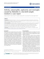
Fig.2 Histopathological micrograph of brain
tissue showing (A) Mononuclear Infiltration
and (B) Vaccuolation suggestive of meningitis
570
Int.J.Curr.Microbiol.App.Sci (2017) 6(7): 569-571
The present findings confirm that the E. coli
is the causative agent of meningitis in calf,
similar findings were recorded by Rodotits
et.al. (2000).
Histopatholocal and Histochemical
Technique, 3rd Edn, Butter Werth and
Co. Ltd., pp. 29-221
Cruickshank et al., (1997): Medical
Microbiology. Vol, 2nd, 12th Edn,
Churchil Livingstone. New York,
p.202-203.
Elefthenios M. (2006): E. coli infection
section
7
Epicture.
htp;//www.emedicine.com/med/topic734.
htm. Accessed March 10, 2007.
Green S.L and Smith L.L (1992): j. Am, vet
Med (1992): Assoc 201(1):125-8.
Kenneth Toder (2002): Toder Online text
book of Bacteriological, Kenneth Toder
University of Wisconsin-Madison
Department of Bacteriology.
MERKS Vet Mannual (2006): Bacteriological
Meningitis and Meningoencephalitis
often affects neonatal calfs caused by E.
coli; Merks and co. JNC. White House
Station N.J, USA.
O.M Radostits, (2000): Text Book of
Veterinary Medine 9th Edn, ELBS
London, p. (538).
Vegad, J.L. (1995): In Text Book of
Veterinary General Pathology 1st edn,
Vikas Publishing House, New Delhi,
p.276.
Microscopic observations of H and E stained
section revealed that diffused, degenerative,
necrotic and necrobiotic changes in neuron
(Fig. 2). Most of the neurons were completely
disappeared in the section leaving empty
spaces, the characteristics changes such as
condensation,
swelling,
chromatolysis,
occasional satellitosis and neurophagia were
evident. Most of the cerebral capillaries
revealed sever congestion and at places there
were
lymphocytic
aggregation.
Histopathological findings are in concurrence
with the findings of Vegad et al., (1995)
Suggestive of diffused meningitis.
A case of calf meningitis was confirmed by
histopathological examination and isolation,
identification of E. coli from the brain tissue.
References
Bingen. E.E. Denamur, Brahimi and J.Elion.
(1996): Clin Infect.Dis.22; 152156(Medicine).
Cullingn (1974): A Hand Book of
How to cite this article:
Kumbhar, G.B. 2017. Microbiological and Histopathological Investigation of Calf Meningitis
Caused by Escherichia Coli. Int.J.Curr.Microbiol.App.Sci. 6(7): 569-571.
doi: />
571