Knockdown of autophagy-related protein 5, ATG5, decreases oxidative stress and has an opposing effect on camptothecin-induced cytotoxicity in osteosarcoma cells
Bạn đang xem bản rút gọn của tài liệu. Xem và tải ngay bản đầy đủ của tài liệu tại đây (1.68 MB, 12 trang )
Hollomon et al. BMC Cancer 2013, 13:500
/>
RESEARCH ARTICLE
Open Access
Knockdown of autophagy-related protein 5,
ATG5, decreases oxidative stress and has an
opposing effect on camptothecin-induced
cytotoxicity in osteosarcoma cells
Mario G Hollomon1,2*, Nancy Gordon1, Janice M Santiago-O’Farrill1 and Eugenie S Kleinerman1
Abstract
Background: Autophagy induction can increase or decrease anticancer drug efficacy. Anticancer drug-induced
autophagy induction is poorly characterized in osteosarcoma (OS). In this study, we investigated the impact of
autophagy inhibition on camptothecin (CPT)-induced cytotoxicity in OS.
Methods: Autophagy-inhibited DLM8 and K7M3 metastatic murine OS cell lines were generated by infection with
lentiviral shRNA directed against the essential autophagy protein ATG5. Knockdown of ATG5 protein expression and
inhibition of autophagy was confirmed by immunoblot of ATG5 and LC3II proteins, respectively. Metabolic activity
was determined by MTT assay and cell viability was determined by trypan blue exclusion. Acridine orange staining
and immunoblotting for LC3II protein expression were used to determine autophagy induction. Oxidative stress
was assessed by staining cells with HE and DCFH-DA followed by flow cytometry analysis. Mitochondrial membrane
potential was determined by staining cells with TMRE followed by flow cytometry analysis. Immunoblotting was
used to detect caspase activation, Parp cleavage and p53 phosphorylation.
Results: Autophagy inhibition caused a greater deficit in metabolic activity and cell growth in K7M3 cells
compared to DLM8 cells. K7M3 cells exhibited higher basal autophagy levels than DLM8 cells and non-transformed
murine MCT3 osteoblasts. Autophagy inhibition did not affect CPT-induced DNA damage. Autophagy inhibition
decreased CPT-induced cell death in DLM8 cells while increasing CPT-induced cell death in K7M3 cells. Autophagy
inhibition reduced CPT-induced mitochondrial damage and CPT-induced caspase activation in DLM8 cells.
Buthionine sulfoximine (BSO)-induced cell death was greater in autophagy-competent DLM8 cells and was reversed
by antioxidant pretreatment. Camptothecin-induced and BSO-induced autophagy induction was also reversed by
antioxidant pretreatment. Significantly, autophagy inhibition not only reduced CPT-induced oxidative stress but also
reduced basal oxidative stress.
Conclusions: The results of this study indicate that autophagy inhibition can have an opposing effect on CPTinduced cytotoxicity within OS. The cytoprotective mechanism of autophagy inhibition observed in DLM8 cells
involves reduced CPT-induced oxidative stress and not reduced DNA damage. Our results also reveal the novel
finding that knockdown of ATG5 protein reduces both basal oxidative stress and drug-induced oxidative stress.
Keywords: Autophagy, Osteosarcoma, Camptothecin, Oxidative stress
* Correspondence:
1
Division of Pediatrics, The University of Texas MD Anderson Cancer Center,
Houston, TX 77054, USA
2
Department of Biology, Texas Southern University, Houston, TX 77004, USA
© 2013 Hollomon et al.; licensee BioMed Central Ltd. This is an open access article distributed under the terms of the Creative
Commons Attribution License ( which permits unrestricted use, distribution, and
reproduction in any medium, provided the original work is properly cited.
Hollomon et al. BMC Cancer 2013, 13:500
/>
Background
Autophagy is a lysosomal-dependent process that occurs at
low basal levels to support cellular homeostasis. During periods of nutrient deprivation, autophagy degrades intracellular proteins to serve as substrates for ATP generation.
Autophagy also carries out housekeeping activities such as
clearing the cell of damaged organelles and proteins that result from ordinary cellular metabolic activity. For example,
damaged mitochondria are selectively targeted for autophagy, thus reducing the release of pro-apoptotic mediators
into the cytosol and subsequent cell death [1]. Therefore,
basal levels of autophagy are necessary for cellular
homeostasis.
Autophagic activity above basal levels (hereafter referred
to as autophagy induction) is induced by anticancer drug
treatment. While autophagy inhibition both increases anticancer drug efficacy [2] and decreases anticancer drug efficacy [3,4], the majority of studies indicate that autophagy
inhibition increases anticancer drug efficacy, suggesting that
autophagy induction is a protective response to anticancer
drug treatment. However, unrestrained drug-induced autophagy induction can lead to cell death [5].
Osteosarcoma (OS) is the most prevalent bone
tumor in children. Despite recent advances in the understanding of the molecular basis of OS and new
therapeutic approaches, the mortality rate has declined only modestly. Autophagy modulation as adjuvant therapy to established anticancer therapies is
currently being investigated in clinical trials, but not
in OS [6]. The use of autophagy modulation as adjuvant therapy in OS may prove beneficial. However,
before considering such, the impact of anticancer
drug-induced autophagy induction on cytotoxicity in
OS must be better characterized.
In this study, we investigated the impact of
autophagy inhibition on camptothecin (CPT)-induced
cytotoxicity in OS cells. Camptothecin induces cell
death by inhibiting topoisomerase I resulting in DNA
single-strand breaks and subsequent cell death [7].
Here, we show that autophagy inhibition has an opposing impact on CPT-induced cytotoxicity in two
metastatic murine OS cell lines. Autophagy inhibition
in K7M3 cells increased sensitivity to CPT. In contrast, autophagy inhibition in DLM8 cells decreased
sensitivity to CPT. The mechanism of autophagy
inhibition-mediated protection in DLM8 cells appeared to be reduced CPT-induced oxidative stress
and a reduction in both mitochondrial damage and
caspase activation. To our knowledge, this is the first
report of an opposing effect of anticancer drug treatment on cytotoxicity in autophagy-inhibited OS cells.
Furthermore, we were unable to locate any other report of autophagy inhibition decreasing anticancer
drug-induced oxidative stress.
Page 2 of 12
Methods
Antibodies and reagents
Camptothecin was purchased from ChemWerth
(Woodbridge, CN). LC3 and ATG5 antibodies were purchased from Novus Biologicals (Littleton, CO). Cleaved
PARP, total p53, phospho p53, cleaved caspase-3 and
cleaved caspase-9 antibodies were purchased from Cell
Signaling Technology, Inc. (Danvers, MA). Pan-caspase
inhibitor was purchased from Enzo Life Sciences (Farmingdale, NY). Ripa lysis buffer was purchased from Santa
Cruz Biotechnology, Inc. (Santa Cruz, CA). Acridine orange, 3-(4,5-dimethylthiazolyl-2)-2,5-diphenylthetrazolium bromide (MTT) reagent, Bafilomycin A1 and actin
antibody were purchased from Sigma Aldrich (St. Louis,
MO). Buthionine sulfoximine (BSO) was purchased from
Acros Organics (Morris Plains, NJ). N-acetyl cysteine
(NAC) was purchased from Calbiochem (Billerica, MA).
Fetal bovine serum (FBS) was purchased from Atlanta
Biologicals (Lawrenceville, GA). DMEM cell culture
medium and supplements, dihydroethidium (HE), 2′,7′dichlorofluorescein diacetate (DCFH-DA), carbonyl
cyanide chlorophenylhydrozone (CCCP) and tetramethylrhodamine, ethyl ester (TMRE) were purchased
from Invitrogen (Carlsbad, CA).
Cell lines and cell culture
DLM8 [8] and K7M3 [9] are metastatic murine OS cell
lines. MC3T3 is a non-transformed murine osteoblast
cell line [10]. Cells were cultured in Dulbecco’s modified
eagle medium (DMEM) containing 10% FBS supplemented with antibiotic, non-essential amino acids, glutamine, sodium pyruvate and cultured in an incubator
maintained at 5% CO2 and 37°C. Prior to experimentation, cells were karyotyped and tested for mycoplasm
contamination. Cells were treated with CPT as indicated
in figure legends. Treatments were based on sensitivity
of each cell line to CPT. Where appropriate, cells were
treated with BSO to induce oxidative stress and NAC to
counter oxidative stress.
Lentiviral shRNA (Open Biosystems, Rockford, IL) targeted to autophagy-related protein-5 (ATG5) RNA was
used to knockdown ATG5 protein expression. Two separate ATG5 knockdown cell lines were generated for each cell
line using two different lentiviral shRNA sequences
[TRCN0000099431, TRCN0000099433]. Briefly, lentivirus
was produced by transfecting 293T cells with 7 ug/ml
transfer plasmid [TRCN0000099431 or TRCN0000099433],
5 ug/ml psPAX2 (packaging plasmid) and 4 ug/ml pMD2.
G (envelop plasmid). Forty-eight hours after 293T cell
transfection, supernatant containing lentivirus was collected
and immediately used for infection or stored at −80°C. For
infection, 2 ml of supernatant containing lentivirus was
added to each well of a 6-well plate containing 1×10⁵ cells.
Cells were incubated with lentivirus for 12 h and next
Hollomon et al. BMC Cancer 2013, 13:500
/>
transferred to a 75 mm flask. Assessment of ATG5 protein
knockdown was determined when cells were approximately
70% confluent. Both ATG5 knockdown cell lines showed
similar responses to CPT treatment. Control cells were infected with lentivirus containing empty shRNA vector. Cells
treated with empty shRNA vector are hereafter referred to
as autophagy-competent. ATG5 protein knockdown cells
are hereafter referred to as autophagy-inhibited.
Cell growth, cell metabolic activity and cell viability
determination
Cell growth was determined by seeding 4×10⁴ cells per
well in a 12-well plate followed by cell count at 48 h.
Metabolic activity was assessed by MTT assay. Metabolic
activity converts yellow MTT reagent to a purple formazan. Color intensity is indicative of metabolic activity.
MTT reagent (1mg/ml) was added to cells (3×10³ cells
per well) and incubated for 1 h at 37°C followed by
solubilization of formazan with DMSO followed by determination of formazan color intensity with a microplate reader set at absorbance reading 570 nm.
Absorbance readings of autophagy-inhibited groups were
compared to autophagy-competent groups which were
normalized to one hundred percent. To determine cell
viability, 4×10⁴ cells per well were seeded in 12-well
plates. Following CPT treatment, cell viability was determined by trypan blue exclusion assay using an automated cell counter (Vi-Cell, Beckman Coulter, Miami,
FL). Cells restricting trypan blue entry were considered
viable.
Acidic vesicular organelle (AVO) staining
Acridine orange freely diffuses the membranes of cells
and organelles. Inside acidic vesicles, acridine orange is
protonated and fluoresces bright red. Increased red
fluorescence indicates increased acidic vesicular organelle (AVO) formation [11]. Following CPT treatment,
cell culture medium was removed from the cells and replaced with cell culture medium containing 1ug/ml acridine orange and incubated for 20 min at 37°C. Cells
were then removed, washed twice and fluorescence immediately analyzed using the FL3 channel of a FACSCalibur flow cytometer (Becton Dickinson, San Jose, CA).
Page 3 of 12
transferred onto nitrocellulose membranes (BioRad
Laboratories, Hercules, CA). Membranes were blocked
with 5% nonfat milk then incubated with antibodies
against ATG5, LC3, cleaved caspase-9, cleaved
caspase-3, total p53, phospho p53 or cleaved PARP.
Membranes were then washed and incubated with appropriate secondary antibody conjugated to HRP (GE Healthcare Life Sciences, Piscataway, NJ). Following secondary
antibody incubation, membranes were washed and signal
detected with ECL detection reagent (GE Healthcare Life
Sciences, Piscataway, NJ). Beta-actin protein expression
served as a protein loading control.
Oxidative stress determination
Following drug treatment, cell culture medium was removed from the cells and replaced with cell culture
medium containing 5 uM dihydroethidium (HE) or
5 uM 2′,7′-dichlorofluorescein diacetate (DCFH-DA)
and incubated for 20 min at 37°C to assess superoxide
anion (.O2-) and hydrogen peroxide (H2O2) levels, respectfully. Cells were then removed, washed twice and
fluorescence immediately analyzed using a FACSCalibur
flow cytometer (Becton Dickinson, San Jose, CA). HE
freely diffuses the plasma membrane and is reduced by
intracellular .O2- resulting in a red fluorescence. Intracellular DCFH-DA reacts with H2O2 to give a green fluorescence. Increased HE and DCFH-DA fluorescence
indicates increased .O2- and H2O2 presence, respectively.
Mitochondrial membrane potential (ΔΨm)
Tetramethylrhodamine ethyl ester perchlorate (TMRE)
preferentially stains mitochondria producing red fluorescence and is used as an indicator of mitochondrial
membrane potential (ΔΨm). Decreased TMRE fluorescence is indicative of ΔΨm depolarization and ΔΨm
depolarization is associated with release of pro-apoptotic
mediators [12]. Following CPT treatment, cells were incubated with 25 ng/ml TMRE for 20 min at 37°C to assess ΔΨm. The protonophore carbonylcyanide mchlorophenylhydrozone (CCCP) was used as a positive
control for ΔΨm depolarization and to test TMRE staining efficiency.
Statistical analysis
Western blot
Following drug treatment, supernatant and cells were
collected and centrifuged at 300 g for 5 min at 4°C. The
resultant pellet was lysed with RIPA lysis buffer containing protease and phosphatase inhibitor cocktail and centrifuged at 10,000 g for 10 min at 4°C. Supernatants
were then collected and total protein was determined by
BioRad reagent (BioRad Laboratories, Hercules, CA).
Unless otherwise indicated, 30 ug of protein were resolved in SDS-polyacrylamide gels (SDS-PAGE) and
Results are presented as means ± S.E.M. Experimental
data were analyzed using 2-tailed Student t test. P values
less than 0.05 were considered statistically significant.
Results
CPT decreases metabolic activity, cell growth and induces
cell death
To begin this study, we assessed CPT-induced cytotoxicity in two metastatic murine OS cell lines. Camptothecin caused a dose-dependent decrease in cell viability in
Hollomon et al. BMC Cancer 2013, 13:500
/>
DLM8 and K7M3 cells (Figure 1A). Basal level of autophagy is associated with metabolic homeostasis; therefore, we determined if autophagy inhibition affected
metabolic activity or cell growth. Autophagy inhibition
significantly reduced both metabolic activity and cell
growth in K7M3 cells (Figure 1B and C).
CPT induces apoptosis and autophagy
To determine CPT-induced apoptosis we assessed
markers of apoptosis. Cleaved caspase-3 and cleaved
PARP (Figure 2A) with accompanying cell death indicated CPT-induced apoptotic cell death. Pre-treatment
with pan-caspase inhibitor blocked caspase-3 activation
in both cell lines (Figure 2B) and reversed CPT-induced
cell death in DLM8 cells but not in K7M3 cells
(Figure 2C). Acidic vesicular organelle accumulation was
determined to screen for increased autophagic activity
following CPT treatment. Camptothecin treatment significantly increased AVO production in DLM8 and
K7M3 cells (Figure 3A and B). Autophagy induction was
confirmed by LC3II immunoblot. During autophagy induction, LC3I is converted to LC3II. LC3II protein expression increased in both cell lines following CPT
treatment, confirming increased autophagic activity
(Figure 3C). It is important to note that to measure
LC3II protein levels, 30 ug of total protein from DLM8
were loaded to a SDS-PAGE gel, while only 7.5 ug of
total protein from K7M3 were loaded. Thirty micrograms of total protein from K7M3 resulted in saturation
of the membrane which prevented detection of differences in protein expression between treatment groups.
Camptothecin-induced autophagy induction was also
confirmed by assessment of a second autophagy marker
p62 (Additional file 1: Figure S1). Reduced p62 protein
expression is indicative of autophagy induction. Wildtype cells were treated with Bafilomycin A1 to determine
the functional status of autophagy. Bafilomycin A1 inhibits autophagosome and lysosome fusion causing an
increase in LC3II accumulation. Bafilomycin A1 caused
an increase in LC3II accumulation compared to nontreated cells in both cell lines (Additional file 2: Figure
S2), indicating that autophagy flux was functional in
both cell lines.
Knockdown of ATG5 protein expression has an opposing
impact on cell viability in DLM8 and K7M3 OS cells
Autophagy was inhibited by knocking down the expression of essential autophagy protein ATG5. Knockdown
of ATG5 protein expression and its impact on autophagy
inhibition were confirmed by immunoblot of ATG5 and
LC3II, respectively (Figure 4A). Knockdown of ATG5 reduced CPT-induced AVO formation, thus validating
AVO as a reliable screen for autophagy induction
(Figure 4B). Knockdown of ATG5 protein expression in
Page 4 of 12
DLM8 cells decreased CPT-induced cell death. In contrast, knockdown of ATG5 protein expression in K7M3
cells increased CPT-induced cell death (Figure 4C
and D). Basal levels of autophagy were higher in K7M3
cells compared to DLM8 cells and a nontransformed
osteoblast cell line, suggesting increased dependence
of K7M3 on autophagy for metabolic homeostasis
(Figure 4E). Camptothecin treatment induced similar
level of phosphorylation of p53 at Ser15 in both
autophagy-competent and autophagy-inhibited DLM8
cells, indicating similar levels of CPT-induced DNA
damage (Figure 4F).
Autophagy inhibition decreases CPT-induced oxidative
stress and buthionine sulfoximine (BSO)-induced cell
death
To investigate the impact of autophagy inhibition on
CPT-induced oxidative stress, HE and DCFH-DA probes
were used to access .O2- and H2O2 levels, respectively.
The level of CPT-induced .O2- and H2O2 generation was
greater in autophagy-competent DLM8 cells (Figure 5A
and B). To determine if autophagy-competent DLM8
cells were more sensitive to oxidative stress in general,
cell viability was assessed in autophagy-competent and
autophagy-inhibited DLM8 cells following BSO or combination treatment of BSO and CPT. Buthionine sulfoximine inhibits synthesis of the endogenous antioxidant
glutathione [13], thus increasing oxidative stress levels.
Autophagy-competent DLM8 cells were more sensitive
to BSO-induced cell death and the cotreatment of BSO
and CPT caused greater cell death in autophagycompetent DLM8 cells compared to autophagy-inhibited
DLM8 cells (Figure 5C). Pretreatment with the antioxidant NAC reversed BSO-induced cell death (Figure 5D)
but not CPT-induced cell death (data not shown).
Buthionine sulfoximine treatment increased autophagy
levels, as indicated by increased LC3II levels, in
autophagy-competent DLM8 cells (Figure 5E). N-acetyl
cysteine treatment reversed CPT-induced and BSOinduced autophagy induction in autophagy-competent
DLM8 cells (Figure 5F and G).
Autophagy inhibition decreases CPT-induced
mitochondrial membrane potential (ΔΨm) depolarization
Previously reported CPT-induced mitochondrial damage
[14] prompted an investigation into the impact of autophagy inhibition on mitochondrial damage following
CPT treatment. Camptothecin induced mitochondrial
damage in both autophagy-competent and autophagyinhibited DLM8 cells as determined by ΔΨm depolarization.
However, ΔΨm depolarization was greater in autophagycompetent DLM8 cells compared to autophagy-inhibited
DLM8 cells (Figure 6A), suggesting that mitochondrial damage was less in autophagy-inhibited DLM8 cells following
Hollomon et al. BMC Cancer 2013, 13:500
/>
Page 5 of 12
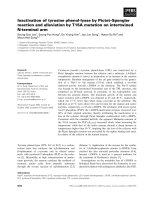
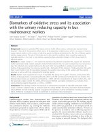
Figure 1 Camptothecin decreases cell viability and metabolic
activity. A, Camptothecin-induced cell death. DLM8 and K7M3 cells
were cultured in 12-well plates and treated with CPT as indicated for
48 h. Cell viability was determined by trypan blue exclusion assay.
*, p < 0.05, compared with same treatment group. B, Impact of
autophagy inhibition on metabolic activity. Cells were grown in a
96-well plate and allowed to grow in normal media to
approximately 70% confluency. MTT assay was used to determine
metabolic activity. Control values were set to one hundred percent.
*, p < 0.05. C, Impact of autophagy inhibition on cell growth. Cells
were grown in 12-well plates in normal media followed by cell
count at 48 h. *, p < 0.05. Data represents the results of at least three
independent experiments, ± SE. p < 0.05 was considered significant.
CPT treatment. Caspase-9 activation and caspase-3 activation was greater in autophagy-competent DLM8 cells compared to autophagy-inhibited DLM8 cells following CPT
treatment (Figure 6B). Caspase-3 activation was greater in
autophagy-inhibited K7M3 cells compared to autophagycompetent K7M3 cells (Figure 6C).
Discussion and conclusions
The protective role of autophagy induction against anticancer therapy is supported by observations that autophagy inhibition increases anticancer drug efficacy [2]. A
literature search returned a limited number of studies
reporting reduced anticancer therapy efficacy in
autophagy-inhibited cells [15]. With autophagy inhibition currently being investigated as adjuvant anticancer
therapy, these limited observations are relevant. In this
study, ATG5 protein expression was knocked down to
inhibit autophagy. Here, we report an opposing effect of
ATG5 knockdown-mediated autophagy inhibition on
CPT-induced cytotoxicity within OS. Autophagy inhibition decreased sensitivity to CPT in DLM8 cells and increased sensitivity to CPT in K7M3 cells. To date, there
are no reports showing an opposing impact of autophagy
inhibition on anticancer therapy within OS.
Following the observation that autophagy inhibition in
K7M3 cells increased sensitivity to CPT, we reasoned
that autophagy plays a greater role in the overall maintenance and metabolic homeostasis in K7M3 cells and
suspected that the basal level of autophagy in K7M3
cells is greater than that of DLM8 cells. Immunoblot
analysis of LC3II confirmed that basal level of autophagy
is higer in K7M3 cells compared to DLM8 cells and
non-transformed murine MC3T3 osteoblasts (Figure 4E).
This finding supports the suggestion that K7M3 cells
have an increased dependence on autophagy for ordinary
metabolic activities. The dependence of K7M3 on autophagy is further supported by the observation that autophagy inhibition significantly decreased both K7M3
cell metabolic activity and cell growth (Figure 4B
and C). It is plausible that increased basal level of autophagy in K7M3 cells is one of several genetic
Hollomon et al. BMC Cancer 2013, 13:500
/>
Page 6 of 12
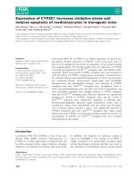
Figure 2 Camptothecin induces caspase activation. Cells were treated with no drug, or CPT or caspase inhibitor plus CPT at doses as
indicated for 48 h. Following drug treatment, cells were lysed and cell lysate immunoblotted for cleaved caspase-3 and cleaved Parp protein
expression. A, Cleaved caspase-3 and cleaved PARP protein expression in wildtype DLM8 and K7M3 cells. B, Pan caspase inhibitor blocks caspase3 activation. Cells were treated with CPT doses indicated in figure for 48 h. Treatment doses were based on cell sensitivity to CPT. C, Caspase
inhibition reverses CPT-induced cell death in DLM8. Wildtype DLM8 and K7M3 cells were pretreated with a pan-caspase inhibitor for 2 h followed
by CPT treatment for 48 h. Control group received no drug and an additional group received CPT only. Cell viability was determined by trypan
blue exclusion assay. *, p < 0.05, compared with control group. Data represents the results of at least three independent experiments, ± SE.
p < 0.05 was considered significant. Actin served as a protein loading control. Immunoblots are representative of immunoblots from at least two
independent experiments.
Hollomon et al. BMC Cancer 2013, 13:500
/>
Page 7 of 12
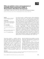
Figure 3 Camptothecin increases autophagic activity. Following 48 h CPT treatment, cells were incubated with the lysosomotropic agent
acridine orange and fluorescence analyzed by flow cytometry. A, Representative flow cytometry analysis of acidic vesicular organelle (AVO)
formation in wildtype DLM8 cells. B, Graph representation of CPT-induced AVO formation in wildtype DLM8 and K7M3 cells. *, p < 0.05, compared
with same treatment group. C, LC3I/LC3II protein expression. Following 48 h CPT treatment, cells were lysed and cell lysate immunoblotted for
LC3I/LC3II protein expression. Increased LC3II expression is indicative of autophagy induction. The expression of treatment group LC3II/actin ratio
was determined by densitometry and compared to control group LC3II/actin ratio which was normalized to the arbitrary value of one. Treatment
group LC3II expression was normalized to control actin levels as needed. 30ug of protein were loaded for DLM8 LC3I/LC3II determination while
only 7.5ug of protein were loaded for K7M3 LC3I/LC3II determination. Actin served as a protein loading control. Data represents the results of
three independent experiments, ± SE. p < 0.05 was considered significant. Immunoblot is representative of immunoblots from three
independent experiments.
influences that contribute to the cancer phenotype and
decreased autophagic capability increases sensitivity to
stresses such as anticancer treatment. Increased dependence on autophagy has been reported for other cancers.
For example, pancreatic cancer cells [16] and Ras
oncogenic-driven cancer cells [17] have been shown to
have increased dependence on autophagy. These two
studies also reported increased basal levels of autophagy.
In this study, autophagy inhibition decreased sensitivity
to CPT in DLM8 cells, which contrasts the more often reported observation that autophagy inhibition increases sensitivity to anticancer drug treatment. Therefore, we were
Hollomon et al. BMC Cancer 2013, 13:500
/>
Page 8 of 12
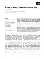
Figure 4 Autophagy inhibition has an opposing impact on CPT-induced cell death. A, ATG5 protein levels in DLM8 and K7M3 cells
following shRNA-mediated knockdown of ATG5. Cells were infected with lentivirus containing empty shRNA vector or lentiviral shRNA targeted
against ATG5 mRNA. Following infection, cells were lysed and total protein collected. To confirm ATG5 protein knockdown and autophagy
inhibition, total protein was immunoblotted for ATG5 and LC3I/C3II protein levels, respectively. Actin served as a protein loading control. B, Acidic
vesicular organelle (AVO) formation. Autophagy-competent and autophagy-inhibited DLM8 cells were treated with CPT for 24 h followed by
assessment of AVO formation. Impact of autophagy inhibition on cell death in C, DLM8 and D, K7M3 OS cells. Autophagy-competent and
autophagy-inhibited DLM8 and K7M3 cells were treated with CPT as indicated for 48 h. Following drug treatment, cell viability was assessed by
trypan blue exclusion. *, p < 0.05, compared with same treatment group. E, Basal levels of autophagy in MC3T3, DLM8 and K7M3 cells. Cells were
untreated and allowed to grow to approximately 70% confluency. Cells were then collected, lysed and total protein immunoblotted for LC3I/
LC3II. 30ug of protein were loaded for each cell line. Actin served as a protein loading control. F, Phosphorylation of p53 in DLM8 cells. Cells
were treated with CPT as indicated for 24 h. Following CPT treatment, cells were lysed and cell lysate probed for phospho p53 and total p53
protein expression. Data represents the results of at least three independent experiments, ± SE. p < 0.05 was considered significant. Immunoblots
are representative of immunoblots from two independent experiments.
particularly interested in the response of autophagyinhibited DLM8 cells to CPT and explored further this cell
line. While it was clear that autophagy inhibition in DLM8
cells decreased CPT-induced cell death compared to
autophagy-competent DLM8 cells, the mechanism was unknown. Considering that the mechanism of action for CPT
is DNA damage [18], we explored the impact of autophagy
inhibition on CPT-induced DNA damage as a possible
mechanism for decreased sensitivity to CPT in autophagyinhibited DLM8 cells. DNA damage as determined by
phosphorylation of p53 at Ser15 [19] was unchanged between autophagy-competent and autophagy-inhibited
Hollomon et al. BMC Cancer 2013, 13:500
/>
Page 9 of 12
Figure 5 Autophagy inhibition decreases CPT-induced oxidative stress and buthionine sulfoximine (BSO)-induced cell death. Cells were
treated with CPT for 24 h followed by incubation with HE or DCFH-DA. A, Camptothecin-induced .O2-. *, p < 0.05. B, Camptothecin-induced H2O2.
*, p < 0.05. C, Autophagy-competent cells are more sensitive to BSO-induced cell death. Cells were pretreated with 1mM BSO for 2 h followed by
48 h CPT treatment. Cells received a second 1mM BSO treatment 12 h into the CPT treatment. Following CPT treatment, cell viability was
determined by trypan blue exclusion. *, p < 0.05, compared with control group. D The antioxidant NAC reverses BSO-induced cell death. Cells
were pretreated with NAC for 2 h prior to BSO treatment. Cells received a second 1mM BSO treatment 12 h into the BSO treatment. Following
48 h BSO treatment, cell viability was determined by trypan blue exclusion. *, p < 0.05, compared with control group. E, BSO treatment increases
LC3II levels in autophagy-competent DLM8 cells. Cells were treated with 1mM BSO for times indicated in figure. F, NAC pretreatment inhibits
CPT-induced autophagy induction in autophagy-competent DLM8 cells. Cells received no drug, NAC, CPT or combination as indicated in figure
for 48 h. For combination groups, cells were pretreated with NAC for 2 h. G, NAC pretreatment inhibits BSO-induced autophagy induction in
autophagy-competent DLM8 cells. Cells received no drug, NAC, 1mM BSO or combination for 6 h. For combination group, cells were pretreated
with NAC for 2 h. Following drug treatment, cells were lysed and total protein immunoblotted for LC3I/LC3II protein expression. Actin served as a
protein loading control. Data represents the results of three independent experiments, ± SE. p < 0.05 was considered significant. Immunoblots are
representative of immunoblots from at least two independent experiments.
DLM8 cells (Figure 4F). We also assessed the impact of autophagy inhibition on DLM8 cell growth. Autophagy inhibition did not significantly impact cell growth of DLM8 cells
(Figure 1C). This is relevant because the mechanism of action for CPT is DNA damage that occurs during cell division. Had autophagy inhibition significantly reduced
DLM8 cell growth, this would support the suggestion that
autophagy inhibition-mediated protection is due to reduced
cell division. Together, this set of data suggests that the autophagy inhibition-mediated protection observed in this
study was not due to reduced DNA damage or reduced cell
division.
Hollomon et al. BMC Cancer 2013, 13:500
/>
Page 10 of 12
Figure 6 CPT induces mitochondrial membrane potential and induces caspase-9 activation. Tetramethylrhodamine, ethyl ester, perchlorate
(TMRE) was used to determine mitochondrial membrane potential (ΔΨm). A, Mitochondrial membrane potential depolarization in autophagycompetent and autophagy-inhibited DLM8 cells following 24 h CPT treatment. Following 24 h CPT treatment, cells were incubated with TMRE
followed by flow cytometry analysis. Decreased TMRE fluorescence is indicative of decreased ΔΨm and increased release of pro-apoptotic
molecules into the cytosol. Cells were incubated with the membrane uncoupler carbonylcyanide m-chlorophenylhydrazone (CCCP) prior to TMRE
incubation to depolarize the mitochondrial membrane and serve as a positive control. Open histogram represents CCCP + TMRE treatment. Filled
histogram represents cells incubated with TMRE only. Percentage values represent degree of mitochondrial membrane depolarization. Data is
representative of results from two independent experiments. B, Caspase-9 activation and caspase-3 activation is reduced in autophagy-inhibited
DLM8 cells. C, Caspase-3 activation is increased in autophagy-inhibited K7M3 cells. Cells were treated with CPT for 48 h. Cells were next collected,
lysed and total protein immunoblotted for cleaved caspase-9 or cleaved caspase-3. Actin served as a protein loading control. Immunoblots are
representative of immunoblots from at least two independent experiments.
Previous reports of CPT-induced oxidative stress [20]
led us to investigate the impact of autophagy inhibition
on CPT-induced oxidative stress as a contributing factor
to the observed autophagy inhibition-mediated protection. Oxidative stress, as determined by generation
of .O2- and H2O2, was higher in autophagy-competent
DLM8 cells compared to autophagy-inhibited DLM8
cells following CPT treatment, indicating that autophagy
inhibition decreased CPT-induced oxidative stress.
Autophagy inhibition also reduced basal oxidative stress
level. To our knowledge, this is the first report of autophagy inhibition-mediated reduced basal oxidative
stress as well as autophagy inhibition-mediated reduced
anticancer drug-induced oxidative stress.
Increased levels of CPT-induced oxidative stress
coupled with increased CPT-induced cell death in
autophagy-competent DLM8 cells led us to determine if
autophagy-competent DLM8 cells are more sensitive to
Hollomon et al. BMC Cancer 2013, 13:500
/>
oxidative stress. The use of BSO allowed for the investigation of the impact of oxidative stress alone on cell
death and autophgay induction. Autophagy-competent
DLM8 cells were more sensitive than autophagyinhibited DLM8 cells to BSO-induced cell death. In
agreement with Martinez-Outschonnra et al. [21], BSO
also induced autophagy. BSO-induced cell death and
BSO-induced autophagy induction were reversed by
NAC pretreatment indicating a link between increased
oxidative stress and both cell death and autophagy
induction.
Basal levels of autophagy have been previously reported to differ among cancer cell lines [17] and here we
report varying basal levels of autophagy in two metastatic murine OS cell lines. Considering these reports, it
is plausible that the threshold level of autophagy induction that causes autopahgic cell death also varies for different cancers or even different cell lines within the
same type of cancer. Camptothecin-induced DNA damage and CPT-induced oxidative stress together may have
caused autophagy induction in autophagy-competent
DLM8 cells that exceeded the threshold level necessary
to cause autophagic cell death. Considering this, autophagy inhibition would reduce or delay CPT-induced autophagic cell death, making autophagy-inhibited DLM8
cells less sensitive to CPT-induced cell death. Therefore,
one explanation for decreased sensitivity of autophagyinhibited DLM8 cells compared to autophagy-competent
DLM8 cells is reduced CPT-induced autopahgic cell
death. Camptothecin-induced oxidative stress was lower
in autophagy-inhibited DLM8 cells compared to
autophagy-competent DLM8 cells. Therefore, an alternative explanation for decreased sensitivity to CPT in
autophagy-inhibited DLM8 cells is reduced oxidative
stress-induced cell death unrelated to autophagic cell
death. At this point in our investigation, we are unable
to present data supporting an explanation for lower oxidative stress in autophagy-inhibited DLM8 cells. However, the endogenous antioxidant catalase is a reported
target of selective autophagy [22] and we suspect that
autophagy inhibition may affect levels of endogenous
antioxidants.
With observed CPT-induced oxidative stress in this study
and reports of oxidative stress induced mitochondrial damage [23], we investigated the impact of CPT on mitochondria. In agreement with a previous study [24], CPT caused
ΔΨm depolarization in both autophagy-competent and
autophagy-inhibited DLM8 cells (Figure 6A). However,
ΔΨm depolarization was greater in autophagy-competent
DLM8 cells, suggesting increased mitochondrial damage.
Mitochondrial membrane potential depolarization and
mitochondrial damage is associated with caspase-9 activation and caspase-3 activation [25]. Immunoblot confirmed
increased caspase-9 activation and caspase-3 activation in
Page 11 of 12
autophagy-competent DLM8 cells compared to autophagyinhibited DLM8 cells (Figure 6B). Thus, the observed mitochondrial damage was likely an upstream event of caspase
activation and likely contributed to increased cell death in
autophagy-competent cells. Conversely, caspase-3 activation was higher in autophagy-inhibited K7M3 cells compared to autophagy-competent cells (Figure 6C). This
observation suggests that autophagy inhibition increased
the sensitivity of K7M3 to CPT-induced apoptosis.
In conclusion, we show that autophagy inhibition can
have an opposing impact on the response of OS cells following CPT treatment. Our data suggest that the protective mechanism of autophagy inhibition involves both
reduced oxidative stress and mitochondrial damage. The
results of this study reminds us that autophagy inhibition can decrease the efficacy of anticancer drug therapy
and underscores the need to better understand and predict the response of autophagy-modulated cancer cells
to anticancer drug therapy.
Additional files
Additional file 1: Figure S1. Camptothecin treatment decreases p62
protein expression. Reduced p62 protein expression is indicative of
autophagy induction. Wildtype DLM8 and K7M3 cells were treated with
CPT for 48 h. Cells were next collected, lysed and 30ug of total protein
immunoblotted for p62. Actin served as a protein loading control.
Immunoblots are representative of immunoblots from at least two
independent experiments.
Additional file 2: Figure S2. Bafilomycin A1 treatment increases LC3II
protein expression. Wildtype DLM8 and K7M3 cells were treated with
Bafilomycin A1 to determine the functional status of autophagy.
Bafilomycin A1 inhibits autophagosome and lysosome fusion causing an
increase in LC3II accumulation. Wildtype DLM8 and K7M3 cells were
treated with Bafilomycin A1 for 48 h. Immunoblots are representative of
immunoblots from at least two independent experiments.
Abbreviations
OS: Osteosarcoma; NAC: N-acetyl cysteine; BSO: buthionine sulfoximine;
CPT: Camptothecin; HE: Dihydroethidium; DCFH-DA: 2′,7′-dichlorofluorescein
diacetate; ATG5: Autophagy-related protein-5; AVO: Acidic vesicular organelle;
ΔΨm: Mitochondrial membrane potential.
Competing interests
The authors declare that they have no competing interest.
Authors’ contributions
MGH conceived the study, carried out experiments, carried out data analysis
and wrote the manuscript. NG assisted with project conception. JMS assisted
with experiments and preparation of manuscript. ESK served as project
supervisor. All authors read and approved the final manuscript.
Acknowledgements
This work was supported by National Cancer Institute grant R01CA042992
(ESK) and Legends of Friendswood Research Award (MGH). We thank Dr.
Joya Chandra for critical reading and comments regarding the manuscript.
Received: 23 May 2013 Accepted: 21 October 2013
Published: 26 October 2013
Hollomon et al. BMC Cancer 2013, 13:500
/>
References
1. Okamoto K, Kondo-Okamoto N, Ohsumi Y: Mitochondria-anchored
receptor Atg32 mediates degradation of mitochondria via selective
autophagy. Dev Cell 2009, 17:87–97.
2. Gupta A, Roy S, Lazar AJ, Wang WL, AcAuliffe JC, Reynoso D, McMahon J,
Taguchi T, Floris G, Debiec-Rychter M, Schoffski P, Trent JA, Debnath J,
Rubin BP: Autophagy inhibition and antimalarials promote cell death in
gastrointestinal stromal tumor (GIST). Proc Natl Acad Sci USA 2010,
107(32):14333–14338.
3. Lin CI, Whang EE, Abramson MA, Jiang X, Price BD, Donner DB, Moore FD
Jr, Ruan DT: Autophagy: a new target for advanced papillary thyroid
cancer therapy. Surgery 2009, 146(6):1208–1214.
4. Dupere-Richer D, Kinal M, Menasehe V, Nielsen TH, del Rincon S, Pettersson
R, Miller WH: Vorinostat-induced autophagy switches from a deathpromoting to a cytoprotective signal to drive acquired resistance.
Cell Death Dis 2013, 4:e486–e496.
5. Baehrecke EH: Autophagy: dual roles in life and death? Nat Rev Mol Cell
Biol 2005, 6:505–510.
6. Chen S, Rehman SK, Zhang W, Wen A, Yao L, Zhang J: Autophagy is a
therapeutic target in anticancer drug resistance. Biochim Biophys Acta
2010, 1806(2):220–229.
7. Qing-Yong L, Yuan-Gang Z, Rong-Zhen S, Li-Ping Y: Review camptothecin:
current perspectives. Curr Med Chem 2006, 13:2021–2039.
8. Asai T, Ueda T, Itoh K, Yoshioka K, Aoki Y, Mori S, Yoshikawa H:
Establishment and characterization of a murine osteosarcoma cell line
(LM8) with high metastatic potential to the lung. Int J Cancer 1998,
76:418–422.
9. Khanna C, Prehn J, Yeung C, Caylor J, Tsokos M, Helman L: An orthotopic
model of murine osteosarcoma with clonally related variants differing in
pulmonary metastatic potential. Clin Exp Metastasis 2000, 18:261–271.
10. Sudo H, Kodama HA, Amagai Y, Yamamoto S, Kasai S: In vitro
differentiation and calcification of a new clonal osteogenic cell line
derived from newborn mouse calvarin. J Cell Biol 1983, 96:191–198.
11. Paglin S, Hollister T, Delohery T, Hackett N, McMahill M, Sphicas E, Domingo
D, Yahalom J: A novel response of cancer cells to radiation involves
autophagy and formation of acidic vesicles. Cancer Res 2001,
61(2):439–444.
12. Heiskanen KM, Bhat MB, Wang HW, Ma J, Nieminen AL: Mitochondria
depolarization accompanies cytochrome c release during apoptosis in
PC6 cells. J Biol Chem 1999, 274(9):5654–5658.
13. Drew R, Miners JO: The effects of buthionine sulphoximine (BSO) on
glutathione depletion and xenobiotic biotransformation.
Biochem Pharmacol 1984, 33(4):2989–2994.
14. Yuan Q, Ray RM, Johnson LR: Polyamine depletion prevents
camptothecin-induced apoptosis by inhibiting release of cytochrome c.
Am J Physiol Cell Physiol 2002, 282(6):C1290–C1297.
15. Voss V, Senft C, Lang V, Ronellenfitsch MW, Steinbach JP, Seifert V, Kogel D:
The pan-Bcl-2 inhibitor(−)-gossypol triggers autophagic cell death in
malignant glioma. Mol Cancer Res 2010, 7:1002–1016.
16. Yang S, Wang X, Contino G, Liesa M, Sahin E, Ying H, Bause A, Li Y, Stommel
JM, Dell’antonio G, Mautner J, Tonon G, Haigis M, Shirihai OS, Doglioni C,
Bardeesy N, Kimmelman AC: Pancreatic cancers require autophagy for
tumor growth. Genes Dev 2011, 25(7):717–729.
17. Guo JY, Chen HY, Mathew R, Fan J, Strohecker AM, Karsli-Uzunbas G,
Kamphorst JJ, Chen G, Lemons JM, Karantza V, Coller HA, Dipaola RS,
Gelinas C, Rabinowitz JD, White E: Activated ras requires autophagy to
maintain oxidative metabolism and tumorigenesis. Genes Dev 2011,
25(5):460–470.
18. Avemann K, Knipper R, Koller T, Sogo JM: Camptothecin specific inhibitor
of type I DNA topoisomerase, induces DNA breakage at replication forks.
Mol Cell Biol 1988, 33(7):3026–3034.
19. Shieh SY, Ikeda M, Taya Y, Prives C: DNA damage-induced phosphorylation
of p53 alleviates inhibition by MDM2. Cell 1997, 91(3):325–334.
20. Timur M, Akbas S, Ozben T: The effect of topotecan on oxidative stress in
MCF-7 human breast cancer cell line. Acta Biochim Pol 2005,
52(4):897–902.
21. Martinez-Outschoorn UE, Trimmer C, Lin Z, Whitaker-Menezes D, Chiavarina
B, Zhou J, Wang C, Pavlides S, Martinez-Cantarin M, Capozza F, Witkiewicz K,
Flomenberg N, Howell A, Pestell R, Caro J, Lisanti M, Sotgia F: Autophagy in
cancer associated fibroblasts promotes tumor cell survival: role of
Page 12 of 12
22.
23.
24.
25.
hypoxia, HIF1 induction and NFκB activation in the tumor stromal
microenvironment. Cell Cycle 2010, 9(17):3514–3533.
Yu L, Wan F, Dutta S, Welsh S, Liu Z, Freundt E, Baehrecke E, Lenardo M:
Autophagic programmed cell death by selective catalase degradation.
Proc Natl Acad Sci USA 2006, 103(13):4952–4957.
Zaragoza A, Diez-Fernandez C, Alvarez AM, Andres D, Cascales M:
Mitochondrial involvement in cocaine-treated rat hepatocytes: effect of
N-acetylcysteine and deferoxamine. Br J Pharmacol 2001,
132(5):1063–1070.
Wang LM, Li QY, Zu YG, Fu YJ, Chen LY, Lv HY, Yao LP, Jiang SG: Antiproliferative and pro-apoptotic effect of CPT13, a novel camptothecin
analog, on human colon cancer HCT8 cell line. Chem Biol Interact 2008,
176(2–3):165–172.
Miura T, Chiba M, Kasai K, Nozaka H, Nakamura T, Shoji T, Kanda T, Ohtake Y,
Sato T: Apple procyanidins induce tumor cell apoptosis through
mitochondrial pathway activation of caspase-3. Carcinogenesis 2008,
29:585–593.
doi:10.1186/1471-2407-13-500
Cite this article as: Hollomon et al.: Knockdown of autophagy-related
protein 5, ATG5, decreases oxidative stress and has an opposing effect
on camptothecin-induced cytotoxicity in osteosarcoma cells. BMC Cancer
2013 13:500.
Submit your next manuscript to BioMed Central
and take full advantage of:
• Convenient online submission
• Thorough peer review
• No space constraints or color figure charges
• Immediate publication on acceptance
• Inclusion in PubMed, CAS, Scopus and Google Scholar
• Research which is freely available for redistribution
Submit your manuscript at
www.biomedcentral.com/submit