IGF-1 receptor and IGF binding protein-3 might predict prognosis of patients with resectable pancreatic cancer
Bạn đang xem bản rút gọn của tài liệu. Xem và tải ngay bản đầy đủ của tài liệu tại đây (556.11 KB, 9 trang )
Hirakawa et al. BMC Cancer 2013, 13:392
/>
RESEARCH ARTICLE
Open Access
IGF-1 receptor and IGF binding protein-3 might
predict prognosis of patients with resectable
pancreatic cancer
Toshiki Hirakawa1, Masakazu Yashiro1,2*, Akihiro Murata1, Keiichiro Hirata1, Kenjiro Kimura1, Ryosuke Amano1,
Nobuya Yamada1, Bunzo Nakata1 and Kosei Hirakawa1
Abstract
Background: The present study aimed to elucidate the clinicopathologic role of insulin-like growth factor-1
receptor (IGF1R) and IGF binding protein-3 (IGFBP3) in patients with pancreatic cancer. The function of IGFBP3 is
controversial, because both inhibition and facilitation of the action of IGF as well as IGF-independent effects have
been reported. In this study, IGF1R and IGFBP3 expression was examined, and their potential roles as prognostic
markers in patients with pancreatic cancer were evaluated.
Methods: Clinicopathological features of 122 patients with curatively resected pancreatic cancer were
retrospectively reviewed, and expression of IGF1R and IGFBP3 was immunohistochemically analyzed.
Results: Expression of IGF1R and IGFBP3 was observed in 50 (41.0%) and 37 (30.3%) patients, respectively. IGF1R
expression was significantly associated with histological grade (p = 0.037). IGFBP3 expression had a significant
association with tumor location (p = 0.023), and a significant inverse association with venous invasion (p = 0.037).
Tumors with IGF1R-positive and IGFBP3-negative expression (n = 32) were significantly frequently Stage II and III
(p = 0.011). The prognosis for IGF1R positive patients was significantly poorer than that for IGF1R negative
patients (p = 0.0181). IGFBP3 protein expression did not correlate significantly with patient survival. The subset of
patients with both positive IGF1R and negative IGFBP3 had worse overall survival (8.8 months versus 12.6
months, respectively, p < 0.001).
Conclusion: IGF1R signaling might be associated with tumor aggressiveness, and IGFBP3 might show
antiproliferative effects in pancreatic cancer. Both high IGF1R expression and low IGFBP3 expression represent
useful prognostic markers for patients with curatively resected pancreatic cancer.
Keywords: Pancreatic cancer, IGF1R, IGFBP3, Prognosis
Background
Pancreatic ductal adenocarcinoma (PDAC) is one of the
most lethal solid tumors, and carries an extremely poor
prognosis [1]. Although the management and treatment
of patients with pancreatic cancer have improved over
the last few decades, the overall 5-year survival rate
remains less than 5% [2]. Long-term survival is rare,
even in patients who undergo a histologically curative
* Correspondence:
1
Department of Surgical Oncology, Osaka City University Graduate School of
Medicine, 1-4-3 Asahi-machi, Osaka, Abeno-ku, Japan
2
Oncology Institute of Geriatrics and Medical Science, Osaka City University
Graduate School of Medicine, 1-4-3 Asahi-machi, Osaka, Abeno-ku 545-8585,
Japan
operation, with overall 5-year survival rates ranging
from 10% to 25% [3,4]. The high mortality rate associated
with pancreatic cancer is known to be due to extensive
invasion into surrounding tissues and metastasis to distant
organs at the time of diagnosis (or even after a curative
operation); however, the molecular mechanisms of the
highly aggressive nature of PDAC remains unclear [5].
Previous studies have shown an association between
progression of PDAC and overexpression of several
growth factors (and their receptors) including insulinlike growth factor (IGF), vascular endothelial growth
factor (VEGF), fibroblast growth factor (FGF), and
platelet-derived growth factor (PDGF) [6-8]. Most of
© 2013 Hirakawa et al.; licensee BioMed Central Ltd. This is an Open Access article distributed under the terms of the Creative
Commons Attribution License ( which permits unrestricted use, distribution, and
reproduction in any medium, provided the original work is properly cited.
Hirakawa et al. BMC Cancer 2013, 13:392
/>
the cellular effects of IGF-I and IGF-II are mediated by
the IGF-I receptor (IGF1R). Binding of the ligand to
IGF1R leads to tyrosine phosphorylation of the major
receptor substrate followed by activation of certain
downstream signaling cascades [9]. The IGFs have been
implicated through IGF1R in the pathogenesis, cell proliferation, and cell survival of many cancers [10,11]. IGF-1,
which is produced primarily by the liver, is known to play
an important role in the regulation of cell proliferation,
differentiation, and apoptosis [10-12]. Clinical studies in
colorectal, esophageal, and pancreatic cancers have shown
that IGF1R signaling correlates with increased tumor
growth and malignancy in vitro [8,13,14].
The IGF system is a complex network of growth factors
(IGF-I and IGF-II), cell surface trans-membrane receptors
(IGF1R), and high affinity IGF-binding proteins (IGFBPs)
that play an important role in normal cellular growth and
development, and disruption of the balance of this system
has been implicated in the etiology and progression of
breast and other cancers [15]. Activation of the IGF system stimulates proliferation, differentiation, angiogenesis,
metastasis, survival, and resistance to anticancer therapies
in many cancers [16], supporting the idea that the IGF
system is an attractive therapeutic target. The biological
actions of IGFs are modulated by a family of IGFBPs in
the local tissue microenvironment [17,18]. IGFBP3 is part
of the family of six IGFBPs that bind the peptide growth
factors IGF-I and IGF-II with high affinity and regulate
their bioactivity [19]. IGFBP3 is the most abundant IGFBP,
being present in almost all tissues. IGFBP3 inhibits IGF1R
mediated signaling by preventing the interaction of IGFs
with IGF1R. IGFBP3 regulates the mitogenic action of
IGFs or inhibits their antiapoptotic effects through IGFdependent and IGF-independent mechanisms [20,21].
However, there are few evidences of an association between IGFBP3 and enhanced cell proliferation. These findings have encouraged investigators to investigate whether
IGFBP3 plays a positive or negative role in IGF-promoted
tumor development.
Although serum levels of IGF-I are generally considered
to be a positive risk factor for development of colorectal
cancer, the role of IGFBP3 appears less clear. Both the
inhibition and activation of cellular functions by these
proteins have been demonstrated to depend on cell type
[22]. The present study examined IGF1R and cell
surface-associated IGFBP3 expression in patients with
pancreatic cancer.
Methods
Patients
A total of 122 patients who had undergone resection of a
primary pancreatic tumor at the Department of Surgical
Oncology, Osaka City University Hospital were included.
The pathologic diagnoses and classifications were made
Page 2 of 9
according to the UICC Classification of Malignant
Tumors [23]. No patients had hematogenous metastases
or peritoneal dissemination before surgery. Histological
findings are according to the classification of pancreatic
carcinoma in Japan Pancreas Society [24]. Patients’ characteristics are shown in Table 1. The median age of patients
was 68 years (range 33–84 years). A total of 79 patients
(64.8%) died during the follow-up period, and the majority
of patients were male (67.2%), and Stage II (78.7%). The
observation period is overall survival time that was set in
days as the period from the time of resection until the
Table 1 Patients’ clinicopathological characteristics
Clinicopathologic characteristics
n = 122
Gender
Male
66
Female
56
Age (years)
Median
68
Range
33-84
Tumor location
proximal
81
distal
41
Tumor differentiation
Grade 1
35
Grade 2
67
Grade 3
17
Grade 4
3
Tumor stromal volume
Medullary type (med)
1
Intermediate type (int)
85
Scirrhous type (sci)
36
T category
T1
11
T2
15
T3
84
T4
12
N category
N0
56
N1
66
I
15
II
95
III
12
IV
0
Stage
TNM classification is according to the International Union against Cancer
(UICC, 2003).
Medullary type (med): scanty stroma, Intermediate type (int): the quantity of
stroma is intermediate between the two above types, Scirrhous type (sci):
abundant stroma.
Hirakawa et al. BMC Cancer 2013, 13:392
/>
Page 3 of 9
time of death. The study protocol conformed to the ethical
guidelines of the Declaration of Helsinki (1975). This
study was approved by the Osaka City University ethics
committee. Informed consent was obtained from all
patients prior to entry.
Immunohistochemical techniques
Sections of paraffin-embedded tissue (4-μm thick) were
prepared. Immunohistochemical staining for IGF1R and
IGFBP3 was performed using the avidin-biotin-peroxidase
complex method. In brief, the deparaffinized and hydrated
tissues were heated for 10 min at 105°C in Target Retrieval
Solution (Dako, Carpinteria, CA, USA). Then, the slides
were allowed to cool for 20 min on a lab bench in the
Target Retrieval Solution at 25°C. The slides were incubated overnight at 4°C with 5 μg/mL of antihuman IGF1R
mouse monoclonal antibody (Abcam, Cambridge, MA,
USA) and 5 μg/mL of antihuman IGFBP3 rabbit polyclonal antibody (Abcam, Cambridge, MA, USA).
Immunohistochemical determination
All slides were examined by two of the authors who were
blinded to clinical data. The final evaluation of ambiguous
cases was decided after discussion between the two
authors. For determination of IGF1Rand IGFBP3 protein
immunoreactivity, the cytoplasm and membrane staining
intensity and patterns were evaluated according to the following scale. Immunoreactivity for IGF1R was evaluated
in the neoplastic epithelial cells using a combined scoring
system based on the sum of the staining intensity and the
percentage of positive cells. Scores from 0–3 were given
for the staining intensity and the percentage of positive
cells as follows: score of 0, no staining is observed, or is
A
IGF1R
observed in less than 10% of the tumor cells; score of 1+,
weak staining is detected in 10% or more of the tumor
cells; score of 2+, moderate staining is observed in 10% or
more of the tumor cells; and score of 3+, strong staining
is observed in 10% or more of the tumor cells. Scores of
0 and 1+ were considered to be negative for IGF1R
overexpression, while scores of 2+ and 3+ were considered to be positive for IGF1R overexpression. Immunoreactivity for IGFBP3 was evaluated in the neoplastic
epithelial cells using a combined scoring system based
on the sum of the staining intensity and the percentage
of positive cells. For determination of IGFBP3 protein
immunoreactivity, staining of antibody was considered
positive if >10% of tumor cells were stained.
Statistical analysis
The χ2-test or Fisher’s exact test was used to determine
the significance of the differences between the covariates.
Survival durations were calculated using the Kaplan-Meier
method and were analyzed by the log-rank test to compare the cumulative survival durations in the patient
groups. The Cox proportional hazards model was used for
the univariate and multivariate analyses. All analyses were
performed using SPSS software (SPSS Japan, Tokyo,
Japan). A P-value < 0.05 was considered to represent statistical significance.
Results
Expression of IGF1R and IGFBP3
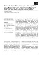
Tumors with positive IGF1R protein showed cytoplasmic
staining. Typical images of positive immunostaining for
IGF1R in cancer cells are shown in Figure 1A. Overall,
seven cases had a score of 0, 69 cases had a score of 1+,
B
3+
2+
positive
1+
0
negative
IGFBP3
positive
negative
Figure 1 IGF1R and IGFBP3 expression in pancreatic cancer. A, Representative IGF1R staining quantified with scores of 0 to 3+ according to
staining intensity. (Original magnification X 200). IGF1R was mainly expressed in the cytoplasm of pancreatic cancer cells. B, IGFBP3 was expressed
in the cell membrane and the cytoplasm of pancreatic cancer cells.
Hirakawa et al. BMC Cancer 2013, 13:392
/>
Page 4 of 9
Table 2 Association between IGF1R & IFGBP3 expression and clinicopathological factors in resectable pancreatic cancer
IGF1R expression
Characteristics
positive
negative
n = 50
n = 72
<60
7
18
≧60
43
54
Male
25
31
Female
25
41
T1, T2
9
17
T3, T4
41
55
IGFBP3 expression
p-value
positive
negative
n = 37
n = 85
10
15
27
70
15
41
22
44
7
19
30
66
Both IGF1R (+) and IGFBP3 (−)
p-value
positive
others
n = 32
n = 90
3
22
29
68
16
40
16
50
3
23
29
67
p-value
Age
0.139
0.238
0.07
Gender
0.449
0.433
0.588
T category
0.457
0.670
N category
0.077
N0
23
33
N1
27
39
I
4
11
II & III
46
61
0.986
20
36
17
49
5
10
32
75
0.233
14
42
18
48
0
15
32
75
0.776
Stage
Tumor location
0.273
0.787
0.011
proximal
31
50
30
51
18
63
distal
19
22
7
34
14
27
Grade1, 2
46
56
34
68
28
74
Grade 3, 4
4
16
3
17
4
16
negative
24
42
19
47
15
51
positive
26
30
18
38
17
39
negative
43
67
33
77
27
83
positive
7
5
4
8
5
7
36
71
28
79
1
14
4
11
16
43
16
43
0.241
21
42
0.456
16
47
27
59
19
67
0.087
10
26
0.692
13
23
Tumor differentiation
0.392
0.037
0.023
0.082
0.157
0.588
Lymphatic invasion
0.260
0.688
0.340
Arterial invasion
0.198
0.754
0.200
Venous invasion
negative
45
62
positive
5
10
negative
21
38
positive
29
34
0.52
0.037
0.967
Intrapancreatic nerve invasion
0.829
Tumor stromal volume
med & int
31
55
sci
19
17
negative
32
53
positive
18
19
0.108
IGFBP3 expression
0.256
Medullary type (med): scanty stroma, Intermediate type (int): the quantity of stroma is intermediate between the two above types, Scirrhous type (sci): abundant
stroma.
Hirakawa et al. BMC Cancer 2013, 13:392
/>
Page 5 of 9
23 cases had a score of 2+, and 27 cases had a score of
3+. Thus, 50 cases (41%) were positive for IGF1R
overexpression. Most of the positive staining was observed
in the cytoplasm, while two cases showed positive staining
in both membranes and cytoplasm. In contrast, no or
weak staining was seen in the cytoplasm of pancreatic
duct cells and acinar cells, and there was no staining in
the membranes. Figure 1B shows a representative picture
of IGFBP3 staining. IGFBP3 was mainly expressed in the
cytoplasm of cancer cells. Eighty-five cases of PDAC
showed negative IGFBP3 expression, whereas 37 cases
were positive.
Clinicopathological association of IGF1R and IGFBP3
expression
Table 2 shows the association of clinicopathological
characteristics and IGF1R or/and IGFBP3 expression.
IGF1R expression had a significant association with
histological grade (Fisher’s exact test, p = 0.037). Stromal volume tended to be more abundant in PDAC with
IGF1R overexpression, but no significant difference
was observed (χ2 test, p = 0.087). IGFBP3 expression
had a significant association with tumor location (χ2 test,
Relationship between clinicopathological features and
tumors with IGF1R-positive and IGFBP3-negative
expression
Among the 50 patients with positive IGF1R expression,
32 patients (64.0%) had negative IGFBP3 expression.
Tumors with IGF1R-positive and IGFBP3-negative expression (n = 32) were significantly frequently found to
have Stage II and III cancer (χ2 test, p = 0.011) compared
to the other groups (n = 90). Tumors with IGF1R-positive
and IGFBP3-negative expression tended to be in older
patients (Fisher’s exact test, p = 0.07) and advanced T stage
(χ2 test, 0.077). Among the 72 patients with negative
IGF1R, 53 patients (73.6%) showed negative IGFBP3 expression, whereas 19 patients (26.4%) had positive IGFBP3
expression. No association was found between IGF1R and
IGFBP3 expression.
1.0
IGF1R
0.8
0.6
Negative (n=72)
0.4
p = 0.018
0.2
Positive (n=50)
0
1
2
3
4
Probability of overall survival
Probability of overall survival
1.0
p = 0.023), and a significant inverse association with
venous invasion (Fisher’s exact test, p = 0.037). IGFBP3
expression tended to be frequent in differentiated PDAC
in histological grade, but no significant difference was
observed (χ2 test, p = 0.082).
IGFBP3
0.8
Positive (n=37)
0.6
p =0.079
0.4
0.2
5
Years after operation
Negative (n=85)
0
1
3
4
5
1.0
IGF1R and IGFBP3
0.8
0.6
Other groups
(n=90)
0.4
p <0.001
0.2
IGF1R-positive
and IGFBP3-negative (n=32 )
0
1
2
3
4
Years after operation
5
Probability of overall survival
1.0
Probability of overall survival
2
Years after operation
IGF1R and IGFBP3
0.8
IGF1R-negative and
IGFBP3-positive (n=19)
0.6
0.4
p =0.218
0.2
Other groups (n=103)
0
1
2
3
4
5
Years after operation
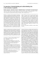
Figure 2 Overall survival of patients based on IGF1R and IGFBP3 expression. The survival curve shows the Kaplan-Meier overall survival
curves in relation to the IGF1R and IGFBP3 levels in patients with pancreatic cancer. A statistically significant difference in survival was observed
between patients with IGF1R-positive and IGF1R-negative tumors (p = 0.018). The prognosis of patients with IGF1R-positive and IGFBP3-negative
patients showed a significant correlation with overall survival (p < 0.001). IGFBP3 expression alone tended to be associated with overall survival
(p = 0.079). The co-expression of IGF1R-negative and IGFBP3-positive PDAC was not associated with overall survival (p = 0.218).
Hirakawa et al. BMC Cancer 2013, 13:392
/>
Page 6 of 9
Survival
Kaplan-Meier survival analyses showed a significantly
poorer overall survival in the IGF1R-positive group
compared to the IGF1R-negative group (p = 0.018).
Moreover, the prognosis of patients with IGF1R-positive
and IGFBP3-negative PDAC was significantly poorer
than that of other patients (p < 0.001). In contrast, the
prognosis of patients with IGF1R-negative and IGFBP3positive PDAC was not significantly correlation with
overall survival (p = 0.218), while IGFBP3 expression alone
tended to be associated with overall survival (p = 0.079)
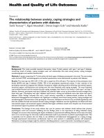
(Figure 2). Figure 3 shows the overall survival stratified for
IGF1R and IGFBP3 expression in cancer cells according to
clinical stage II status. The prognosis for IGF1R positive
patients with stage II tumors was significantly (p = 0.0080)
poorer than that for IGF1R negative patients, while no
significant difference in the prognosis was found between
the IGF1R expression in either stage I or III tumors
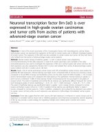
(data not shown). On univariate analysis, three factors,
IGF1R overexpression, IGF1R-positive and IGFBP3negative expression, and lymph node metastasis, were
significantly associated with worse overall survival.
Because IGF1R status is deeply associated with IGF1R
and IGFBP3 status, multivariate analysis was performed
with two factors: IGF1R-positive and IGFBP3-negative
expression, and lymph node metastasis. The multivariate survival analysis indicated that IGF1R-positive and
IGFBP3-negative expression, along with lymph node
metastasis, were independent prognostic indicators
(Table 3). IGF1R-positive and IGFBP3-negative expression and lymph node metastasis were independent predictors of poor prognosis.
Discussion
The present study analyzed the immunohistochemical
expression of IGF1R and IGFBP3 with clinicopathological
variables and the correlation with overall survival in 122
patients with PDAC. IGF1R expression had a significant
association with histological grade of tumor differentiation, and also tended to be associated with abundant
stroma. These findings suggest that the IGF1R signaling
system might be correlated with histopathologic features
of PDAC. It has been reported that IGF1 is produced from
stromal cells [11]. There might be an interaction between
cancer cells and stromal cells via IGF/IGF1R signaling.
PDAC patients with IGF1R-positive expression showed
significantly poorer survival, compared to the IGF1Rnegative group (Figure 2). The present findings suggest
1.0
IGF1R
0.8
Stage II
0.6
Negative (n=56)
0.4
p = 0.008
Positive (n=39)
0.2
0
1
2
3
4
Probability of overall survival
Probability of overall survival
1.0
5
IGFBP3
0.8
0.6
Positive (n=28)
0.4
p =0.188
0.2
Negative (n=67)
0
Years after operation
IGF1R and IGFBP3
0.8
0.6
Other groups
(n=68)
p =0.0009
0.4
0.2
IGF1R-positive and
IGFBP3 negative (n=27 )
0
1
2
3
4
Years after operation
2
3
4
5
5
1.0
Probability of overall survival
Probability of overall survival
1.0
1
Years after operation
IGF1R and IGFBP3
0.8
IGF1R-negative
and IGFBP3-positive (n=16)
0.6
0.4
p =0.121
0.2
Other groups (n=79)
0
1
2
3
4
5
Years after operation
Figure 3 Overall survival stratified by IGF1R and IGFBP3 expression in cancer cells in patients with clinical stage II tumors. Prognosis of
IGF1R-positive cancer was significantly poorer (p = 0.008) than that of IGF1R-negative cancer in the stage II group. Analysis of prognosis of patients
with IGF1R-positive and IGFBP3-negative tumors shows a significant correlation with overall survival (p = 0.0009) in patients with stage II tumors.
Hirakawa et al. BMC Cancer 2013, 13:392
/>
Page 7 of 9
Table 3 Univariate and multivariate survival analyses in pancreatic cancer
Variable
Univariate analysis
Multivariate analysis
Hazard ratio
95% CI
p-value
1.714
1.091-2.693
0.020
1.561
0.946-2.576
0.081
3.101
1.843-5.218
< 0.001
male vs female
0.975
0.623-1.525
0.911
≧ 60 vs <60
1.444
0.822-2.537
0.201
1.844
0.997-3.413
0.051
1.79
1.106-2.736
0.017
1.108
0.671-1.736
0.752
1.332
0.855-2.075
0.205
1.209
0.554-2.637
0.634
1.579
0.812-3.071
0.178
1.452
0.930-2.267
0.101
1.452
0.826-2.551
0.195
0.724
0.453-1.156
0.176
Hazard ratio
95% CI
p-value
3.060
1.823-5.138
< 0.001
1.718
1.092-2.702
0.019
IGF1R expression
Positive vs negative
IGFBP3 low expression
Positive vs negative
IGF1R (+) & IGFBP3 (−)
Positive vs negative
Gender
Age
T category
3, 4 vs 1,2
Lymph node metastasis
Positive vs negative
Tumor location
proximal vs distal
Lymphatic invasion
Positive vs negative
Arterial invasion
Positive vs negative
Venous invasion
Positive vs negative
Intrapancreatic nerve invasion
Positive vs negative
Tumor differentiation
grade 1,2 vs grade 3,4
Tumor stromal volume
med/int vs scir
that the IGF1R signaling system might be correlated with
tumor aggressiveness in PDAC, as has been previously
reported [25,26].
IGF bioavailability is regulated by a family of six IGFbinding proteins (IGFBP), of which IGFBP3 is the major
IGF carrier protein [17]. The function of IGFBP3 is controversial. IGFBP3 has been shown to produce either inhibition [27-29] or potentiation [30-32] of IGF effects. The
direction of the effect may depend on the cell type [27]. In
this study, favorable survival in the IGFBP3-positive group
was noted, but statistical significance was not obtained
(Figure 2). IGFBP3 expression had an inverse association
with venous invasion. These findings suggest that IGFBP3
might show antiproliferative effects in PDAC. IGFBP3 expression had a significant association with proximal tumors. Most insulin is secreted from the distal pancreas.
IGFBP3 expression might be associated with lesions
involving insulin secretion.
Next, the significance of the combination of IGF1R expression and IGFBP3 expression was evaluated. Tumors
with IGF1R-positive and IGFBP3-negative expression were
significantly frequently found at an advanced clinical stage
(II or III), compared to the other groups. The prognosis of
patients with IGF1R-positive and IGFBP3-negative PDAC
was poorer than that of other groups, especially in patients
with stage II tumors (Figure 3). The IGF1R-positive and
IGFBP3-negative subgroup was the group with the
worst prognosis (Figures 2 & 3). These findings suggest
that IGFBP3 could produce inhibition of IGF effects.
Decreased IGFBP3 production and increased IGF1R expression in pancreas tumors might enhance the tumorigenesis and cell motility as previously reported [26,33,34].
Hirakawa et al. BMC Cancer 2013, 13:392
/>
Prediction of prognosis in patients with operable PDAC is
important to determine the adjuvant therapy. This is especially true in patients with stage II tumors, because the
local recurrence rate of PDAC is high, even in patients
with curative R0 operations. The present study suggests
that combined evaluation of IGF1R expression and
IGFBP3 expression is a useful prognostic factor in pancreatic cancer, especially with clinical stage II tumors.
Although IGFBP3 is the major IGF carrier protein,
some paper reported that IGFBP3 has IGF-independent
antiproliferative and proapoptotic effects [20,21]. The
inhibition of IGF1-induced functions by cell surfaceassociated IGFBP3 have been reported [27,29]; however,
the relationship between membrane-associated IGFBPs
and IGF1R signaling is less well understood. Therefor
significance of co-expression of IGFBP3 and IGF1R in
PDAC remains obscure. We then analyzed the significance of IGF1R-negative and IGFBP3-positive group
with respect to overall survival (in the right bottom diagram of Figures 2 & 3), which might clarify whether
IGFBP3 is IGF1/IGF1R signaling-independent or not.
Although IGFBP3 expression alone tended to be associated with overall survival (p = 0.079), co-expression of
IGF1R-negative and IGFBP3-positive PDAC was not
associated with overall survival (p = 0.218). These data
suggested that the function of IGFBP3 might be
dependent on IGF1R expression.
Conclusion
IGF1R signaling might be associated with tumor aggressiveness, and IGFBP3 might show antiproliferative effects
in pancreatic cancer. Both high IGF1R expression and low
IGFBP3 expression represent useful prognostic markers
for patients with curatively resected pancreatic cancer.
Page 8 of 9
3.
4.
5.
6.
7.
8.
9.
10.
11.
12.
13.
14.
15.
16.
17.
Competing interests
All of the authors have no conflicts of interest to disclose.
18.
Authors’ contributions
TH: study design, data analysis, material sampling, paper preparation. MY:
study design, data analysis, interpretation of data, paper preparation. AM, KH,
KK, RA, NY and BN: material sampling. KH: data analysis, interpretation. All
authors read and approved the final manuscript.
19.
20.
21.
Sources of support
This study was supported in part, by the National Cancer Center Research
and Development Fund (23-A-9), and by Grants-in Aid for Scientific Research
(KAKENHI, Nos. 20591573, 22390262, and 23390329) from the Ministry of
Education, Science, Sports, Culture and Technology of Japan.
22.
23.
Received: 14 March 2013 Accepted: 15 August 2013
Published: 21 August 2013
24.
References
1. Jemal A, Siegel R, Xu J, Ward E: Cancer statistics, 2010. CA Cancer J Clin
2010, 60(5):277–300.
2. Cho K, Ishiwata T, Uchida E, Nakazawa N, Korc M, Naito Z, Tajiri T: Enhanced
expression of keratinocyte growth factor and its receptor correlates with
venous invasion in pancreatic cancer. Am J Pathol 2007, 170(6):1964–1974.
25.
26.
Cleary SP, Gryfe R, Guindi M, Greig P, Smith L, Mackenzie R, Strasberg S,
Hanna S, Taylor B, Langer B, et al: Prognostic factors in resected
pancreatic adenocarcinoma: analysis of actual 5-year survivors. J Am Coll
Surg 2004, 198(5):722–731.
Sohn TA, Yeo CJ, Cameron JL, Koniaris L, Kaushal S, Abrams RA, Sauter PK,
Coleman J, Hruban RH, Lillemoe KD: Resected adenocarcinoma of the
pancreas-616 patients: results, outcomes, and prognostic indicators.
J Gastrointest Surg 2000, 4(6):567–579.
Spannuth WA, Nick AM, Jennings NB, Armaiz-Pena GN, Mangala LS, Danes
CG, Lin YG, Merritt WM, Thaker PH, Kamat AA, et al: Functional significance
of VEGFR-2 on ovarian cancer cells. Int J Cancer 2009, 124(5):1045–1053.
Doi Y, Yashiro M, Yamada N, Amano R, Noda S, Hirakawa K: VEGF-A/VEGFR2 signaling plays an important role for the motility of pancreas cancer
cells. Ann Surg Oncol 2012, 19(8):2733–2743.
Ozawa F, Friess H, Tempia-Caliera A, Kleeff J, Buchler MW: Growth factors
and their receptors in pancreatic cancer. Teratog Carcinog Mutagen 2001,
21(1):27–44.
Bergmann U, Funatomi H, Yokoyama M, Beger HG, Korc M: Insulin-like
growth factor I overexpression in human pancreatic cancer: evidence for
autocrine and paracrine roles. Cancer Res 1995, 55(10):2007–2011.
LeRoith D, Werner H, Beitner-Johnson D, Roberts CT Jr: Molecular and
cellular aspects of the insulin-like growth factor I receptor. Endocr Rev
1995, 16(2):143–163.
Furstenberger G, Senn HJ: Insulin-like growth factors and cancer. Lancet
Oncol 2002, 3(5):298–302.
Pollak MN, Schernhammer ES, Hankinson SE: Insulin-like growth factors
and neoplasia. Nat Rev Cancer 2004, 4(7):505–518.
Pollak M: Insulin-like growth factor physiology and cancer risk. Eur J
Cancer 2000, 36(10):1224–1228.
Lahm H, Suardet L, Laurent PL, Fischer JR, Ceyhan A, Givel JC, Odartchenko
N: Growth regulation and co-stimulation of human colorectal cancer cell
lines by insulin-like growth factor I, II and transforming growth factor
alpha. Br J Cancer 1992, 65(3):341–346.
Chen SC, Chou CK, Wong FH, Chang CM, Hu CP: Overexpression of
epidermal growth factor and insulin-like growth factor-I receptors and
autocrine stimulation in human esophageal carcinoma cells. Cancer Res
1991, 51(7):1898–1903.
Renehan AG, Zwahlen M, Minder C, O’Dwyer ST, Shalet SM, Egger M:
Insulin-like growth factor (IGF)-I, IGF binding protein-3, and cancer risk:
systematic review and meta-regression analysis. Lancet 2004, 363
(9418):1346–1353.
Samani AA, Yakar S, LeRoith D, Brodt P: The role of the IGF system in
cancer growth and metastasis: overview and recent insights. Endocr Rev
2007, 28(1):20–47.
Butt AJ, Fraley KA, Firth SM, Baxter RC: IGF-binding protein-3-induced
growth inhibition and apoptosis do not require cell surface binding and
nuclear translocation in human breast cancer cells. Endocrinology 2002,
143(7):2693–2699.
Hwa V, Oh Y, Rosenfeld RG: Insulin-like growth factor binding proteins: a
proposed superfamily. Acta Paediatr Suppl 1999, 88(428):37–45.
Firth SM, Baxter RC: Cellular actions of the insulin-like growth factor
binding proteins. Endocr Rev 2002, 23(6):824–854.
Baxter RC: Insulin-like growth factor (IGF)-binding proteins: interactions
with IGFs and intrinsic bioactivities. Am J Physiol Endocrinol Metab 2000,
278(6):E967–976.
Baxter RC: Signalling pathways involved in antiproliferative effects of
IGFBP-3: a review. Mol Pathol 2001, 54(3):145–148.
Kelley KM, Oh Y, Gargosky SE, Gucev Z, Matsumoto T, Hwa V, Ng L, Simpson
DM, Rosenfeld RG: Insulin-like growth factor-binding proteins (IGFBPs)
and their regulatory dynamics. Int J Biochem Cell Biol 1996, 28(6):619–637.
Sobin LH: TNM, sixth edition: new developments in general concepts
and rules. Semin Surg Oncol 2003, 21(1):19–22.
Isaji S, Kawarada Y, Uemoto S: Classification of pancreatic cancer:
comparison of Japanese and UICC classifications. Pancreas 2004,
28(3):231–234.
Liu JP, Baker J, Perkins AS, Robertson EJ, Efstratiadis A: Mice carrying null
mutations of the genes encoding insulin-like growth factor I (Igf-1) and
type 1 IGF receptor (Igf1r). Cell 1993, 75(1):59–72.
Baserga R, Hongo A, Rubini M, Prisco M, Valentinis B: The IGF-I receptor in
cell growth, transformation and apoptosis. Biochim Biophys Acta 1997,
1332(3):F105–126.
Hirakawa et al. BMC Cancer 2013, 13:392
/>
Page 9 of 9
27. Rogers J, Wiltrout L, Nanu L, Fant ME: Developmentally regulated
expression of IGF binding protein-3 (IGFBP-3) in human placental
fibroblasts: effect of exogenous IGFBP-3 on IGF-1 action. Regul Pept 1996,
61(3):189–195.
28. Oh Y, Muller HL, Lamson G, Rosenfeld RG: Insulin-like growth factor (IGF)independent action of IGF-binding protein-3 in Hs578T human breast
cancer cells. Cell surface binding and growth inhibition. J Biol Chem 1993,
268(20):14964–14971.
29. Samaras SE, Hammond JM: Insulin-like growth factor binding protein-3
inhibits porcine granulosa cell function in vitro. Am J Physiol 1995,
268(6 Pt 1):E1057–1064.
30. Chevalier X, Tyler JA: Production of binding proteins and role of the
insulin-like growth factor I binding protein 3 in human articular cartilage
explants. Brit J Rheumatol 1996, 35(6):515–522.
31. Conover CA, Clarkson JT, Bale LK: Factors regulating insulin-like growth
factor-binding protein-3 binding, processing, and potentiation of insulinlike growth factor action. Endocrinology 1996, 137(6):2286–2292.
32. Chen JC, Shao ZM, Sheikh MS, Hussain A, LeRoith D, Roberts CT Jr, Fontana
JA: Insulin-like growth factor-binding protein enhancement of insulinlike growth factor-I (IGF-I)-mediated DNA synthesis and IGF-I binding in
a human breast carcinoma cell line. J Cell Physiol 1994, 158(1):69–78.
33. Baker J, Liu JP, Robertson EJ, Efstratiadis A: Role of insulin-like growth
factors in embryonic and postnatal growth. Cell 1993, 75(1):73–82.
34. Mauro L, Salerno M, Morelli C, Boterberg T, Bracke ME, Surmacz E: Role of
the IGF-I receptor in the regulation of cell-cell adhesion: implications in
cancer development and progression. J Cell Physiol 2003, 194(2):108–116.
doi:10.1186/1471-2407-13-392
Cite this article as: Hirakawa et al.: IGF-1 receptor and IGF binding
protein-3 might predict prognosis of patients with resectable pancreatic
cancer. BMC Cancer 2013 13:392.
Submit your next manuscript to BioMed Central
and take full advantage of:
• Convenient online submission
• Thorough peer review
• No space constraints or color figure charges
• Immediate publication on acceptance
• Inclusion in PubMed, CAS, Scopus and Google Scholar
• Research which is freely available for redistribution
Submit your manuscript at
www.biomedcentral.com/submit